Embed Size (px)
Citation preview

Tib
MD
R
vqdsnibsmtlsetao
qt
elGcttag(f(tcH
0
Biochemical and Biophysical Research Communications 280, 363–367 (2001)
doi:10.1006/bbrc.2000.4082, available online at http://www.idealibrary.com on
wo Isoforms of Trimming Glucosidase II Existn Mammalian Tissues and Cell Linesut Not in Yeast and Insect Cells
artin Ziak,1 Mirjam Meier, Kay-Sara Etter, and Jurgen Rothivision of Cell and Molecular Pathology, Department of Pathology, University of Zurich, CH-8091 Zurich, Switzerland
eceived November 13, 2000
a subunit composition of glucosidase II was obtained inSg
pmtcwdam
Isoalhg(tih4
ioaIo
M
acSmiar
We previously cloned glucosidase II and provided inivo evidence for its involvement in protein foldinguality control. DNA-sequencing of different clonesemonstrated the existence of two isoforms of gluco-idase II which differed by 66 nucleotides due to alter-ative splicing. The existence of two enzyme isoforms
n various organs of pig and rat as well as human,ovine, rat, and mouse cell lines could be demon-trated by RT-PCR and Western blotting. Further-ore, the two isoforms of glucosidase II could be de-
ected in embryonic and postnatal rat kidney andiver. In yeast, Saccharomyces cerevisiae, and in in-ects, Drosophila S2 cells, only one isoforms of thenzyme was detectable. The ubiquitous occurrence ofhe two glucosidase II isoforms in mammalian tissuesnd cell lines might be indicative of a special functionf each isoform. © 2001 Academic Press
Key Words: glucosidase II; enzyme isoforms; proteinuality control; endoplasmic reticulum; N-glycosyla-ion; yeast; insect; mammalian tissue.
N-glycosylation of proteins is initiated in thendoplasmic reticulum (ER) with the transfer of aipid-linked precursor, dolichol pyrophosphatelc3Man9(GlcNAc)2, to an asparagine residue of nas-
ent polypeptide chains. Subsequently, ER locatedrimming glucosidase I and glucosidase II, respec-ively, remove the terminal a1,2-linked glucose residuend the two a1,3 glucose residues of newly synthesizedlycoproteins to obtain the Man9(GlcNAc)2 structure1, 2). In addition, ER a-mannosidase I can trim theour a1,2-linked mannose residues to yield the Man5
GlcNAc)2 structure (2, 3). Trombetta et al. (4) reportedhat glucosidase II from rat liver is composed of aatalytic a-subunit and a tightly bound noncatalyticDEL-containing b-subunit. Further evidence for such
1 To whom correspondence should be addressed. Fax: 141 1 255 447. E-mail: [email protected].
363
. pombe by the disruption of the gene either encodinglucosidase II a or glucosidase II b (5).Glucosidase II, in concert with UDP-glucose:glyco-
rotein glucosyltransferase and chaperones, and ERannosidase I have been shown to be involved in pro-
ein folding control and degradation of misfolded gly-oproteins, respectively (for review; 6, 7). In harmonyith the involvement of glucosidase II in such a fun-amental cellular control mechanism, enzyme activitynd immunoreactivity have been detected in yeast andammalian cells (8–10).Recently, we isolated the cDNA encoding glucosidase
I by screening a pig liver cDNA library (11). DNA-equencing demonstrated the existence of two isoformsf pig liver glucosidase II. An extra sequence of 22mino acid residues was inserted at position 188 of pigiver glucosidase II (11) due to alternative splicing. Weave separately expressed both isoforms of pig liverlucosidase II in a glucosidase II-deficient yeast strain12) under the GAL1 promoter in pYES2 in order toest if both isoforms were enzymatically active. Bothsoforms were detectable by Western blotting and ex-ibited enzymatic activity with the artificial substrate-methylumbelliferyl-a-D-glucopyranoside (8).Here, we report the ubiquitous occurrence of the two
soforms of glucosidase II in various adult rat and pigrgans as well as embryonic and postnatal rat tissuesnd cell lines of human, bovine, mouse and rat origin.n insect cells and Saccharomyces cerevisiae, however,nly one isoform was detectable.
ATERIALS AND METHODS
Materials. Frozen embryonic day 20, postnatal days 2, 7, 14 anddult rat kidneys and livers were purchased from Pel Freez Biologi-als (Rogers, AR). Dulbecco’s modified Eagle medium (DMEM),chneider’s Drosophila medium, fetal bovine serum (FBS), Taq poly-erase from Life Technologies (Basel, Switzerland), protease inhib-
tor cocktail tablets from Roche Diagnostics (Rotkreuz, Switzerland)nd TRI reagent from Lucerna Chem (Luzern, Switzerland). AMVeverse transcriptase, random primers and dNTPs were purchased
0006-291X/01 $35.00Copyright © 2001 by Academic PressAll rights of reproduction in any form reserved.
from Promega (Wallisellen, Switzerland), Omniscript reverse tran-sSapIR
wC(lA
c1dg(w
rcwoR5rugmsgl5CGCotTG1ewtfoscwp
4erwPpBmtfi1ru
R
(deaLbacclpaei
satutWvsaafddswfiprnw
a(afPv
Vol. 280, No. 1, 2001 BIOCHEMICAL AND BIOPHYSICAL RESEARCH COMMUNICATIONS
criptase and QIAquick PCR-purification kit from QIAGEN (Basel,witzerland). Yeast nitrogen base without amino acids, casaminocids were from Difco Laboratories (Basel, Switzerland). Alkalinehosphatase-conjugated goat anti-rabbit IgG, and goat anti-mousegG (all affinity-purified) were purchased from Jackson Immuno-esearch Laboratories, Inc. (West Grove, PA).
Cell lines. The mouse lymphoma cell lines BW5147 and PHAR2.7ere kindly provided by Dr. I. Trowbridge (Salk Institute, San Diego,A) and Drosophila S2 cells were obtained from Dr. W. G. Gehring
Biozentrum, University of Basel, Switzerland). Human HepG2, rativer clone 9 and BRL 3A and bovine MDBK cells were obtained frommerican Type Culture Collection (Manassas, VA).
Strains and medium. The following yeast strains were used: S.erevisiae SS328 which is wild type for glucosidase II: MATa ade2-01 ura3-52 his 3D200 lys2-801. YG427 which is a glucosidase IIeficient strain: MATa ade2-101 ura3-52 his 3D200 lys2-801 Dls2<KanMX (12). S. cerevisae were grown at 30°C in YPD medium2% Bacto-Peptone, 1% yeast extract, 2% glucose). Drosophila cellsere grown in Schneider’s Drosophila medium at 25°C.
RT-PCR. Total RNA from frozen embryonic, postnatal and adultat organs as well as from the various mammalian cell lines, insectells and yeast were isolated with the TRI reagent (13). Total RNAas reverse transcribed by AMV reverse transcriptase using randomligonucleotide primers. The 20 ml reaction mixture contained 1 mg ofNA, 20 U of RNAsin, 0.5 mg of random primer, 1 mM of each dNTP,mM MgCl2, and 20 U of AMV reverse transcriptase in the supplied
everse transcription buffer. Alternatively, cDNA was synthesizedsing total RNA, omniscript reverse transcriptase and random oli-onucleotide primers following the protocol of the manufacturer. A 5l aliquot of the reaction mixture was used as template in theubsequent PCR-amplification with the following species specificlucosidase II oligonucleotides (A–C, mammalian tissues and cellines; D–F, S. cerevisiae; G–I, Drosophila): oligonucleotide A,9-CAAGAGGCAGCGAAGCATAC; oligonucleotide B, 59-TACCTC-ACGCTGTTGTCAT; oligonucleotide C, 59-CTCTGCCTTCCCCTG-ATCTCCTCT; oligonucleotide D, 59-TAACAAGGAAAACCAT-ACC; oligonucleotide E, 59-TTCACCGAGACCACCAGCAC;ligonucleotide F, 59-GAAAAGTCCAGAGCCACAGC; oligonucleo-ide G, 59-AGAGAATGCCAACCAACAGC; oligonucleotide H, 59-GTCTGCAGTGGAGTTTGAA; oligonucleotide I, 59- AGAACCCAT-AAAGAGAAAT. The PCR was performed in a reaction volume of00 ml containing 1.5 mM MgCl2, 0.5 mM of each primer, 200 mM ofach dNTP and 2.5 U of Taq polymerase. The cycling parametersere set as follows: 60 s at 94°C (1 cycle), then 60 s at 94°C, 60 s at
he appropriate annealing temperature and 60 s at 72°C (30 cycles)ollowed by a final extension at 72°C. PCR products derived from pigrgans were separated using a 5% polyacrylamide TBE gel and silvertained. All other PCR products were resolved in 2% agarose gelontaining ethidium bromide. For DNA-sequencing PCR-productsere separated in 2% agarose gels and the bands were excised andurified using the QIAquick PCR-purification.
SDS-PAGE and immunoblotting. Tissues were homogenized at°C in PBS (pH 7.4) containing protease inhibitors and proteinsxtracted with 1% Triton X-100 for 1 h at 4°C. Cell debris wasemoved by centrifugation at 10,000g for 10 min at 4°C. The extractsere mixed with Laemmli buffer and boiled for 5 min at 100°C (14).roteins (150 mg/lane) were resolved by SDS–PAGE using 7.5%olyacrylamide gels. Protein concentrations were determined by theradford procedure (15). The gels were transferred to nitrocelluloseembranes using a semi-dry blotting apparatus. For immunoblot-
ing, the membranes were conditioned with PBS containing 1% de-atted milk powder and 0.05% Tween 20 for 1 h at room temperature,ncubated with rabbit anti-pig liver glucosidase II antibodies (10) for8 h at 4°C, followed by alkaline phosphatase-conjugated goat anti-abbit IgG antibodies (0.12 mg/ml). Color reaction was performedsing nitroblue tetrazolium/BCIP-phosphate as substrates.
364
ESULTS
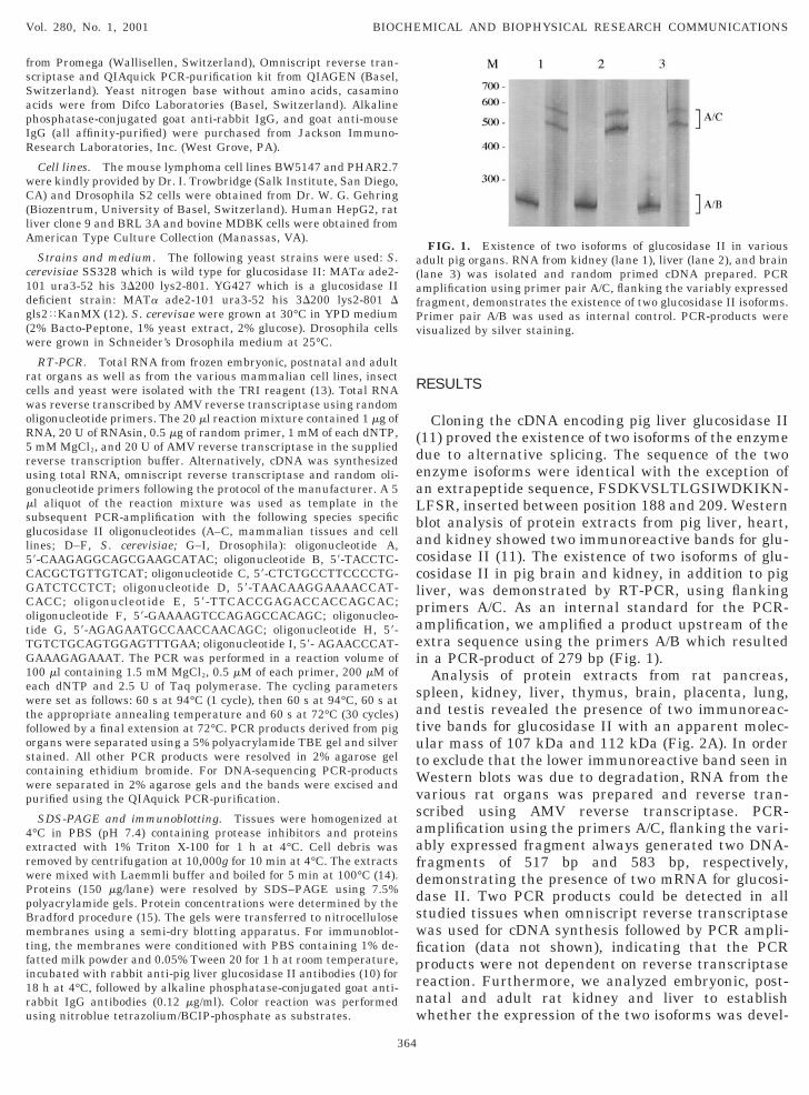
Cloning the cDNA encoding pig liver glucosidase II11) proved the existence of two isoforms of the enzymeue to alternative splicing. The sequence of the twonzyme isoforms were identical with the exception ofn extrapeptide sequence, FSDKVSLTLGSIWDKIKN-FSR, inserted between position 188 and 209. Westernlot analysis of protein extracts from pig liver, heart,nd kidney showed two immunoreactive bands for glu-osidase II (11). The existence of two isoforms of glu-osidase II in pig brain and kidney, in addition to pigiver, was demonstrated by RT-PCR, using flankingrimers A/C. As an internal standard for the PCR-mplification, we amplified a product upstream of thextra sequence using the primers A/B which resultedn a PCR-product of 279 bp (Fig. 1).
Analysis of protein extracts from rat pancreas,pleen, kidney, liver, thymus, brain, placenta, lung,nd testis revealed the presence of two immunoreac-ive bands for glucosidase II with an apparent molec-lar mass of 107 kDa and 112 kDa (Fig. 2A). In ordero exclude that the lower immunoreactive band seen in
estern blots was due to degradation, RNA from thearious rat organs was prepared and reverse tran-cribed using AMV reverse transcriptase. PCR-mplification using the primers A/C, flanking the vari-bly expressed fragment always generated two DNA-ragments of 517 bp and 583 bp, respectively,emonstrating the presence of two mRNA for glucosi-ase II. Two PCR products could be detected in alltudied tissues when omniscript reverse transcriptaseas used for cDNA synthesis followed by PCR ampli-cation (data not shown), indicating that the PCRroducts were not dependent on reverse transcriptaseeaction. Furthermore, we analyzed embryonic, post-atal and adult rat kidney and liver to establishhether the expression of the two isoforms was devel-
FIG. 1. Existence of two isoforms of glucosidase II in variousdult pig organs. RNA from kidney (lane 1), liver (lane 2), and brainlane 3) was isolated and random primed cDNA prepared. PCRmplification using primer pair A/C, flanking the variably expressedragment, demonstrates the existence of two glucosidase II isoforms.rimer pair A/B was used as internal control. PCR-products wereisualized by silver staining.

odcsioadsqKPc
Beeta4gFgp4gcediPtswtqAtwS
(pttatts(v
pppa
hrpHM(ifDapP
Vol. 280, No. 1, 2001 BIOCHEMICAL AND BIOPHYSICAL RESEARCH COMMUNICATIONS
pmentally regulated. Embryonic day 20 and postnatalay 7 kidneys showed a high amount of the long glu-osidase II isoform, whereas in the adult kidney thehort isoform was prominently present (Fig. 3A). Bothsoforms of glucosidase II could be detected in embry-nic, postnatal and adult rat liver (Fig. 3B). The shortnd the long PCR products obtained from embryonicay 20, postnatal day 7 and adult rat kidney wereubjected to DNA sequencing. The amino acid se-uence of the extrapeptide, FSDKVSLTLGSIWD-IKNLFSR, as deduced from the DNA sequence of theCR product was identical in all samples. In variousell lines such as human HepG2, rat liver clone 9 and
FIG. 2. Two isoforms of glucosidase II exist in various rat organs.A) Western blot analysis of protein extracts (150 mg/lane) fromancreas (lane 1), spleen (lane 2), kidney (lane 3), liver (lane 4),hymus (lane 5), brain (lane 6), placenta (lane 7), lung (lane 8), andestis (lane 9) revealed two immunoreactive bands. (B) RT-PCRmplification was performed using the primer pair A/C and reverse-ranscribed RNA isolated from testis (lane 1), adrenal gland (lane 2),hymus (lane 3), spleen (lane 4), pancreas (lane 5), placenta (lane 6),tomach (lane 7), heart (lane 8), small intestine (lane 9), and ovarylane 10). PCR-Products were separated in a 2% agarose gel andisualized by ethidium bromide staining.
FIG. 3. Existence of two isoforms of glucosidase II in embryonic,ostnatal, and adult rat kidney and liver. Random primed cDNArepared from RNA from kidney (A) or liver (B) was amplified usingrimers A/C: embryonic day 20 (lane 1), postnatal day 7 (lane 2), anddult (lane 3). Primer pair A/B was used as internal control.
365
RL 3A, bovine MDBK and mouse BW5147 cells thexistence of the two isoforms of glucosidase II could bestablished as well. Like the parental BW5147 cells,he glucosidase II-deficient mutant cell line PHAR2.7lso showed the presence of two PCR products (Fig.A). This indicates that at least the 59 end of thelucosidase II gene is present in this mutant cell line.urthermore, the RT-PCR assay was used to investi-ate whether the two glucosidase II isoforms are alsoresent in S. cerevisiae and Drosophila S2 cells (Fig.B). Although both contain glucosidase II activity, theene coding for gls2 has been only identified in S.erevisiae (GenEMBL Accession No. Z36098). In S. cer-visiae SS328, one isoform of glucosidase II could beetected when the primers D/F were used. As expected,n the glucosidase II deficient yeast strain, YG427, noCR-product could be detected, using the primers men-ioned above (data not shown). Using the amino acidequence derived from the pig liver glucosidase II (8)e searched the Drosophila gene data bank and iden-
ified an open reading frame of Drosophila with a se-uence identity of 46% (GenEMBL Accession No.E003122). Based on this DNA sequence, we designed
he Drosophila specific oligonucleotides G, H and I,hich were used for PCR amplification. In Drosophila2 cells one DNA-fragment of 486 bp was detected
FIG. 4. (A) Two isoforms of glucosidase II are detectable inuman, bovine, mouse, and rat cell lines. RNA was extracted andeverse transcribed using random primers. PCR amplification waserformed using the primers A/C and A/B as a control. HumanepG2 (lane 1), rat clone 9 (lane 2), rat BRL 3A (lane 3), bovineDBK (lane 4), mouse BW5147 (lane 5), and mouse PHAR2.7 cells
lane 6). (B) One isoform of glucosidase II is detectable in yeast andnsect cells. Reverse transcribed cDNA, prepared from RNA isolatedrom S. cerevisiae SS328, was amplified using the primers D/F and/E as control (lane 1). RNA from Drosophila S2 cells was isolatednd random primed cDNA prepared. PCR amplification using primerair G/I demonstrates the existence of one glucosidase II isoform.rimer pair G/H was used as internal control (lane 2).

using the primers G/I. These data demonstrate, that inSo
D
dgtppaNisktldaecPfiitp
woeotl
lmNnwak(arat(Rb
atgc
outermost of the two glucose residues occurs fastertqafiD(gTbtisstghqe(isrNtcoieTiit
ptittipsbeRtctcc
ttrgt
Vol. 280, No. 1, 2001 BIOCHEMICAL AND BIOPHYSICAL RESEARCH COMMUNICATIONS
. cerevisiae and Drosophila S2 cells only one isoformf glucosidase II is present.
ISCUSSION
The results of the present study provide strong evi-ence for the widespread occurrence of two isoforms oflucosidase II. Due to alternative splicing, an addi-ional peptide of 22 amino acid residues is inserted intoosition 188 of pig liver glucosidase II (8, 11, andresent study). The additional peptide did not showny significant homology to known protein sequences.ot unexpected, the amino acid sequence of the peptide
dentified in pig liver was identical to the amino acidequence found in embryonic, postnatal and adult ratidneys. Recently, Arendt et al. (16) reported the exis-ence of splice variants of glucosidase II in mouse Tymphoma cells. Interestingly, the additional peptideetected in pig liver as well as in embryonic, postnatalnd adult rat kidney showed 90% identity to a variablyxpressed segment, box A1, in the mouse T lymphomaells (16). During the preparation of our manuscript,elletier et al. (17) reported the existence of two iso-
orms of the a subunit of human glucosidase II differ-ng in 22 amino acid residues. The extrapeptide foundn human glucosidase II (17) showed 95% identity tohe extrapeptide of the pig and rat enzyme (8, 11, andresent study).We designed species specific oligonucleotides, whichere used to amplify a possible extrapeptide sequencef glucosidase II in S. cerevisiae and Drosophila. How-ver, in S. cerevisiae and in Drosophila S2 cells onlyne isoform of glucosidase II was detectable, indicatinghat the two isoforms of the enzyme must have arisenater in evolution.
Glucosidase II is ubiquitously expressed in mamma-ian tissues (4, 11, 18). However, the expression level
ay vary as demonstrated for various pig tissues byorthern and Western blot analysis (11). Two immu-oreactive bands migrating at about 107 and 112 kDaere detectable when extracts of pig tissues were an-lyzed by Western blotting. The intensity of the 107Da band was always stronger than the 112 kDa band11). In agreement with our observations, immunoblotnalysis of crude microsomes prepared from pig brain,at liver, calf liver, and monkey kidney cells using annti-pig liver glucosidase II antibody showed two reac-ive bands with a molecular mass of 107 and 112 kDa18). These data, together with the results obtained byT-PCR, might indicate that the two immunoreactiveands represent the two isoforms of glucosidase II.Glucosidase II sequentially cleaves the two inner
1,3-linked glucose residues. However, it is noteworthyhat these glucose residues are either a1,3-linked tolucose, or a1,3-linked to mannose. Furthermore, pulsehase studies demonstrated that the removal of the
366
han the removal of the inner one (19). Three majoruestions arise. First, are both isoforms enzymaticallyctive; second, do they have different substrate speci-city; and third, are there differences in their kinetics?uring the purification of glucosidase II from rat liver
20) or pig liver (11) it was not possible to separate thelucosidase II isoforms by their physical properties.herefore, the measured enzymatic activity could note attributed to one of the isoforms. In order to answerhe first question we have expressed the cDNA encod-ng either the long or short isoform of pig liver gluco-idase II in a glucosidase II-deficient S. cerevisiaetrain. Both isoforms showed enzymatic activity, usinghe artificial substrate 4-methylumbelliferyl a-D-lucoside (8). Expression of a1/b and a2/b isoforms ofuman glucosidase II in Sf9 insect cells and subse-uent enzyme purification resulted in a heteroenzymexhibiting p-nitrophenyl a-D-glucopyranoside activity17). Both isoforms, a1 and a2, showed the same spec-ficity for the Glc2Man9GlcNAc1 and Glc1Man9GlcNAc1
ubstrates (17). Although Pelletier et al. (17) have pu-ified the recombinant human glucosidase II by Ni21-TA and anion exchange chromatography, obviously
he possibility that the recombinant human a subunitan form a complex with the endogenous glucosidase IIf Sf9 cells cannot be excluded. It has been shown thatn pig kidney (10) and pig liver (18) glucosidase IIxists as a tetrameric complex under native conditions.herefore, the measured glucosidase II activities and
soform specificity of human glucosidase II expressedn Sf9 cells (17) might not be unequivocally attributedo the recombinant enzyme.
The two glucosidase II isoforms exist in embryonic,ostnatal and adult mammalian tissues. Furthermore,he sequence of the additional 22 amino acid residuess highly conserved in rat, pig and human. Thus, it isempting to speculate that this high level of conserva-ion might be indicative of different functions of eachsoform. It has been speculated that the additionaleptide might modulate protein-protein interactionsuch as the association between the a- and the-subunit of glucosidase II or an interaction of thextrapeptide with other ER proteins or substrates (16).ecently, Arendt et al. (21) demonstrated a strong in-
eraction between glucosidase II and CD45 in mouse Tells. Subsequently, the same authors demonstrated inransfection experiments that the glucosidase isoformontaining the alternatively spliced sequence Box A1 isapable of binding to CD45 (22).Monoglucosylated oligosaccharides play an impor-
ant role with regard to folding of proteins present inhe ER (6, 7). Experiments are now in progress toeveal the possible functional role of the two isoforms oflucosidase II in this quality control mechanism andheir subcellular distributions.

ACKNOWLEDGMENTS
csMsG
R
9. Lucocq, J. M., Brada, D., and Roth, J. (1986) J. Cell Biol. 102,
1
1
1
1111
1
11
2
2
2
Vol. 280, No. 1, 2001 BIOCHEMICAL AND BIOPHYSICAL RESEARCH COMMUNICATIONS
We thank Christian Zuber for helpful discussions as well as forritical reading of the manuscript, Dieter Zimmermann for DNAequencing, and Markus Aebi and Stefan te Heesen (Institute oficrobiology, ETH Zurich) for instructions handling yeast cells. This
tudy was supported by the Swiss National Science Foundationrant 31-50835.97.
EFERENCES
1. Kornfeld, R., and Kornfeld, S. (1985) Annu. Rev. Biochem. 54,631–664.
2. Moremen, K. W., Trimble, R. B., and Herscovics, A. (1994) Gly-cobiology 4, 113–125.
3. Gonzalez, D. S., Karaveg, K., Vandersall-Nairn, A. S., Lal, A.,and Moremen, K. W. (1999) J. Biol. Chem. 274, 21375–21386.
4. Trombetta, E. S., Simons, J. F., and Helenius, A. (1996) J. Biol.Chem. 271, 27509–27516.
5. D’Alessio, C., Fernandez, F., Trombetta, E. S., and Parodi, A. J.(1999) J. Biol. Chem. 274, 25899–25905.
6. Ellgaard, L., Molinari, M., and Helenius, A. (1999) Science 286,1882–1888.
7. Parodi, A. J. (2000) Annu. Rev. Biochem. 69, 69–93.8. Ziak, M., Meier, M., and Roth, J. (1999) Mol. Biol. Cell 10 S,
414a.
367
2137–2146.0. Brada, D., and Dubach, U. C. (1984) Eur. J. Biochem. 141,
149–156.1. Flura, T., Brada, D., Ziak, M., and Roth, J. (1997) Glycobiology 7,
617–624.2. Jakob, C. A., Burda, P., te Heesen, S., Aebi, M., and Roth, J.
(1998) Glycobiology 8, 155–164.3. Chomczynski, P. (1993) BioTechniques 15, 532–537.4. Laemmli, U. (1970) Nature 227, 680–685.5. Bradford, M. M. (1976) Anal. Biochem. 72, 248–250.6. Arendt, C. W., Dawicki, W., and Ostergaard, H. L. (1999) Glyco-
biology 9, 277–283.7. Pelletier, M. F., Marcil, A., Sevigny, G., Jakob, C. A., Tessier,
D. C., Chevet, E., Menard, R., Bergeron, J. J., and Thomas, D. Y.(2000) Glycobiology 10, 815–827.
8. Hentges, A., and Bause, E. (1997) Biol. Chem. 378, 1031–1038.9. Hubbard, S., and Robbins, P. (1979) J. Biol. Chem. 254, 4568–
4576.0. Hirano, K., Ziak, M., Kamoshita, K., Sukenaga, Y., Kametani, S.,
Shiga, Y., Roth, J., and Akanuma, H. (2000) Glycobiology, inpress.
1. Arendt, C. W., and Ostergaard, H. L. (1997) J. Biol. Chem. 272,13117–13125.
2. Baldwin, T. A., Gogela-Spehar, M., and Ostergaard, H. L. (2000)J. Biol. Chem. 275, 32071–32076.