Embed Size (px)
Citation preview
Chemical Physics Letters 477 (2009) 179–183
Contents lists available at ScienceDirect
Chemical Physics Letters
journal homepage: www.elsevier .com/ locate /cplet t
Two distinct branch–stem interfacial structures of silver dendrites with verticaland slanted branchings
Shaochun Tang, Sascha Vongehr, Xiangkang Meng *
National Laboratory of Solid State Microstructures, Department of Materials Science and Engineering, Nanjing University, Nanjing 210093, People’s Republic of China
a r t i c l e i n f o
Article history:Received 12 May 2009In final form 26 June 2009Available online 1 July 2009
0009-2614/$ - see front matter � 2009 Elsevier B.V. Adoi:10.1016/j.cplett.2009.06.087
* Corresponding author. Fax: +86 25 8359 5535.E-mail address: [email protected] (X. Meng).
a b s t r a c t
Silver dendrites synthesized by sonoelectrochemical deposition are investigated via transmission elec-tron microscopy (TEM) in order to clarify the formation of specific branching angles. Two kinds of den-drites are found to form simultaneously; one has slanted angles while the other shows orthogonalbranching. Investigation of the branch–stem interfacial structures reveals that the vertical branchesattach at a characteristic transition layer around the main stem, while the slanted growth involves twin-ning induced dislocations. It is suggested that oriented attachment of nanoparticles followed by epitaxialgrowth leads to obliqueness, while crystallization of amorphous phase involving grain rotation andrealignment results in vertical branching.
� 2009 Elsevier B.V. All rights reserved.
1. Introduction
Much effort has been devoted to understand non-equilibriumgrowth phenomena theoretically and experimentally [1,2]. Amongsuch phenomena, dendrites are one of the most intensively inves-tigated topics due to their attractive structures and large surfacearea [3], both leading to many applications, for example in cataly-sis [4], chemical/biochemical sensors [5] and surface-enhancedRaman scattering [6]. Detailed description of the structure isimportant to understand the underlying growth mechanisms,which in turn is crucial to achieve designable structures (e.g. withwell defined branch–stem angles) resulting in desired properties.Many studies have focused on the mechanisms of dendritic growth[1,2,7–10]. Some novel growth scenarios have been recently pro-posed. Ding et al. [9] reported a double-interface growth mode ofsilver in a replacement reaction where dendritic growth was con-trolled by the formation of an amorphous phase and its crystalliza-tion. Cheng et al. [8] demonstrated that dendritic structure canemerge from oriented attachment of nanoparticles. Ostwald ripen-ing forms the initial particles and also smoothes the morphologyafter attachment. Dislocations are known to originate at the bond-ing interface in the oriented attachment of nanoparticles [7,8].
As for growth under non-equilibrium conditions, the distancefrom equilibrium is important [11,12] but generally not easily con-trolled [13]. Electrochemistry is in this context especially conve-nient, because the ‘distance’ can be tuned continuously andreversibly by the electrode potential. Particular attention has beenfocused on the study of the formation and growth of silver
ll rights reserved.
dendrites by various sonoelectrochemical methods with the assis-tance of organic capping agents such as nitrilotriacetate [14], gela-tin [15] and poly (vinyl pyrrolidone) [16]. Generally, branchingangles vary in a range from 15� to 90� [5]. Many factors affectingstructure have been discussed [2,13,16], but the detailed growthmechanisms are still unknown. It remains unclear how thebranches emerge from the stems; how the interplay of nucleationand growth at the microscopic scale results in specific densities,branching angles and so on.
In this work, we describe the discovery and investigation of twobranch–stem interfacial structures occurring simultaneously in asonoelectrochemical process involving silver: the surfactant-free,constant-current method leads to both, vertical branching and alsoa slanted growth, sometimes even along the same stem. High res-olution transmission electron microscopy (HRTEM) reveals twodistinct microstructures underlying the different interfaces. Theorigin of such dissimilar interfacial structures will be discussedin light of the many new proposals concerning non-equilibriumgrowth.
2. Experiment
Silver dendrites were fabricated by a sonoelectrochemical reac-tion with silver perchloride solutions as electrolytes. The simpletwo-electrode setup was described previously [16–19]. Two iden-tical silver sheets (2 cm � 2 cm) were cleaned with acetone, hydro-chloric acid and distilled water to remove surface contamination.The sheets were fixed on the two opposing sides of an electro-chemical cell 4 cm apart in order to serve as the working cathodeand counter anode. The electrolyte concentration (C), current den-sity (J), and the reaction time (t) were about 1.0 � 10�4 to 5 � 10�3
180 S. Tang et al. / Chemical Physics Letters 477 (2009) 179–183
mol/L, 1.25 to 2.5 mA/cm2 and 10 to 20 min, respectively. Electrol-ysis was conducted at room temperature under continuous 40 kHzultrasonic vibration at 50 W power. Products were precipitatedand purified by five centrifugation/rinsing/redispersion steps withdeionized water and ethanol.
The products were collected, dried at 40 �C for 12 h in anoven, and characterized by X-ray diffraction (XRD) with a D/Max-RA X-ray diffractometer (using Cu Ka = 1.5418 Å radiation).Transmission electron microscopy (TEM) samples were preparedby dripping suspensions of the dendrites onto a carbon filmcovering a copper grid and subsequent drying. TEM measurementwas performed with an FEI TECNAI F20 microscope operating at200 kV.
3. Results and discussion
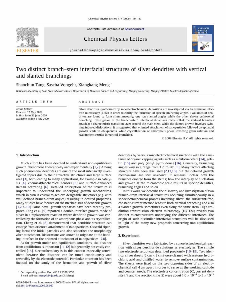
Fig. 1 shows an XRD pattern of the as-synthesized product froma typical experiment (C, V and t were 1.1 � 10�3 mol/L, 1.25 mA/cm2 and 20 min). The XRD pattern reveals that these Ag dendritesare cubic Ag (a = 4.09 Å, JCPDS 04-0783). The diffraction peaks ob-served can be indexed as (1 1 1), (2 0 0), (2 2 0) and (3 1 1) planesof the face-centered cubic (fcc) structure, respectively. An absenceof other peaks indicates a high purity of the silver.
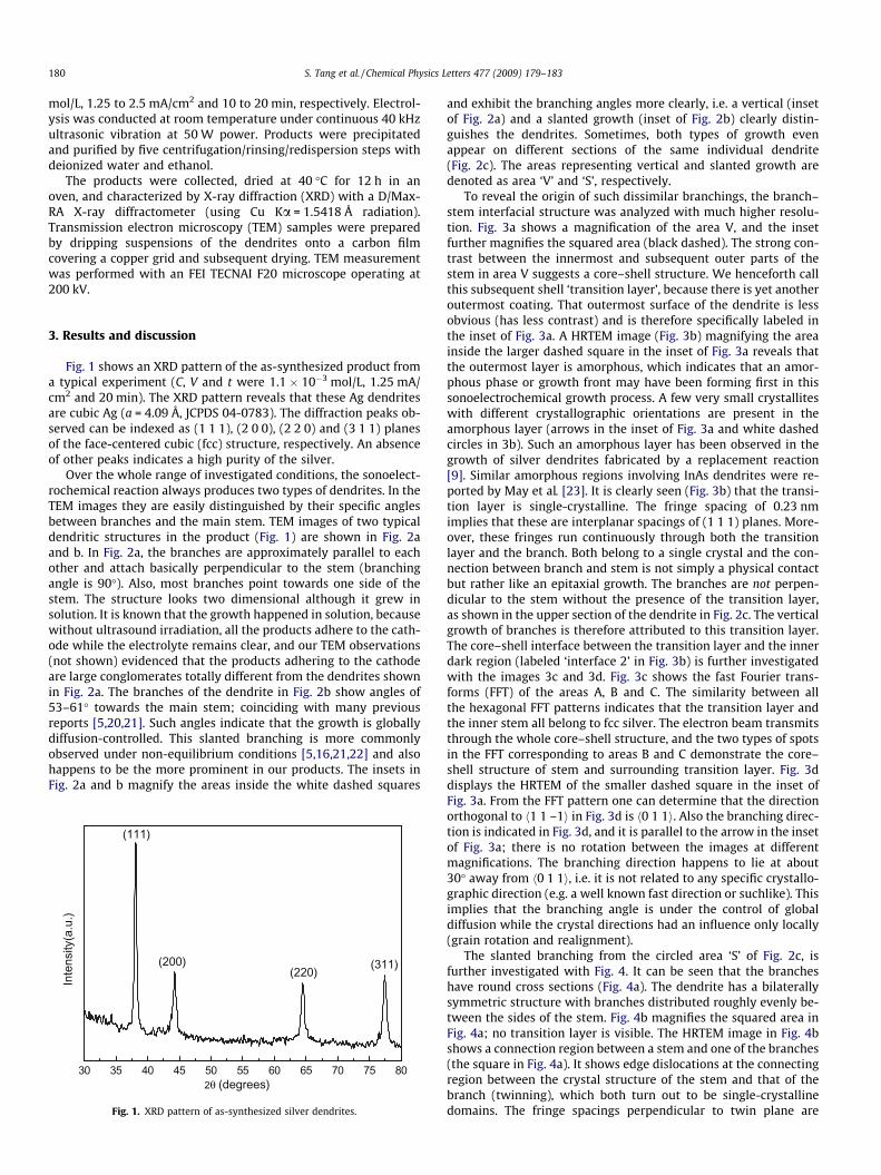
Over the whole range of investigated conditions, the sonoelect-rochemical reaction always produces two types of dendrites. In theTEM images they are easily distinguished by their specific anglesbetween branches and the main stem. TEM images of two typicaldendritic structures in the product (Fig. 1) are shown in Fig. 2aand b. In Fig. 2a, the branches are approximately parallel to eachother and attach basically perpendicular to the stem (branchingangle is 90�). Also, most branches point towards one side of thestem. The structure looks two dimensional although it grew insolution. It is known that the growth happened in solution, becausewithout ultrasound irradiation, all the products adhere to the cath-ode while the electrolyte remains clear, and our TEM observations(not shown) evidenced that the products adhering to the cathodeare large conglomerates totally different from the dendrites shownin Fig. 2a. The branches of the dendrite in Fig. 2b show angles of53–61� towards the main stem; coinciding with many previousreports [5,20,21]. Such angles indicate that the growth is globallydiffusion-controlled. This slanted branching is more commonlyobserved under non-equilibrium conditions [5,16,21,22] and alsohappens to be the more prominent in our products. The insets inFig. 2a and b magnify the areas inside the white dashed squares
30 35 40 45 50 55 60 65 70 75 80θ (degrees)
Intensity(a.u.)
(111)
(200)(220) (311)
Fig. 1. XRD pattern of as-synthesized silver dendrites.
and exhibit the branching angles more clearly, i.e. a vertical (insetof Fig. 2a) and a slanted growth (inset of Fig. 2b) clearly distin-guishes the dendrites. Sometimes, both types of growth evenappear on different sections of the same individual dendrite(Fig. 2c). The areas representing vertical and slanted growth aredenoted as area ‘V’ and ‘S’, respectively.
To reveal the origin of such dissimilar branchings, the branch–stem interfacial structure was analyzed with much higher resolu-tion. Fig. 3a shows a magnification of the area V, and the insetfurther magnifies the squared area (black dashed). The strong con-trast between the innermost and subsequent outer parts of thestem in area V suggests a core–shell structure. We henceforth callthis subsequent shell ‘transition layer’, because there is yet anotheroutermost coating. That outermost surface of the dendrite is lessobvious (has less contrast) and is therefore specifically labeled inthe inset of Fig. 3a. A HRTEM image (Fig. 3b) magnifying the areainside the larger dashed square in the inset of Fig. 3a reveals thatthe outermost layer is amorphous, which indicates that an amor-phous phase or growth front may have been forming first in thissonoelectrochemical growth process. A few very small crystalliteswith different crystallographic orientations are present in theamorphous layer (arrows in the inset of Fig. 3a and white dashedcircles in 3b). Such an amorphous layer has been observed in thegrowth of silver dendrites fabricated by a replacement reaction[9]. Similar amorphous regions involving InAs dendrites were re-ported by May et al. [23]. It is clearly seen (Fig. 3b) that the transi-tion layer is single-crystalline. The fringe spacing of 0.23 nmimplies that these are interplanar spacings of (1 1 1) planes. More-over, these fringes run continuously through both the transitionlayer and the branch. Both belong to a single crystal and the con-nection between branch and stem is not simply a physical contactbut rather like an epitaxial growth. The branches are not perpen-dicular to the stem without the presence of the transition layer,as shown in the upper section of the dendrite in Fig. 2c. The verticalgrowth of branches is therefore attributed to this transition layer.The core–shell interface between the transition layer and the innerdark region (labeled ‘interface 2’ in Fig. 3b) is further investigatedwith the images 3c and 3d. Fig. 3c shows the fast Fourier trans-forms (FFT) of the areas A, B and C. The similarity between allthe hexagonal FFT patterns indicates that the transition layer andthe inner stem all belong to fcc silver. The electron beam transmitsthrough the whole core–shell structure, and the two types of spotsin the FFT corresponding to areas B and C demonstrate the core–shell structure of stem and surrounding transition layer. Fig. 3ddisplays the HRTEM of the smaller dashed square in the inset ofFig. 3a. From the FFT pattern one can determine that the directionorthogonal to h1 1 –1i in Fig. 3d is h0 1 1i. Also the branching direc-tion is indicated in Fig. 3d, and it is parallel to the arrow in the insetof Fig. 3a; there is no rotation between the images at differentmagnifications. The branching direction happens to lie at about30� away from h0 1 1i, i.e. it is not related to any specific crystallo-graphic direction (e.g. a well known fast direction or suchlike). Thisimplies that the branching angle is under the control of globaldiffusion while the crystal directions had an influence only locally(grain rotation and realignment).
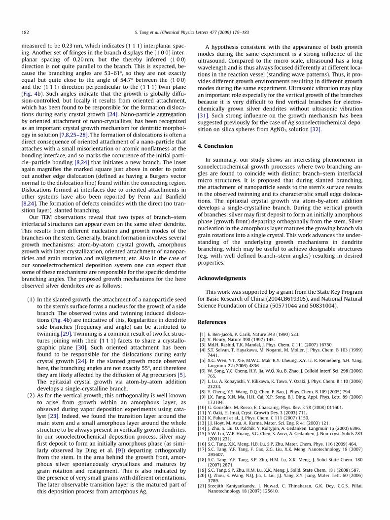
The slanted branching from the circled area ‘S’ of Fig. 2c, isfurther investigated with Fig. 4. It can be seen that the brancheshave round cross sections (Fig. 4a). The dendrite has a bilaterallysymmetric structure with branches distributed roughly evenly be-tween the sides of the stem. Fig. 4b magnifies the squared area inFig. 4a; no transition layer is visible. The HRTEM image in Fig. 4bshows a connection region between a stem and one of the branches(the square in Fig. 4a). It shows edge dislocations at the connectingregion between the crystal structure of the stem and that of thebranch (twinning), which both turn out to be single-crystallinedomains. The fringe spacings perpendicular to twin plane are
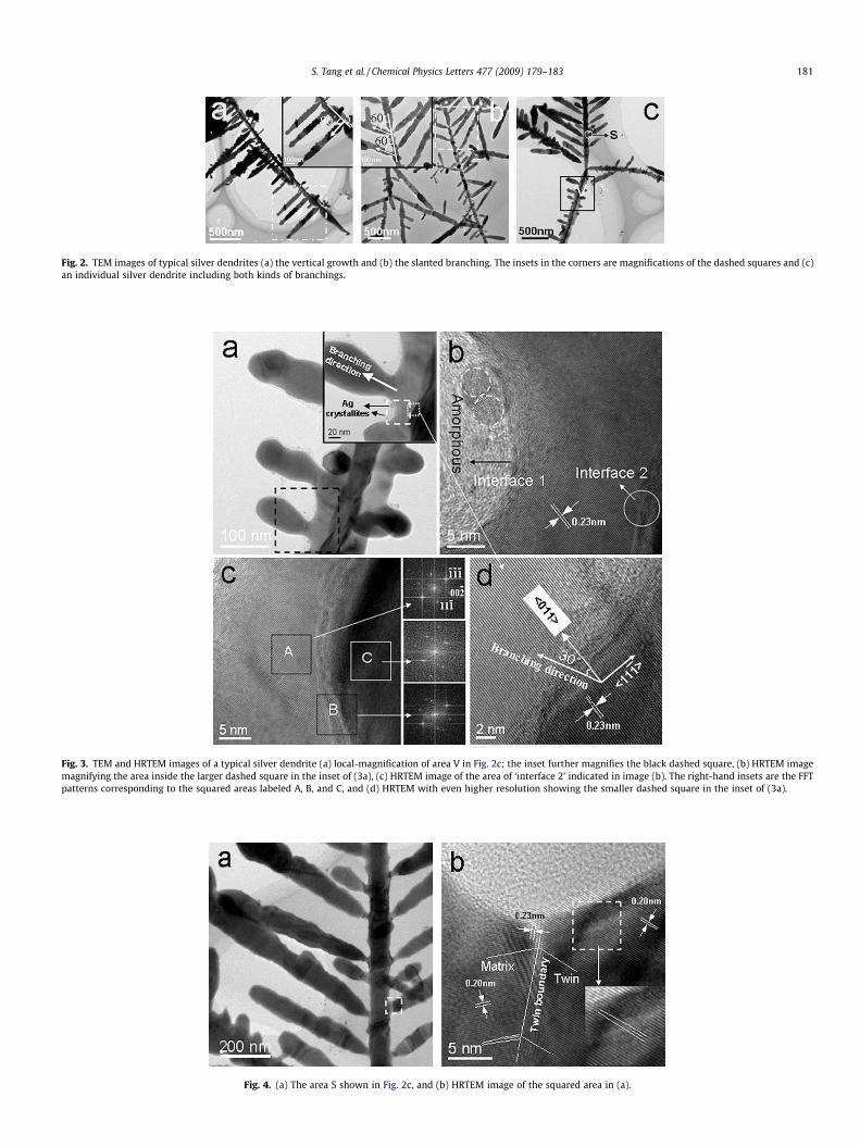
Fig. 2. TEM images of typical silver dendrites (a) the vertical growth and (b) the slanted branching. The insets in the corners are magnifications of the dashed squares and (c)an individual silver dendrite including both kinds of branchings.
Fig. 3. TEM and HRTEM images of a typical silver dendrite (a) local-magnification of area V in Fig. 2c; the inset further magnifies the black dashed square, (b) HRTEM imagemagnifying the area inside the larger dashed square in the inset of (3a), (c) HRTEM image of the area of ‘interface 2’ indicated in image (b). The right-hand insets are the FFTpatterns corresponding to the squared areas labeled A, B, and C, and (d) HRTEM with even higher resolution showing the smaller dashed square in the inset of (3a).
Fig. 4. (a) The area S shown in Fig. 2c, and (b) HRTEM image of the squared area in (a).
S. Tang et al. / Chemical Physics Letters 477 (2009) 179–183 181
182 S. Tang et al. / Chemical Physics Letters 477 (2009) 179–183
measured to be 0.23 nm, which indicates (1 1 1) interplanar spac-ing. Another set of fringes in the branch displays the (1 0 0) inter-planar spacing of 0.20 nm, but the thereby inferred h1 0 0idirection is not quite parallel to the branch. This is expected, be-cause the branching angles are 53–61�, so they are not exactlyequal but quite close to the angle of 54.7� between the h1 0 0iand the h1 1 1i direction perpendicular to the (1 1 1) twin plane(Fig. 4b). Such angles indicate that the growth is globally diffu-sion-controlled, but locally it results from oriented attachment,which has been found to be responsible for the formation disloca-tions during early crystal growth [24]. Nano-particle aggregationby oriented attachment of nano-crystallites, has been recognizedas an important crystal growth mechanism for dentritic morphol-ogy in solution [7,8,25–28]. The formation of dislocations is often adirect consequence of oriented attachment of a nano-particle thatattaches with a small misorientation or atomic nonflatness at thebonding interface, and so marks the occurrence of the initial parti-cle–particle bonding [8,24] that initiates a new branch. The insetagain magnifies the marked square just above in order to pointout another edge dislocation (defined as having a Burgers vectornormal to the dislocation line) found within the connecting region.Dislocations formed at interfaces due to oriented attachments inother systems have also been reported by Penn and Banfield[8,24]. The formation of defects coincides with the direct (no tran-sition layer), slanted branching.
Our TEM observations reveal that two types of branch–steminterfacial structures can appear even on the same silver dendrite.This results from different nucleation and growth modes of thebranches on the stem. Generally, branch formation involves severalgrowth mechanisms: atom-by-atom crystal growth, amorphousgrowth with later crystallization, oriented attachment of nanopar-ticles and grain rotation and realignment, etc. Also in the case ofour sonoelectrochemical deposition system one can expect thatsome of these mechanisms are responsible for the specific dendritebranching angles. The proposed growth mechanisms for the hereobserved silver dendrites are as follows:
(1) In the slanted growth, the attachment of a nanoparticle seedto the stem’s surface forms a nucleus for the growth of a sidebranch. The observed twins and twinning induced disloca-tions (Fig. 4b) are indicative of this. Regularities in dendriteside branches (frequency and angle) can be attributed totwinning [29]. Twinning is a common result of two fcc struc-tures joining with their {1 1 1} facets to share a crystallo-graphic plane [30]. Such oriented attachment has beenfound to be responsible for the dislocations during earlycrystal growth [24]. In the slanted growth mode observedhere, the branching angles are not exactly 55�, and thereforethey are likely affected by the diffusion of Ag precursors [5].The epitaxial crystal growth via atom-by-atom additiondevelops a single-crystalline branch.
(2) As for the vertical growth, this orthogonality is well knownto arise from growth within an amorphous layer, asobserved during vapor deposition experiments using cata-lyst [23]. Indeed, we found the transition layer around themain stem and a small amorphous layer around the wholestructure to be always present in vertically grown dendrites.In our sonoelectrochemical deposition process, silver mayfirst deposit to form an initially amorphous phase (as simi-larly observed by Ding et al. [9]) departing orthogonallyfrom the stem. In the area behind the growth front, amor-phous silver spontaneously crystallizes and matures bygrain rotation and realignment. This is also indicated bythe presence of very small grains with different orientations.The later observable transition layer is the matured part ofthis deposition process from amorphous Ag.
A hypothesis consistent with the appearance of both growthmodes during the same experiment is a strong influence of theultrasound. Compared to the micro scale, ultrasound has a longwavelength and is thus always focused differently at different loca-tions in the reaction vessel (standing wave patterns). Thus, it pro-vides different growth environments resulting in different growthmodes during the same experiment. Ultrasonic vibration may playan important role especially for the vertical growth of the branchesbecause it is very difficult to find vertical branches for electro-chemically grown silver dendrites without ultrasonic vibration[31]. Such strong influence on the growth mechanism has beensuggested previously for the case of Ag sonoelectrochemical depo-sition on silica spheres from AgNO3 solution [32].
4. Conclusion
In summary, our study shows an interesting phenomenon insonoelectrochemical growth processes where two branching an-gles are found to coincide with distinct branch–stem interfacialmicro structures. It is proposed that during slanted branching,the attachment of nanoparticle seeds to the stem’s surface resultsin the observed twinning and its characteristic small edge disloca-tions. The epitaxial crystal growth via atom-by-atom additiondevelops a single-crystalline branch. During the vertical growthof branches, silver may first deposit to form an initially amorphousphase (growth front) departing orthogonally from the stem. Silvernucleation in the amorphous layer matures the growing branch viagrain rotations into a single crystal. This work advances the under-standing of the underlying growth mechanisms in dendritebranching, which may be useful to achieve designable structures(e.g. with well defined branch–stem angles) resulting in desiredproperties.
Acknowledgments
This work was supported by a grant from the State Key Programfor Basic Research of China (2004CB619305), and National NaturalScience Foundation of China (50571044 and 50831004).
References
[1] E. Ben-Jacob, P. Garik, Nature 343 (1990) 523.[2] V. Fleury, Nature 390 (1997) 145.[3] Md.H. Rashid, T.K. Mandal, J. Phys. Chem. C 111 (2007) 16750.[4] S.T. Selvan, T. Hayakawa, M. Nogami, M. Moller, J. Phys. Chem. B 103 (1999)
7441.[5] X.G. Wen, Y.T. Xie, M.W.C. Mak, K.Y. Cheung, X.Y. Li, R. Renneberg, S.H. Yang,
Langmuir 22 (2006) 4836.[6] W. Song, Y.C. Cheng, H.Y. Jia, W.Q. Xu, B. Zhao, J. Colloid Interf. Sci. 298 (2006)
765.[7] L. Lu, A. Kobayashi, Y. Kikkawa, K. Tawa, Y. Ozaki, J. Phys. Chem. B 110 (2006)
23234.[8] Y. Cheng, Y.S. Wang, D.Q. Chen, F. Bao, J. Phys. Chem. B 109 (2005) 794.[9] J.X. Fang, X.N. Ma, H.H. Cai, X.P. Song, B.J. Ding, Appl. Phys. Lett. 89 (2006)
173104.[10] G. González, M. Rosso, E. Chassaing, Phys. Rev. E 78 (2008) 011601.[11] Y. Oaki, H. Imai, Cryst. Growth Des. 3 (2003) 711.[12] K. Fukami et al., J. Phys. Chem. C 111 (2007) 1150.[13] J.J. Hoyt, M. Asta, A. Karma, Mater. Sci. Eng. R 41 (2003) 121.[14] J. Zhu, S. Liu, O. Palchik, Y. Koltypin, A. Gedanken, Langmuir 16 (2000) 6396.[15] S.W. Liu, W.P. Huang, S.G. Chen, S. Avivi, A. Gedanken, J. Non-cryst. Solids 283
(2001) 231.[16] S.C. Tang, X.K. Meng, H.B. Lu, S.P. Zhu, Mater. Chem. Phys. 116 (2009) 464.[17] S.C. Tang, Y.F. Tang, F. Gao, Z.G. Liu, X.K. Meng, Nanotechnology 18 (2007)
295607.[18] S.C. Tang, Y.F. Tang, S.P. Zhu, H.M. Lu, X.K. Meng, J. Solid State Chem. 180
(2007) 2871.[19] S.C. Tang, S.P. Zhu, H.M. Lu, X.K. Meng, J. Solid. State Chem. 181 (2008) 587.[20] Q. Zhou, S. Wang, N.Q. Jia, L. Liu, J.J. Yang, Z.Y. Jiang, Mater. Lett. 60 (2006)
3789.[21] Sreejith Kaniyankandy, J. Nuwad, C. Thinaharan, G.K. Dey, C.G.S. Pillai,
Nanotechnology 18 (2007) 125610.

S. Tang et al. / Chemical Physics Letters 477 (2009) 179–183 183
[22] J.X. Fang, H.J. You, C. Zhu, P. Kong, M. Shi, X.P. Song, B.J. Ding, Chem. Phys. Lett.439 (2007) 204.
[23] S.J. May, J.G. Zheng, B.W. Wessels, L.J. Lauhon, Adv. Mater. 17 (2005)598.
[24] R.L. Penn, J.F. Banfield, Science 281 (1998) 969.[25] A. Doughterty, P.D. Kaplan, J.P. Gollub, Phys. Rev. Lett. 58 (1987) 652.[26] R. Pieters, J.S. Langer, Phys. Rev. Lett. 56 (1987) 1948.[27] D.E. Zhang, X.J. Zhang, X.M. Ni, J.M. Song, H.G. Zheng, Chem. Phys. Lett. 430
(2006) 326.
[28] R. He, X.F. Qian, J. Yin, Z.K. Zhu, Chem. Phys. Lett. 369 (2003) 454.[29] A.R. Despic, K.I. Popov, Transport controlled deposition and dissolution of
metals, modern aspects of electrochemistry, No. 7, in: B.E. Conway, J.OM.Bockris (Eds.), Plenum Press, New York, 1972 (Chapter 4).
[30] Z.L. Wang, J. Phys. Chem. B 104 (2000) 1153.[31] J.X. Fang, H. Hahn, R. Krupke, F. Schramm, T. Scherer, B.J. Ding, X.P. Song, Chem.
Commun. 1130 (2009).[32] V.G. Pol, D.N. Srivastava, O. Palchik, V. Palchik, M.A. Slifkin, A.M. Weiss, A.
Gedanken, Langmuir 18 (2002) 3352.