Embed Size (px)
Citation preview

1
Cells:Chapt. 4
Two Basic Types of Cells
• Prokaryotes:– prounounced: pro-carry-oats
• Eukaryotes– Proun: you-carry-oats
A. Prokaryotes
Small, simple cells (relative to eukaryotes)Size: about 1 µm (1 micron)No internal membrane-bounded organellesNo nucleusSimple cell division
Contain the; 1. true bacteria & 2. archaebacteria
2
1. True Bacteria = Eubacteria
• Majority of bacteria
• Examples include: E. coli, Lactobacillus (yoghurt), Lyme disease
Eubacteria
•Peptidoglycan cell walls (carbos & AA)
•Separated into Gram + and - forms
Text pg. 58
Bacteria in the Environment
A) An acid hot spring in Yellowstone is rich in iron and sulfur.B) A black smoker chimney in the deep sea emits iron sulfides
at very high temperatures (270 to 380 degrees C).
example:Iron
utilizing Baceria
A B

3
2. Archaebacteria
• Live in extreme environments: high salt, high temps
• Different cell wall• Very different
membrane lipids• Unusual nucleic acid
sequence
Archaea types:
Based on their physiology, Archae can be organized into three types:
• Methanogens (prokaryotes that produce methane); • Extreme halophiles (prokaryotes that live at very
high concentrations of salt (NaCl); • Extreme (hyper) thermophiles (prokaryotes that live
at very high temperatures).
All archaea have features that distinguish them from Bacteria (i.e., no murein in cell wall, ether-linked membrane lipids, etc.). And, these prokaryotes exhibit unique structural or biochemical attributes which adapt them to their particular habitats.
B. Eukaryotes
• Bigger cells: 10-100 µm• True nucleus• Membrane-bounded
structures inside. Called organelles
• Divide by a complex, well-organized mitotic process
Liver Cell 9,400x
4
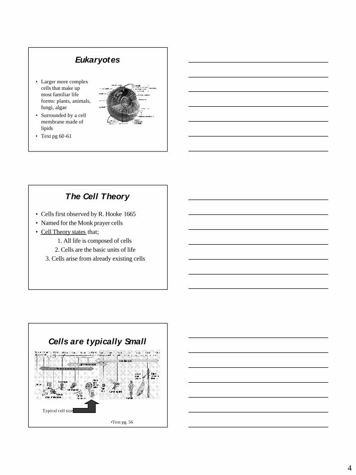
Eukaryotes
• Larger more complex cells that make up most familiar life forms: plants, animals, fungi, algae
• Surrounded by a cell membrane made of lipids
• Text pg 60-61
The Cell Theory
• Cells first observed by R. Hooke 1665• Named for the Monk prayer cells• Cell Theory states that;
1. All life is composed of cells2. Cells are the basic units of life
3. Cells arise from already existing cells
Cells are typically Small
Typical cell size
•Text pg. 56

5
Why are Cells Small?
• Cells must exchange gases & other molecules with environment…
• Nutrients in, Wastes out• As size increases, the rate of diffusion
exchange slows down….• This is due to the ratio of surface area to
volume
Surface Area to Volume
• Cell surface area is important in taking in nutrients
• Sfc area increases as the square of cell diameter
• But… entire cell volume needs to be fed• And, cell volume increases as the cube of
cell diameter
Consider 2 Cells...
6
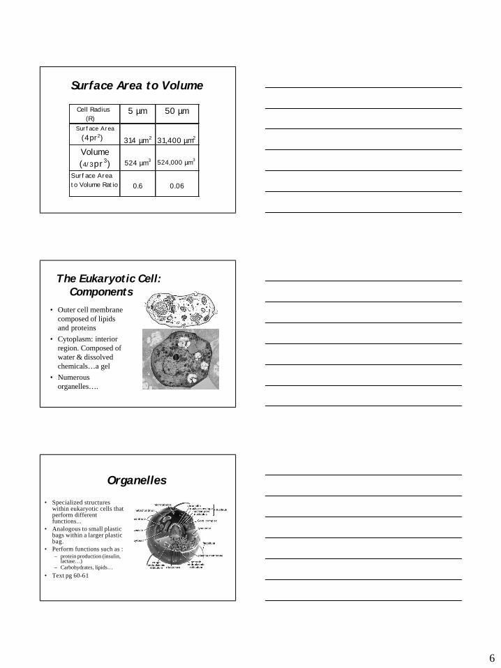
Surface Area to Volume
Cell Radius (R)
5 µm 50 µm
Surface Area (4pr2)
314 µm2
31,400 µm2
Volume (4/3pr3)
524 µm3
524,000 µm3
Surface Area to Volume Ratio
0.6
0.06
The Eukaryotic Cell: Components
• Outer cell membrane composed of lipids and proteins
• Cytoplasm: interior region. Composed of water & dissolved chemicals…a gel
• Numerous organelles….
Organelles
• Specialized structures within eukaryotic cells that perform different functions...
• Analogous to small plastic bags within a larger plastic bag.
• Perform functions such as :– protein production (insulin,
lactase…)– Carbohydrates, lipids…
• Text pg 60-61

7
Organelles of Note:The Nucleus
• Contains the genetic material (DNA), controls protein synthesis.
DNA --> RNA --> Protein• Surrounded by a double
membrane with pores
• Contains the chromosomes= fibers of coiled DNA & protein
• Text pg. 62
Chromosomes
All Chromosomes from a single cell
One chromosomePulled apart
A single chromosomeShowing the amount of DNA within
Mitochondria• Generate cellular energy in the
form of ATP molecules• ATP is generated by the
systematic breakdown of glucose = cell respiration
• Also, surrounded by 2 membrane layers
• Contain their own DNA!• A typical liver cell may have
1,700 mitoch.• All your mitoch. come from
your mother..• Text pg. 68

8
Chloroplasts
• Found in plants, algae and some bacteria. Responsible for capturing sunlight and converting it to food = photosynthesis.
• Surrounded by 2 membranes
• And…contain DNA
• Text pg. 69
Ribosomes
• Size ~20nm• Made of two subunits
(large and small)• Composed of RNA
and over 30 proteins• Come in two
sizes…80S and 70S• S units =
Sedimentation speed
Ribosomes• DNA --> RNA --> Protein• The RNA to Protein step
(termed translation) is done on cytoplasmicprotein/RNA particles termed ribosomes.
• Contain the protein synthesis machinery
• Ribosomes bind to RNA and produce protein.
9
Endoplasmic Reticulum = ER
• Cytoplasm is packed w. membrane system which move molecules about the cell and to outside
• An internal cellular subway system
• Outer sfc of ER may be smooth (SER)
• Or Rough (RER) • ER functions in lipid and
protein synthesis and transport
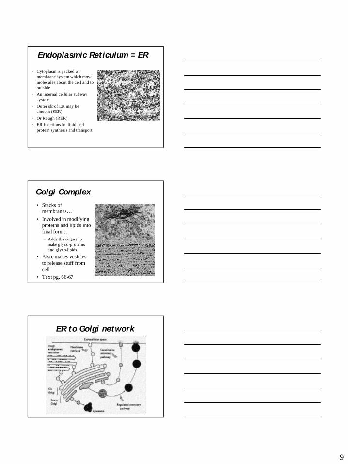
Golgi Complex• Stacks of
membranes…• Involved in modifying
proteins and lipids into final form…– Adds the sugars to
make glyco-proteins and glyco-lipids
• Also, makes vesicles to release stuff from cell
• Text pg. 66-67
ER to Golgi network
10
Lysosomes• important in breaking
down bacteria and old cell components
• contains many digestive enzymes
• The ‘garbage disposal’ or ‘recycling unit’ of a cell
• Malfunctioning lysosomesresult in some diseases (Tay-Sachs disease)
• Or may self-destruct cell such as in asbestosis
• Text pg 67
Cytoskeleton
• Composed of 3 filamentous proteins:
MicrotubulesMicrofilaments
Intermediate filaments
• All produce a complex network of structural fibers within cell
• Text pgs. 72-76
The specimen is human lung cell double-stained to expose microtubules and actin microfilaments using a mixture of FITC and rhodamine-phalloidin . Photo taken with an Olympus microscope.
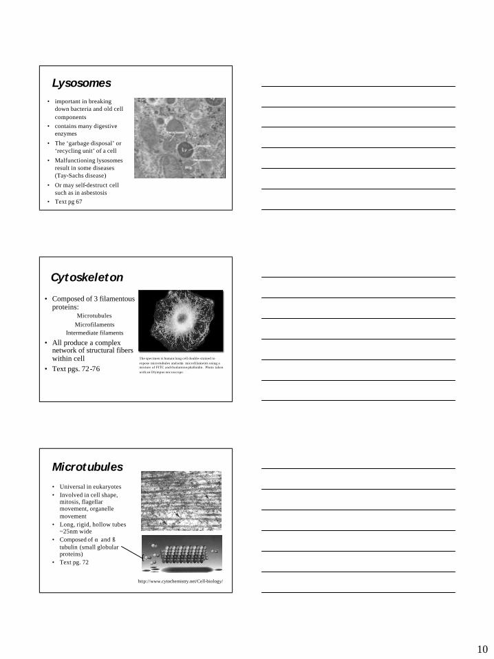
Microtubules
• Universal in eukaryotes• Involved in cell shape,
mitosis, flagellarmovement, organelle movement
• Long, rigid, hollow tubes ~25nm wide
• Composed of α and ß tubulin (small globular proteins)
• Text pg. 72
http://www.cytochemistry.net/Cell-biology/
11
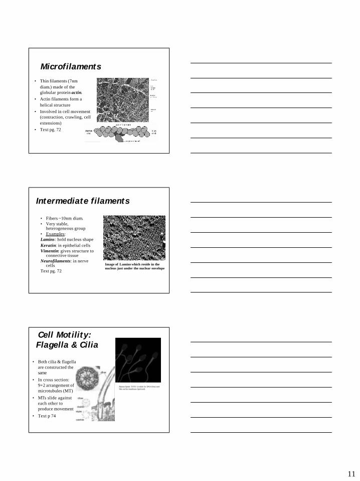
Microfilaments
• Thin filaments (7nm diam.) made of the globular protein actin.
• Actin filaments form a helical structure
• Involved in cell movement (contraction, crawling, cell extensions)
• Text pg. 72
Intermediate filaments
• Fibers ~10nm diam.• Very stable,
heterogeneous group• Examples:Lamins : hold nucleus shapeKeratin: in epithelial cells Vimentin: gives structure to
connective tissueNeurofilaments: in nerve
cells Text pg. 72
Image of Lamins which reside in the nucleus just under the nuclear envelope
Cell Motility:Flagella & Cilia
• Both cilia & flagella are constructed the same
• In cross section: 9+2 arrangement of microtubules (MT)
• MTs slide against each other to produce movement
• Text p 74
Human Sperm: TOTO -3 iodide for DNA (blue) and Nile red for membrane lipid (red)

12
How Flagella Move a Cell
Possible Origins of Eukaryotic Cells
Infolding of outer membranes Uptake of prokaryotes
• Text pg 70
Endosymbiosis
• Theory that eukaryotic cells arose from an early prokaryote (1) engulfing a second, smaller prokaryote (2)
• The internalized #2 was not digested but became a symbiote.
• Today’s mitochondria & chloroplasts may have arisen this way
• Text pg. 70
13
Evidence for Endosymbiosis
• Double membrane around both organelles• Both organelles have their own DNA• Both organelles have smaller (70S)
ribosomes…• Both organelles divide by simple fission