Embed Size (px)
Citation preview
tvpjournal.com | January/February 2015 | TODAY’S VETERINARY PRACTICE
PRACTICAL DENTISTRY Peer Reviewed
65
Periodontal disease is the number one problem in small animal medicine.1,2 In fact, the classic university study reported that, by 2 years of age, 80% of dogs and 70% of cats have some form of periodontal disease.3
In contrast, a recent large clinical study from general practices reported that by 10 years of age, only 24% of patients were clinically diagnosed with periodontal disease.4 Therefore, it appears this disease is signifi cantly under diagnosed, likely due to lack of dental education.
The signifi cant adverse consequences of periodontal disease (Table) show why diagnosis is so important.5,6
Veterinarians have many misconceptions about periodontal disease. This article outlines some common pitfalls encountered by veterinarians and provides some practical tips that can be implemented in general practice. (See Dental De� nitions on page 67.)
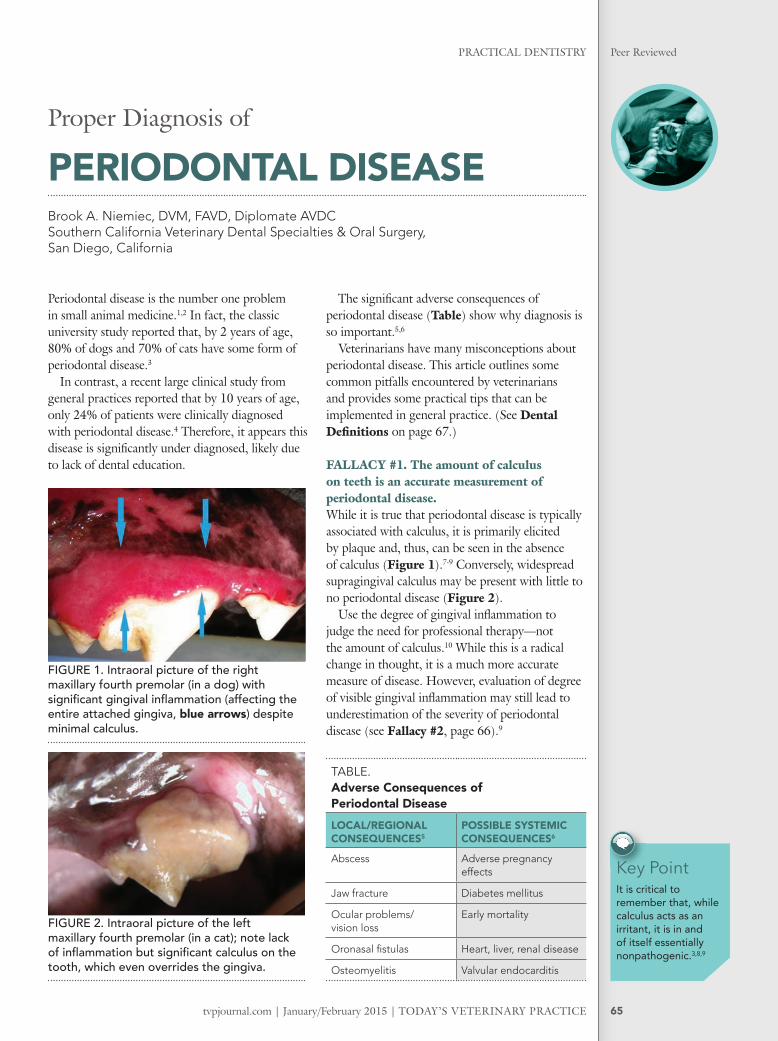
FALLACY #1. The amount of calculus on teeth is an accurate measurement of periodontal disease.While it is true that periodontal disease is typically associated with calculus, it is primarily elicited by plaque and, thus, can be seen in the absence of calculus (Figure 1).7-9 Conversely, widespread supragingival calculus may be present with little to no periodontal disease (Figure 2).
Use the degree of gingival infl ammation to judge the need for professional therapy—not the amount of calculus.10 While this is a radical change in thought, it is a much more accurate measure of disease. However, evaluation of degree of visible gingival infl ammation may still lead to underestimation of the severity of periodontal disease (see Fallacy #2, page 66).9
Proper Diagnosis of
PERIODONTAL DISEASEBrook A. Niemiec, DVM, FAVD, Diplomate AVDCSouthern California Veterinary Dental Specialties & Oral Surgery,San Diego, California
Key PointIt is critical to remember that, while calculus acts as an irritant, it is in and of itself essentially nonpathogenic.3,8,9
FIGURE 1. Intraoral picture of the right maxillary fourth premolar (in a dog) with signifi cant gingival infl ammation (affecting the entire attached gingiva, blue arrows) despite minimal calculus.
FIGURE 2. Intraoral picture of the left maxillary fourth premolar (in a cat); note lack of infl ammation but signifi cant calculus on the tooth, which even overrides the gingiva.
TABLE. Adverse Consequences of Periodontal DiseaseLOCAL/REGIONAL CONSEQUENCES5
POSSIBLE SYSTEMIC CONSEQUENCES6
Abscess Adverse pregnancy effects
Jaw fracture Diabetes mellitus
Ocular problems/vision loss
Early mortality
Oronasal fi stulas Heart, liver, renal disease
Osteomyelitis Valvular endocarditis
TODAY’S VETERINARY PRACTICE | January/February 2015 | tvpjournal.com
PRACTICAL DENTISTRYPeer Reviewed
66
PRACTICAL TIP: Lights/solutions that identify plaque/calculus can be used in conscious patients in the examination room to demonstrate the level of calculus (Figure 3). Plaque and early calculus can be invisible to the naked eye in natural light. Therefore, they can easily be missed, especially on wet teeth. Plaque-disclosing solutions and lights allow veterinarians to visually identify plaque/calculus.
FALLACY #2. Color change of the gingiva is the � rst sign of periodontal disease.The fi rst clinical sign of gingivitis was believed to be color change of the gingiva, termed marginal gingivitis.11,12 While this is a reliable sign of disease, it is now known that increased gingival bleeding on probing or brushing occurs fi rst.11,13 In fact, bleeding is a more objective measure of infl ammation than subtle color change. In addition, gingival color change is not a reliable indicator in dark pigmented patients (Figure 4).
PRACTICAL TIP: Consider carefully probing or brushing tractable patients’ teeth on conscious examination to demonstrate level of infl ammation. In addition, ask clients about a history of bleeding during brushing or after chewing hard/rough toys.8,12,14 If either of these are positive, a diagnosis of early gingivitis can be made despite a lack of gingival color change.10
FALLACY #3. A visual oral examination is suf� cient for diagnosis of periodontal disease.This is completely untrue for both conscious and anesthetized examinations. Signifi cant gingivitis can exist without periodontal pockets and, conversely, deep periodontal pockets can be present without signifi cant gingival infl ammation (Figure 5).9
Periodontal ProbeNormal sulcal depths in:12,15-17
• Dogs are 0 mm to 3 mm• Cats are 0 mm to 0.5 mm.
This is not common knowledge in most veterinary hospitals, and emphasizes the point that periodontal disease cannot be accurately diagnosed without a periodontal probe, which determines sulcal depth and identifi es pockets.12,18,19 Various periodontal probes are available; one such probe
A CFIGURE 3. Plaque-disclosing light used on maxillary incisors (dog): The teeth appear to be fairly clean (A), but the light reveals extensive plaque accumulation (B). Plaque-disclosing solution applied to a maxillary fourth premolar (C).
FIGURE 4. Intraoral picture of patient with dark gingiva and minimal to no calculus; however, signifi cant bleeding is apparent upon probing, indicating that gingivitis is present.
FIGURE 5. Deep (15-mm) periodontal pocket on right mandibular fi rst molar (in a dog) despite relatively normal appearance of gingiva; only periodontal probing under general anesthesia allowed identifi cation of this pocket.
B
tvpjournal.com | January/February 2015 | TODAY’S VETERINARY PRACTICE
PRACTICAL DENTISTRY Peer Reviewed
67
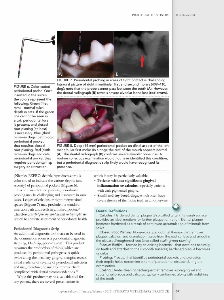
(Niemiec EXPRO, dentalaireproducts.com) is color coded to indicate the various depths (and severity) of periodontal pockets (Figure 6).
Even in anesthetized patients, periodontal probing may be challenging and inaccurate in some cases. Ledges of calculus or tight interproximal spaces (Figure 7) may preclude the standard insertion path and result in a missed pocket.20 Therefore, careful probing and dental radiographs are critical to accurate assessment of periodontal health.
Periodontal Diagnostic StripAn additional diagnostic tool that can be used in the examination room is a periodontal diagnostic strip (eg, OraStrip, perio-dx.com). This product measures the production of thiols, which are produced by periodontal pathogens. A quick swipe along the maxillary gingival margins reveals visual evidence of severity of periodontal infection and may, therefore, be used to improve client compliance with dental recommendations.21
While this product may be a valuable tool for any patient, there are several presentations in
which it may be particularly valuable:• Patients without significant gingival
inflammation or calculus, especially patients with dark pigmented gingiva
• Small and toy breed dogs, which often have severe disease of the molar teeth in an otherwise
A BFIGURE 7. Periodontal probing in areas of tight contact is challenging. Intraoral picture of right mandibular fi rst and second molars (409–410, dog); note that the probe cannot pass between the teeth (A). However, the dental radiograph (B) reveals severe alveolar bone loss (red arrow).
FIGURE 6. Color-coded periodontal probe. Once inserted in the sulcus, the colors represent the following: Green (fi rst mm)—normal sulcal depth in cats. If the green line cannot be seen in a cat, periodontal loss is present, and closed root planing (at least) is necessary. Blue (third mm)—in dogs, pathologic periodontal pocket that requires closed root planing. Red (sixth mm)—in dogs and cats, periodontal pocket that requires periodontal fl ap surgery or extraction.
A BFIGURE 8. Deep (14-mm) periodontal pocket on distal aspect of the left mandibular fi rst molar (in a dog); the rest of the mouth appears normal (A). The dental radiograph (B) confi rms severe alveolar bone loss. A routine conscious examination would not have identifi ed this condition, but a periodontal diagnostic strip likely would have recognized its presence.
Dental Defi nitionsCalculus: Hardened dental plaque (also called tartar); its rough surface
provides an ideal medium for further plaque formation. Dental plaque becomes hardened as a result of continued accumulation of minerals from saliva
Closed Root Planing: Nonsurgical periodontal therapy that removes plaque, calculus, and granulation tissue from the root surface and smooths the diseased/roughened root (also called scaling/root planing)
Plaque: Biofi lm—formed by colonizing bacteria—that develops naturally on teeth and attaches to their smooth surfaces; hardened plaque becomes calculus
Probing: Process that identifi es periodontal pockets and evaluates their depth; helps determine extent of periodontal disease during oral examination
Scaling: Dental cleaning technique that removes supragingival and subgingival plaque and calculus; typically performed along with polishing of the teeth
TODAY’S VETERINARY PRACTICE | January/February 2015 | tvpjournal.com
PRACTICAL DENTISTRYPeer Reviewed
68
fairly healthy mouth; this disease is typically difficult or impossible to completely evaluate on conscious oral examination, but infection is demonstrated on the test strip (Figure 8, page 67).
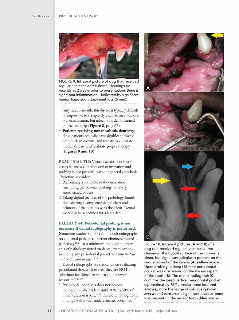
• Patients receiving nonanesthesia dentistry; these patients typically have significant disease despite clean crowns, and test strips elucidate hidden disease and facilitate proper therapy (Figures 9 and 10).
PRACTICAL TIP: Visual examination is not accurate, and a complete oral examination and probing is not possible, without general anesthesia. Therefore, consider: 1. Performing a complete oral examination
(including periodontal probing) on every anesthetized patient
2. Taking digital pictures of the pathology found; then sharing a completed dental chart and printout of the pictures with the client. Dental work can be scheduled for a later date.
FALLACY #4. Periodontal probing is not necessary if dental radiography is performed.Numerous studies support full-mouth radiographs on all dental patients to further eliminate missed pathology.22-24 At a minimum, radiograph every area of pathology noted on dental examination, including any periodontal pocket > 3 mm in dogs and > 0.5 mm in cats.12,15-17
Dental radiographs are critical when evaluating periodontal disease; however, they are NOT a substitute for clinical examination for several reasons.15,19,25-29
1. Periodontal bone loss does not become radiographically evident until 30% to 50% of mineralization is lost;25,30 therefore, radiographic fi ndings will always underestimate bone loss.15,31
Figure 10. Intraoral pictures (A and B) of a dog that received regular anesthesia-free cleanings; the buccal surface of the crowns is clean, but signifi cant calculus is present on the lingual aspect of the canine (A, yellow arrow). Upon probing, a deep (16-mm) periodontal pocket was discovered on the mesial aspect of the tooth (B). The dental radiograph (C) confi rms the deep vertical periodontal pocket (approximately 75% alveolar bone loss, red arrows); note the ledge of calculus (yellow arrow) and concurrent signifi cant alveolar bone loss present on the incisor teeth (blue arrow).
FIGURE 9. Intraoral picture of dog that received regular anesthesia-free dental cleanings (as recently as 2 weeks prior to presentation); there is signifi cant infl ammation—indicated by signifi cant hemorrhage and attachment loss (6 mm).
A
B
C
tvpjournal.com | January/February 2015 | TODAY’S VETERINARY PRACTICE
PRACTICAL DENTISTRY Peer Reviewed
69
2. Dental radiographs are a 2-dimensional image of a 3-dimensional space; any overlying structures can easily obscure periodontal pockets (Figure 11).
3. The fi rst stage of furcation exposure (F1) is not evident radiographically.
4. Errors of angulation can greatly affect the radiographic appearance of alveolar bone loss.
PRACTICAL TIP: Expose full-mouth radiographs on all small breed (< 15 lb) dogs because: 1. Periodontal disease is more prevalent in these dogs2. They are more likely to have heavy calculus and
tight contacts that may complicate probing.
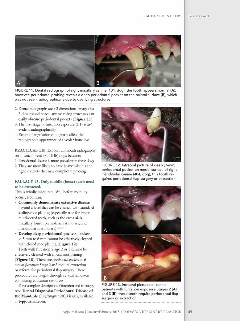
FALLACY #5. Only mobile (loose) teeth need to be extracted.This is wholly inaccurate. Well before mobility occurs, teeth can: • Commonly demonstrate extensive disease
beyond a level that can be cleaned with standard scaling/root planing (especially true for larger, multirooted teeth, such as the carnassials, maxillary fourth premolars/fi rst molars, and mandibular fi rst molars)3,32-35
• Develop deep periodontal pockets; pockets > 5 mm to 6 mm cannot be effectively cleaned with closed root planing (Figure 12).Teeth with furcation Stage 2 or 3 cannot be
effectively cleaned with closed root planing (Figure 13). Therefore, teeth with pockets > 6 mm or furcation Stage 2 or 3 require extraction or referral for periodontal fl ap surgery. These procedures are taught through several hands-on continuing education resources.
For a complete description of furcation and its stages, read Dental Diagnosis: Periodontal Disease of the Mandible (July/August 2013 issue), available at tvpjournal.com.
FIGURE 11. Dental radiograph of right maxillary canine (104, dog); the tooth appears normal (A); however, periodontal probing reveals a deep periodontal pocket on the palatal surface (B), which was not seen radiographically due to overlying structures.
A B
FIGURE 12. Intraoral picture of deep (9-mm) periodontal pocket on mesial surface of right mandibular canine (404, dog); this tooth re-quires periodontal fl ap surgery or extraction.
FIGURE 13. Intraoral pictures of canine patients with furcation exposure Stages 2 (A) and 3 (B); these teeth require periodontal fl ap surgery or extraction.
A
B
TODAY’S VETERINARY PRACTICE | January/February 2015 | tvpjournal.com
PRACTICAL DENTISTRYPeer Reviewed
70
PRACTICAL TIP: Educate your clients about the various treatments required, or available, for periodontal disease prior to the surgical appointment, and discuss cost. This avoids miscommunication and frustration related to clients’ decisions about dental procedures.
IN SUMMARYPeriodontal disease is a very common, but misunderstood disease process that has numerous deleterious consequences both regionally within the oral cavity and systemically.5,6 Utilization of the tools and information provided in this article will aid clinicians in diagnosing and treating this common but signifi cant disease process.
Read Proper Treatment of Periodontal Disease in an upcoming issue of Today’s Veterinary Practice.
References1. University of Minnesota Center for Companion Animal Health.
National companion animal study. Uplinks, 1996, p 3.2. Lund EM, Armstrong PJ, Kirk CA. Health status and population
characteristics of dogs and cats examined at private veterinary practices in the United States. JAVMA 1999; 214:1336-1341.
3. Wiggs RB, Lobprise HB. Periodontology. In Wiggs RB, Lobprise HB (eds): Veterinary Dentistry Principles and Practice. Philadelphia: Lippincott–Raven, 1997, pp 186-231.
4. Banfi eld Pet Hospital. State of Pet Health 2014 Report. Available at stateofpethealth.com.
5. Niemiec BA. Local and regional effects of periodontal disease. In Niemiec BA (ed): Veterinary Periodontology. Ames, IA: Wiley–Blackwell, 2012, pp 69-80.
6. Niemiec BA. Systemic manifestations of periodontal disease. In Niemiec BA (ed): Veterinary Periodontology. Ames, IA: Wiley–Blackwell, 2012, pp 80-91.
7. Hinrichs JE. The role of dental calculus and other predisposing factors. In Newman MG, Takei HH, Klokkevold PR, Carranza FA (eds): Carranza’s Clinical Periodontology. St. Louis: WB Saunders, 2006, pp 170-192.
8. Niemiec BA. Periodontal disease. Top Companion Anim Med 2008; 23(2):72-80.
9. Niemiec BA. Etiology and pathogenesis of periodontal disease. In Niemiec BA (ed): Veterinary Periodontology. Ames, IA: Wiley–Blackwell, 2012, pp 18-34.
10. Niemiec BA. Gingivitis. In Niemiec BA (ed): Veterinary Periodontology. Ames, IA: Wiley–Blackwell, 2012, pp 41-50.
11. Fiorellini JP, Ishikawa SO, Kim DM. Clinical features of gingivitis. In Newman MG, Takei HH, Klokkevold PR, Carranza FA (eds): Carranza’s Clinical Periodontology. St. Louis: WB Saunders, 2006, pp 362-372.
12. Debowes LJ. Problems with the gingiva. In Niemiec BA (ed): Small Animal Dental, Oral and Maxillofacial Disease—A Colour Handbook. London: Manson, 2010, pp 159-181.
13. Meitner SW, Zander HA, Iker HP, Polson AM. Identifi cation of infl amed gingival surfaces. J Clin Periodontol 1979; 6(2):93-97.
14. Amato R, Caton J, Polson A, Espeland M. Interproximal gingival infl ammation related to the conversion of a bleeding to a nonbleeding state. J Periodontol 1986; 57(2):63-68.
15. Niemiec BA. Veterinary dental radiology. In Niemiec BA (ed): Small Animal Dental, Oral and Maxillofacial Disease, A Colour Handbook. London: Manson, 2010, pp 63-87.
16. Wiggs RB, Lobprise HB. Oral exam and diagnosis. In Wiggs RB, Lobprise HB (eds): Veterinary Dentistry, Principles and Practice. Philadelphia: Lippincott–Raven, 1997, pp 87-103.
17. Bellows J. Periodontal equipment, materials, and techniques.
Small Animal Dental Equipment, Materials, and Techniques: A Primer. Ames, IA: Blackwell, 2004, pp 115-173.
18. Carranza FA, Takei HH. Clinical diagnosis. In Newman MG, Takei HH, Klokkevold PR, Carranza FA (eds): Carranza’s Clinical Periodontology. St. Louis: WB Saunders, 2006, pp 540-560.
19. Tetradis S, Carranza FA, Fazio RC, Takei HH. Radiographic aids in the diagnosis of periodontal disease. In Newman MG, Takei HH, Klokkevold PR, Carranza FA (eds): Carranza’s Clinical Periodontology. St. Louis: WB Saunders, 2006, pp 561-578.
20. Niemiec BA. Advanced non-surgical therapy. In Niemiec BA (ed): Veterinary Periodontology. Ames, IA: Wiley–Blackwell, 2012, pp 154-169.
21. Manfra Maretta S, Leesman M, Burgess-Cassler A, et al. Pilot evaluation of a novel test strip for the assessment of dissolved thiol levels, as an indicator of canine gingival health and periodontal status. Can Vet J 2012; 53(12):1260-1265.
22. Tsugawa AJ, Verstraete FJ. How to obtain and interpret periodontal radiographs in dogs. Clin Tech Small Anim Pract 2000; 15(4):204-210.
23. Verstraete FJ, Kass PH, Terpak CH. Diagnostic value of full-mouth radiography in cats. Am J Vet Res 1998; 59(6):692-695.
24. Verstraete FJ, Kass PH, Terpak CH. Diagnostic value of full-mouth radiography in dogs. Am J Vet Res 1998; 59(6):686-691.
25. Niemiec BA. Case based dental radiology. Top Companion Anim Med 2009; 24(1):4-19.
26. Ramadan AB, Mitchell DF. A roentgenographic study of experimental bone destruction. Oral Surg Oral Med Oral Pathol 1962; 15:934-943.
27. Bender IB, Seltzer S. Roentgenographic and direct observation of experimental lesions in bone: I. 1961. J Endod 2003; 29(11):702-706.
28. Bender IB, Seltzer S. Roentgenographic and direct observation of experimental lesions in bone: II. 1961. J Endod 2003; 29(11):707-712.
29. Gawor JG. Dental radiology for periodontal disease. In Niemiec BA (ed): Veterinary Periodontology. Ames, IA: Wiley–Blackwell, 2012, pp 107-128.
30. Mulligan TW, Aller MS, Williams CA. Interpretation of periodontal disease. In Mulligan TW, Aller MS (eds): Atlas of Canine and Feline Dental Radiography. Trenton, NJ: Veterinary Learning Systems, 1998, pp 104-123.
31. Rees TD, Biggs NL, Collings CK. Radiographic interpretation of periodontal osseous lesions. Oral Surg Oral Med Oral Pathol 1971; 32(1):141-153.
32. Carranza FA, Takei HH. Phase II periodontal therapy. In Newman MG, Takei HH, Klokkevold PR, Carranza FA (eds): Carranza’s Clinical Periodontology. St. Louis: WB Saunders, 2006, pp 881-886.
33. Perry DA, Schmid MO, Takei HH. Phase I periodontal therapy. In Newman MG, Takei HH, Klokkevold PR, Carranza FA (eds): Carranza’s Clinical Periodontology. St. Louis: WB Saunders, 2006, pp 722-727.
34. Caffesse RG, Sweeney PL, Smith BA. Scaling and root planing with and without periodontal fl ap surgery. J Clin Periodontol 1986; 13(3):205-210.
35. Holmstrom SE, Frost P, Eisner ER. Periodontal therapy and surgery. Veterinary Dental Techniques: for the Small Animal Practitioner, 2nd ed. Philadelphia: Saunders, 1998, pp 167-213.
BROOK A. NIEMIECBrook A. Niemiec, DVM, FAVD, Diplomate AVDC, is Chief of Staff of Southern California Veterinary Dental Specialties, with offi ces in San Diego and Murrieta, California, and Las Vegas, Nevada. He lectures extensively at national and international conferences. He received his DVM from University of California–Davis.