Embed Size (px)
Citation preview


Tutorial 12Sections 009/010
TA: Greydon GilmorePhysiology 2130
Dec 3rd, 2019

Your TA reminding you…
• 2nd Quiz (1%)• Opens: Dec 2nd @ 4pm• Closes: Dec 4th @ 4pm
• 2nd Midterm (15%)• When: Dec 19th @ 9am-10am• Room Assignments:
• ABBA-GANE: Alumni Hall 15• GHAB-POSA: Alumni Hall 201• PRIM-WOOD: Alumni Hall Stage• WU-ZIA: Somerville House 2316
• 2nd Midterm review session• When: Monday Dec 16th from 6-8pm • Where: Auditorium B University Hospital, 3rd floor

Today
• Group work activity
• Learning Catalytics Question
• Almost finish cardiovascular anatomy

Group Work

Crossword Puzzle!

Learning Catalytic Question

The Cardiovascular System:Mechanical Performance of the Heart
Chapter 7: Professor Stavraky

Heart Rate (HR)
• Average: 70 bpm
• Lower HR = “healthier” (i.e. athletes: 45 bpm)
• Max HR = 220 – age
• Controlled by autonomic nervous system➢ PS-NS: decreases HR
➢ S-NS: increases HR

Stroke Volume = EDV -ESV
• End-Diastolic Volume (EDV): volume of blood in ventricles at end of ventricular diastole (just before they contract; end of Phase 1)
• End-Systolic Volume (ESV): volume of blood in ventricles at end of ventricular systole (just after contraction; end of Phase 3)
• Stroke volume = EDV –ESV
= 160 ml –90 ml
= 70 ml
• Altering either EDV or ESV will change stroke volume
Cardiac output can be determined by which of the following formulas?
A. HR – SV
B. HR divided by SV
C. HR + SV
D. HR x SV
Cardiac output can be determined by which of the following formulas?
A. HR – SV
B. HR divided by SV
C. HR + SV
D. HR x SV

Cardiac Output (CO)• Volume of blood pumped by each ventricle per minute
• CO = Heart Rate x Stroke Volume• Heart Rate = Beats per minute• Stroke Volume = Amount of blood pumped by each ventricle per beat
• At rest:• CO = 5 L/min• HR = 70 beat/min • SV = 70-80 mL/beat • CO = (70 beat/min)(0.07 L/beat) = 4.9 L/min
• During exercise:• CO can increase to 20-40 L/min • How? By changing HR and/or SV!

Which of the following is INCORRECT regarding diastole (filling of the heart)?
a. Atrioventricular valves are open.
b. Semilunar valves are closed.
c. Blood is flowing from the atria into the ventricles.
d. Pressure in the ventricles is greater than in the atria.

Which of the following is INCORRECT regarding diastole (filling of the heart)?
a. Atrioventricular valves are open.
b. Semilunar valves are closed.
c. Blood is flowing from the atria into the ventricles.
d. Pressure in the ventricles is greater than in the atria.

Overall Control of SV by ANS
• Stroke volume is amount of blood pumped by each ventricle per beat
• Two factors that affect stroke volume:➢ ANS➢ Preload (End diastolic volume)
• PS-NS decreases SV➢ Ca2+ flow into cardiac cells➢ force of contraction
• S-NS increases SV➢ Ca2+ flow into cardiac cells➢ force of contraction

Stroke Volume
• During exercise, the S-NS is activated:➢ Heart contracts more forcefully and ejects
more blood
➢ Thus, ESV decreases
• Meanwhile, the heart is filling with more blood➢ Thus, EDV increases

Stroke Volume and Preload
• Preload: The “load” on the cardiac muscle before contraction
• This “load” comes from the blood in the ventricles that stretches the ventricular muscle➢ Thus, higher EDV = greater preload

PNS Effect on HR
• PNS innervates SA and AV nodes through vagus nerve➢ PNS releases Ach, which binds to
receptors on cells of SA and AV nodes
• K+ permeability (i.e. more exits cell) and Ca2+ permeability (i.e. less enters cell)
• Net effect: ➢ K+ = HYPERPOLARIZATION➢ Ca2+ = Decreases slope of pacemaker
potential

SNS Effect on HR• SNS innervates SA, AV nodes and
ventricular muscles➢ SNS releases NE, which binds to
receptors on cells of nodes and muscle
• Na+ and Ca2+ permeability (i.e. more enters cell)
• Net effect: DEPOLARIZATION and increased slope of pacemaker potential

Which graph represents sympathetic influence on heart rate (in both cases the
light grey line is under resting conditions)?
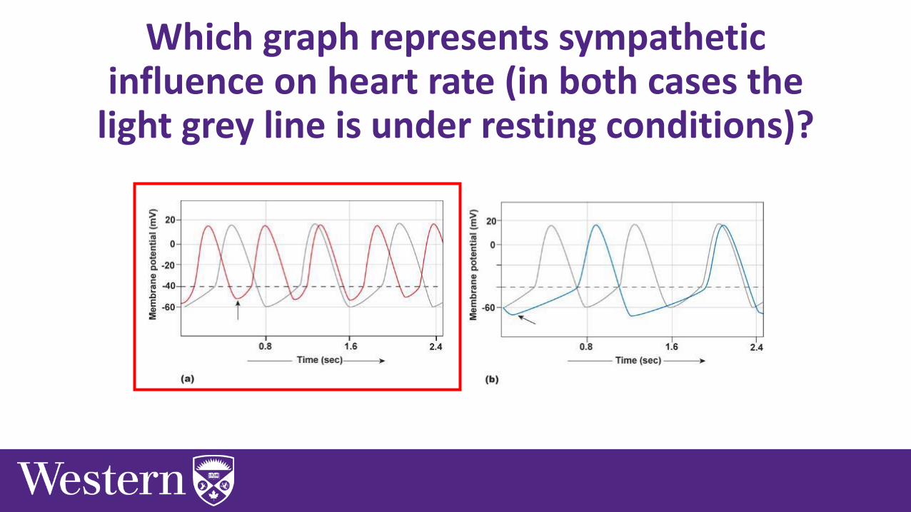
Which graph represents sympathetic influence on heart rate (in both cases the
light grey line is under resting conditions)?

Summary of ANS Control of Heart Rate
PSNS• Acetylcholine released onto these areas
➢ Increase K+, decrease Ca2+ permeabilities➢ Decreases slope of pacemaker potential
SNS• Release norepinephrine onto these areas (indirect:
epinephrine)• Increases heart rate and force of contraction
➢ Increase Na and Ca permeability➢ Increase slope of pacemaker potential

Frank-Starling Law
An increase in EDV = An increase in preload
Increases the stretch of myocardial cells
Increases the force of contraction of these cells when the heart contracts
Increases the amount of blood ejected
Increases Stroke Volume (Increases Cardiac Output)
• Frank-Starling Law states that “an increase in EDV will increase stroke volume”

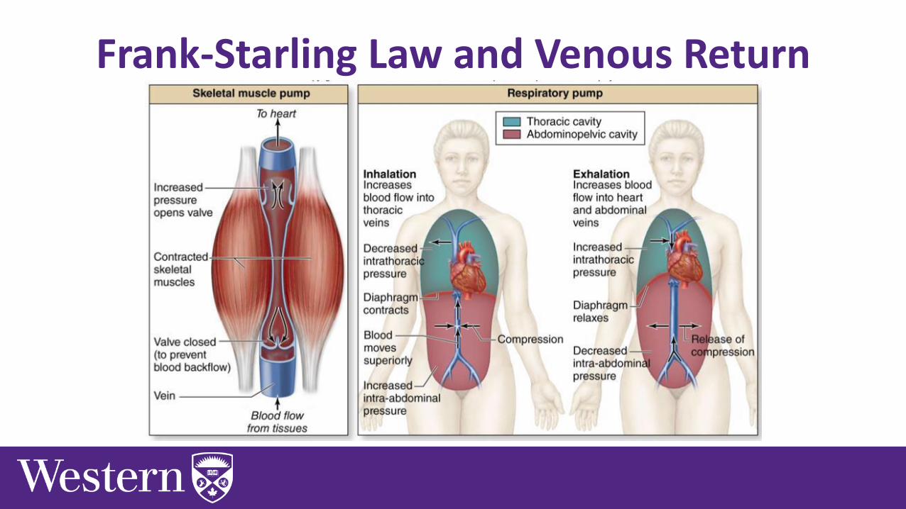
Frank-Starling Law and Venous Return
• How to increase EDV? Increase venous return to the heart!
• During dynamic exercise:
1. Muscle Pump: Contracted skeletal muscle around veins pushes blood to heart
2. Respiratory Pump: Changes in pressure during breathing pushes blood towards the heart
3. S-NS: Constriction of veins squeezes blood to heart
Frank-Starling Law and Venous Return

The aortic semilunar valve prevents blood from returning to the _____.
A. left ventricle
B. Aorta
C. Right ventricle
D. Left atrium

The aortic semilunar valve prevents blood from returning to the _____.
A. left ventricle
B. Aorta
C. Right ventricle
D. Left atrium

The Cardiovascular System:Vascular Function
Chapter 7: Dr. Stavraky

Blood Vessels
• Structural properties of vessels are what contribute to the blood pressure characteristics seen in circulation

Vessel Constriction and Blood Flow
• As the radius decreases the pressure gradient increases.

Relationship Between Pressure, Flow and Resistance (Page 232)
𝑹𝒆𝒔𝒊𝒔𝒕𝒂𝒏𝒄𝒆 =𝑳 𝜼
𝒓𝟒
𝑹𝒆𝒔𝒊𝒔𝒕𝒂𝒏𝒄𝒆 =𝟏
𝒓𝟒
𝑩𝒍𝒐𝒐𝒅 𝑭𝒍𝒐𝒘 =𝑷𝟏−𝑷𝟐
𝟏
𝒓𝟒
= 𝑷𝟏 − 𝑷𝟐 ∗ 𝒓𝟒
L = length of vessel𝜂 = viscosity of the fluidr = radius of the vessel
A small change in radius will have a LARGE effect on blood flow
Just know this part of the equation

Relationship Between Pressure, Flow and Resistance
𝑩𝒍𝒐𝒐𝒅 𝑭𝒍𝒐𝒘 = 𝑷𝟏 − 𝑷𝟐 ∗ 𝒓𝟒
𝑩𝒍𝒐𝒐𝒅 𝑭𝒍𝒐𝒘 = 𝟒 − 𝟐 ∗ 𝟏𝟒
𝑩𝒍𝒐𝒐𝒅 𝑭𝒍𝒐𝒘 = 𝟐 L/min
𝑩𝒍𝒐𝒐𝒅 𝑭𝒍𝒐𝒘 = 𝑷𝟏 − 𝑷𝟐 ∗ 𝒓𝟒
𝑩𝒍𝒐𝒐𝒅 𝑭𝒍𝒐𝒘 = 𝟏𝟎 − 𝟐 ∗ 𝟎. 𝟓𝟒
𝑩𝒍𝒐𝒐𝒅 𝑭𝒍𝒐𝒘 = 𝟎. 𝟓 L/min
A small change in radius will have a LARGE effect on blood flow

Defining terms• Blood velocity (cm/sec): speed at which
blood is moving through particular blood vessel• Fluid flows faster through a narrow tube
than a larger tube
• As cross sectional area increases mean velocity decreases
• Blood flow (L/min): volume of blood moving through set of vessels.

Overall blood flow does not change• Blood velocity can change but total blood flow needs to
remain constant• If you have 5L of blood you can’t add or subtract… unless you have a wound

Arteries and Veins• Contain three layers:➢ Outer Layer – Tunica externa
▪ Fibrous connective tissue
➢ Middle Layer – Tunica media▪ Smooth muscle and elastic tissue
➢ Inner Layer – Tunica interna▪ Endothelial cells
• Veins contain valves
• Capillaries have single layer of endothelial cells

Aorta and Large ArteriesBlood Characteristics Structure Purpose
Aorta/Large Arteries
- High blood pressure- 80-120 mmHg- High blood velocity
- Large diameter- Elastic tissue- Thin walls▪ Easily distended▪ Low resistance to blood flow▪ Small drop in blood pressure
- ‘Shock absorbers’- Distribute the blood

CapillariesBlood Characteristics Structure Purpose
Capillaries
- Low blood pressure- Small drop in blood
pressure- Very low blood velocity (1-
2 cm/sec)
- One endothelial cell thick- Large cross sectional area- Very large surface area▪ Diffusion of gas, nutrients and
waste
- Exchange vessels

VeinsBlood Characteristics Structure Purpose
Veins
- Low blood pressure- Low to medium blood
velocity (5-10 cm/sec)
- Very thin walls with large diameter
- Contain valves- Some elastic tissue- Smoth of smooth muscle
innervated by ANS▪ Vasoconstriction/dilation
- Capacitance vessels: 70% of TBV

Starling Forces• Two hydrostatic pressures➢ Capillary hydrostatic pressure
➢ Interstitial fluid hydrostatic pressure
• Two osmotic pressures➢ Plasma osmotic pressure
➢ Interstitial osmotic pressure

Exchange In Capillaries• Diffusion
➢ Down concentration gradients
➢ Oxygen, CO2, O2, lipid soluble substances
• Filtration and reabsorption (Starling forces)➢ Filtration: movement of fluid out of capillary
➢ Reabsorption: movement of fluid back into capillary

Capillary Hydrostatic Pressure (Pc)• Pressure exerted by fluid in the capillary
• Pressure drives fluid OUT of capillary and is generated by ventricular systole (Filtration)
25 mmHg10 mmHg
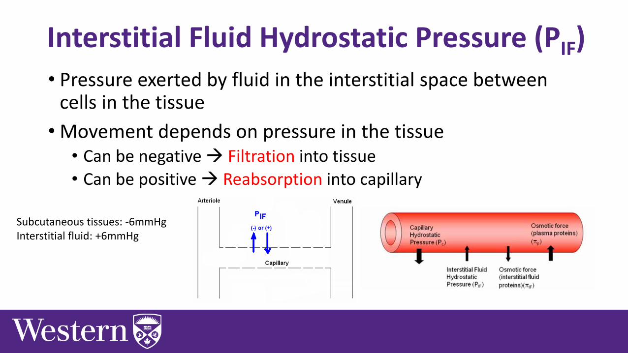
Interstitial Fluid Hydrostatic Pressure (PIF)• Pressure exerted by fluid in the interstitial space between
cells in the tissue
• Movement depends on pressure in the tissue• Can be negative → Filtration into tissue
• Can be positive → Reabsorption into capillary
Subcutaneous tissues: -6mmHgInterstitial fluid: +6mmHg

Interstitial Osmotic Pressure (𝝅IF)• Pressure caused by osmosis due to few proteins in interstitial
fluid (5mmHg)
• Pressure drives fluid OUT of capillary and into tissue (Filtration)

Plasma Osmotic Pressure (𝝅P)• Pressure caused by osmosis due to proteins in plasma
(28mmHg)
• Pressure drives fluid INTO capillary (Reabsorption)

Balance of Starling Forces• Starling-Landis equation used to calculate net
fluid movement (NFM) across capillary bed𝑁𝐹𝑀 = 𝐾𝑓 𝑃𝑐 − 𝑃𝐼𝐹 − 𝜋𝑃 − 𝜋𝐼𝐹
• 𝐾𝑓 is filtration coefficient, which represents permeability of capillary (assume 1)𝑁𝐹𝑀 = 1 25 − (−6) − 28 − (+5)𝑁𝐹𝑀 = +8𝑚𝑚𝐻𝑔
• If positive filtration OUT of capillary, if negative reabsorption INTO capillary

PC = 10, PIF = 1, πIF = 5, πC = 28
•𝑁𝐹𝑀 = 𝐾𝑓 𝑃𝑐 − 𝑃𝐼𝐹 − 𝜋𝑃 − 𝜋𝐼𝐹•𝑁𝐹𝑀 = 1 10 − (1) − 28 − 5
•𝑁𝐹𝑀 = 1 9 − 23
•𝑁𝐹𝑀 = −14
• Thus, reabsorption into plasma from interstitial fluid

Next Tutorial (Jan 14th)
• Have a great New Year!

What Questions Do You Have?
You can ask in the Owl forums as well!
Also anonymously ask questions in the online dropbox!!