Embed Size (px)
Citation preview
Tumorigenesis in the multiple intestinal neoplasiamouse: Redundancy of negative regulators andspecificity of modifiersRichard B. Halberg*, Darren S. Katzung*, Peter D. Hoff†, Amy R. Moser*, Carolyn E. Cole‡, Ronald A. Lubet§,Lawrence A. Donehower¶, Russell F. Jacoby‡, and William F. Dove*i**
*McArdle Laboratory for Cancer Research, †Department of Biostatistics, and iLaboratory of Genetics, University of Wisconsin, Madison, WI 53706;‡University of Wisconsin Comprehensive Cancer Center, Madison, WI 53792; §Chemoprevention Branch, Division of Cancer Prevention,National Cancer Institute, Bethesda, MD 20892; and ¶Department of Molecular Virology and Microbiologyand Department of Molecular and Cellular Biology, Baylor College of Medicine, Houston, TX 77030
Contributed by William F. Dove, December 31, 1999
The interaction between mutations in the tumor-suppressor genesApc and p53 was studied in congenic mouse strains to minimize theinfluence of polymorphic modifiers. The multiplicity and invasive-ness of intestinal adenomas of ApcMin/1 (Min) mice was enhancedby deficiency for p53. In addition, the occurrence of desmoidfibromas was strongly enhanced by p53 deficiency. The geneticmodifier Mom1 and the pharmacological agents piroxicam anddifluoromethylornithine each reduced intestinal adenoma multi-plicity in the absence of p53 function. Mom1 showed no influenceon the development of desmoid fibromas, whereas the combina-tion of piroxicam and difluoromethylornithine exerted a moderateeffect. The ensemble of tumor suppressors and modifiers of aneoplastic process can be usefully analyzed in respect to tissuespecificity and synergy.
Genes of several kinds regulate normal and neoplastic growthin the mammal. Positive and negative regulators can act
either cell autonomously or nonautonomously to alter the pro-liferative potential of the stem cell that establishes the normal orneoplastic lineage. Individual genes in the ensemble can affectthe transition between normal and neoplastic growth andyor themaintenance of one of the growth states. One important class ofgenes has been identified by loss-of-function alleles transmittedthrough the germline in heterozygous form, each predisposing acarrier to a particular spectrum of neoplasms. In these tumors,it is commonly found that the remaining wild-type allele hasbeen lost or inactivated. The gene in question, commonly calleda ‘‘tumor suppressor,’’ is formally a cell-autonomous negativeregulator of the neoplastic state.
Humans and mice heterozygous for germline mutations thatinactivate the Adenomatous polyposis coli (APCyApc) gene de-velop only a limited range of neoplasms, including intestinaladenomas and desmoid fibromas (1). Because this gene is widelyexpressed (2), this restriction in neoplastic histotype is surpris-ing. Does the limited range of neoplasia reflect cooperationbetween APCyApc and other negative regulators?
The p53 gene is a negative regulator that is mutated in a broadrange of human neoplasms, but its gene product is constitutivelyactive in only a few cell types (3, 4). For example, the loss of p53adenocarcinomas in the human intestine, but the protein be-comes detectable in this tissue after stress such as ionizingradiation (5). Does an interaction between Apc and p53 affectintestinal neoplasia in experimental models of cancer (6)?Preliminary studies tested whether a lack of p53 activity affectedneoplasia in the intestine of Min (multiple intestinal neoplasia)mice heterozygous for the Min nonsense allele of Apc, but nosignificant effect was observed (7–10). Those studies utilizedpopulations of mice with heterogeneous genetic backgrounds. Inthis report, we reinvestigate whether a tissue-specific or stage-specific interaction between Apc and p53 affects the spectrum ofneoplasms in mice, by using a homogeneous genetic background.
A central issue addressed in these experiments is whether themajor negative regulators each act singly in controlling neoplasiaof a particular histotype, as implied by the ‘‘gatekeeper hypoth-esis’’ (11).
Beyond these major negative regulators, a number of otherfactors have been found to inf luence intestinal neoplasia inhumans and mice. Such modifiers can act to either promote orretard tumor growth, thereby affecting tumor multiplicity inexperimental models. The genetic modifier Mom1 encodes asecretory phospholipase, Pla2g2a, expressed throughout theintestinal tract. The active allele of Pla2g2a leads to a reductionin the growth rate and multiplicity of intestinal adenomas inthe Min mouse. Similarly, pharmacological agents can affecttumorigenesis. Piroxicam, a nonsteroidal antiinf lammatoryagent that inhibits cyclooxygenase-1 and -2, and dif luorometh-ylornithine (DFMO), a suicide substrate of ornithine decar-boxylase, each reduce the multiplicity of intestinal adenomas.The strongest inhibition is seen with a combination of piroxi-cam and DFMO (12). In the present study, we have askedwhether the action of either Mom1 or the piroxicamyDFMOcombination is tissue specific. Further, we have asked whethereither of these growth-inhibitory actions depends on p53activity.
Materials and MethodsMice. The C57BLy6–p53 congenic mouse strain (B6-p53) wasfounded by a (129ySv 3 B6) F2 female that carried a targeteddisruption of the p53 gene (13). Designating the founder femaleas N1, an expanded N10 intercross population was produced bymating Apc1/1 Mom1R/S p531/2 females to ApcMin/1 Mom1R/S
p531/2, ApcMin/1 Mom1R/S p532/2, and ApcMin/1 Mom1R/R
p532/2 males. Mice were housed as described previously (14).
Drug Treatment. After weaning at approximately 30 days of age,animals were housed in groups of one to five in microisolatorcages under fluorescent lighting on a 12-hr cycle and weighedonce per week. Tap water was available ad libitum for theduration of the experiment and was replaced weekly. The micewere treated with the appropriate drug or control vehicle mixedin the defined synthetic AIN-93G diet (Dyets, Bethlehem, PA),then killed after the specified duration of treatment.
The chemoprotective agents were stable for at least 7 days ina standard rodent diet at the concentrations used in these
Abbreviation: DFMO, difluoromethylornithine.
**To whom reprint requests should be addressed. E-mail: [email protected].
The publication costs of this article were defrayed in part by page charge payment. Thisarticle must therefore be hereby marked “advertisement” in accordance with 18 U.S.C.§1734 solely to indicate this fact.
Article published online before print: Proc. Natl. Acad. Sci. USA, 10.1073ypnas.050585597.Article and publication date are at www.pnas.orgycgiydoiy10.1073ypnas.050585597
PNAS u March 28, 2000 u vol. 97 u no. 7 u 3461–3466
MED
ICA
LSC
IEN
CES
studies. New batches were prepared weekly by thorough mixingof the diet with the indicated doses of drug and were stored untiluse in sealed containers at 4°C. Fresh diet was added to protectedfeeders three times weekly and was completely changed after thefeeders were emptied once weekly.
Piroxicam (CAS no. 36322–90-4) was purchased from Sigma,and DFMO (CAS no. 70052–12-9) was a gift from Ajit Verma(University of Wisconsin–Madison). Piroxicam at the intendedconcentration was mixed in the diet beginning at approximately30 days of age; the mice were killed at age 90 days after 2 mo oftreatment. Mice treated with DFMO were given water mixedwith the intended concentration of that drug from age 30 to 90days. When administered to mice after weaning, these cycloox-ygenase and decarboxylase inhibitors were not overtly toxic atthe doses used.
DNA Preparation. Mice were anesthetized with ether, and 250 mlof blood was collected from the retroorbital sinus. DNA wasisolated from blood as described previously (14).
Genotyping. Mice were genotyped to identify carriers of the Minallele of Apc, the resistance allele of Mom1, and the knockoutallele of p53 with PCR assays as described (14–16). In somecases, tail DNA was prepared from neonates to verify indepen-dently the p53 genotype by Southern blot analysis, as describedpreviously (13).
Tumor Counts. All mice were killed at 90 days by CO2 asphyxia-tion. The intestinal tract was removed, washed with PBS, openedlongitudinally, and laid out as described previously (6). In thismethod, 4-cm sections from the proximal, medial, and distalregions of the small intestine and the entire colon were exam-ined. The number of tumors was scored with a dissectingmicroscope by a single observer blind to the genotype of the miceor their treatment group. The samples were then fixed in 10%buffered formalin, washed in 70% ethanol, and stored in thissolution. Tumor counts were verified by a second observer in asubset of the postfixed samples.
At sacrifice, the number of desmoid fibromas was scored bya single observer blind to the genotype of the mice or theirtreatment group. The abdominal body wall was then removed,stretched, fixed overnight in 10% formalin, washed in 70%ethanol, and stored in this solution. Tumor counts wereverified by a second observer for a subset of the postfixedsamples. To facilitate scoring, the tissue was stained with Fastgreen, a dye that detects collagen. Samples were incubated for30 min each in the following series: distilled water; acetatebuffer (1 M acetic acidy7.2 mM sodium acetatey0.25% for-malin); 5% (wtyvol) phosphotungstic acid in distilled water;Fast green [1 M acetic acidy110 mM sodium acetatey0.05%(wtyvol) Fast green]; acetate buffer; 10 mM sulfuric acid; 50%ethanol; and 70% ethanol.
Histological Analysis of Intestinal Tumors. The largest tumors wereisolated from sections of the small intestine and the entire colon.After fixation in 10% buffered formalin, the tumors wereembedded in paraffin, sectioned, and stained with hematoxylinand eosin. Sections were analyzed by light microscopy for signsof progression by H. C. Pitot (McArdle Lab), also blind to thegenotype of the samples.
Histological Analysis of Desmoid Fibromas. Desmoid fibromas wereisolated from postfixed abdominal body walls. The tumors wereembedded in paraffin, sectioned, and stained with either hema-toxylin and eosin or Masson’s trichrome.
Resultsp53 Effect on Multiplicity of Intestinal Adenomas. Does a lack of p53activity affect the multiplicity of adenomas in the intestine of theMin mouse? Min mice homozygous for a null allele of p53developed significantly more intestinal adenomas than thosehomozygous for the wild-type allele of p53, regardless of theMom1 genotype (Table 1). Thus, p53 negatively regulates thedevelopment of adenomas in the intestine of the Min mouse.
Min mice heterozygous for a null allele of p53 (ApcMin/1
p531/2) also develop more tumors than those homozygous forthe wild-type allele of p53 (Table 1). The difference is at theborderline of statistical significance for two of the Mom1classes.
One interpretation for the effect of the p53 genotype is thata polymorphic modifier, linked to the p53 locus, affects thedevelopment of intestinal adenomas in the Min mouse (14). Inderiving the B6-p53 congenic animals, a region from chromo-some 11 of the 129 genome was moved onto the B6 geneticbackground. We have tested this hypothesis and found noevidence for such a modifier. The extent of the 129ySvEvgenome carried by the p53 mutant line was assessed bygenotyping Min mice heterozygous for a null allele of p53 withmarkers distributed along chromosome 11. This analysis indi-cated that the introgressed region of the 129ySvEv genome inthe B6-p53 congenic strain extends at least from 19.7 cM to thep53 locus at 39 cM on chromosome 11. To test whether apolymorphic modifier was present in this region of the 129genome, B6 females were mated to (129ySvPas 3 B6)F1 Minmales. At least 20 resulting progeny were scored for thenumber of adenomas and genotyped with markers from chro-mosome 11. Tumor multiplicity was comparable between Minmice that were heterozygous for the 129ySvPas allele and thosethat are homozygous for the B6 allele at each locus (data notshown). Thus, the heterozygous effect of the p53 genotypecannot be explained by a dominant polymorphic modifier ofintestinal tumorigenesis that is linked to the p53 locus.
p53 Effect on the Progression of Intestinal Adenomas. Mutation ofp53 has been correlated with the progression of tumors in thehuman colon (17). To investigate whether a lack of p53 functionenhances tumor progression in Min mice, the largest tumors
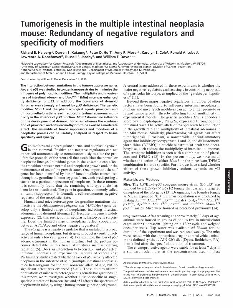
Table 1. Effect of p53 on intestinal tumor multiplicity in Min mice
Mom1S/S Mom1R/S Mom1R/R Treated*
N Tumor count P-value† N Tumor count P-value† N Tumor count P-value† N Tumor count P-value‡
p531/1 20 32 6 11 NA 20 13 6 5 NA 19 5 6 4 NA 5 5 6 2 0.0007p531/2 19 37 6 9 0.072 21 15 6 7 0.403 20 8 6 4 0.053 10 6 6 4 0.00001p532/2 21 45 6 13 0.0014 14 26 6 8 0.00002 16 13 6 6 0.00008 4 12 6 8 0.003
Mice were killed at 90 days of age. Tumor counts are shown as means 6 SD.*Mom1S/S mice were treated with piroxicam and DFMO (see Materials and Methods).†P-values were calculated by using two-sided Wilcoxon rank sum tests compared with p531/1 mice of the same Mom1 genotype.‡P-values were calculated by using two-sided Wilcoxon rank sum tests compared with untreated mice of the same p53 genotype.
3462 u www.pnas.org Halberg et al.
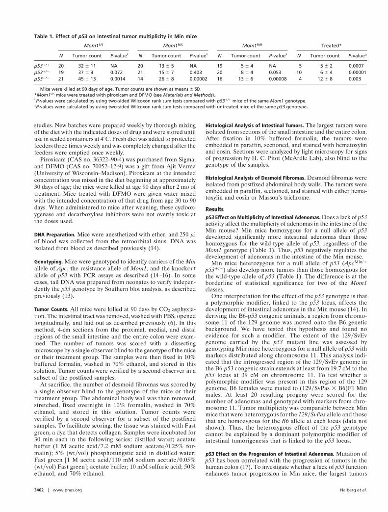
Fig. 1. Intestinal tumors exhibit signs of progression in ApcMin/1 p532/2 mice. Tumors were isolated from ApcMin/1 Mom1S/S mice carrying zero, one, or two copiesof the p53 knockout allele and stained with hematoxylin and eosin. Most tumors from the small intestine and colon of p53-deficient mice were typical adenomas(A and D, respectively). However, signs of progression were exhibited by some of the tumors isolated from the proximal region of the small intestine and thecolon (B and E, respectively). The regions that exhibited signs of progression are boxed in B and E and are shown at higher magnification in C and F. Themagnification was the same for A, B, D, and E, with the scale bar in A representing 1 mm. C and F are 34 of B and E, respectively.
Halberg et al. PNAS u March 28, 2000 u vol. 97 u no. 7 u 3463
MED
ICA
LSC
IEN
CES
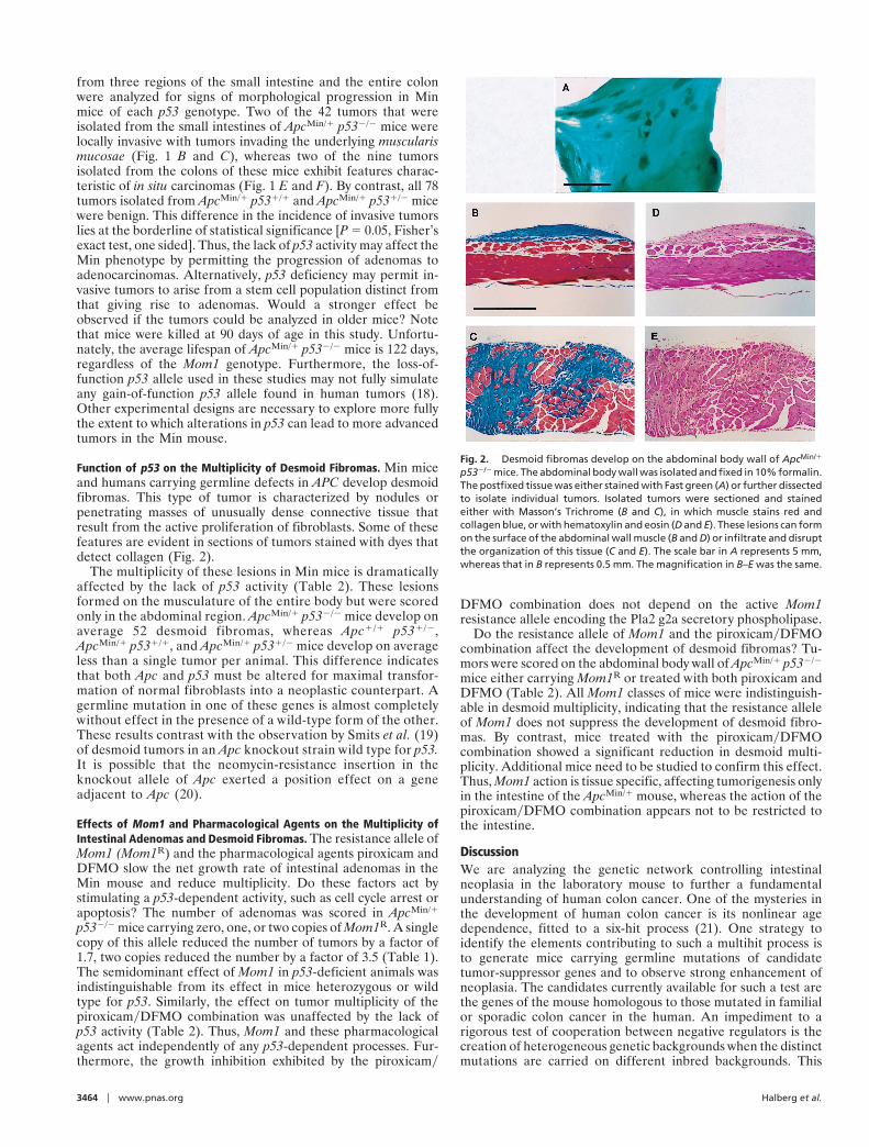
from three regions of the small intestine and the entire colonwere analyzed for signs of morphological progression in Minmice of each p53 genotype. Two of the 42 tumors that wereisolated from the small intestines of ApcMin/1 p532/2 mice werelocally invasive with tumors invading the underlying muscularismucosae (Fig. 1 B and C), whereas two of the nine tumorsisolated from the colons of these mice exhibit features charac-teristic of in situ carcinomas (Fig. 1 E and F). By contrast, all 78tumors isolated from ApcMin/1 p531/1 and ApcMin/1 p531/2 micewere benign. This difference in the incidence of invasive tumorslies at the borderline of statistical significance [P 5 0.05, Fisher’sexact test, one sided]. Thus, the lack of p53 activity may affect theMin phenotype by permitting the progression of adenomas toadenocarcinomas. Alternatively, p53 deficiency may permit in-vasive tumors to arise from a stem cell population distinct fromthat giving rise to adenomas. Would a stronger effect beobserved if the tumors could be analyzed in older mice? Notethat mice were killed at 90 days of age in this study. Unfortu-nately, the average lifespan of ApcMin/1 p532/2 mice is 122 days,regardless of the Mom1 genotype. Furthermore, the loss-of-function p53 allele used in these studies may not fully simulateany gain-of-function p53 allele found in human tumors (18).Other experimental designs are necessary to explore more fullythe extent to which alterations in p53 can lead to more advancedtumors in the Min mouse.
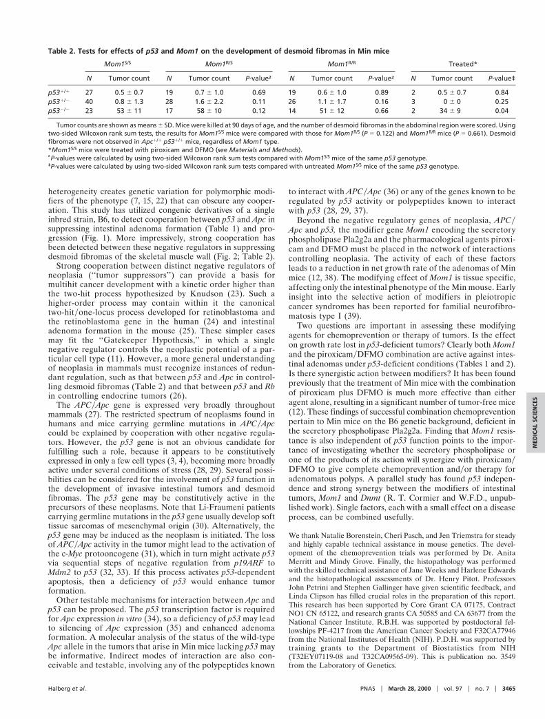
Function of p53 on the Multiplicity of Desmoid Fibromas. Min miceand humans carrying germline defects in APC develop desmoidfibromas. This type of tumor is characterized by nodules orpenetrating masses of unusually dense connective tissue thatresult from the active proliferation of fibroblasts. Some of thesefeatures are evident in sections of tumors stained with dyes thatdetect collagen (Fig. 2).
The multiplicity of these lesions in Min mice is dramaticallyaffected by the lack of p53 activity (Table 2). These lesionsformed on the musculature of the entire body but were scoredonly in the abdominal region. ApcMin/1 p532/2 mice develop onaverage 52 desmoid fibromas, whereas Apc1/1 p531/2,ApcMin/1 p531/1, and ApcMin/1 p531/2 mice develop on averageless than a single tumor per animal. This difference indicatesthat both Apc and p53 must be altered for maximal transfor-mation of normal fibroblasts into a neoplastic counterpart. Agermline mutation in one of these genes is almost completelywithout effect in the presence of a wild-type form of the other.These results contrast with the observation by Smits et al. (19)of desmoid tumors in an Apc knockout strain wild type for p53.It is possible that the neomycin-resistance insertion in theknockout allele of Apc exerted a position effect on a geneadjacent to Apc (20).
Effects of Mom1 and Pharmacological Agents on the Multiplicity ofIntestinal Adenomas and Desmoid Fibromas. The resistance allele ofMom1 (Mom1R) and the pharmacological agents piroxicam andDFMO slow the net growth rate of intestinal adenomas in theMin mouse and reduce multiplicity. Do these factors act bystimulating a p53-dependent activity, such as cell cycle arrest orapoptosis? The number of adenomas was scored in ApcMin/1
p532/2 mice carrying zero, one, or two copies of Mom1R. A singlecopy of this allele reduced the number of tumors by a factor of1.7, two copies reduced the number by a factor of 3.5 (Table 1).The semidominant effect of Mom1 in p53-deficient animals wasindistinguishable from its effect in mice heterozygous or wildtype for p53. Similarly, the effect on tumor multiplicity of thepiroxicamyDFMO combination was unaffected by the lack ofp53 activity (Table 2). Thus, Mom1 and these pharmacologicalagents act independently of any p53-dependent processes. Fur-thermore, the growth inhibition exhibited by the piroxicamy
DFMO combination does not depend on the active Mom1resistance allele encoding the Pla2 g2a secretory phospholipase.
Do the resistance allele of Mom1 and the piroxicamyDFMOcombination affect the development of desmoid fibromas? Tu-mors were scored on the abdominal body wall of ApcMin/1 p532/2
mice either carrying Mom1R or treated with both piroxicam andDFMO (Table 2). All Mom1 classes of mice were indistinguish-able in desmoid multiplicity, indicating that the resistance alleleof Mom1 does not suppress the development of desmoid fibro-mas. By contrast, mice treated with the piroxicamyDFMOcombination showed a significant reduction in desmoid multi-plicity. Additional mice need to be studied to confirm this effect.Thus, Mom1 action is tissue specific, affecting tumorigenesis onlyin the intestine of the ApcMin/1 mouse, whereas the action of thepiroxicamyDFMO combination appears not to be restricted tothe intestine.
DiscussionWe are analyzing the genetic network controlling intestinalneoplasia in the laboratory mouse to further a fundamentalunderstanding of human colon cancer. One of the mysteries inthe development of human colon cancer is its nonlinear agedependence, fitted to a six-hit process (21). One strategy toidentify the elements contributing to such a multihit process isto generate mice carrying germline mutations of candidatetumor-suppressor genes and to observe strong enhancement ofneoplasia. The candidates currently available for such a test arethe genes of the mouse homologous to those mutated in familialor sporadic colon cancer in the human. An impediment to arigorous test of cooperation between negative regulators is thecreation of heterogeneous genetic backgrounds when the distinctmutations are carried on different inbred backgrounds. This
Fig. 2. Desmoid fibromas develop on the abdominal body wall of ApcMin/1
p532/2 mice. The abdominal body wall was isolated and fixed in 10% formalin.The postfixed tissue was either stained with Fast green (A) or further dissectedto isolate individual tumors. Isolated tumors were sectioned and stainedeither with Masson’s Trichrome (B and C), in which muscle stains red andcollagen blue, or with hematoxylin and eosin (D and E). These lesions can formon the surface of the abdominal wall muscle (B and D) or infiltrate and disruptthe organization of this tissue (C and E). The scale bar in A represents 5 mm,whereas that in B represents 0.5 mm. The magnification in B–E was the same.
3464 u www.pnas.org Halberg et al.
heterogeneity creates genetic variation for polymorphic modi-fiers of the phenotype (7, 15, 22) that can obscure any cooper-ation. This study has utilized congenic derivatives of a singleinbred strain, B6, to detect cooperation between p53 and Apc insuppressing intestinal adenoma formation (Table 1) and pro-gression (Fig. 1). More impressively, strong cooperation hasbeen detected between these negative regulators in suppressingdesmoid fibromas of the skeletal muscle wall (Fig. 2; Table 2).
Strong cooperation between distinct negative regulators ofneoplasia (‘‘tumor suppressors’’) can provide a basis formultihit cancer development with a kinetic order higher thanthe two-hit process hypothesized by Knudson (23). Such ahigher-order process may contain within it the canonicaltwo-hityone-locus process developed for retinoblastoma andthe retinoblastoma gene in the human (24) and intestinaladenoma formation in the mouse (25). These simpler casesmay fit the ‘‘Gatekeeper Hypothesis,’’ in which a singlenegative regulator controls the neoplastic potential of a par-ticular cell type (11). However, a more general understandingof neoplasia in mammals must recognize instances of redun-dant regulation, such as that between p53 and Apc in control-ling desmoid fibromas (Table 2) and that between p53 and Rbin controlling endocrine tumors (26).
The APCyApc gene is expressed very broadly throughoutmammals (27). The restricted spectrum of neoplasms found inhumans and mice carrying germline mutations in APCyApccould be explained by cooperation with other negative regula-tors. However, the p53 gene is not an obvious candidate forfulfilling such a role, because it appears to be constitutivelyexpressed in only a few cell types (3, 4), becoming more broadlyactive under several conditions of stress (28, 29). Several possi-bilities can be considered for the involvement of p53 function inthe development of invasive intestinal tumors and desmoidfibromas. The p53 gene may be constitutively active in theprecursors of these neoplasms. Note that Li-Fraumeni patientscarrying germline mutations in the p53 gene usually develop softtissue sarcomas of mesenchymal origin (30). Alternatively, thep53 gene may be induced as the neoplasm is initiated. The lossof APCyApc activity in the tumor might lead to the activation ofthe c-Myc protooncogene (31), which in turn might activate p53via sequential steps of negative regulation from p19ARF toMdm2 to p53 (32, 33). If this process activates p53-dependentapoptosis, then a deficiency of p53 would enhance tumorformation.
Other testable mechanisms for interaction between Apc andp53 can be proposed. The p53 transcription factor is requiredfor Apc expression in vitro (34), so a deficiency of p53 may leadto silencing of Apc expression (35) and enhanced adenomaformation. A molecular analysis of the status of the wild-typeApc allele in the tumors that arise in Min mice lacking p53 maybe informative. Indirect modes of interaction are also con-ceivable and testable, involving any of the polypeptides known
to interact with APCyApc (36) or any of the genes known to beregulated by p53 activity or polypeptides known to interactwith p53 (28, 29, 37).
Beyond the negative regulatory genes of neoplasia, APCyApc and p53, the modifier gene Mom1 encoding the secretoryphospholipase Pla2g2a and the pharmacological agents piroxi-cam and DFMO must be placed in the network of interactionscontrolling neoplasia. The activity of each of these factorsleads to a reduction in net growth rate of the adenomas of Minmice (12, 38). The modifying effect of Mom1 is tissue specific,affecting only the intestinal phenotype of the Min mouse. Earlyinsight into the selective action of modifiers in pleiotropiccancer syndromes has been reported for familial neurofibro-matosis type I (39).
Two questions are important in assessing these modifyingagents for chemoprevention or therapy of tumors. Is the effecton growth rate lost in p53-deficient tumors? Clearly both Mom1and the piroxicamyDFMO combination are active against intes-tinal adenomas under p53-deficient conditions (Tables 1 and 2).Is there synergistic action between modifiers? It has been foundpreviously that the treatment of Min mice with the combinationof piroxicam plus DFMO is much more effective than eitheragent alone, resulting in a significant number of tumor-free mice(12). These findings of successful combination chemopreventionpertain to Min mice on the B6 genetic background, deficient inthe secretory phospholipase Pla2g2a. Finding that Mom1 resis-tance is also independent of p53 function points to the impor-tance of investigating whether the secretory phospholipase orone of the products of its action will synergize with piroxicamyDFMO to give complete chemoprevention andyor therapy foradenomatous polyps. A parallel study has found p53 indepen-dence and strong synergy between the modifiers of intestinaltumors, Mom1 and Dnmt (R. T. Cormier and W.F.D., unpub-lished work). Single factors, each with a small effect on a diseaseprocess, can be combined usefully.
We thank Natalie Borenstein, Cheri Pasch, and Jen Triemstra for steadyand highly capable technical assistance in mouse genetics. The devel-opment of the chemoprevention trials was performed by Dr. AnitaMerritt and Mindy Grove. Finally, the histopathology was performedwith the skilled technical assistance of Jane Weeks and Harlene Edwardsand the histopathological assessments of Dr. Henry Pitot. ProfessorsJohn Petrini and Stephen Gallinger have given scientific feedback, andLinda Clipson has filled crucial roles in the preparation of this report.This research has been supported by Core Grant CA 07175, ContractNO1 CN 65122, and research grants CA 50585 and CA 63677 from theNational Cancer Institute. R.B.H. was supported by postdoctoral fel-lowships PF-4217 from the American Cancer Society and F32CA77946from the National Institutes of Health (NIH). P.D.H. was supported bytraining grants to the Department of Biostatistics from NIH(T32EY07119-08 and T32CA09565-09). This is publication no. 3549from the Laboratory of Genetics.
Table 2. Tests for effects of p53 and Mom1 on the development of desmoid fibromas in Min mice
Mom1S/S Mom1R/S Mom1R/R Treated*
N Tumor count N Tumor count P-value† N Tumor count P-value† N Tumor count P-value‡
p531/1 27 0.5 6 0.7 19 0.7 6 1.0 0.69 19 0.6 6 1.0 0.89 2 0.5 6 0.7 0.84p531/2 40 0.8 6 1.3 28 1.6 6 2.2 0.11 26 1.1 6 1.7 0.16 3 0 6 0 0.25p532/2 23 53 6 11 17 58 6 10 0.12 14 51 6 12 0.66 2 34 6 9 0.04
Tumor counts are shown as means 6 SD. Mice were killed at 90 days of age, and the number of desmoid fibromas in the abdominal region were scored. Usingtwo-sided Wilcoxon rank sum tests, the results for Mom1S/S mice were compared with those for Mom1R/S (P 5 0.122) and Mom1R/R mice (P 5 0.661). Desmoidfibromas were not observed in Apc1/1 p531/1 mice, regardless of Mom1 type.*Mom1S/S mice were treated with piroxicam and DFMO (see Materials and Methods).†P-values were calculated by using two-sided Wilcoxon rank sum tests compared with Mom1S/S mice of the same p53 genotype.‡P-values were calculated by using two-sided Wilcoxon rank sum tests compared with untreated Mom1S/S mice of the same p53 genotype.
Halberg et al. PNAS u March 28, 2000 u vol. 97 u no. 7 u 3465
MED
ICA
LSC
IEN
CES
1. Bulow, S. (1987) Dan. Med. Bull. 34, 1–15.2. Smith, M. L., Chen, I. T., Zhan, Q., Bae, I., Chen, C. Y., Gilmer, T. M.,
Kastan, M. B., O’Connor, P. M. & Fornace, A. J. (1994) Science 266,1376–1380.
3. MacCallum, D. E., Hupp, T. R., Midgley, C. A., Stuart, D., Campbell, S. J.,Harper, A., Walsh, F. S., Wright, E. G., Balmain, A., Lane, D. P., et al. (1996)Oncogene 13, 2575–2587.
4. Komarova, E. A., Diatchenko, L., Rokhlin, O. W., Hill, J. E., Wang, Z. J.,Krivokrysenko, V. I., Feinstein, E. & Gudkov, A. V. (1998) Oncogene 17,1089–1096.
5. Merritt, A. J., Potten, C. S., Kemp, C. J., Hickman, J. A., Balmain, A., Lane,D. P. & Hall, P. A. (1994) Cancer Res. 54, 614–617.
6. Moser, A. R., Pitot, H. C. & Dove, W. F. (1990) Science 247, 322–324.7. Dove, W. F., Luongo, C., Connelly, C. S., Gould, K. A., Shoemaker, A. R.,
Moser, A. R. & Gardner, R. L. (1994) Cold Spring Harbor Symp. Quant. Biol.59, 501–508.
8. Clarke, A. R., Cummings, M. C. & Harrison, D. J. (1995) Oncogene 11,1913–1920.
9. Fazeli, A., Steen, R. G., Dickinson, S. L., Bautista, D., Dietrich, W. F., Bronson,R. T., Bresalier, R. S., Lander, E. S., Costa, J. & Weinberg, R. A. (1997) Proc.Natl. Acad. Sci. USA 94, 10199–10204.
10. Fodde, R. & Khan, P. M. (1995) Crit. Rev. Oncog. 6, 291–303.11. Kinzler, K. E. & Vogelstein, B. (1998) in The Genetic Basis of Human Cancer,
eds. Vogelstein, B. & Kinzler, K. E. (McGraw–Hill, New York), pp. 241–242.12. Jacoby, R. F., Cole, C. E., Tutsch, K., Newton, M. A., Kelloff, G., Hawk, E. T.
& Lubet, R. A. (2000) Cancer Res., in press.13. Donehower, L. A., Harvey, M., Slagle, B. L., McArthur, M. J., Montgomery,
C. A., Jr., Butel, J. S. & Bradley, A. (1992) Nature (London) 356, 215–221.14. Gould, K. A., Dietrich, W. F., Borenstein, N., Lander, E. S. & Dove, W. F.
(1996) Genetics 144, 1769–1776.15. Dietrich, W. F., Lander, E. S., Smith, J. S., Moser, A. R., Gould, K. A., Luongo,
C., Borenstein, N. & Dove, W. F. (1993) Cell 75, 631–639.16. Timme, T. L. & Thompson, T. C. (1994) BioTechniques 17, 460–463.17. Fearon, E. R. & Vogelstein, B. (1990) Cell 61, 759–767.18. White, A. E., Livanos, E. M. & Tlsty, T. D. (1994) Genes Dev. 8, 666–677.
19. Smits, R., Van Oordt, W., Luz, A., Zurcher, C., Jagmohan-Changur, S.,Breukel, C., Khan, P. M. & Fodde, R. (1998) Gastroenterology 114, 275–283.
20. Gerard, M., Chen, J. Y., Gronemeyer, H., Chambon, P., Duboule, D. &Zakany, J. (1996) Genes Dev. 10, 2326–2334.
21. Armitage, P. & Doll, R. (1954) Br. J. Cancer 8, 1–12.22. Cormier, R. T., Hong, K. H., Halberg, R. B., Hawkins, T. L., Richardson, P.,
Mulherkar, R., Dove, W. F. & Lander, E. S. (1997) Nat. Genet. 17, 88–91.23. Knudson, A. G., Jr. (1971) Proc. Natl. Acad. Sci. USA 68, 820–823.24. Knudson, A. G., Jr. (1978) Semin. Oncol. 5, 57–60.25. Luongo, C. & Dove, W. F. (1996) Genes Chromosomes Cancer 17, 194–198.26. Harvey, M., Vogel, H., Morris, D., Bradley, A., Bernstein, A. & Donehower,
L. A. (1995) Nat. Genet. 9, 305–311.27. Smith, K. J., Johnson, K. A., Bryan, T. M., Hill, D. E., Markowitz, S., Willson,
J. K. V., Paraskeva, C., Petersen, G. M., Hamilton, S. R., Vogelstein, B., et al.(1993) Proc. Natl. Acad. Sci. USA 90, 2846–2850.
28. Oren, M. (1999) J. Biol. Chem. 274, 36031–36034.29. Prives, C. & Hall, P. A. (1999) J. Pathol. 187, 112–126.30. Strong, L. C., Williams, W. R. & Tainsky, M. A. (1992) Am. J. Epidemiol. 135,
190–199.31. He, T. C., Chan, T. A., Vogelstein, B. & Kinzler, K. W. (1999) Cell 99, 335–345.32. Zindy, F., Eischen, C. M., Randle, D. H., Kamijo, T., Cleveland, J. L., Sherr,
C. J. & Roussel, M. F. (1998) Genes Dev. 12, 2424–2433.33. Sherr, C. J. (1998) Genes Dev. 12, 2984–2991.34. Narayan, S. & Jaiswal, A. S. (1997) J. Biol. Chem. 272, 30619–30622.35. Shoemaker, A. R., Moser, A. R., Midgley, C. A., Clipson, L., Newton, M. A.
& Dove, W. F. (1998) Proc. Natl. Acad. Sci. USA 95, 10826–10831.36. Polakis, P. (1997) Biochim. Biophys. Acta 1332, F127–F148.37. Yu, J., Zhang, L., Hwang, P. M., Rago, C., Kinzler, K. W. & Vogelstein, B.
(1999) Proc. Natl. Acad. Sci. USA 96, 14517–14522.38. Gould, K. A., Luongo, C., Moser, A. R., McNeley, M. K., Borenstein, N.,
Shedlovsky, A., Dove, W. F., Hong, K., Dietrich, W. F. & Lander, E. S. (1996)Genetics 144, 1777–1785.
39. Easton, D. F., Ponder, M. A., Huson, S. M. & Ponder, B. A. J. (1993) Am. J.Hum. Genet. 53, 305–313.
3466 u www.pnas.org Halberg et al.