Embed Size (px)
Citation preview
T
ADJaa Nb Nc Nd Ae Nf Swg Sh Ni Sk
Veterinary Microbiology 173 (2014) 318–322
A
Art
Re
Re
Ac
Ke
Tu
Fra
Do
Lem
Ha
Hu
*
htt
03
ularaemia in Norwegian dogs
nne Nordstoga a,*, Kjell Handeland b, Tone Bjordal Johansen c, Lena Iversen d,olores Gavier-Widen e,f, Roland Mattsson e, Kjersti Wik-Larssen g,n Egil Afset g,h, Rune Næverdal i, Arve Lund b
orwegian Veterinary Institute, Department of Laboratory Services, Immunology Section, Pb 750 Sentrum, N-0106 Oslo, Norway
orwegian Veterinary Institute, Department of Health Surveillance, Pb 750 Sentrum, N-0106 Oslo, Norway
orwegian Veterinary Institute, Department of Laboratory Services, Bacteriology Section, Pb 750 Sentrum, N-0106 Oslo, Norway
lta Veterinary Clinic, Altahøyden 2, 9513 Alta, Norway
ational Veterinary Institute, Department of Pathology and Wildlife Diseases, SE-751 89 Uppsala, Sweden
edish University of Agricultural Sciences, Department of Biomedical Sciences and Veterinary Public Health,Box 7028, 750 07 Uppsala, Sweden
t. Olavs Hospital, Department of Medical Microbiology, Postbox 3250 Sluppen, NO-7006 Trondheim, Norway
orwegian University of Science and Technology, Faculty of Medicine, Olav Kyrres gate 9, NO-7489 Trondheim, Norway
edsmo Veterinary Clinic, Riisveien 7, 2007 Kjeller, Norway
1. Introduction
Tularaemia is a zoonotic bacterial disease caused byFrancisella tularensis. The two most important subspeciesare F. tularensis tularensis (type A) occurring in North
R T I C L E I N F O
icle history:
ceived 31 January 2014
ceived in revised form 19 June 2014
cepted 28 June 2014
ywords:
laraemia
ncisella tularensis subspecies holarctica
gs
mings
res
mans
A B S T R A C T
We describe tularaemia in a Norwegian dog caused by Francisella tularensis subspecies
holarctica. A Hamilton Hound and his owner developed tulaeremia after hunting an
infected mountain hare (Lepus timidus). The dog showed signs of lethargy, anorexia and
fever during a period two to four days after hunting and thereafter fully recovered. Its
antibody titers increased 32-fold from one to three weeks post exposure. Thereafter, the
titer declined and leveled off at moderate positive values up to one year after exposure
(end of study). This is believed to be the first case report of clinical F. tularensis subspecies
holarctica infection in a European dog.
In 2011, enormous numbers of Norway lemmings (Lemmus lemmus) occurred in
Finnmark, the northernmost county of Norway and many dogs caught and swallowed
lemmings. Some of these dogs developed non-specific signs of disease and the owners
consulted a veterinary surgeon, who suspected tularaemia. In order to investigate this
hypothesis, serum samples from 33 dogs were examined for antibodies to F. tularensis. The
dogs were allocated into three groups: Dogs from Finnmark that became sick (Group 1) or
remained healthy following contact with lemmings (Group 2), and healthy control dogs
from Oslo without known contact with lemmings (Group 3). All the serum samples were
analyzed with a tube agglutination assay. Among dogs exposed to lemmings, 10/11 and
3/12 were antibody positive in Group 1 and Group 2, respectively, whereas none of the
control dogs (n = 10) were positive for antibodies against F. tularensis. These results
strongly indicate that the non-specific disease seen in the dogs in Finnmark was linked to
F. tularensis infection acquired through contact with lemmings.
� 2014 Elsevier B.V. All rights reserved.
Corresponding author. Tel.: +47 23 21 63 08.
E-mail address: [email protected] (A. Nordstoga).
Contents lists available at ScienceDirect
Veterinary Microbiology
jou r nal h o mep ag e: w ww .e ls evier . co m/lo c ate /vetm i c
p://dx.doi.org/10.1016/j.vetmic.2014.06.031
78-1135/� 2014 Elsevier B.V. All rights reserved.
AEtiHvpHti
sn1hforNeycawwtudwa
(FrEa1L
nnTsthVtufooshsfoos
dmrinfo
2
2
a
A. Nordstoga et al. / Veterinary Microbiology 173 (2014) 318–322 319
merica and F. tularensis holarctica (type B) found inurope, North America and Asia (World Health Organiza-on, 2007). F. tularensis has a very broad host range.owever, the susceptibility and sensitivity to infectionaries greatly between animal species, and tularaemia isrimarily a disease of lagomorphs and rodents (Hopla andopla, 1994). In Norway, the mountain hare (Lepus
midus) is the only wild lagomorph present. This harepecies is very sensitive to F. tularensis infection andormally dies within a few days post exposure (Borg et al.,969; Morner and Sandstedt, 1983). Thus, the mountainare should be regarded a good sentinel but not reservoirr F. tularensis in the Norwegian nature. The main animal
eservoirs are considered to be rodents, particularly theorway lemming (Lemmus lemmus) that occurs innormous numbers in certain years (so-called lemmingears). A human disease called lemming fever, almostertainly tularaemia, was described more than 100 yearsgo in Norway (Horne, 1911). This disease was associatedith lemming years and contamination of local drinkingater sources. Also later reported human outbreaks oflaraemia in Norway have been associated with high
ensities of lemmings and contamination of drinkingater (Berdal et al., 2000; Fossum et al., 2002; Larssen et
l., 2011).Dogs are considered relatively resistant to F. tularensis
oley and Nieto, 2010) and to the authors’ knowledge noeports of canine tularaemia have been published fromurope. However, in the USA, canine infection has beenssociated with subclinical or mild disease (Calhoun et al.,955; Schmid et al., 1983; Gustafson and DeBowes, 1996;eighton et al., 2001; Meinkoth et al., 2004).
During the summer and autumn 2011, enormousumbers of Norway lemmings occurred in Finnmark, theorthernmost county in Norway (Rolf Ims, University ofromsø, personal communication). At the same time,everal cases of tularaemia in the mountain hare were for
e first time diagnosed in this county by the Norwegianeterinary Institute (NVI) and 46 cases of humanlaraemia were recorded (Norwegian Surveillance Systemr Communicable Diseases). In the same period, many dog
wners consulted veterinarians in Finnmark with dogshowing non-specific signs of illness. The dogs had aistory of exposure to lemmings and tularaemia wasuspected. Serum samples from four dogs were examinedr antibodies to F. tularensis and all tested positive. These
bservations indicated a relationship between clinicaligns and infection with F. tularensis.
This paper presents a case report of natural infection in aog and its owner after exposure to a tularaemia-sickountain hare during hunting. In addition we report the
esults of a serological pilot study on F. tularensis conducted groups of dogs after exposure to lemmings in 2011 and,r comparison, in dogs with no history of such contact.
. Materials and methods
.1. Case report
During hunting (September 2012) in Central Norway andult mountain hare was caught and killed by the hunting
dog, a 1.5-year-old Hamilton Hound. The hare was dressedby the hunter the same day and hung. A few days later thedog and the hunter became sick and the hunter suspectedtularaemia. The hare carcass was delivered for post mortemexamination at the NVI six days after hunting and the hunterwas advised to take immediate contact with his physicianfor medical examination. He was also encouraged to visit aveterinary clinic to have his dog examined and bloodsampled for serological testing on tularaemia.
The hare was submitted to the NVI as an evisceratedcarcass with no internal organs and examined followingstandard procedures. The femurs were split and the bonemarrow was inspected. Bone marrow samples wereobtained for histopathological and immunohistochemicalexaminations as well as for bacterial culturing. Samples forhistopathology and immunohistochemistry were fixed in10% buffered formalin, embedded in paraffin and sectionedat 5 mm. Slides for histopathological examination werestained with haematoxylin and eosin, whereas those forimmunohistochemical examination were processed usinga commercial anti-F. tularensis primary monoclonal anti-body (C86316M, BIO, Maine, USA). Bacterial culturing wasperformed on chocolate agar plates incubated in 10% CO2
at 37 8C. Typical colonies were verified by realtime PCR onbacterial DNA from inactivated colony material usingprimers and probes specific for F. tularensis subspeciesholarctica (realtime PCR developed at the Swedish Institutefor Communicable Disease Control, unpublished).
The hunter was examined by his physician at day 6 afterhunting. He was given antibiotic treatment on clinicalsuspicion of tularaemia. Paired blood samples taken on theday of consultation and 3 weeks later were analyzed forF. tularensis antibodies by in-house microagglutination(Bevanger et al., 1988) as well as IgM and IgG ELISA (SerionELISA Classic Francisella tularensis IgM/IgG, Serion immu-nodiagnostica, Wurzburg, Germany). Data from the hunteris included after informed written consent.
The hunting dog was examined at the veterinary clinicat day 7 after hunting. Blood samples were drawn from thedog for tularaemia serology, clinical biochemistry andhematology. Swab samples were obtained from the tonsils.Thereafter, convalescent-phase sera were sampled twoweeks after the first sampling and subsequently at 3, 6, 9and 12 months post exposure.
2.2. Pilot study
Three groups of dogs were included in the serologicalpilot study. Group 1 comprised dogs from Finnmark thatbecame sick shortly after exposure to lemmings. They wereexamined in the acute stage at a veterinary clinic andtreated with antibiotics. Some of the dogs were bloodsampled during the consultation, whereas others were re-called and blood sampled in a period varying from 1 weekto 8 months after the disease consultation. Group 2included dogs from Finnmark with no history of clinicaldisease after exposure to lemmings. All dogs in groups 1and 2 had, according to their owners, been observed whilecatching and swallowing lemmings. Group 3 comprisedhealthy dogs from the city of Oslo that reportedly neverhad been in areas with lemmings.
2.2
acVe19Frsatoanpo50NOtuinsh
Fig
a 1
inf
Fig
co
A. Nordstoga et al. / Veterinary Microbiology 173 (2014) 318–322320
.1. Detection of antibodies
The Widal tube agglutination test was performedcording to a routine standard procedure at the Nationalterinary Institute, Uppsala, Sweden (Francis and Evans,26; Omland et al., 1977; Koskela and Salminen, 1985).
om each serum sample 100 ml were diluted in 900 mlline to a concentration of 1:10 and titrated in 2-fold steps
1:640. One tube with 500 ml of diluted positive serumd one tube with 500 ml of saline were included assitive and negative controls, respectively. To each tube0 ml of Francisella tularensis antigen (Reagensia AB ART
1102, Solna, Sweden) diluted at 1:60, was added. Thebes were incubated at 37 8C for 24 h followed by a secondcubation of 24 h at room temperature. The tubes wereaken slightly and the results obtained after each
incubation step were recorded. The titer of the serumwas expressed as the reciprocal value of the last dilutionwith a clear supernatant and flocculent agglutination atthe bottom of the tube. A titer of �10 was regarded as apositive result.
3. Results
3.1. Case report
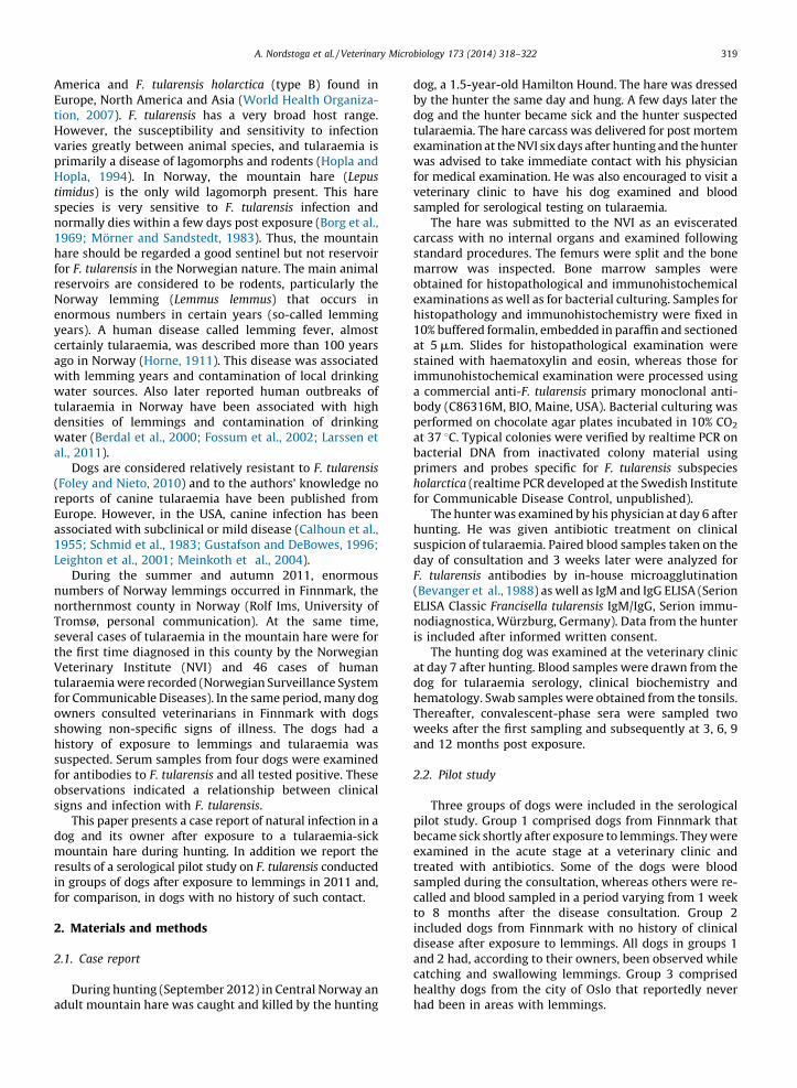
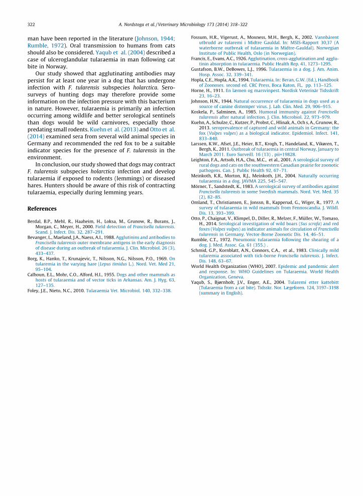
The hunting dog became sick two days after killing thehare and the signs increased in severity on day 3. Accordingto the owner, signs included lethargy, loss of appetite,raised hair-coat and fever (>39.5 8C). The dog’s conditionimproved on day 4 with full recovery on day 5. Clinicalexamination by the veterinary surgeon on day 7 afterhunting revealed no signs of disease except for slightlyenlarged pharyngeal, prescapular and popliteal lymphnodes. Blood biochemistry and hematology values werewithin normal ranges. Agglutinating antibody titer againstF. tularensis in serum samples obtained on day 7 afterhunting was 20 and peaked at 640 two weeks later.Thereafter, the titer stabilized at around 80 for up to 1 yearafter exposure (end of study) (Fig. 1).
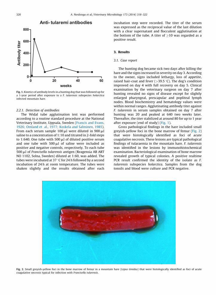
Gross pathological findings in the hare included smallgreyish-yellow foci in the bone marrow of femur (Fig. 2)that were histologically identified as foci of acutecoagulative necrosis. These lesions are typical pathologicalfindings of tularaemia in the mountain hare. F. tularensis
was identified in the lesions by immunohistochemicalexamination. Bacteriological examination of bone marrowrevealed growth of typical colonies. A positive realtimePCR result confirmed the identity of the isolate as F.
tularensis subspecies holarctica. Samples from the dogtonsils and blood were culture and PCR negative.
. 1. Kinetics of antibody levels in a hunting dog that was followed up for
-year period after exposure to a F. tularensis subspecies holarctica
ected mountain hare.
. 2. Small greyish-yellow foci in the bone marrow of femur in a mountain hare (Lepus timidus) that were histologically identified as foci of acute
agulative necrosis typical for infection with Francisella tularensis.
dinnhde1sS
3
v1optithaagoda
4
tubh
h(tliink(uTh
T
A
th
A. Nordstoga et al. / Veterinary Microbiology 173 (2014) 318–322 321
The hunter became sick 4 days after hunting andescribed his symptoms as moderate and influenza-like,cluding fever, headache and swollen axillary lymph
odes. Skin eruptions on his hands occurred on day 7 afterunting. The tularaemia diagnosis was confirmed byemonstration of seroconversion (from negative to mod-rate titer in the IgM and IgG ELISA, and from negative to28 in the microagglutination test) of paired serumamples (National reference laboratory for tularaemia,t. Olavs Hospital, Trondheim, Norway).
.2. Pilot study
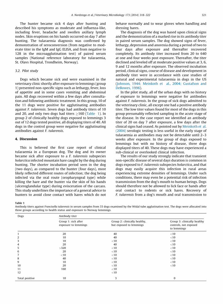
Dogs which became sick and were examined in theeterinary clinic shortly after exposure to lemmings (group) presented non-specific signs such as lethargy, fever, lossf appetite and in some cases vomiting and abdominalain. All dogs recovered within a few days after consulta-on and following antibiotic treatment. In this group, 10 ofe 11 dogs were positive for agglutinating antibodies
gainst F. tularensis. Seven dogs had antibody titers of 10nd 20, and only two dogs had titers �160 (Table 1). Inroup 2 of clinically healthy dogs exposed to lemmings 3ut of 12 dogs tested positive, all displaying titers of 40. Allogs in the control group were negative for agglutinatingntibodies against F. tularensis.
. Discussion
This is believed the first case report of clinicallaraemia in a European dog. The dog and its owner
ecame sick after exposure to a F. tularensis subspeciesolarctica infected mountain hare caught by the dog duringunting. The shorter incubation period seen in the dogwo days), as compared to the hunter (four days), mostkely reflected different routes of infection; the dog beingfected via the oral route (oropharyngeal type) while
illing the hare and the hunter via the skin of his handslceroglandular type) during evisceration of the carcass.
his study underlines the importance of a general advice tounters to avoid close contact with hares which do not
behave normally and to wear gloves when handling anddressing hares.
The diagnosis of the dog was based upon clinical signsand the demonstration of a marked rise in its antibody titerin paired serum samples. The dog showed signs of fever,lethargy, depression and anorexia during a period of two tofour days after exposure and thereafter recoveredcompletely. Its antibody titer increased from 20 to 640at one and four weeks post exposure. Thereafter, the titerdeclined and leveled off at moderate positive values at 3, 6,9 and 12 months after exposure. The observed incubationperiod, clinical signs, course of disease and development inantibody titer were in accordance with case studies ofnatural and experimental tularaemia in dogs in the US(Johnson, 1944; Meinkoth et al., 2004; Gustafson andDeBowes, 1996).
In the pilot study, all of the urban dogs with no historyof exposure to lemmings were negative for antibodiesagainst F. tularensis. In the group of sick dogs admitted tothe veterinary clinic, all except one had a positive antibodytiter. The low titer values found for most of the dogs in thisgroup presumably reflected sampling in the acute stage ofthe disease. In the case report we identified an antibodytiter of 20 on day 7 after exposure, a few days after theclinical signs had ceased. As pointed out by Meinkoth et al.(2004) serologic testing is less useful in the early stage oftularaemia as antibodies may not be detectable until 2–3weeks after exposure. In the group of dogs exposed tolemmings but with no history of disease, three dogsdisplayed titers of 40. These dogs may have experienced asub-clinical or overlooked clinical infection.
The results of our study strongly indicate that transientnon-specific disease of several days duration is common indogs exposed to F. tularensis subspecies holarctica, and thatdogs may easily acquire this infection in rural areasexperiencing extreme densities of lemmings. Under suchconditions, there may even be a potential risk of infectiontransmission from the dog’s mouth to human beings. Dogsshould therefore not be allowed to lick face or hands afteroral contact to rodents or sick hares. Recovery ofF. tularensis from a dog’s mouth and oral transmission to
able 1
ntibody titers against Francisella tularensis in serum samples from 33 dogs examined by the Widal tube agglutination test. The dogs were allocated into
ree groups according to health status and exposure to Norway lemmings.
Dogs Antibody titer
Group 1: sick after
exposure to lemmings
Group 2: clinically healthy,
but exposed to lemmings
Group 3: clinically healthy
controls, not exposed
to lemmings
1 20 40 <10
2 10 40 <10
3 10 <10 <10
4 20 40 <10
5 >320 <10 <10
6 20 <10 <10
7 <10 <10 <10
8 40 <10 <10
9 10 <10 <10
10 20 <10 <10
11 160 <10
12 <10
Total positive 10 3 0
mRushcabi
peinsuininocthpr(2Geinen
F.
tuhatu
Re
Be
Be
Bo
Ca
Fo
A. Nordstoga et al. / Veterinary Microbiology 173 (2014) 318–322322
an have been reported in the literature (Johnson, 1944;mble, 1972). Oral transmission to humans from catsould also be considered. Yaqub et al. (2004) described ase of ulceroglandular tularaemia in man following catte in Norway.
Our study showed that agglutinating antibodies mayrsist for at least one year in a dog that has undergonefection with F. tularensis subspecies holarctica. Sero-rveys of hunting dogs may therefore provide someformation on the infection pressure with this bacterium nature. However, tularaemia is primarily an infectioncurring among wildlife and better serological sentinelsan dogs would be wild carnivores, especially thoseedating small rodents. Kuehn et al. (2013) and Otto et al.014) examined sera from several wild animal species inrmany and recommended the red fox to be a suitable
dicator species for the presence of F. tularensis in thevironment.In conclusion, our study showed that dogs may contracttularensis subspecies holarctica infection and developlaraemia if exposed to rodents (lemmings) or diseasedres. Hunters should be aware of this risk of contractinglaraemia, especially during lemming years.
ferences
rdal, B.P., Mehl, R., Haaheim, H., Loksa, M., Grunow, R., Burans, J.,Morgan, C., Meyer, H., 2000. Field detection of Francisella tularensis.Scand. J. Infect. Dis. 32, 287–291.
vanger, L., Maeland, J.A., Naess, A.I., 1988. Agglutinins and antibodies toFrancisella tularensis outer membrane antigens in the early diagnosisof disease during an outbreak of tularaemia. J. Clin. Microbiol. 26 (3),433–437.
rg, K., Hanko, T., Krunajevic, T., Nilsson, N.G., Nilsson, P.O., 1969. Ontularemia in the varying hare (Lepus timidus L.). Nord. Vet. Med 21,95–104.
lhoun, E.L., Mohr, C.O., Alford, H.I., 1955. Dogs and other mammals ashosts of tularaemia and of vector ticks in Arkansas. Am. J. Hyg. 63,127–135.
ley, J.E., Nieto, N.C., 2010. Tularaemia Vet. Microbiol. 140, 332–338.
Fossum, H.R., Vigerust, A., Moxness, M.H., Bergh, K., 2002. Vannbarentutbrudd av tularemi i Midtre Gauldal. In: MSIS-Rapport 30,37 (Awaterborne outbreak of tularaemia in Midtre-Gauldal). NorwegianInstitute of Public Health, Oslo (in Norwegian).
Francis, E., Evans, A.C., 1926. Agglutination, cross-agglutination and agglu-tinin absorption in tularaemia. Public Health Rep. 41, 1273–1295.
Gustafson, B.W., DeBowes, L.J., 1996. Tularaemia in a dog. J. Am. Anim.Hosp. Assoc. 32, 339–341.
Hopla, C.E., Hopla, A.K., 1994. Tularaemia. In: Beran, G.W. (Ed.), Handbookof Zoonoses. second ed. CRC Press, Boca Raton, FL, pp. 113–125.
Horne, H., 1911. En læmen og marsvinpest. Nordisk Veterinar Tidsskrift23, 16–23.
Johnson, H.N., 1944. Natural occurrence of tularaemia in dogs used as asource of canine distemper virus. J. Lab. Clin. Med. 29, 906–915.
Koskela, P., Salminen, A., 1985. Humoral immunity against Francisellatularensis after natural infection. J. Clin. Microbiol. 22, 973–979.
Kuehn, A., Schulze, C., Kutzer, P., Probst, C., Hlinak, A., Och s, A., Grunow, R.,2013. seroprevalence of captured and wild animals in Germany: thefox (Vulpes vulpes) as a biological indicator. Epidemiol. Infect. 141,833–840.
Larssen, K.W., Afset, J.E., Heier, B.T., Krogh, T., Handeland, K., Vikøren, T.,Bergh, K., 2011. Outbreak of tularaemia in central Norway, January toMarch 2011. Euro Surveill. 16 (13) , pii=19828.
Leighton, F.A., Artsob, H.A., Chu, M.C., et al., 2001. A serological survey ofrural dogs and cats on the southwestern Canadian prairie for zoonoticpathogens. Can. J. Public Health 92, 67–71.
Meinkoth, K.R., Morton, R.J., Meinkoth, J.H., 2004. Naturally occurringtularaemia in a dog. JAVMA 225, 545–547.
Morner, T., Sandstedt, K., 1983. A serological survey of antibodies againstFrancisella tularensis in some Swedish mammals. Nord. Vet. Med. 35(2), 82–85.
Omland, T., Christiansen, E., Jonssn, B., Kapperud, G., Wiger, R., 1977. Asurvey of tularaemia in wild mammals from Fennoscandia. J. Wildl.Dis. 13, 393–399.
Otto, P., Chaignat, V., Klimpel, D., Diller, R., Melzer, F., Muller, W., Tomaso,H., 2014. Serological investigation of wild boars (Sus scrofa) and redfoxes (Vulpes vulpes) as indicator animals for circulation of Francisellatularensis in Germany. Vector-Borne Zoonotic Dis. 14, 46–51.
Rumble, C.T., 1972. Pneumonic tularaemia following the shearing of adog. J. Med. Assoc. Ga. 61 (355.) .
Schmid, G.P., Kornblatt, A.N., Connors, C.A., et al., 1983. Clinically mildtularemia associated with tick-borne Francisella tularensis. J. Infect.Dis. 148, 63–67.
World Health Organization (WHO), 2007. Epidemic and pandemic alertand response. In: WHO Guidelines on Tularaemia. World HealthOrganization, Geneva.
Yaqub, S., Bjørnholt, J.V., Enger, A.E., 2004. Tularemi etter kattebitt(Tularaemia from a cat bite). Tidsskr. Nor. Lægeforen. 124, 3197–3198(summary in English).