Embed Size (px)
Citation preview

TUBERCULOUS PNEUMONIA
Dr Etienne Leroy-Terquem Centre hospitalier de Meulan les Mureaux. France
French-cambodian association for pneumology (OFCP)

Common adult TB Basic radiological images:
• Nodule • Infiltrate • Cavity • Tuberculous pneumonia
Tuberculous pneumonia(1)
• This is an alveolar image: non-homogenous, not clearly limited, except if contact with scissura, with aeric bronchogram
• The association with other tuberculous lesions is very frequent: adenopathies, nodules and infiltrates, especially in AIDS patients
• The lesions are often bilateral

Tuberculous pneumonia (2) • The research of AFB is most often positive in sputum,
because these lesions are very rich in tuberculous bacilli.
• The spontaneous evolution is the constitution of cavitation and destruction of the lung tissue, retraction and fibrosis: >>> important sequela if treatment is too late.
• Tuberculous pneumonia is frequent in AIDS cases. In this case the pneumonia is as frequent in the inferior lobes as the superior, and is often associated with adenopathies. The excavation is infrequent in cases of severe immunodepression.

Aeric bronchogram

Bilateral tuberculous pneumonia with mediastinal hilar adenopathies and adenopathies in superior mediastinum.
AFB positive in sputum (HIV+).
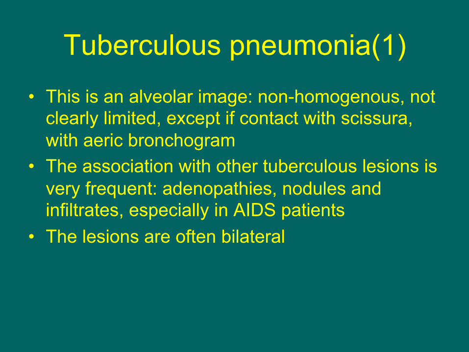
AFB ++ in sputum: right superior lobe pneumonia. Notice the beginning of the lobe retraction and controlateral nodules: the
association is highly indicative of TB.
Man, 30 years old. Dyspnea, fever, cough and weight loss over two months.
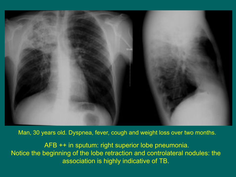
Chest x-ray at the end of treatment. Retractile evolution with ascension of the right hilus.
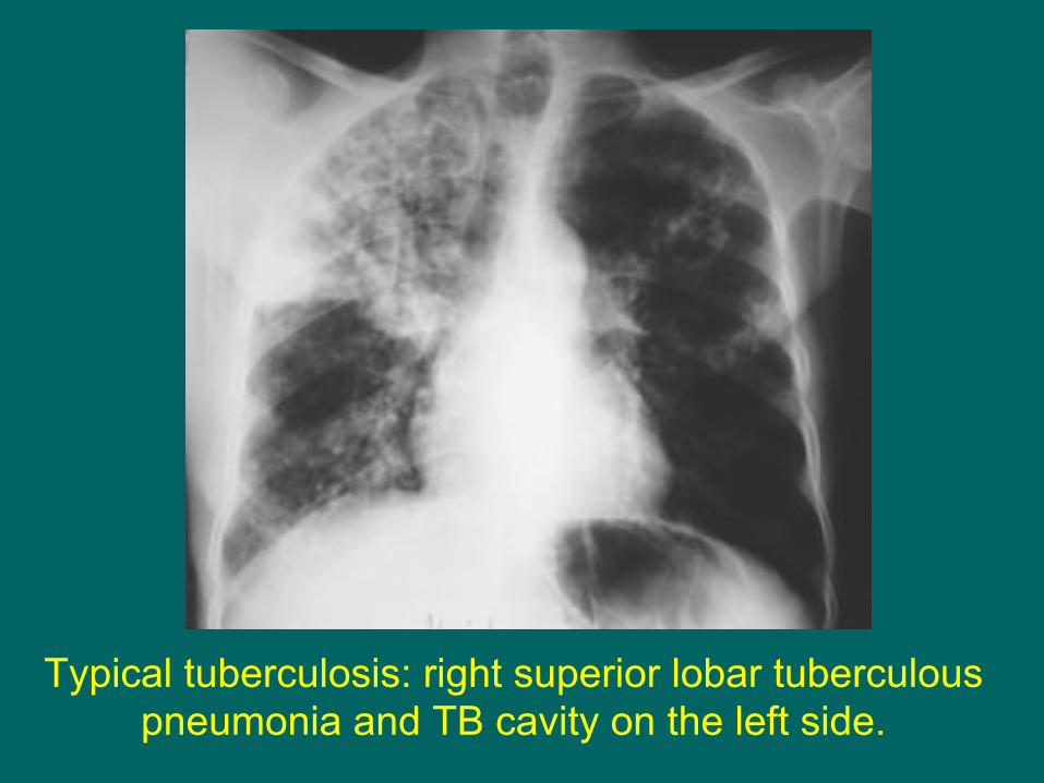
Typical tuberculosis: right superior lobar tuberculous pneumonia and TB cavity on the left side.
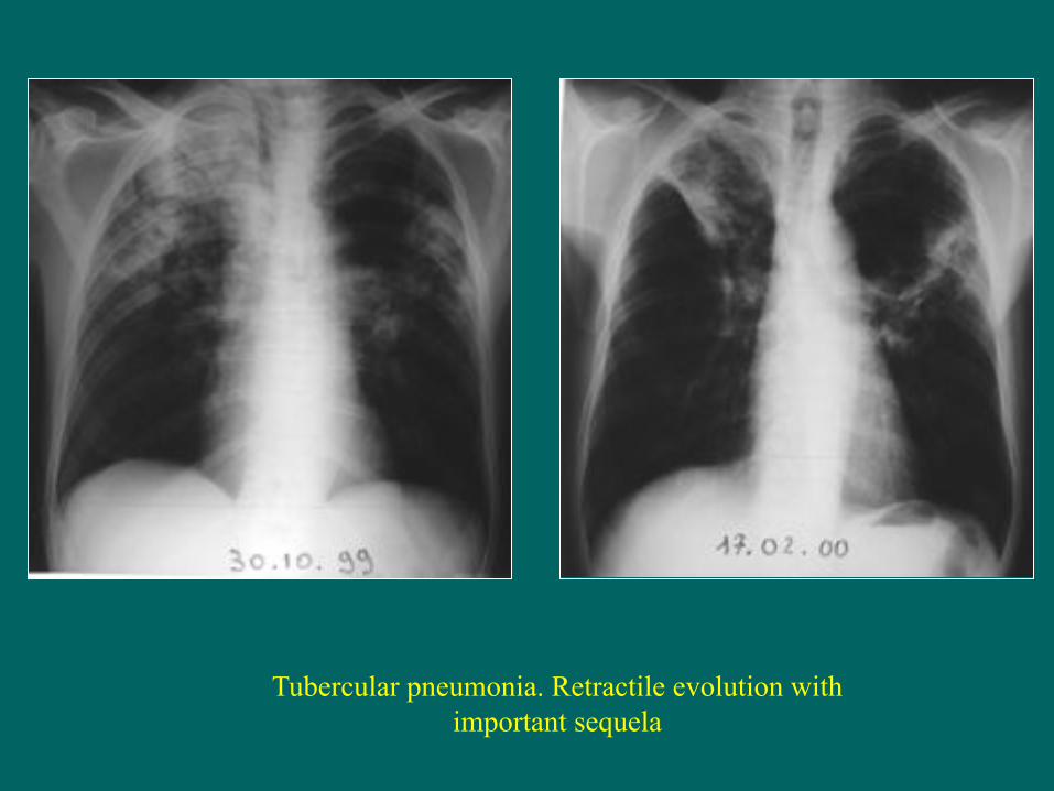
Tubercular pneumonia. Retractile evolution with important sequela
© OFCP
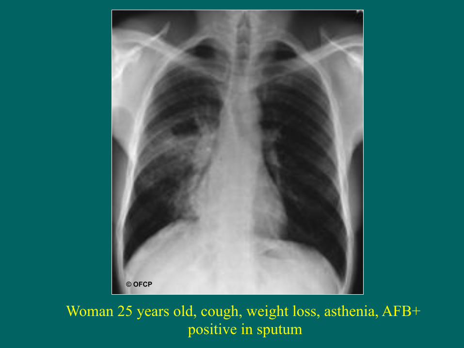
Woman 25 years old, cough, weight loss, asthenia, AFB+ positive in sputum
© OFCP © OFCP
© OFCP© OFCP
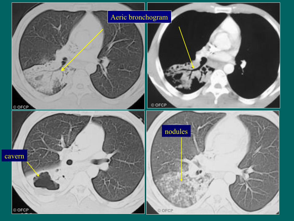
Aeric bronchogram
cavern
nodules
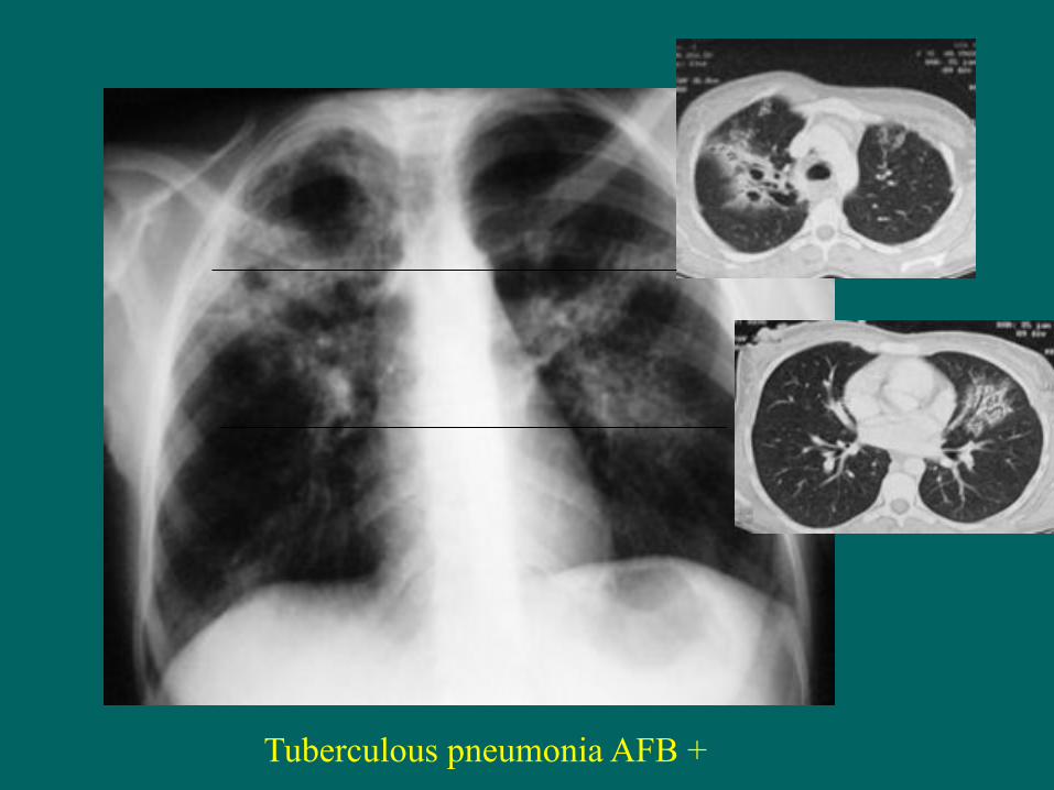
Tuberculous pneumonia AFB +
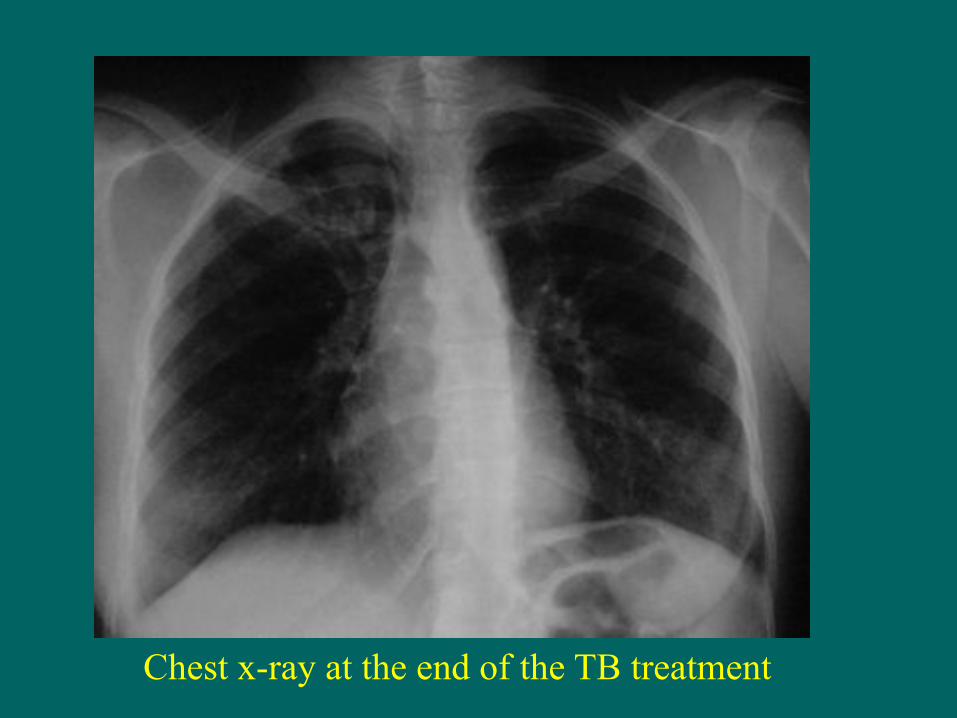
Chest x-ray at the end of the TB treatment
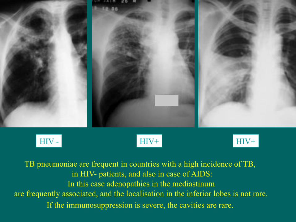
TB pneumoniae are frequent in countries with a high incidence of TB, in HIV- patients, and also in case of AIDS: In this case adenopathies in the mediastinum
are frequently associated, and the localisation in the inferior lobes is not rare. If the immunosuppression is severe, the cavities are rare.
HIV - HIV+ HIV+

-Tuberculous pneumonia. HIV+, CD4< 100. - Bilateral lesions - Localisation in middle lobe and left inf. - Latero-tracheal adenopathy - no cavitation
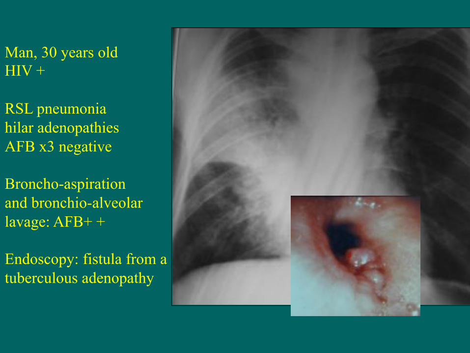
Man, 30 years old HIV + RSL pneumonia hilar adenopathies AFB x3 negative Broncho-aspiration and bronchio-alveolar lavage: AFB+ + Endoscopy: fistula from a tuberculous adenopathy

Man HIV+, miliary, medastinal adenopathies, right pneumonia AFB+

25.04.01
Man, 25 years old, AEG, T° 39°C, cough, AFB +, treatment 2RHZE alveolar opacities, with bronchogram, left superior lobe predominant

Failure after 2 months of treatment, with persistant AFB + Antibiogram: resistance to R and H ( “MDR“ TB).
Modification of the treatment (quinolones, cycloserine, Pyrazinamide, ethambutol). Favourable evolution with retraction.
22.06.01 28.12.01 Evolution with 2 months of RHZE

But all pneumoniae are not tuberculous. The clinical context is vital for diagnosis…

Young man, no pathological antecedents, sudden onset of symptoms with fever, chills, thoracic pain
Acute lobar pneumonia
(streptococcus pneumoniae)

HIV+ context, subacute evolution, adenopathies: it is not an acute lobar pneumonia consequent to bacterial infection
with strep.pneumoniae. It is a tuberculous pneumonia.

Young woman, good health, 39-40°C fever for 48h, non-productive cough and right thoracic pain:
Acute pneumonia (probable infection with strep. pneumoniae)…
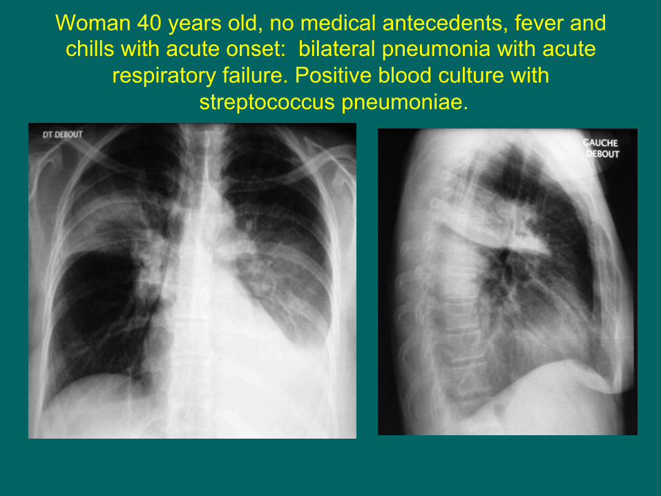
Woman 40 years old, no medical antecedents, fever and chills with acute onset: bilateral pneumonia with acute
respiratory failure. Positive blood culture with streptococcus pneumoniae.

In cases of AIDS, if severe dyspnea, normal or subnormal auscultation, and diffuse non-excavated pneumonia, think
PNEUMOCYSTOSIS
Garde Républicain 35 aTrouvé par ses camarades dans sa chambreconfus, obnubilé,ayant vomit° 40°
LBA : P. P. cariniicariniiBrossage protégé Nelson G :St. aureusSt. aureus
HomosexuelSIDA
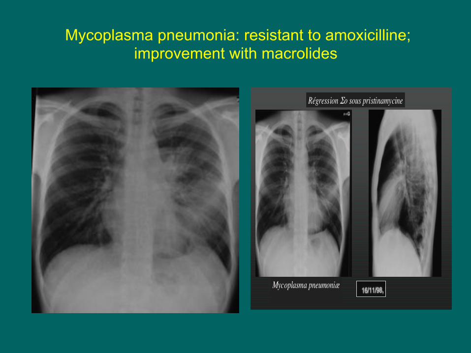
Mycoplasma pneumonia: resistant to amoxicilline; improvement with macrolides
Régression !o sous pristinamycine
Mycoplasma pneumoniæ

Legionnaire’s disease
Young man, severe dyspnea and fever, headache, abdominal pain, No improvement with amoxicillin…
Conclusion1
• Pneumonia is a frequent clinical manifestation of tuberculosis in countries with a high incidence of TB
• The lesions are often bilateral and associated with other lesions: nodules, adenopathies, cavities.
• AFB in sputum are often positive, but do not neglect
the causes of false negatives: salivary sputum, patient too weak for reliable sputum, technical error, treatment begun before sampling.

Conclusions 2
• The tuberculous pneumoniae are frequent in cases of AIDS: All the lobes can be affected (particularly the inferior lobes) and are often associated with bulky adenopathies. In cases of severe immunodepression, cavitation is rare.
• Differential diagnosis with the other infectious pneumoniae is only possible with the history-taking and clinical examination, which must always be associated with the analysis of the chest radiography.


![Pneumonia [Harrison's]](https://img.pdfslide.us/doc/110x75/54515befb1af9f83248b46c1/pneumonia-harrisons.jpg)