Embed Size (px)
Citation preview
REVIEW
TRPV1 and TRPA1 in cutaneous neurogenicand chronic inflammation: pro-inflammatoryresponse induced by their activation and theirsensitization
Olivier Gouin1,2, Killian L’Herondelle1, Nicolas Lebonvallet1, Christelle Le Gall-Ianotto1, Mehdi Sakka1,Virginie Buhé1, Emmanuelle Plée-Gautier1, Jean-Luc Carré1, Luc Lefeuvre2, Laurent Misery1&,Raphaele Le Garrec1
1 Laboratory on Interaction Neurons-Keratinocytes (LINK), University of Western Brittany, 29200 Brest, France2 Uriage Dermatological Laboratories, 92400 Courbevoie, France& Correspondence: [email protected] (L. Misery)
Received January 9, 2017 Accepted February 28, 2017
ABSTRACT
Cutaneous neurogenic inflammation (CNI) is inflamma-tion that is induced (or enhanced) in the skin by therelease of neuropeptides from sensory nerve endings.Clinical manifestations are mainly sensory and vasculardisorders such as pruritus and erythema. Transientreceptor potential vanilloid 1 and ankyrin 1 (TRPV1 andTRPA1, respectively) are non-selective cation channelsknown to specifically participate in pain and CNI. BothTRPV1 and TRPA1 are co-expressed in a large subset ofsensory nerves, where they integrate numerous noxiousstimuli. It is now clear that the expression of bothchannels also extends far beyond the sensory nerves inthe skin, occuring also in keratinocytes, mast cells,dendritic cells, and endothelial cells. In these non-neu-ronal cells, TRPV1 and TRPA1 also act as nociceptivesensors and potentiate the inflammatory process. Thisreview discusses the role of TRPV1 and TRPA1 in themodulation of inflammatory genes that leads to ormaintains CNI in sensory neurons and non-neuronalskin cells. In addition, this review provides a summary ofcurrent research on the intracellular sensitization path-ways of both TRP channels by other endogenousinflammatory mediators that promote the self-mainte-nance of CNI.
KEYWORDS sensory nerve, neurogenic skininflammation, inflammatory gene regulation, pruritus
INTRODUCTION
CNI definition, induction, and self-maintenance
Cutaneous neurogenic inflammation (CNI) is the inflamma-tion induced (or enhanced) by an excessive release ofneuropeptides such as calcitonin gene-related peptide(CGRP) and tachykinins (mainly substance P, SP) in the skinfrom locally or antidromically activated sensory nerve end-ings (Herbert and Holzer, 2002; Roosterman et al., 2006;Gouin et al., 2015).
CNI can be induced through the direct activation ofreceptors on sensory nerve endings by mechanical skininjuries and other exogenous stimuli, such as exposure of theskin to injurious heat or cold, ultraviolet (physical factors),chemical irritants or allergens. Endogenous stimuli, includingosmotic or pH changes in the skin, can also initiate CNI(Herbert and Holzer, 2002; Roosterman et al., 2006). Thereleased neuropeptides act on skin cells that express cog-nate neuropeptide receptors, including microvascular cellsand resident mast cells, leading to degranulation, vasodila-tion, and extravasation of plasma proteins and leukocytes.Some clinical manifestations of acute CNI are localized pru-ritus, redness, heat, and edema (Herbert and Holzer 2002;Roosterman et al., 2006; Teresiak-Mikołajczak et al., 2013).
Moreover, neuropeptides and mast cell-released media-tors can act on other neighboring target cells, includingkeratinocytes, dendritic cells, neutrophils and fibroblasts,leading to the disruption of skin homeostasis, with abnormalskin growth, differentiation, and/or immunomodulation (Shim
© The Author(s) 2017. This article is an open access publication
Protein Cell 2017, 8(9):644–661DOI 10.1007/s13238-017-0395-5 Protein&Cell
Protein
&Cell
et al., 2007; Trevisani et al., 2007; Wilson et al., 2013b;Patricio et al., 2015).
Furthermore, a variety of neurotrophic (e.g. nerve growthfactor, NGF) or inflammatory mediators (e.g. proteases, his-tamine, cytokines, prostanoids) or endocannabinoids (e.g.anandamide) released by skin cells/nerve fibers or chemoat-tracted cells are able to further activate or sensitize thesesensory receptors (Briot et al., 2009; Vellani et al., 2010;Roostermanet al., 2006;Riol-Blancoet al., 2014;Wilsonet al.,2013b; Wei et al., 2012). Such positive feedback loops con-tribute to the enhancement of the inflammatory process andthus to self-maintained CNI (Gouin et al., 2015).
Therefore, CNI appears to be a multi-cellular network withmultiple, multi-directional interactions leading to a viciouscircle of processes that results in chronic inflammation(Gouin et al., 2015). Indeed, CNI is frequently involved inchronic inflammatory skin disorders, including psoriasis,atopic dermatitis (AD) (Kubanov et al., 2015; Smolyannikovaet al., 2015), sensitive skin (Costa et al., 2014), rosacea(Kürkçüoğlu and Alaybeyi 1991; Salem et al., 2013), andhypertrophic scars (Akaishi et al., 2008; Kwak et al., 2014).
Role of PARs and TRPs
Although the exocytosis of neuropeptides from large dense-core vesicles differs from that of classical neurotransmittersfrom small clear vesicles, it is also triggered by a rise in thecytosolic Ca2+ concentration (Huang and Neher 1996;Zupanc 1996; Jans et al., 2004). Two primary pathways leadto increased Ca2+ concentration in the cytosol, the Ca2+
influx associated with the opening of plasmalemmal Ca2+
channels and the Ca2+ released from intracellular stores (theendoplasmic reticulum and mitochondria). In addition tovoltage-gated Ca2+ channels, cutaneous sensory nervesexpress cationic channels and G protein-coupled receptors(GPCRs), the activation of which can lead directly or indi-rectly to an increase in cytosolic Ca2+. In addition to theinduction of neuropeptide release, an increase in cytosolicCa2+ can also drive the regulation of the expression ofseveral inflammatory genes, such as those encoding neu-ropeptides, cytokines, growth factors, prostaglandins (PG),and matrix metalloproteinases (MMPs), which have a pos-sible role in chronic cutaneous inflammation.
Interestingly, cationic channels expressed by cutaneousnerveendings include some transient receptor potential (TRP)channels known to be involved in neuropeptide exocytosisand skin disorders that have neurogenic mechanisms. In thisreview we focus on two of these channels including TRPV1and TRPA1 (TRP subfamily vanilloid 1 and TRP subfamilyankyrin 1, respectively) (Xie 2009; Wei et al., 2010; Boillatet al., 2014; Horváth et al., 2015). Other TRP channels havealso been found to be involved in CNI and pruritus such asTRPV3 (Lin et al., 2012) and TRPV4 (Zhao et al., 2014;Rajasekhar et al., 2015;Akiyamaet al., 2016;Kimet al., 2016).In addition, the sensory nerves in the skin also expressGPCRs, of which the protease-activated receptor (PAR)
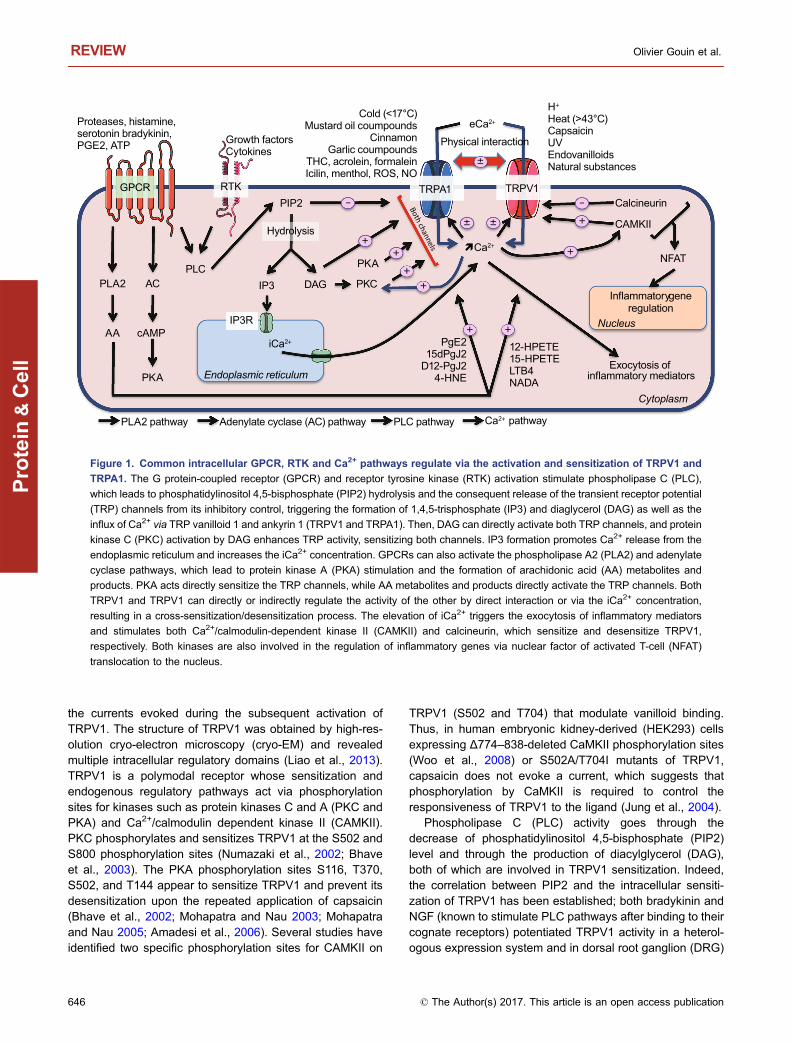
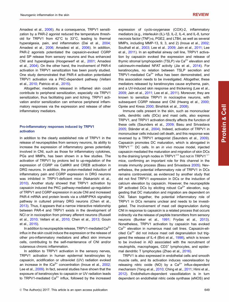
family in particular is known to be involved in CNI, especiallyPAR-2 and PAR-4 (Cocks and Moffatt 2000). Thus, TRPV1,TRPA1, PAR-2, and PAR-4 are associated not only with a risein the intracellular Ca2+ concentration (iCa2+) with the subse-quent exocytosis of neuropeptides but also with pro-inflam-matory gene expression (Fig. 1). Moreover, following theactivation of oneof these receptors, intracellular pathways canlead to the sensitization of one of these channels.
In addition to their expression by sensory nerves, TRPV1,TRPA1, PAR-2, and PAR-4 are found in resident skin cells orcells recruited during CNI. Thus, they could contribute to theintense and narrow communication within the skin betweensensory nerve endings, skin, and immune cells.
This review discusses the role of TRPV1 and TRPA1activation or signaling pathways associated with their sen-sitization in the self-maintenance of CNI through the induc-tion of a pro-inflammatory response in the skin.
TRPV1
A cationic channel with multiple direct roles in CNIinduction and self-maintenance
TRPV1 is a nociceptive cationic (mainly Ca2+) channelresponsive tohigh temperature (>43°C) (Boillat et al., 2014). Inaddition to excessive heat, various exogenous and endoge-nous triggering factors can directly activate or sensitizeTRPV1 (Table 1). TRPV1 was initially described in cutaneousC- andAδ-type sensory nerve endings, where it plays a criticalrole. It can be activated by the natural agonist capsaicin(Jancsó et al., 1967; Caterina et al., 1997). High temperatureand capsaicin have been demonstrated to activate sensorynerves and induce neurogenic inflammation (Jancsó et al.,1967; Caterina et al., 1997). Indeed, TRPV1 activation bythese direct activators allows the entry of Ca2+, leading to therelease of neuropeptides, including SP (Andreev et al., 2012)and CGRP (Boillat et al., 2014), that control edema and caninduce (or enhance) neurogenic inflammation (Szallasi andBlumberg 1989; Zygmunt et al., 1999; Steinhoff et al., 2003;Roosterman et al., 2006; Vincent et al., 2013).
Furthermore, endogenousmediators produced or releasedduringCNI (eicosanoids, acidosis, ATP, histamine, bradykinin,NGF) that further sensitize or activate TRPV1 on skinnerve terminals contribute to the self-maintenance of CNI(Roosterman et al., 2006). Moreover, TRPV1 was found to beexpressed in skin cells (keratinocytes, dermal mast cells,dendritic cells, sebocytes, dermal blood vessels, hair follicles,and sweat glands), where it acts as a pain and chemicalsensor (Ständer et al., 2004).
TRPV1 sensitization/desensitization by endogenousmodulators following intracellular pathway activation
Intracellular pathways of TRPV1 sensitization
The TRPV1 sensitization process is an intracellular mecha-nism that facilitates the gating of the channel or strengthens
Neurogenic inflammation in the skin REVIEW
© The Author(s) 2017. This article is an open access publication 645
Protein
&Cell
the currents evoked during the subsequent activation ofTRPV1. The structure of TRPV1 was obtained by high-res-olution cryo-electron microscopy (cryo-EM) and revealedmultiple intracellular regulatory domains (Liao et al., 2013).TRPV1 is a polymodal receptor whose sensitization andendogenous regulatory pathways act via phosphorylationsites for kinases such as protein kinases C and A (PKC andPKA) and Ca2+/calmodulin dependent kinase II (CAMKII).PKC phosphorylates and sensitizes TRPV1 at the S502 andS800 phosphorylation sites (Numazaki et al., 2002; Bhaveet al., 2003). The PKA phosphorylation sites S116, T370,S502, and T144 appear to sensitize TRPV1 and prevent itsdesensitization upon the repeated application of capsaicin(Bhave et al., 2002; Mohapatra and Nau 2003; Mohapatraand Nau 2005; Amadesi et al., 2006). Several studies haveidentified two specific phosphorylation sites for CAMKII on
TRPV1 (S502 and T704) that modulate vanilloid binding.Thus, in human embryonic kidney-derived (HEK293) cellsexpressing Δ774–838-deleted CaMKII phosphorylation sites(Woo et al., 2008) or S502A/T704I mutants of TRPV1,capsaicin does not evoke a current, which suggests thatphosphorylation by CaMKII is required to control theresponsiveness of TRPV1 to the ligand (Jung et al., 2004).
Phospholipase C (PLC) activity goes through thedecrease of phosphatidylinositol 4,5-bisphosphate (PIP2)level and through the production of diacylglycerol (DAG),both of which are involved in TRPV1 sensitization. Indeed,the correlation between PIP2 and the intracellular sensiti-zation of TRPV1 has been established; both bradykinin andNGF (known to stimulate PLC pathways after binding to theircognate receptors) potentiated TRPV1 activity in a heterol-ogous expression system and in dorsal root ganglion (DRG)
PLC
�Ca2+
eCa2+
GPCR TRPV1TRPA1RTK
Proteases, histamine, serotonin bradykinin, PGE2, ATP Growth factors
Cytokines
Hydrolysis± ±
-
DAGIP3
+
PKC +
iCa2+
Physical interaction
±
PLA2
AAIP3R
12-HPETE15-HPETELTB4NADA
PgE215dPgJ2
D12-PgJ24-HNE
+ +
Endoplasmic reticulum
CAMKII
+
+
+
Calcineurin-
Nucleus
Inflammatorygeneregulation
Exocytosis of inflammatory mediators
cAMP
PKA
PKA+
PLA2 pathway Adenylate cyclase (AC) pathway
AC
PLC pathway Ca2+ pathway
H+
Heat (>43°C)CapsaicinUVEndovanilloidsNatural substances
PIP2
Cytoplasm
NFAT
Cold (<17°C)Mustard oil coumpounds
CinnamonGarlic coumpounds
THC, acrolein, formaleinIcilin, menthol, ROS, NO
Figure 1. Common intracellular GPCR, RTK and Ca2+ pathways regulate via the activation and sensitization of TRPV1 and
TRPA1. The G protein-coupled receptor (GPCR) and receptor tyrosine kinase (RTK) activation stimulate phospholipase C (PLC),
which leads to phosphatidylinositol 4,5-bisphosphate (PIP2) hydrolysis and the consequent release of the transient receptor potential
(TRP) channels from its inhibitory control, triggering the formation of 1,4,5-trisphosphate (IP3) and diaglycerol (DAG) as well as the
influx of Ca2+ via TRP vanilloid 1 and ankyrin 1 (TRPV1 and TRPA1). Then, DAG can directly activate both TRP channels, and protein
kinase C (PKC) activation by DAG enhances TRP activity, sensitizing both channels. IP3 formation promotes Ca2+ release from the
endoplasmic reticulum and increases the iCa2+ concentration. GPCRs can also activate the phospholipase A2 (PLA2) and adenylate
cyclase pathways, which lead to protein kinase A (PKA) stimulation and the formation of arachidonic acid (AA) metabolites and
products. PKA acts directly sensitize the TRP channels, while AA metabolites and products directly activate the TRP channels. Both
TRPV1 and TRPV1 can directly or indirectly regulate the activity of the other by direct interaction or via the iCa2+ concentration,
resulting in a cross-sensitization/desensitization process. The elevation of iCa2+ triggers the exocytosis of inflammatory mediators
and stimulates both Ca2+/calmodulin-dependent kinase II (CAMKII) and calcineurin, which sensitize and desensitize TRPV1,
respectively. Both kinases are also involved in the regulation of inflammatory genes via nuclear factor of activated T-cell (NFAT)
translocation to the nucleus.
REVIEW Olivier Gouin et al.
646 © The Author(s) 2017. This article is an open access publication
Protein
&Cell
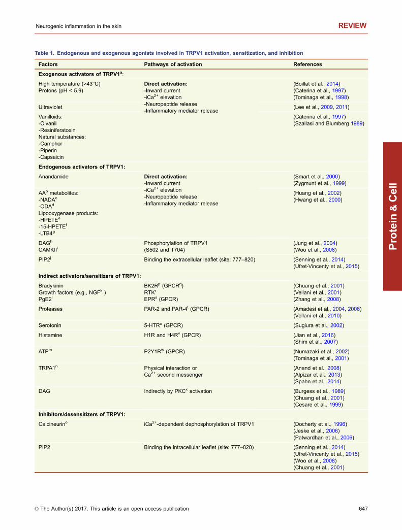
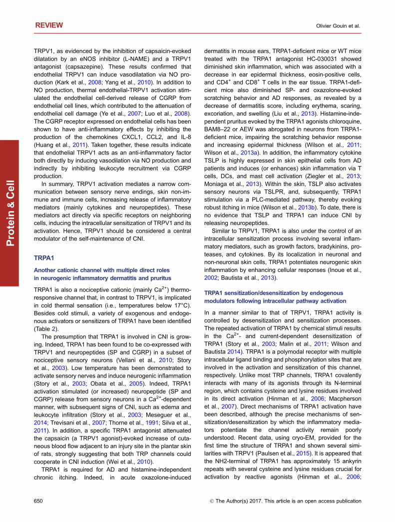
Table 1. Endogenous and exogenous agonists involved in TRPV1 activation, sensitization, and inhibition
Factors Pathways of activation References
Exogenous activators of TRPV1a:
High temperature (>43°C)Protons (pH < 5.9)
Direct activation:-Inward current-iCa2+ elevation-Neuropeptide release-Inflammatory mediator release
(Boillat et al., 2014)(Caterina et al., 1997)(Tominaga et al., 1998)
Ultraviolet (Lee et al., 2009, 2011)
Vanilloids:-Olvanil-ResiniferatoxinNatural substances:-Camphor-Piperin-Capsaicin
(Caterina et al., 1997)(Szallasi and Blumberg 1989)
Endogenous activators of TRPV1:
Anandamide Direct activation:-Inward current-iCa2+ elevation-Neuropeptide release-Inflammatory mediator release
(Smart et al., 2000)(Zygmunt et al., 1999)
AAb metabolites:-NADAc
-ODAd
Lipooxygenase products:-HPETEe
-15-HPETEf
-LTB4g
(Huang et al., 2002)(Hwang et al., 2000)
DAGh
CAMKIIiPhosphorylation of TRPV1(S502 and T704)
(Jung et al., 2004)(Woo et al., 2008)
PIP2j Binding the extracellular leaflet (site: 777–820) (Senning et al., 2014)(Ufret-Vincenty et al., 2015)
Indirect activators/sensitizers of TRPV1:
BradykininGrowth factors (e.g., NGFk )PgE2l
BK2Rp (GPCRq)RTKr
EPRs (GPCR)
(Chuang et al., 2001)(Vellani et al., 2001)(Zhang et al., 2008)
Proteases PAR-2 and PAR-4t (GPCR) (Amadesi et al., 2004, 2006)(Vellani et al., 2010)
Serotonin 5-HTRu (GPCR) (Sugiura et al., 2002)
Histamine H1R and H4Rv (GPCR) (Jian et al., 2016)(Shim et al., 2007)
ATPm P2Y1Rw (GPCR) (Numazaki et al., 2002)(Tominaga et al., 2001)
TRPA1n Physical interaction orCa2+ second messenger
(Anand et al., 2008)(Alpizar et al., 2013)(Spahn et al., 2014)
DAG Indirectly by PKCx activation (Burgess et al., 1989)(Chuang et al., 2001)(Cesare et al., 1999)
Inhibitors/desensitizers of TRPV1:
Calcineurino iCa2+-dependent dephosphorylation of TRPV1 (Docherty et al., 1996)(Jeske et al., 2006)(Patwardhan et al., 2006)
PIP2 Binding the intracellular leaflet (site: 777–820) (Senning et al., 2014)(Ufret-Vincenty et al., 2015)(Woo et al., 2008)(Chuang et al., 2001)
Neurogenic inflammation in the skin REVIEW
© The Author(s) 2017. This article is an open access publication 647
Protein
&Cell
neurons (Chuang et al., 2001). The role of PIP2 as a TRPV1inhibitor or “desensitizer” after its depletion by sequestrationor PLC hydrolysis has been subsequently shown to beessential for TRPV1 sensitization (Prescott and Julius 2003).In addition, the lack of a PIP2 binding site (786–828)increased the strong inward current evoked by a DAG ana-log and capsaicin in a Δ774–838 deletion mutant of TRPV1(Woo et al., 2008). However, PIP2 could activate TRPV1 bybinding the extracellular leaflet (777–820), while it has aninhibitor effect by binding the intracellular leaflet (682–725)(Senning et al., 2014; Ufret-Vincenty et al., 2015). Interest-ingly, in addition to its ability to directly activate TRPV1 via aPKC-independent pathway by binding to the capsaicinbinding site at Y511; (Woo et al., 2008)), DAG could alsocontribute to the indirect sensitization of TRPV1 via PKCphosphorylation at the S502 and S801 sites (Burgess et al.,1989; Cesare et al., 1999; Chuang et al., 2001) (Numazakiet al., 2002; Bhave et al., 2003).
Finally, it might be noticed that the prolonged or repeatedactivation of TRPV1 induces a desensitization or inhibitionprocess. It has been observed that the repeated activation ofTRPV1 by chemical stimuli results in its desensitization by aCa2+-dependent process (Chuang et al., 2001; Bhave et al.,2002; Dai et al., 2004). Protein phosphatase 2B (calcineurin)acts as a desensitizer of TRPV1, as indicated by the inhi-bition of calcineurin by cyclosporine or CsA-CyP, which havebeen shown to inhibit the desensitization of TRPV1 inducedby capsaicin (Docherty et al., 1996; Mohapatra and Nau2005). The cannabinoid WIN 55,212-2 seems to play an anti-inflammatory and analgesic role via the inhibition of TRPV1by dephosphorylating the T144 and T370 sites in a Ca2+/calcineurin-dependent manner, reducing the release ofCGRP (Patwardhan et al., 2006; Jeske et al., 2006). Thesedata show the potential effects of calcineurin inhibitors onTRPV1 desensitization and suggest cannabinoids as newtherapeutic drugs for CNI, hyperalgesia, itching, and pain.
Inflammatory mediators that use TRPV1 intracellularsensitization pathways
Following tissue damage, endogenously released inflam-matory mediators (ATP, bradykinin, serotonin PGs, NGF,
chemokines, histamine or proteases) can regulate TRPV1activity via intracellular pathways associated with theirspecific GPCR. For example, in transfected HEK293 cellsand DRG neurons, ATP increased capsaicin-inducedTRPV1 activation via the purinergic receptor P2Y1, whichpotentiated TRPV1 activity in a PKC-dependent pathway(Tominaga et al., 2001; Numazaki et al., 2002). In the sameway, in a Ca2+- and PLC/PKC-dependent manner, bradyki-nin, serotonin (G protein-coupled 5-HT receptor), PgE2 (Gprotein-coupled EP receptors), and NGF enhanced cap-saicin-, heat-, proton-, and anandamine-evoked currents(Vellani et al., 2001; Sugiura et al., 2002; Zhang et al., 2008;Wilson et al., 2011). Similarly, the chemokine CCL3 wasfound to sensitize TRPV1 by increasing the heat-, anan-damide-, and capsaicin-evoked Ca2+ influx through a PLC-and PKC-pathway in HEK293 cells and DRG neurons(Zhang et al., 2005).
Finally, histamine enhanced the capsaicin-mediatedinward current, increased the Ca2+ level and then induceditching via sensory neurons, and this effect was abolished byTRPV1 antagonists and in TRPV1-deficient mice (Shimet al., 2007). These mechanisms appeared to involvephospholipase A2 (PLA2)-, lipoxygenase (NDGA)- and PLC-dependent pathways, with the subsequent sensitization andactivation of TRPV1 (Jian et al., 2016). These studies sug-gest that TRPV1 is involved in histamine-dependent itchingwith arachidonic acid metabolites, primarily 12-hydroxye-icosatetraenoic (12-HETE), acting as a central participant(Shim et al., 2007).
Because of their ability to cleave and thus activatespecific PARs, endogenous proteases play a specific role inCNI. Both PAR-2 and PAR-4 are known to induce CNI andtherefore to be involved in several skin disorders, such aspruritus and AD (Asfaha et al., 2007; Briot et al., 2009; Vel-lani et al., 2010; Fu et al., 2014). In addition to their ability toinduce CNI via the release of SP and CGRP, it has clearlybeen established that PAR-2 and PAR-4 activation couldlead to TRPV1 sensitization. Patch clamp and Ca2+ imagingassays in HEK293 cells that co-express TRPV1 and PAR-2and DRG neurons revealed that PAR-2 activation potenti-ates TRPV1 via phosphorylation by PKC- and PKA-depen-dent pathways (Amadesi et al., 2004; Dai et al., 2004;
Table 1. continued
Factors Pathways of activation References
TRPA1 Physical interaction oriCa2+ second messenger
(Akopian et al., 2007)(Patil et al., 2010)
Notes: a transient receptor potential vanilloid 1; b arachidonic acid; c N-arachidonoyl-dopamine ; d N-oleoyl dopamine;e 12-hydroperoxyeicosatetraenoic acid; f 15-hydroperoxy-eicosatetraenoic acid; g leukotrien B4; h diacylglycerol; i Ca2+/
calmodulin-dependent kinase II; j phosphatidylinositol 4,5-bisphosphate; (iCa2+) intercellular Ca2+; k nerve growth factor;l prostaglandin E2; m adenosine triphosphate; n transient receptor potential ankyrin 1; o protein phosphatase 2B; p bradykinin B2 receptor;q G protein-coupled receptor; r receptor tyrosine kinase; s E prostanoid receptor; t protease-activated receptor-2 and 4;u 5-hydroxytryptamine receptor; v histamine receptors 1 and 4; w purinergic P2Y1 receptor; x protein kinase C.
REVIEW Olivier Gouin et al.
648 © The Author(s) 2017. This article is an open access publication
Protein
&Cell
Amadesi et al., 2006). As a consequence, TRPV1 sensiti-zation by a PAR-2 agonist reduced the temperature thresh-old for TRPV1 from 42°C to 33°C, leading to thermalhyperalgesia, pain, and inflammation (Dai et al., 2004;Amadesi et al., 2006; Amadesi et al., 2006). In addition,PAR-2 agonists potentiated the capsaicin-evoked CGRPand SP release from sensory neurons and thus enhancedCNI and hyperalgesia (Hoogerwerf et al., 2001; Amadesiet al., 2004). On the other hand, the involvement of PAR-4activation in TRPV1 sensitization has been poorly studied.One study demonstrated that PAR-4 activation potentiatedTRPV1 activation via a PKC-dependent pathway (Vellaniet al., 2010; Patricio et al., 2015).
Altogether, mediators released in inflamed skin couldcontribute to peripheral sensitization, especially via TRPV1sensitization, thus facilitating pain and itching. TRPV1 acti-vation and/or sensitization can enhance peripheral inflam-matory responses via the expression and release of otherinflammatory mediators.
Pro-inflammatory responses induced by TRPV1activation
In addition to the clearly established role of TRPV1 in therelease of neuropeptides from sensory neurons, its ability toincrease the expression of inflammatory genes potentiallyinvolved in CNI, such as those for inflammatory cytokines,PGs and MMPs, has been shown in a few studies. Theactivation of TRPV1 by protons led to up-regulation of theexpression of CGRP via CaMKII and CREB activation inDRG neurons. In addition, the proton-mediated induction ofinflammatory pain and CGRP expression in DRG neuronswas inhibited in TRPV1-deficient mice (Nakanishi et al.,2010). Another study showed that TRPV1 activation bycapsaicin induced the PKC pathway-mediated up-regulationof TRPV1 and CGRP expression in acute CNI and increasedPAR-4 mRNA and protein levels via a cAMP/PKA signalingpathway in cultured primary DRG neurons (Chen et al.,2013). Thus, it appears that a narrow interactive relationshipbetween PAR-4 and TRPV1 exists in the development ofNCI or in nociception from primary afferent neurons (Russellet al., 2010; Vellani et al., 2010; Chen et al., 2013; Gouinet al., 2015).
In addition to neuropeptide release, TRPV1-mediatedCa2+
influx in the skin could induce the expression or the release ofother pro-inflammatory mediators and affect skin immunecells, contributing to the self-maintenance of CNI and/orcutaneous chronic inflammation.
In addition to TRPV1 activation in the sensory nerves,TRPV1 activation in human epidermal keratinocytes bycapsaicin, acidification or ultraviolet (UV) radiation evokedan increase in the iCa2+ concentration (Inoue et al., 2002;Lee et al., 2009). In fact, several studies have shown that theexposure of keratinocytes to capsaicin or UV radiation leadsto TRPV1-mediated Ca2+ influx, followed by the increased
expression of cyclo-oxygenase (COX)-2, inflammatorymediators (e.g., interleukin (IL)-1β, IL-2, IL-4, and IL-8, tumornecrosis factor (TNF)-α, PGE2, and LTB4, as well as severalMMPs, including MMP-13, 9, 3, and 2) (Inoue et al., 2002;Southall et al., 2003; Lee et al.. 2009; Jain et al., 2011; Leeet al., 2011). In an epithelial airway cell line, TRPV1 activa-tion by capsaicin evoked the expression and release ofthymic stromal lymphopoietin (TSLP) via Ca2+ elevation andcalcineurin-mediated NFAT activity (Jia et al., 2014). Forkeratinocytes, no relation between TSLP secretion andTRPV1-mediated Ca2+ influx has been demonstrated, andthis association needs to be investigated. Altogether, thesemediators released by keratinocytes cause erythema, pain,and a UV-induced skin response and thickening (Lee et al.,2009; Jain et al., 2011; Lee et al., 2011). Moreover, they areknown to sensitize TRPV1 in nociceptive neurons, withsubsequent CGRP release and CNI (Hwang et al., 2000;Oprée and Kress 2000; Binshtok et al., 2008).
Immune cells present in the skin, such as mononuclearcells, dendritic cells (DCs) and mast cells, also expressTRPV1, and TRPV1 activation directly affects the function ofthese cells (Saunders et al., 2009; Basu and Srivastava2005; Ständer et al., 2004). Indeed, activation of TRPV1 inmononuclear cells induced cell death, and this response wasreversed by a TRPV1 antagonist (Saunders et al., 2009).Capsaicin promotes DC maturation, which is abrogated inTRPV1−/− DC cells. In an in vivo mouse model, injectedcapsaicin mediated the maturation and migration of skin DCsto the draining lymph nodes in TRPV1+/+ but not in TRPV1−/−
mice, confirming an important role for this channel in theinnate immunity process (Basu and Srivastava 2005). Nev-ertheless, the potential inflammatory role of TRPV1 in DCsremains controversial, as evidenced by another study thatdid not find TRPV1 expression in DCs or the induction ofcalcium elevation by capsaicin. However, the neuropeptideSP activated DCs by eliciting robust Ca2+ elevation, sug-gesting that DC maturation and migration are dependent onCNI. Taken together, the potential inflammatory role ofTRPV1 in DCs remains unclear and needs to be investi-gated. The involvement of mast cell degranulation duringCNI in response to capsaicin is a related process that occursindirectly via the release of peptide transmitters from sensoryneurons (Bunker et al., 1991; Frydas et al., 2013).Nonetheless, TRPV1 activation by capsaicin has evokedCa2+ elevation in numerous mast cell lines. Capsaicin-eli-cited Ca2+ did not induce mast cell degranulation but trig-gered the release of IL-4 (Bı́ró et al., 1998), which is knownto be involved in AD associated with the recruitment ofneutrophils, macrophages, CD3+ lymphocytes, and epider-mal dendritic T lymphocytes (Zhao et al., 2016).
TRPV1 is also expressed in endothelial cells and smoothmuscle cells, and its activation induces vasorelaxation byreleasing nitric oxide (NO) by a Ca2+ influx-dependentmechanism (Yang et al., 2010; Ching et al., 2011; Himi et al.,2012). Endothelium-dependent vasodilatation is in turndependent on endothelial nitric oxide synthase (eNOS) and
Neurogenic inflammation in the skin REVIEW
© The Author(s) 2017. This article is an open access publication 649
Protein
&Cell
TRPV1, as evidenced by the inhibition of capsaicin-evokeddilatation by an eNOS inhibitor (L-NAME) and a TRPV1antagonist (capsazepine). These results confirmed thatendothelial TRPV1 can induce vasodilatation via NO pro-duction (Kark et al., 2008; Yang et al., 2010). In addition toNO production, thermal endothelial-TRPV1 activation stim-ulated the endothelial cell-derived release of CGRP fromendothelial cell lines, which contributed to the attenuation ofendothelial cell damage (Ye et al., 2007; Luo et al., 2008).The CGRP receptor expressed on endothelial cells has beenshown to have anti-inflammatory effects by inhibiting theproduction of the chemokines CXCL1, CCL2, and IL-8(Huang et al., 2011). Taken together, these results indicatethat endothelial TRPV1 acts as an anti-inflammatory factorboth directly by inducing vasodilation via NO production andindirectly by inhibiting leukocyte recruitment via CGRPproduction.
In summary, TRPV1 activation mediates a narrow com-munication between sensory nerve endings, skin non-im-mune and immune cells, increasing release of inflammatorymediators (mainly cytokines and neuropeptides). Thesemediators act directly via specific receptors on neighboringcells, inducing the intracellular sensitization of TRPV1 and itsactivation. Hence, TRPV1 should be considered a centralmodulator of the self-maintenance of CNI.
TRPA1
Another cationic channel with multiple direct rolesin neurogenic inflammatory dermatitis and pruritus
TRPA1 is also a nociceptive cationic (mainly Ca2+) thermo-responsive channel that, in contrast to TRPV1, is implicatedin cold thermal sensation (i.e., temperatures below 17°C).Besides cold stimuli, a variety of exogenous and endoge-nous activators or sensitizers of TRPA1 have been identified(Table 2).
The presumption that TRPA1 is involved in CNI is grow-ing. Indeed, TRPA1 has been found to be co-expressed withTRPV1 and neuropeptides (SP and CGRP) in a subset ofnociceptive sensory neurons (Vellani et al., 2010; Storyet al., 2003). Low temperature has been demonstrated toactivate sensory nerves and induce neurogenic inflammation(Story et al., 2003; Obata et al., 2005). Indeed, TRPA1activation stimulated (or increased) neuropeptide (SP andCGRP) release from sensory neurons in a Ca2+-dependentmanner, with subsequent signs of CNI, such as edema andleukocyte infiltration (Story et al., 2003; Meseguer et al.,2014; Trevisani et al., 2007; Thorne et al., 1991; Silva et al.,2011). In addition, a specific TRPA1 antagonist attenuatedthe capsaicin (a TRPV1 agonist)-evoked increase of cuta-neous blood flow adjacent to an injury site in the plantar skinof rats, strongly suggesting that both TRP channels couldcooperate in CNI induction (Wei et al., 2010).
TRPA1 is required for AD and histamine-independentchronic itching. Indeed, in acute oxazolone-induced
dermatitis in mouse ears, TRPA1-deficient mice or WT micetreated with the TRPA1 antagonist HC-030031 showeddiminished skin inflammation, which was associated with adecrease in ear epidermal thickness, eosin-positive cells,and CD4+ and CD8+ T cells in the ear tissue. TRPA1-defi-cient mice also diminished SP- and oxazolone-evokedscratching behavior and AD responses, as revealed by adecrease of dermatitis score, including erythema, scaring,excoriation, and swelling (Liu et al., 2013). Histamine-inde-pendent pruritus evoked by the TRPA1 agonists chloroquine,BAM8–22 or AEW was abrogated in neurons from TRPA1-deficient mice, impairing the scratching behavior responseand increasing epidermal thickness (Wilson et al., 2011;Wilson et al., 2013a). In addition, the inflammatory cytokineTSLP is highly expressed in skin epithelial cells from ADpatients and induces (or enhances) skin inflammation via Tcells, DCs, and mast cell activation (Ziegler et al., 2013;Moniaga et al., 2013). Within the skin, TSLP also activatessensory neurons via TSLPR, and, subsequently, TRPA1stimulation via a PLC-mediated pathway, thereby evokingrobust itching in mice (Wilson et al., 2013b). To date, there isno evidence that TSLP and TRPA1 can induce CNI byreleasing neuropeptides.
Similar to TRPV1, TRPA1 is also under the control of anintracellular sensitization process involving several inflam-matory mediators, such as growth factors, bradykinins, pro-teases, and cytokines. By its localization in neuronal andnon-neuronal skin cells, TRPA1 potentiates neurogenic skininflammation by enhancing cellular responses (Inoue et al.,2002; Bautista et al., 2013).
TRPA1 sensitization/desensitization by endogenousmodulators following intracellular pathway activation
In a manner similar to that of TRPV1, TRPA1 activity iscontrolled by desensitization and sensitization processes.The repeated activation of TRPA1 by chemical stimuli resultsin the Ca2+- and current-dependent desensitization ofTRPA1 (Story et al., 2003; Malin et al., 2011; Wilson andBautista 2014). TRPA1 is a polymodal receptor with multipleintracellular ligand binding and phosphorylation sites that areinvolved in the activation and sensitization of this channel,respectively. Unlike most TRP channels, TRPA1 covalentlyinteracts with many of its agonists through its N-terminalregion, which contains cysteine and lysine residues involvedin its direct activation (Hinman et al., 2006; Macphersonet al., 2007). Direct mechanisms of TRPA1 activation havebeen described, although the precise mechanisms of sen-sitization/desensitization by which the inflammatory media-tors potentiate the channel activity remain poorlyunderstood. Recent data, using cryo-EM, provided for thefirst time the structure of TRPA1 and shown several simi-larities with TRPV1 (Paulsen et al., 2015). It is appeared thatthe NH2-terminal of TRPA1 has approximately 15 ankyrinrepeats with several cysteine and lysine residues crucial foractivation by reactive agonists (Hinman et al., 2006;
REVIEW Olivier Gouin et al.
650 © The Author(s) 2017. This article is an open access publication
Protein
&Cell
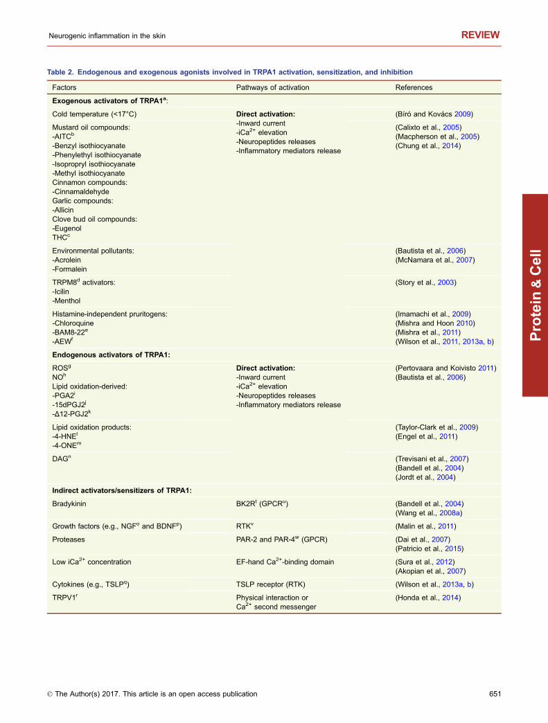
Table 2. Endogenous and exogenous agonists involved in TRPA1 activation, sensitization, and inhibition
Factors Pathways of activation References
Exogenous activators of TRPA1a:
Cold temperature (<17°C) Direct activation:-Inward current-iCa2+ elevation-Neuropeptides releases-Inflammatory mediators release
(Bíró and Kovács 2009)
Mustard oil compounds:-AITCb
-Benzyl isothiocyanate-Phenylethyl isothiocyanate-Isopropryl isothiocyanate-Methyl isothiocyanateCinnamon compounds:-CinnamaldehydeGarlic compounds:-AllicinClove bud oil compounds:-EugenolTHCc
(Calixto et al., 2005)(Macpherson et al., 2005)(Chung et al., 2014)
Environmental pollutants:-Acrolein-Formalein
(Bautista et al., 2006)(McNamara et al., 2007)
TRPM8d activators:-Icilin-Menthol
(Story et al., 2003)
Histamine-independent pruritogens:-Chloroquine-BAM8-22e
-AEWf
(Imamachi et al., 2009)(Mishra and Hoon 2010)(Mishra et al., 2011)(Wilson et al., 2011, 2013a, b)
Endogenous activators of TRPA1:
ROSg
NOh
Lipid oxidation-derived:-PGA2i
-15dPGJ2j
-Δ12-PGJ2k
Direct activation:-Inward current-iCa2+ elevation-Neuropeptides releases-Inflammatory mediators release
(Pertovaara and Koivisto 2011)(Bautista et al., 2006)
Lipid oxidation products:-4-HNEl
-4-ONEm
(Taylor-Clark et al., 2009)(Engel et al., 2011)
DAGn (Trevisani et al., 2007)(Bandell et al., 2004)(Jordt et al., 2004)
Indirect activators/sensitizers of TRPA1:
Bradykinin BK2Rt (GPCRu) (Bandell et al., 2004)(Wang et al., 2008a)
Growth factors (e.g., NGFo and BDNFp) RTKv (Malin et al., 2011)
Proteases PAR-2 and PAR-4w (GPCR) (Dai et al., 2007)(Patricio et al., 2015)
Low iCa2+ concentration EF-hand Ca2+-binding domain (Sura et al., 2012)(Akopian et al., 2007)
Cytokines (e.g., TSLPq) TSLP receptor (RTK) (Wilson et al., 2013a, b)
TRPV1r Physical interaction orCa2+ second messenger
(Honda et al., 2014)
Neurogenic inflammation in the skin REVIEW
© The Author(s) 2017. This article is an open access publication 651
Protein
&Cell
Macpherson et al., 2007). A EF-hand domain implicated incalcium-dependent gating was also found in its NH2 region(Zurborg et al., 2007; Doerner et al., 2007). Other specificdomains have also been described such as a PIP2 bindingsite as well as CAMKII and PKC phosphorisation site (Choiet al., 2014). The transmembrane domain S6 are essentialfor gating by antagonists and agonists (Chen et al., 2008).
iCa2+ potentiates TRPA1 activation
Growing evidence suggests that Ca2+ regulates TRPA1 bypotentializing and inactivating its activity at low and highintracellular concentrations, respectively (Jordt et al., 2004;Doerner et al., 2007). On the one hand, Ca2+ potentiatedcinnamaldehyde-, AITC-, and carvacrol-evoked currents inHEK293 cells expressing TRPA1. In addition, an increase iniCa2+ also seems to directly act on the activation of TRPA1by eliciting a current in a PLC-independent manner andserves as a co-agonist with icilin that directly interacts withTRPA1 (Zurborg et al., 2007; Doerner et al., 2007; Wanget al., 2008b). On the other hand, Ca2+ could also inactivateTRPA1. Indeed, even though high extracellular Ca2+ con-centrations could enhance a cinnamaldehyde-evoked cur-rent, they could also induce rapid inactivation, which has notbeen found at low extracellular Ca2+ concentrations (Wanget al., 2008b). Hence, Ca2+ appears to be a crucial factor forthe regulation of TRPA1 activity, suggesting that other TRPchannels and GPCRs could modulate TRPA1 activity bymobilizing iCa2+.
Intracellular sensitizers of TRPA1
Several studies suggest that TRP channels may be poten-tiated by endogenous DAG and IP3, the products of PIP2breakdown, by Ca2+ release from internal stores, by PKCactivation or by the formation of endogenous lipid oxidation-derived and products in sensory neurons (Wang et al.,2008a; Mizumura et al., 2009). TRPA1-expressing CHO
cells were also sensitive to DAG, arachidonic acid, and iCa2+
(Jordt et al., 2004; Bandell et al., 2004). PIP2 has an inhi-bitory effect not only on TRPV1 activity (refer to section In-tracellular pathways of TRPV1 sensitization) but also onTRPA1 activity, since PIP2 has been shown to inhibit AITC-and mustard oil-evoked currents (Kim et al., 2008; Kara-shima et al., 2008). Moreover, the reduction of the PIP2 levelat the membrane by polyphosphatase (e.g., PPPi), PIP2antibody or phenylarsine oxide enhanced the AITC- andmustard oil-evoked currents via TRPA1 sensitization (Kimet al., 2008; Karashima et al., 2008). Taken together, thesedata suggest that TRPA1 activity could be sensitized by theactivation of numerous GPCRs or receptor tyrosine kinases(RTKs) via the cAMP/PKA and PLC/PKC pathways followingCa2+ elevation (Taylor-Clark et al., 2008; Andersson et al.,2008; Taylor-Clark et al., 2009)
TRPA1 sensitization by inflammatory mediators
Several inflammatory mediators, such as growth factors,bradykinins, proteases, and, more recently, the inflammatorycytokine TSLP, have been found to act indirectly on TRPA1activity and expression through specific receptor-dependentsignaling pathways (Diogenes et al., 2007; Dai et al., 2007;Wang et al., 2008a; Malin et al., 2011). It has now beenclearly established that TRPA1 in sensory neurons mediatesthe CNI responses to bradykinin (Jordt et al., 2004; Bandellet al., 2004; Bautista et al., 2006; Wang et al., 2008a), NGF,and brain-derived neurotrophic factor (BDNF) (Malin et al.,2011) by potentiating the activity of the channel and impair-ing its desensitization after repeated activation. In addition,NGF has been found to up-regulate the expression ofTRPA1 mRNA via p38 MAPK activation in trigeminal gangliaand sensory neurons. Taken together, these mediatorsenhance the activity and prevent the desensitization ofTRPA1 and consequently facilitate pain, hyperalgesia, andallodynia (Obata et al., 2005; Diogenes et al., 2007). TSLP
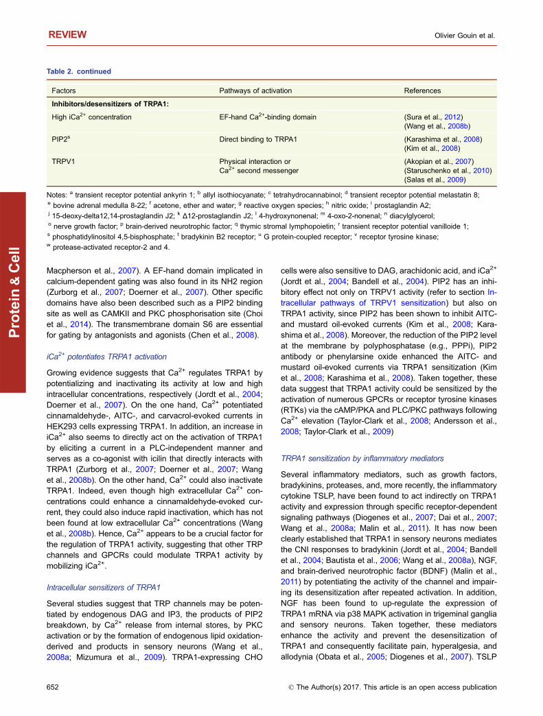
Table 2. continued
Factors Pathways of activation References
Inhibitors/desensitizers of TRPA1:
High iCa2+ concentration EF-hand Ca2+-binding domain (Sura et al., 2012)(Wang et al., 2008b)
PIP2s Direct binding to TRPA1 (Karashima et al., 2008)(Kim et al., 2008)
TRPV1 Physical interaction orCa2+ second messenger
(Akopian et al., 2007)(Staruschenko et al., 2010)(Salas et al., 2009)
Notes: a transient receptor potential ankyrin 1; b allyl isothiocyanate; c tetrahydrocannabinol; d transient receptor potential melastatin 8;e bovine adrenal medulla 8-22; f acetone, ether and water; g reactive oxygen species; h nitric oxide; i prostaglandin A2;j 15-deoxy-delta12,14-prostaglandin J2; k Δ12-prostaglandin J2; l 4-hydroxynonenal; m 4-oxo-2-nonenal; n diacylglycerol;o nerve growth factor; p brain-derived neurotrophic factor; q thymic stromal lymphopoietin; r transient receptor potential vanilloide 1;s phosphatidylinositol 4,5-bisphosphate; t bradykinin B2 receptor; u G protein-coupled receptor; v receptor tyrosine kinase;w protease-activated receptor-2 and 4.
REVIEW Olivier Gouin et al.
652 © The Author(s) 2017. This article is an open access publication
Protein
&Cell
released from keratinocytes potentiated TRPA1 activity bybinding to its specific receptor (TSLPR) on sensory neuronsin the skin of AD patients and in mouse models of AD. WhileTSLP-evoked sensory neuron activation and consequentchronic itching in AD has been established, the role of TSLP-triggered neuropeptide release in neurons and the subse-quent CNI remain an open question (Wilson et al., 2013a, b).
While the involvement of endogenous proteases inTRPV1 sensitization via PAR-2 and PAR-4 activation is wellestablished (refer to section Inflammatory mediators that useTRPV1 intracellular sensitization pathways), the implicationof these proteases in TRPA1 sensitization remains poorlyunderstood. PAR2 activation has been demonstrated toenhance TRPA1 agonist-evoked pain behavior in rats viaPLC activation and subsequent PIP2 hydrolysis (Dai et al.,2007). In addition, PAR-2-evoked mechanical and coldallodynia, as well as heat hyperalgesia, was found to bedependent on the PKA and PKC signaling pathways, whichsensitize TRPA1 (Chen et al., 2011). The involvement ofPAR-4 in the sensitization of TRPA1 is poorly studied, but arecent finding demonstrated its potential role in pruritus: thedorsal intradermal administration of the PAR-4 agonist pep-tide AYPGKF-NH2 elicited intense scratching behavior via aSP release in mice, which was abolished by the TRPV1 andTRPA1 antagonists SB366791 and HC-030031, respectively(Patricio et al., 2015), but not in TRPA1-deficient mice. Anopposite pattern in PAR-4-evoked itching via TRPA1 bycompensatory mechanisms in TRPA1− deficient mice hasbeen hypothesized (Patricio et al., 2015; Petrus et al., 2007).Consequently, PAR-4 activation could sensitize TRPA1 incollaboration with TRPV1 and elicit itching behavior via therelease of neuropeptides from sensory neurons; however,some points still require clarification, mainly the intracellularpathways underlying the TRPV1/TRPA1 sensitization viaPAR-4 activation (Patricio et al., 2015).
Finally, the intracellular pathways for many inflammatorymediators could sensitize TRPA1 in injured skin, contributingto channel activity and impairing the TRPA1 desensitization,thereby enhancing peripheral inflammatory responses viathe production of other inflammatory mediators.
A pro-inflammatory response induced by TRPA1activation
In addition to its ability to induce the release of neuropep-tides from sensory neurons, TRPA1 seems to play animportant role in the modulation of numerous genes thatcould amplify the cutaneous inflammatory process. Theinvolvement of TRPA1 in the regulation of 1,843 (2,423probe sets) genes in whole trigeminal ganglia isolated fromAEW-treated mice has been established (Wilson et al.,2013a). In this model, TRPA1 activation by AEW highly up-or down-regulated the expression of several cytokines,plasmatic receptors, and ion channels and affected growthregulation, intracellular pathways, and immune cell speci-ficity proteins. For example, AEW up-regulated the
inflammatory bradykinin receptor (BK2R) by 2-fold and up-regulated several itch receptors such as MRGPR and PAR-2in the trigeminal ganglia neurons (Wilson et al., 2013a).These three receptors sensitize and open TRPA1. Thus, theup-regulation of itch and inflammatory receptors in sensoryneurons strongly indicates a role for TRPA1 in chronic pru-ritus and CNI via the enhancement of sensory neuron sen-sitivity to itch and inflammatory mediators.
Growing evidence indicates that TRPA1 acts as aninflammatory regulator. The results of recent studies corre-lated the activation of TRPA1 with expressional changes inthe skin, as associated with cutaneous inflammation, AD,and pain that is blocked in TRPA1-deficient mice. Indeed,TRPA1 activation by oxazolone induced chronic dermatitis inmouse ears and concomitant up-regulation of inflammatorycytokines (i.e., IL-1β, IL-4, and IL-16 and chemokine (C-X-Cmotif) ligand 2 (CXCL-2)), neuropeptides (i.e., SP andendothelin (ET-1)), nerve growth factor (NGF), and neuro-transmitters (i.e., serotonin) known to induce AD, pruritus,and pain and increase nerve fiber density (Liu et al., 2013).Additional studies established the expressional change of9,340 genes linked to itching associated with TRPA1 in skinbiopsies from AEW-treated mice, showing that TRPA1 up-regulated the expression of 79% of itch-related genes knownto be involved in the initiation and maintenance of chronicitching as well as pruriginous skin disorders (e.g., the IL-31receptor (IL-31RA), aquaporin 3, and IL-33 (Wilson et al.,2013a; Nakahigashi et al., 2011; Nobbe et al., 2012; Olssonet al., 2006; Sonkoly et al., 2006). In these studies, theexpressional changes induced by oxazolone or AEW wereabrogated in the TRPA1−/− mice and in WT mice treated withTRPA1 antagonists (Wilson et al., 2013a; Liu et al., 2013).These data strongly suggest the involvement of TRPA1 inpruritus.
TRPA1 is also expressed in the basal keratinocytes,where it acts as a pain, thermal, and chemical sensor (Ato-yan et al., 2009). TRPA1 was found to modulate inflamma-tory gene expression in keratinocytes by increasing theexpression of IL-1α and IL-1β (Atoyan et al., 2009) andcause the secretion of PgE2 (Jain et al., 2011). Both IL-1 andPgE2 are known to be involved in skin inflammation anditching by decreasing the mechanical threshold and thethermal responsiveness of the sensory nerve endings, whichcould facilitate CNI (Binshtok et al., 2008). In addition, inkeratinocytes, TRPA1 activation increases heat shock pro-tein (HSP) 27, which is known to up-regulate the expressionof inflammatory cytokines such as IL-1β, TNF-α, and IL-6 inthe skin during murine allergic contact hypersensitivity(Yusuf et al., 2009; Atoyan et al., 2009). Altogether, thesefindings indicate that TRPA1 activation contributes to theproduction of several cytokines from keratinocytes thatdirectly trigger or enhance CNI by acting on neighboringtarget cells.
In addition to keratinocytes, TRPA1 acts on skin immunecells, but it appears to have an anti-inflammatory role in mono-cytes/macrophages. The TRPA1 activators cinnamaldehyde
Neurogenic inflammation in the skin REVIEW
© The Author(s) 2017. This article is an open access publication 653
Protein
&Cell
and carvacol have been found to inhibit bacterial lipopolysac-charides (LPS)-induced nuclear factor-kappa B (NF-κB)-medi-ated promoter activity and reduce the expression of iNOS,COX2, and TNF-α and subsequent NO production in macro-phages (Chao et al., 2008; Romano et al., 2013). Similarly,cinnamaldehyde inhibited bacterial LPS- and lipoteichoic acid(LTA)-mediated IL-1β, IL-6, and TNF-α release in murine mac-rophages and human blood monocytes, presumably throughROS production (Hsu and Wen 2002; Chao et al., 2008;Romanoetal., 2013). Furthermore, an increase inTNF-αaswellas the induction of both kinnitric oxide synthases (iNOS) andCOX2, which are involved in the formation of NO and PGE2,respectively, are known to act in the pathogenesis of severalinflammatory skin diseases, such as AD, psoriasis, and pruritus(Ormerod et al., 1998; Kim et al., 2015; Ostadhadi et al., 2015;Ahn et al., 2016; Sereflican et al., 2016). It would thereforeappear that TRPA1 acts as an anti-inflammatory actor inmonocytes and macrophages, but these data are controversial(Billeter et al., 2015). The activation of TRPA1 by cinnamalde-hyde acts on adaptive immunity by suppressing lymphoprolif-eration and promoting T-cell maturation, as revealed by T-celldifferentiation from CD4 and CD8 double-positive cells to CD4or CD8 single-positive cells in LPS-treated mouse splenocytes(Koh et al., 1998). These results imply that TRPA1 couldenhance inflammation and immunoreactivity by directly actingon lymphocyte differentiation.
TRPA1 activation by AITC has been found to triggervasodilatation in rat cerebral arteries in an endothelium-de-pendent mechanism. TRPA1-mediated vasodilation triggersCa2+ influx in rat endothelial arteries through Ca2+-activatedpotassium channels in endothelial cells and inwardly recti-fying potassium channels in arterial myocytes (Earley et al.,2009; Qian et al., 2013). This mechanism suggests that thischannel could also act in edema and vasodilatation duringCNI, but this hypothesis must be verified. However, anotherstudy established that TRPA1 activation by cinnamaldehydeplayed an anti-inflammatory role by suppressing the attach-ment of leukocytes to endothelial cells (Liao et al., 2008).
To conclude, TRPA1 activation and sensitization mediateskin inflammation by increasing the release of inflammatorymediators, although their role in immune cells remains to beclarified. In turn, secreted factors mediate the intracellularsensitization of TRPA1, thus facilitating its activation, whichin turn contributes to enhancement of the self-maintenanceof CNI.
CROSS-REGULATION BETWEEN TRPV1 ANDTRPA1
In addition to co-expression in a subset of sensory nervesand non-neuronal cells (keratinocytes), a functional interac-tion between TRPA1 and TRPV1 (cross-sensitization/de-sensitization) has been established, which suggests thecooperation between these channels to promote inflamma-tory thermal hyperalgesia, CNI, and pain (Bautista et al.,
2006; Akopian et al., 2007; Anand et al., 2008; Patil et al.,2010; Aubdool and Brain 2011; Spahn et al., 2014; Fischeret al., 2014).
This cross-communication between TRPA1 and TRPV1appears to involve the concentration of cytosolic Ca2+, oneof the main sensitizers/desensitizers of TRPA1 and TRPV1(Jordt et al., 2004; Jung et al., 2004; Doerner et al., 2007;Woo et al., 2008). It has been shown that capsaicin- andmustard oil-evoked currents desensitized each other viaiCa2+ elevation in TRPV1- and/or TRPA1-expressing CHOcells (Akopian et al., 2007). Another relevant study con-firmed the role of the influx of Ca2+ via TRPV1 as a secondmessenger for the desensitization of TRPA1. Indeed, TRPA1evoked a current in CHO cells expressing TRPA1, and thiscurrent was inhibited by the co-expression of TRPV1 (Patilet al., 2010). In addition, TRPA1 and TRPV1 were found tointeract by forming a physical and functional interaction atthe plasma membrane, as revealed by co-IP and FRETassays in TRPA1- and TRPV1-co-expressing CHO cells, aswell as in sensory neurons (Akopian et al., 2007; Salas et al.,2009; Staruschenko et al., 2010; Fischer et al., 2014). Inthese cells, TRPV1 regulated the desensitization of TRPA1,which appeared to be independent of iCa2+ (Staruschenkoet al., 2010).
On the other hand, evidence has established that theTRPV1 and TRPA1 channels also sensitize each other in aCa2+-dependent signaling pathway. Indeed, bradykinin andfacial capsaicin injections sensitized TRPA1 via TRPV1 in aCa2+-dependent manner in cultured trigeminal neurons frommice and rats, leading to inflammatory cold hyperalgesia(Bautista et al., 2006; Honda et al., 2014). The oppositepattern was also found: TRPA1 could sensitize TRPV1 toAITC-, cinnamaldehyde-, and mustard oil-enhanced heat-and capsaicin-evoked currents and Ca2+ elevation in aCa2+-, cAMP/PKA-, and TRPV1 phosphorylation-dependentmanner in sensory neurons and in HEK293 cells co-ex-pressing TRPV1 and TRPA1 (Anand et al., 2008; Alpizaret al., 2013; Spahn et al., 2014).
Altogether, the TRPV1-TRPA1 interaction and cross-communication via the iCa2+ concentration could inhibit orenhance the ability to a large variety of exogenous andendogenous activators to cause Ca2+ elevation, neuropep-tide release and, consequently, CNI.
CONCLUSIONS AND FUTURE DIRECTIONS
The TRPV1 and TRPA1 channels are at the core of theinflammatory process that occurs in various cutaneousneurogenic disorders that are pruritic diseases, such aspsoriasis, AD, and Netherton syndrome. It is now clear thatboth TRP channels act far beyond the sensory nerves bypotentiating the pathology of numerous skin disorders innon-neuronal skin cells, such as keratinocytes, mast cells,dendritic cells, and blood vessels. We still have a great dealto learn about these channels, notably about the change ofgene expression in sensory neurons and non-neuronal skin
REVIEW Olivier Gouin et al.
654 © The Author(s) 2017. This article is an open access publication
Protein
&Cell
cells, especially in relation to their role in the induction andself-maintenance of CNI (in vitro and in vivo). A great deal ofknowledge about their interaction, their cross-sensitization,and their sensitization by inflammatory mediators awaitsdiscovery, especially in relation to the potentiation and thechronicitization of skin disorders associated with CNI.Although the molecular mechanisms of TRPV1 and TRPA1sensitization induced during CNI and itching are still poorlyunderstood, their clinical relevance in pruritus, inflammatorythermal hyperalgesia, and pain are indisputable.
Targeting one or more of the intracellular signaling path-ways of TRP channel sensitization may allow new opportu-nities for the treatment of skin disorders associated with CNI.Further studies are necessary to test the potential effects oftreatment with protein signaling inhibitors in neurogenicinflamed skin. A new and interesting approach could also beto focus on the effect of the iCa2+ concentration to mediateTRP desensitization and thus block the self-maintenance ofCNI.
ACKNOWLEDGEMENTS
This work was supported by the Uriage Dermatological Laboratories,
Courbevoie, France. The authors wish to thank the entire team of the
Laboratory of Neurosciences of Brest (EA4685). O.G. has received
research grants from Uriage. NL has received research grants from
BASF, Johnson & Johnson. C.L.G.I. has received research grants
from Beiersdorf, Clarins, L’Oréal, Natura, Pierre Fabre. L.L. has
worked for Uriage. L.M. has received research grants from Almirall,
BASF, Beiersdorf, Bioderma, Celgene, Clarins, Expanscience, Gal-
derma, Johnson & Johnson, L’Oréal, Natura, Pierre Fabre, Sofibel,
Solabia, Uriage.
ABBREVIATIONS
12-HETE, 12-hydroxyeicosatetraenoic; AD, atopic dermatitis; BDNF,
brain-derived neurotrophic factor; CAMKII, Ca2+/calmodulin depen-
dent kinase II; CGRP, calcitonin gene-related peptide; CNI, cuta-
neous neurogenic inflammation; COX-2, cyclo-oxygenase-2; cryo-
EM, cryo-electron microscopy; DAG, diacylglycerol; DCs, dendritic
cells; DRG, dorsal root ganglion; eNOS, endothelial nitric oxide
synthase; GPCRs, G protein-coupled receptors; HSP, heat shock
protein; iCa2+, intracellular Ca2+ concentration; IL, interleukin; LPS,
lipopolysaccharides; LTA, lipoteichoic acid; MMPs, matrix metallo-
proteinases; NF-κB, nuclear factor-kappa B; NGF, nerve growth
factor; PAR, protease-activated receptor; PG, prostaglandins; PIP2,
phosphatidylinositol 4,5-bisphosphate; PKA, protein kinases A;
PKC, protein kinases C; PLA2, phospholipase A2; PLC, phospho-
lipase C; RTKs, receptor tyrosine kinases; SP, substance P; TNF-α,
tumor necrosis factor-α, TRP, transient receptor potential; TRPA1,
transient receptor potential ankyrin 1; TRPV1, transient receptor
potential vanilloid 1; TSLP, thymic stromal lymphopoietin; UV,
ultraviolet
COMPLIANCE WITH ETHICS GUIDELINES
The authors declare that they have no conflict of interest.
AUTHORS’ CONTRIBUTIONS
O.G. drafted the manuscript. R.L.G., K.L., N.L., C.L.G.I., and L.M.
helped to draft the manuscript. All authors reviewed and agreed to
the final version of the manuscript.
OPEN ACCESS
This article is distributed under the terms of the Creative Commons
Attribution 4.0 International License (http://creativecommons.org/
licenses/by/4.0/), which permits unrestricted use, distribution, and
reproduction in any medium, provided you give appropriate credit to
the original author(s) and the source, provide a link to the Creative
Commons license, and indicate if changes were made.
REFERENCES
Ahn S, Siddiqi MH, Aceituno VC et al (2016) Ginsenoside Rg5:Rk1
attenuates TNF-α/IFN-γ-induced production of thymus- and
activation-regulated chemokine (TARC/CCL17) and LPS-induced
NO production via downregulation of NF-κB/p38 MAPK/STAT1
signaling in human keratinocytes and macrophages. In Vitro Cell
Dev Biol Anim 52:287–295. doi:10.1007/s11626-015-9983-yAkaishi S, Ogawa R, Hyakusoku H (2008) Keloid and hypertrophic
scar: neurogenic inflammation hypotheses. Med Hypotheses
71:32–38. doi:10.1016/j.mehy.2008.01.032
Akiyama T, Ivanov M, Nagamine M et al (2016) Involvement of
TRPV4 in serotonin-evoked scratching. J Invest Dermatol
136:154–160. doi:10.1038/JID.2015.388Akopian AN, Ruparel NB, Jeske NA, Hargreaves KM (2007)
Transient receptor potential TRPA1 channel desensitization in
sensory neurons is agonist dependent and regulated by TRPV1-
directed internalization. J Physiol 583:175–193. doi:10.1113/
jphysiol.2007.133231
Alpizar YA, Boonen B, Gees M et al (2013) Allyl isothiocyanate
sensitizes TRPV1 to heat stimulation. Pflüg Arch—Eur J Physiol
466:507–515. doi:10.1007/s00424-013-1334-9Amadesi S, Nie J, Vergnolle N et al (2004) Protease-activated
receptor 2 sensitizes the capsaicin receptor transient receptor
potential vanilloid receptor 1 to induce hyperalgesia. J Neurosci
Off J Soc Neurosci 24:4300–4312. doi:10.1523/JNEUROSCI.
5679-03.2004
Amadesi S, Cottrell GS, Divino L et al (2006) Protease-activated
receptor 2 sensitizes TRPV1 by protein kinase Cε- and A-de-
pendent mechanisms in rats and mice. J Physiol 575:555–571.doi:10.1113/jphysiol.2006.111534
Anand U, Otto WR, Facer P et al (2008) TRPA1 receptor localisation
in the human peripheral nervous system and functional studies in
cultured human and rat sensory neurons. Neurosci Lett 438:221–227. doi:10.1016/j.neulet.2008.04.007
Andersson DA, Gentry C, Moss S, Bevan S (2008) Transient
receptor potential A1 is a sensory receptor for multiple products
of oxidative stress. J Neurosci Off J Soc Neurosci 28:2485–2494.doi:10.1523/JNEUROSCI.5369-07.2008
Neurogenic inflammation in the skin REVIEW
© The Author(s) 2017. This article is an open access publication 655
Protein
&Cell
Andreev YA, Vassilevski AA, Kozlov SA (2012) Molecules to
selectively target receptors for treatment of pain and neurogenic
inflammation. Recent Pat Inflamm Allergy Drug Discov 6:35–45Asfaha S, Cenac N, Houle S et al (2007) Protease-activated
receptor-4: a novel mechanism of inflammatory pain modulation.
Br J Pharmacol 150:176–185. doi:10.1038/sj.bjp.0706975Atoyan R, Shander D, Botchkareva NV (2009) Non-neuronal
expression of transient receptor potential type A1 (TRPA1) in
human skin. J Invest Dermatol 129:2312–2315. doi:10.1038/jid.2009.58
Aubdool AA, Brain SD (2011) Neurovascular aspects of skin
neurogenic inflammation. J Investig Dermatol Symp Proc Soc
Investig Dermatol Inc Eur Soc Dermatol Res 15:33–39. doi:10.1038/jidsymp.2011.8
Bandell M, Story GM, Hwang SW et al (2004) Noxious cold ion
channel TRPA1 is activated by pungent compounds and
bradykinin. Neuron 41:849–857Basu S, Srivastava P (2005) Immunological role of neuronal receptor
vanilloid receptor1expressedondendritic cells. ProcNatl AcadSci
U S A 102:5120–5125. doi:10.1073/pnas.0407780102Bautista DM, Jordt S-E, Nikai T et al (2006) TRPA1 mediates the
inflammatory actions of environmental irritants and proalgesic
agents. Cell 124:1269–1282. doi:10.1016/j.cell.2006.02.023Bautista DM, Pellegrino M, Tsunozaki M (2013) TRPA1: A gate-
keeper for inflammation. Annu Rev Physiol 75:181–200. doi:10.1146/annurev-physiol-030212-183811
Bhave G, Zhu W, Wang H et al (2002) cAMP-dependent protein
kinase regulates desensitization of the capsaicin receptor (VR1)
by direct phosphorylation. Neuron 35:721–731Bhave G, Hu H-J, Glauner KS et al (2003) Protein kinase C
phosphorylation sensitizes but does not activate the capsaicin
receptor transient receptor potential vanilloid 1 (TRPV1). Proc
Natl Acad Sci U S A 100:12480–12485. doi:10.1073/pnas.
2032100100
Billeter AT, Galbraith N, Walker S et al (2015) TRPA1 mediates the
effects of hypothermia on the monocyte inflammatory response.
Surgery 158:646–654. doi:10.1016/j.surg.2015.03.065Binshtok AM, Wang H, Zimmermann K et al (2008) Nociceptors are
interleukin-1β sensors. J Neurosci Off J Soc Neurosci 28:14062–14073. doi:10.1523/JNEUROSCI.3795-08.2008
Bíró T, Kovács L (2009) An “ice-cold” TR(i)P to skin biology: the role
of TRPA1 in human epidermal keratinocytes. J Invest Dermatol
129:2096–2099. doi:10.1038/jid.2009.179Bı ́ró T, Maurer M, Modarres S et al (1998) Characterization of
functional vanilloid receptors expressed by mast cells. Blood
91:1332–1340Boillat A, Alijevic O, Kellenberger S (2014) Calcium entry via TRPV1
but not ASICs induces neuropeptide release from sensory
neurons. Mol Cell Neurosci 61C:13–22. doi:10.1016/j.mcn.
2014.04.007
Briot A, Deraison C, Lacroix M et al (2009) Kallikrein 5 induces
atopic dermatitis-like lesions through PAR2-mediated thymic
stromal lymphopoietin expression in Netherton syndrome.
J Exp Med 206:1135–1147. doi:10.1084/jem.20082242
Bunker CB, Cerio R, Bull HA et al (1991) The effect of capsaicin
application on mast cells in normal human skin. Agents Actions
33:195–196
Burgess GM, Mullaney I, McNeill M et al (1989) Second messengers
involved in the mechanism of action of bradykinin in sensory
neurons in culture. J Neurosci 9:3314–3325Calixto JB, Kassuya CAL, André E, Ferreira J (2005) Contribution of
natural products to the discovery of the transient receptor
potential (TRP) channels family and their functions. Pharmacol
Ther 106:179–208. doi:10.1016/j.pharmthera.2004.11.008
Caterina MJ, Schumacher MA, Tominaga M et al (1997) The
capsaicin receptor: a heat-activated ion channel in the pain
pathway. Nature 389:816–824. doi:10.1038/39807Cesare P, Moriondo A, Vellani V, McNaughton PA (1999) Ion
channels gated by heat. Proc Natl Acad Sci U S A 96:7658–7663Chao LK, Hua K-F, Hsu H-Y et al (2008) Cinnamaldehyde inhibits
pro-inflammatory cytokines secretion from mono-
cytes/macrophages through suppression of intracellular signal-
ing. Food Chem Toxicol Int J Publ Br Ind Biol Res Assoc 46:220–231. doi:10.1016/j.fct.2007.07.016
Chen J, Zhang X-F, Kort ME et al (2008) Molecular determinants of
species-specific activation or blockade of TRPA1 channels.
J Neurosci 28:5063–5071. doi:10.1523/JNEUROSCI.0047-08.
2008
Chen Y, Yang C, Wang ZJ (2011) Proteinase-activated receptor 2
sensitizes transient receptor potential vanilloid 1, transient
receptor potential vanilloid 4, and transient receptor potential
ankyrin 1 in paclitaxel-induced neuropathic pain. Neuroscience
193:440–451. doi:10.1016/j.neuroscience.2011.06.085Chen D, Wang Z, Zhang Z et al (2013) Capsaicin up-regulates
protease-activated receptor-4 mRNA and protein in primary
cultured dorsal root ganglion neurons. Cell Mol Neurobiol
33:337–346. doi:10.1007/s10571-012-9899-yChing L-C, Kou YR, Shyue S-K et al (2011) Molecular mechanisms
of activation of endothelial nitric oxide synthase mediated by
transient receptor potential vanilloid type 1. Cardiovasc Res
91:492–501. doi:10.1093/cvr/cvr104Choi S-I, Yoo S, Lim JY, Hwang SW (2014) Are sensory TRP
channels biological alarms for lipid peroxidation? Int J Mol Sci
15:16430–16457. doi:10.3390/ijms150916430
Chuang HH, Prescott ED, Kong H et al (2001) Bradykinin and nerve
growth factor release the capsaicin receptor from PtdIns(4,5)P2-
mediated inhibition. Nature 411:957–962. doi:10.1038/35082088Chung G, Im ST, Kim YH et al (2014) Activation of transient receptor
potential ankyrin 1 by eugenol. Neuroscience 261:153–160.doi:10.1016/j.neuroscience.2013.12.047
Cocks TM, Moffatt JD (2000) Protease-activated receptors: sentries
for inflammation? Trends Pharmacol Sci 21:103–108. doi:10.
1016/S0165-6147(99)01440-6
Costa A, Eberlin S, Polettini AJ et al (2014) Neuromodulatory and
anti-inflammatory ingredient for sensitive skin: in vitro assess-
ment. Inflamm Allergy Drug Targets 13:191–198Dai Y, Moriyama T, Higashi T et al (2004) Proteinase-activated
receptor 2-mediated potentiation of transient receptor potential
vanilloid subfamily 1 activity reveals a mechanism for proteinase-
induced inflammatory pain. J Neurosci Off J Soc Neurosci
24:4293–4299. doi:10.1523/JNEUROSCI.0454-04.2004
Dai Y, Wang S, Tominaga M et al (2007) Sensitization of TRPA1 by
PAR2 contributes to the sensation of inflammatory pain. J Clin
Invest 117:1979–1987. doi:10.1172/JCI30951
REVIEW Olivier Gouin et al.
656 © The Author(s) 2017. This article is an open access publication
Protein
&Cell

Diogenes A, Akopian AN, Hargreaves KM (2007) NGF up-regulates
TRPA1: implications for orofacial pain. J Dent Res 86:550–555Docherty RJ, Yeats JC, Bevan S, Boddeke HW (1996) Inhibition of
calcineurin inhibits the desensitization of capsaicin-evoked cur-
rents in cultured dorsal root ganglion neurones from adult rats.
Pflüg Arch Eur J Physiol 431:828–837Doerner JF, Gisselmann G, Hatt H, Wetzel CH (2007) Transient
receptor potential channel A1 is directly gated by calcium ions.
J Biol Chem 282:13180–13189. doi:10.1074/jbc.M607849200
Earley S, Gonzales AL, Crnich R (2009) Endothelium-dependent
cerebral artery dilation mediated by TRPA1 and Ca2+-Activated
K+ channels. Circ Res 104:987–994. doi:10.1161/
CIRCRESAHA.108.189530
Engel MA, Leffler A, Niedermirtl F et al (2011) TRPA1 and substance
P mediate colitis in mice. Gastroenterology 141:1346–1358.doi:10.1053/j.gastro.2011.07.002
Fischer MJM, Balasuriya D, Jeggle P et al (2014) Direct evidence for
functional TRPV1/TRPA1 heteromers. Pflugers Arch. doi:10.
1007/s00424-014-1497-z
Frydas S, Varvara G, Murmura G et al (2013) Impact of capsaicin on
mast cell inflammation. Int J Immunopathol Pharmacol 26:597–600
Fu Q, Cheng J, Gao Y et al (2014) Protease-activated receptor 4: a
critical participator in inflammatory response. Inflammation.
doi:10.1007/s10753-014-9999-6
Gouin O, Lebonvallet N, L’Herondelle K et al (2015) Self-mainte-
nance of neurogenic inflammation contributes to a vicious cycle
in skin. Exp Dermatol. doi:10.1111/exd.12798
Herbert MK, Holzer P (2002) Neurogenic inflammation. I. Basic
mechanisms, physiology and pharmacology. Anästhesiol Inten-
sivmed Notfallmedizin Schmerzther AINS 37:314–325. doi:10.
1055/s-2002-32233
Himi N, Hamaguchi A, Hashimoto K et al (2012) Calcium influx
through the TRPV1 channel of endothelial cells (ECs) correlates
with a stronger adhesion between monocytes and ECs. Adv Med
Sci 57:224–229. doi:10.2478/v10039-012-0044-4Hinman A, Chuang H-H, Bautista DM, Julius D (2006) TRP channel
activation by reversible covalent modification. Proc Natl Acad Sci
U S A 103:19564–19568. doi:10.1073/pnas.0609598103Honda K, Shinoda M, Furukawa A et al (2014) TRPA1 contributes to
capsaicin-induced facial cold hyperalgesia in rats. Eur J Oral Sci.
doi:10.1111/eos.12157
Hoogerwerf WA, Zou L, Shenoy M et al (2001) The proteinase-
activated receptor 2 is involved in nociception. J Neurosci Off J
Soc Neurosci 21:9036–9042Horváth G, Kemény Á, Barthó L et al (2015) Effects of some natural
carotenoids on TRPA1- and TRPV1-induced neurogenic inflam-
matory processes in vivo in the mouse skin. J Mol Neurosci MN.
doi:10.1007/s12031-014-0472-7
Hsu H-Y, Wen M-H (2002) Lipopolysaccharide-mediated reactive
oxygen species and signal transduction in the regulation of
interleukin-1 gene expression. J Biol Chem 277:22131–22139.doi:10.1074/jbc.M111883200
Huang L-YM, Neher E (1996) Ca2+-dependent exocytosis in the
somata of dorsal root ganglion neurons. Neuron 17:135–145.doi:10.1016/S0896-6273(00)80287-1
Huang SM, Bisogno T, Trevisani M et al (2002) An endogenous
capsaicin-like substance with high potency at recombinant and
native vanilloid VR1 receptors. Proc Natl Acad Sci U S A
99:8400–8405. doi:10.1073/pnas.122196999Huang J, Stohl LL, Zhou X et al (2011) Calcitonin gene-related
peptide inhibits chemokine production by human dermal
microvascular endothelial cells. Brain Behav Immun 25:787–799. doi:10.1016/j.bbi.2011.02.007
Hwang SW, Cho H, Kwak J et al (2000) Direct activation of capsaicin
receptors by products of lipoxygenases: endogenous capsaicin-
like substances. Proc Natl Acad Sci U S A 97:6155–6160Imamachi N, Park GH, Lee H et al (2009) TRPV1-expressing
primary afferents generate behavioral responses to pruritogens
via multiple mechanisms. Proc Natl Acad Sci U S A 106:11330–11335. doi:10.1073/pnas.0905605106
Inoue K, Koizumi S, Fuziwara S et al (2002) Functional vanilloid
receptors in cultured normal human epidermal keratinocytes.
Biochem Biophys Res Commun 291:124–129. doi:10.1006/bbrc.2002.6393
Jain A, Brönneke S, Kolbe L et al (2011) TRP-channel-specific
cutaneous eicosanoid release patterns. Pain 152:2765–2772.doi:10.1016/j.pain.2011.08.025
Jancsó N, Jancsó-Gábor A, Szolcsányi J (1967) Direct evidence for
neurogenic inflammation and its prevention by denervation and
by pretreatment with capsaicin. Br J Pharmacol Chemother
31:138–151Jans R, Sartor M, Jadot M, Poumay Y (2004) Calcium entry into
keratinocytes induces exocytosis of lysosomes. Arch Dermatol
Res 296:30–41. doi:10.1007/s00403-004-0469-0Jeske NA, Patwardhan AM, Gamper N et al (2006) Cannabinoid
WIN 55,212-2 regulates TRPV1 phosphorylation in sensory
neurons. J Biol Chem 281:32879–32890. doi:10.1074/jbc.
M603220200
Jia X, Zhang H, Cao X et al (2014) Activation of TRPV1 mediates
thymic stromal lymphopoietin release via the Ca2+/NFAT pathway
in airway epithelial cells. FEBS Lett 588:3047–3054. doi:10.1016/j.febslet.2014.06.018
Jian T, Yang N, Yang Y et al (2016) TRPV1 and PLC participate in
histamine H4 receptor-induced itch. Neural Plast 2016:1682972.
doi:10.1155/2016/1682972
Jordt S-E, Bautista DM, Chuang H-H et al (2004) Mustard oils and
cannabinoids excite sensory nerve fibres through the TRP
channel ANKTM1. Nature 427:260–265. doi:10.1038/
nature02282
Jung J, Shin JS, Lee S-Y et al (2004) Phosphorylation of vanilloid
receptor 1 by Ca2+/calmodulin-dependent kinase II regulates its
vanilloid binding. J Biol Chem 279:7048–7054. doi:10.1074/jbc.M311448200
Karashima Y, Prenen J, Meseguer V et al (2008) Modulation of the
transient receptor potential channel TRPA1 by phosphatidylinos-
itol 4,5-biphosphate manipulators. Pflüg Arch Eur J Physiol
457:77–89. doi:10.1007/s00424-008-0493-6Kark T, Bagi Z, Lizanecz E et al (2008) Tissue-specific regulation of
microvascular diameter: opposite functional roles of neuronal and
smooth muscle located vanilloid receptor-1. Mol Pharmacol
73:1405–1412. doi:10.1124/mol.107.043323
Neurogenic inflammation in the skin REVIEW
© The Author(s) 2017. This article is an open access publication 657
Protein
&Cell
Kim D, Cavanaugh EJ, Simkin D (2008) Inhibition of transient
receptor potential A1 channel by phosphatidylinositol-4,5-bispho-
sphate. Am J Physiol Cell Physiol 295:C92–99. doi:10.1152/
ajpcell.00023.2008
Kim CG, Kang M, Lee Y-H et al (2015) Bathing effects of various
seawaters on allergic (atopic) dermatitis-like skin lesions induced
by 2,4-dinitrochlorobenzene in hairless mice. Evid-Based Com-
plement Altern Med ECAM 2015:179185. doi:10.1155/2015/
179185
Kim S, Barry DM, Liu X-Y et al (2016) Facilitation of TRPV4 by
TRPV1 is required for itch transmission in some sensory neuron
populations. Sci Signal 9:ra71. doi:10.1126/scisignal.aaf1047
Koh WS, Yoon SY, Kwon BM et al (1998) Cinnamaldehyde inhibits
lymphocyte proliferation and modulates T-cell differentiation. Int J
Immunopharmacol 20:643–660Kubanov AA, Katunina OR, Chikin VV (2015) Expression of
neuropeptides, neurotrophins, and neurotransmitters in the skin
of patients with atopic dermatitis and psoriasis. Bull Exp Biol Med
159:318–322. doi:10.1007/s10517-015-2951-4Kürkçüoğlu N, Alaybeyi F (1991) Substance P immunoreactivity in
rosacea. J Am Acad Dermatol 25:725–726Kwak IS, Choi YH, Jang YC, Lee YK (2014) Immunohistochemical
analysis of neuropeptides (protein gene product 9.5, substance P
and calcitonin gene-related peptide) in hypertrophic burn scar
with pain and itching. Burns 40:1661–1667. doi:10.1016/j.burns.2014.04.004
Lee YM, Kim YK, Kim KH et al (2009) A novel role for the TRPV1
channel in UV-induced matrix metalloproteinase (MMP)-1 expres-
sion in HaCaTcells. J Cell Physiol 219:766–775. doi:10.1002/jcp.21729
Lee YM, Kang SM, Lee SR et al (2011) Inhibitory effects of TRPV1
blocker on UV-induced responses in the hairless mice. Arch
Dermatol Res 303:727–736. doi:10.1007/s00403-011-1153-9Liao B-C, Hsieh C-W, Liu Y-C et al (2008) Cinnamaldehyde inhibits
the tumor necrosis factor-α-induced expression of cell adhesion
molecules in endothelial cells by suppressing NF-κB activation:
Effects upon IκB and Nrf2. Toxicol Appl Pharmacol 229:161–171.doi:10.1016/j.taap.2008.01.021
Liao M, Cao E, Julius D, Cheng Y (2013) Structure of the TRPV1 ion
channel determined by electron cryo-microscopy. Nature
504:107–112. doi:10.1038/nature12822Lin Z, Chen Q, Lee M et al (2012) Exome sequencing reveals
mutations in TRPV3 as a cause of Olmsted syndrome. Am J Hum
Genet 90:558–564. doi:10.1016/j.ajhg.2012.02.006Liu B, Escalera J, Balakrishna S et al (2013) TRPA1 controls
inflammation and pruritogen responses in allergic contact der-
matitis. FASEB J Off Publ Fed Am Soc Exp Biol 27:3549–3563.doi:10.1096/fj.13-229948
Luo D, Zhang Y-W, Peng W-J et al (2008) Transient receptor
potential vanilloid 1-mediated expression and secretion of
endothelial cell-derived calcitonin gene-related peptide. Regul
Pept 150:66–72. doi:10.1016/j.regpep.2008.05.007Macpherson LJ, Geierstanger BH, Viswanath V et al (2005) The
pungency of garlic: activation of TRPA1 and TRPV1 in response
to allicin. Curr Biol CB 15:929–934. doi:10.1016/j.cub.2005.04.018
Macpherson LJ, Dubin AE, Evans MJ et al (2007) Noxious
compounds activate TRPA1 ion channels through covalent
modification of cysteines. Nature 445:541–545. doi:10.1038/
nature05544
Malin S, Molliver D, Christianson JA et al (2011) TRPV1 and TRPA1
function and modulation are target tissue dependent. J Neurosci
Off J Soc Neurosci 31:10516–10528. doi:10.1523/JNEUROSCI.
2992-10.2011
McNamara CR, Mandel-Brehm J, Bautista DM et al (2007) TRPA1
mediates formalin-induced pain. Proc Natl Acad Sci U S A
104:13525–13530. doi:10.1073/pnas.0705924104Meseguer V, Alpizar YA, Luis E et al (2014) TRPA1 channels
mediate acute neurogenic inflammation and pain produced by
bacterial endotoxins. Nat Commun 5:3125. doi:10.1038/
ncomms4125
Mishra SK, Hoon MA (2010) Ablation of TrpV1 neurons reveals their
selective role in thermal pain sensation. Mol Cell Neurosci
43:157–163. doi:10.1016/j.mcn.2009.10.006
Mishra SK, Tisel SM, Orestes P et al (2011) TRPV1-lineage neurons
are required for thermal sensation. EMBO J 30:582–593. doi:10.1038/emboj.2010.325
Mizumura K, Sugiura T, Katanosaka K et al (2009) Excitation and
sensitization of nociceptors by bradykinin: what do we know? Exp
Brain Res 196:53–65. doi:10.1007/s00221-009-1814-5Mohapatra DP, Nau C (2003) Desensitization of capsaicin-activated
currents in the vanilloid receptor TRPV1 is decreased by the
cyclic AMP-dependent protein kinase pathway. J Biol Chem
278:50080–50090. doi:10.1074/jbc.M306619200
Mohapatra DP, Nau C (2005) Regulation of Ca2+-dependent
desensitization in the vanilloid receptor TRPV1 by calcineurin
and cAMP-dependent protein kinase. J Biol Chem 280:13424–13432. doi:10.1074/jbc.M410917200
Moniaga CS, Jeong SK, Egawa G et al (2013) Protease activity
enhances production of thymic stromal lymphopoietin and
basophil accumulation in flaky tail mice. Am J Pathol 182:841–851. doi:10.1016/j.ajpath.2012.11.039
Nakahigashi K, Kabashima K, Ikoma A et al (2011) Upregulation of
aquaporin-3 is involved in keratinocyte proliferation and epider-
mal hyperplasia. J Invest Dermatol 131:865–873. doi:10.1038/jid.2010.395
Nakanishi M, Hata K, Nagayama T et al (2010) Acid activation of
Trpv1 leads to an up-regulation of calcitonin gene-related peptide
expression in dorsal root ganglion neurons via the CaMK-CREB
cascade: a potential mechanism of inflammatory pain. Mol Biol
Cell 21:2568–2577. doi:10.1091/mbc.E10-01-0049
Nobbe S, Dziunycz P, Mühleisen B et al (2012) IL-31 expression by
inflammatory cells is preferentially elevated in atopic dermatitis.
Acta Derm Venereol 92:24–28. doi:10.2340/00015555-1191Numazaki M, Tominaga T, Toyooka H, Tominaga M (2002) Direct
phosphorylation of capsaicin receptor VR1 by protein kinase
Cepsilon and identification of two target serine residues. J Biol
Chem 277:13375–13378. doi:10.1074/jbc.C200104200Obata K, Katsura H, Mizushima T et al (2005) TRPA1 induced in
sensory neurons contributes to cold hyperalgesia after inflam-
mation and nerve injury. J Clin Invest 115:2393–2401. doi:10.1172/JCI25437
REVIEW Olivier Gouin et al.
658 © The Author(s) 2017. This article is an open access publication
Protein
&Cell
Olsson M, Broberg A, Jernås M et al (2006) Increased expression of
aquaporin 3 in atopic eczema. Allergy 61:1132–1137. doi:10.
1111/j.1398-9995.2006.01151.x
Oprée A, Kress M (2000) Involvement of the proinflammatory
cytokines tumor necrosis factor-alpha, IL-1 beta, and IL-6 but not
IL-8 in the development of heat hyperalgesia: effects on heat-
evoked calcitonin gene-related peptide release from rat skin.
J Neurosci Off J Soc Neurosci 20:6289–6293Ormerod AD, Weller R, Copeland P et al (1998) Detection of nitric
oxide and nitric oxide synthases in psoriasis. Arch Dermatol Res
290:3–8Ostadhadi S, Haj-Mirzaian A, Azimi E et al (2015) Involvement of
nitric oxide in serotonin-induced scratching in mice. Clin Exp
Dermatol 40:647–652. doi:10.1111/ced.12605Patil MJ, Jeske NA, Akopian AN (2010) Transient receptor potential
V1 regulates activation and modulation of transient receptor
potential A1 by Ca2+. Neuroscience 171:1109–1119. doi:10.1016/j.neuroscience.2010.09.031
Patricio ES, Costa R, Figueiredo CP et al (2015) Mechanisms
underlying the scratching behavior induced by the activation of
proteinase-activated receptor-4 in mice. J Invest Dermatol
135:2484–2491. doi:10.1038/jid.2015.183PatwardhanAM,JeskeNA,PriceTJet al (2006)ThecannabinoidWIN
55,212-2 inhibits transient receptor potential vanilloid 1 (TRPV1)
and evokes peripheral antihyperalgesia via calcineurin. Proc Natl
Acad Sci U S A 103:11393–11398. doi:10.1073/pnas.0603861103Paulsen CE, Armache J-P, Gao Y et al (2015) Structure of the
TRPA1 ion channel suggests regulatory mechanisms. Nature
520:511–517. doi:10.1038/nature14367Pertovaara A, Koivisto A (2011) TRPA1 ion channel in the spinal
dorsal horn as a therapeutic target in central pain hypersensitivity
and cutaneous neurogenic inflammation. Eur J Pharmacol
666:1–4. doi:10.1016/j.ejphar.2011.05.027Petrus M, Peier AM, Bandell M et al (2007) A role of TRPA1 in
mechanical hyperalgesia is revealed by pharmacological inhibi-
tion. Mol Pain 3:40. doi:10.1186/1744-8069-3-40
Prescott ED, Julius D (2003) A modular PIP2 binding site as a
determinant of capsaicin receptor sensitivity. Science 300:1284–1288. doi:10.1126/science.1083646
Qian X, Francis M, Solodushko V et al (2013) Recruitment of
dynamic endothelial Ca2+ signals by the TRPA1 channel activator
AITC in rat cerebral arteries. Microcirc N Y N 1994 20:138–148.doi:10.1111/micc.12004
Rajasekhar P, Poole DP, Liedtke W et al (2015) P2Y1 receptor
activation of the TRPV4 ion channel enhances purinergic
signaling in satellite glial cells. J Biol Chem 290:29051–29062.doi:10.1074/jbc.M115.689729
Riol-Blanco L, Ordovas-Montanes J, Perro M et al (2014) Nocicep-
tive sensory neurons drive interleukin-23-mediated psoriasiform
skin inflammation. Nature 510:157–161. doi:10.1038/
nature13199
Romano B, Borrelli F, Fasolino I et al (2013) The cannabinoid TRPA1
agonist cannabichromene inhibits nitric oxide production in
macrophages and ameliorates murine colitis. Br J Pharmacol
169:213–229. doi:10.1111/bph.12120Roosterman D, Goerge T, Schneider SW et al (2006) Neuronal
control of skin function: the skin as a neuroimmunoendocrine
organ. Physiol Rev 86:1309–1379. doi:10.1152/physrev.00026.
2005
Russell FA, Veldhoen VE, Tchitchkan D, McDougall JJ (2010)
Proteinase-activated receptor-4 (PAR4) activation leads to sen-
sitization of rat joint primary afferents via a bradykinin B2
receptor-dependent mechanism. J Neurophysiol 103:155–163.doi:10.1152/jn.00486.2009
Salas MM, Hargreaves KM, Akopian AN (2009) TRPA1-mediated
responses in trigeminal sensory neurons: interaction between
TRPA1 and TRPV1. Eur J Neurosci 29:1568–1578. doi:10.1111/j.1460-9568.2009.06702.x
Salem SAM, Abdel Fattah NSA, Tantawy SMA et al (2013)
Neodymium-yttrium aluminum garnet laser versus pulsed dye
laser in erythemato-telangiectatic rosacea: comparison of clinical
efficacy and effect on cutaneous substance (P) expression.
J Cosmet Dermatol 12:187–194. doi:10.1111/jocd.12048Saunders CI, Fassett RG, Geraghty DP (2009) Up-regulation of
TRPV1 in mononuclear cells of end-stage kidney disease
patients increases susceptibility to N-arachidonoyl-dopamine
(NADA)-induced cell death. Biochim Biophys Acta 1792:1019–1026. doi:10.1016/j.bbadis.2009.07.008
Senning EN, Collins MD, Stratiievska A et al (2014) Regulation of
TRPV1 ion channel by phosphoinositide (4,5)-bisphosphate: the
role of membrane asymmetry. J Biol Chem 289:10999–11006.doi:10.1074/jbc.M114.553180
Sereflican B, Goksugur N, Bugdayci G et al (2016) Serum Visfatin,
Adiponectin, and Tumor Necrosis Factor Alpha (TNF-α) Levels in
Patients with Psoriasis and their Correlation with Disease
Severity. Acta Dermatovenerol Croat ADC 24:13–19Shim W-S, Tak M-H, Lee M-H et al (2007) TRPV1 mediates
histamine-induced itching via the activation of phospholipase A2
and 12-lipoxygenase. J Neurosci Off J Soc Neurosci 27:2331–2337. doi:10.1523/JNEUROSCI.4643-06.2007
Silva CR, Oliveira SM, Rossato MF et al (2011) The involvement of
TRPA1 channel activation in the inflammatory response evoked
by topical application of cinnamaldehyde to mice. Life Sci
88:1077–1087. doi:10.1016/j.lfs.2011.03.017Smart D, Gunthorpe MJ, Jerman JC et al (2000) The endogenous
lipid anandamide is a full agonist at the human vanilloid receptor
(hVR1). Br J Pharmacol 129:227–230. doi:10.1038/sj.bjp.
0703050
Smolyannikova VA, Kubanova AA, Karamova AE et al (2015) Role
of the skin expression of neuropeptides, neurotrophins and their
receptors in the pathogenesis of dermatoses. Arkh Patol 77:33–39
Sonkoly E, Muller A, Lauerma AI et al (2006) IL-31: a new link
between Tcells and pruritus in atopic skin inflammation. J Allergy
Clin Immunol 117:411–417. doi:10.1016/j.jaci.2005.10.033Southall MD, Li T, Gharibova LS et al (2003) Activation of epidermal
vanilloid receptor-1 induces release of proinflammatory mediators
in human keratinocytes. J Pharmacol Exp Ther 304:217–222.doi:10.1124/jpet.102.040675
Spahn V, Stein C, Zöllner C (2014) Modulation of transient receptor
vanilloid 1 activity by transient receptor potential ankyrin 1. Mol
Pharmacol 85:335–344. doi:10.1124/mol.113.088997
Ständer S, Moormann C, Schumacher M et al (2004) Expression of
vanilloid receptor subtype 1 in cutaneous sensory nerve fibers,
Neurogenic inflammation in the skin REVIEW
© The Author(s) 2017. This article is an open access publication 659
Protein
&Cell
mast cells, and epithelial cells of appendage structures. Exp
Dermatol 13:129–139. doi:10.1111/j.0906-6705.2004.0178.xStaruschenko A, Jeske NA, Akopian AN (2010) Contribution of
TRPV1-TRPA1 interaction to the single channel properties of the
TRPA1 channel. J Biol Chem 285:15167–15177. doi:10.1074/jbc.M110.106153
Steinhoff M, Neisius U, Ikoma A et al (2003) Proteinase-activated
receptor-2 mediates itch: a novel pathway for pruritus in human
skin. J Neurosci Off J Soc Neurosci 23:6176–6180Story GM, Peier AM, Reeve AJ et al (2003) ANKTM1, a TRP-like
channel expressed in nociceptive neurons, is activated by cold
temperatures. Cell 112:819–829Sugiura T, Tominaga M, Katsuya H, Mizumura K (2002) Bradykinin
lowers the threshold temperature for heat activation of vanilloid
receptor 1. J Neurophysiol 88:544–548Sura L, Zíma V, Marsakova L et al (2012) C-terminal Acidic cluster is
involved in Ca2+-induced regulation of human transient receptor
potential Ankyrin 1 channel. J Biol Chem 287:18067–18077.doi:10.1074/jbc.M112.341859
Szallasi A, Blumberg PM (1989) Resiniferatoxin, a phorbol-related
diterpene, acts as an ultrapotent analog of capsaicin, the irritant
constituent in red pepper. Neuroscience 30:515–520Taylor-Clark TE, Undem BJ, Macglashan DW et al (2008)
Prostaglandin-induced activation of nociceptive neurons via
direct interaction with transient receptor potential A1 (TRPA1).
Mol Pharmacol 73:274–281. doi:10.1124/mol.107.040832
Taylor-Clark TE, Ghatta S, Bettner W, Undem BJ (2009) Nitrooleic
acid, an endogenous product of nitrative stress, activates
nociceptive sensory nerves via the direct activation of TRPA1.
Mol Pharmacol 75:820–829. doi:10.1124/mol.108.054445
Teresiak-Mikołajczak E, Czarnecka-Operacz M, Jenerowicz D, Silny
W (2013) Neurogenic markers of the inflammatory process in
atopic dermatitis: relation to the severity and pruritus. Postȩpy
Dermatol Alergol 30:286–292. doi:10.5114/pdia.2013.38357Thorne PS, Hawk C, Kaliszewski SD, Guiney PD (1991) The
noninvasive mouse ear swelling assay. II. Testing the contact
sensitizing potency of fragrances. Fundam Appl Toxicol Off J Soc
Toxicol 17:807–820Tominaga M, Caterina MJ, Malmberg AB et al (1998) The cloned
capsaicin receptor integrates multiple pain-producing stimuli.
Neuron 21:531–543Tominaga M, Wada M, Masu M (2001) Potentiation of capsaicin
receptor activity by metabotropic ATP receptors as a possible
mechanism for ATP-evoked pain and hyperalgesia. Proc Natl
Acad Sci U S A 98:6951–6956. doi:10.1073/pnas.111025298Trevisani M, Siemens J, Materazzi S et al (2007) 4-Hydroxynonenal,
an endogenous aldehyde, causes pain and neurogenic inflam-
mation through activation of the irritant receptor TRPA1. Proc Natl
Acad Sci U S A 104:13519–13524. doi:10.1073/pnas.
0705923104
Ufret-Vincenty CA, Klein RM, Collins MD et al (2015) Mechanism for
phosphoinositide selectivity and activation of TRPV1 ion chan-
nels. J Gen Physiol 145:431–442. doi:10.1085/jgp.201511354Vellani V, Mapplebeck S, Moriondo A et al (2001) Protein kinase C
activation potentiates gating of the vanilloid receptor VR1 by
capsaicin, protons, heat and anandamide. J Physiol 534:813–825
Vellani V, Kinsey AM, Prandini M et al (2010) Protease activated
receptors 1 and 4 sensitize TRPV1 in nociceptive neurones. Mol
Pain 6:61. doi:10.1186/1744-8069-6-61
Vincent L, Vang D, Nguyen J et al (2013) Mast cell activation
contributes to sickle cell pathobiology and pain in mice. Blood
122:1853–1862. doi:10.1182/blood-2013-04-498105Wang S, Dai Y, Fukuoka T et al (2008a) Phospholipase C and
protein kinase A mediate bradykinin sensitization of TRPA1: a
molecular mechanism of inflammatory pain. Brain J Neurol
131:1241–1251. doi:10.1093/brain/awn060Wang YY, Chang RB, Waters HN et al (2008b) The nociceptor ion
channel TRPA1 is potentiated and inactivated by permeating
calcium ions. J Biol Chem 283:32691–32703. doi:10.1074/jbc.M803568200
Wei H, Koivisto A, Pertovaara A (2010) Spinal TRPA1 ion channels
contribute to cutaneous neurogenic inflammation in the rat.
Neurosci Lett 479:253–256. doi:10.1016/j.neulet.2010.05.073Wei T, Guo T-Z, Li W-W et al (2012) Keratinocyte expression of
inflammatory mediators plays a crucial role in substance P-in-
duced acute and chronic pain. J Neuroinflammation 9:181.
doi:10.1186/1742-2094-9-181
Wilson SR, Bautista DM (2014) Role of transient receptor potential
channels in acute and chronic itch. In: Carstens E, Akiyama T
(eds) Itch: mechanisms and treatment. CRC Press, Boca Raton
(FL)
Wilson SR, Gerhold KA, Bifolck-Fisher A et al (2011) TRPA1 is
required for histamine-independent, Mas-related G protein-cou-
pled receptor-mediated itch. Nat Neurosci 14:595–602. doi:10.1038/nn.2789
Wilson SR, Nelson AM, Batia L et al (2013a) The ion channel TRPA1
is required for chronic itch. J Neurosci Off J Soc Neurosci
33:9283–9294. doi:10.1523/JNEUROSCI.5318-12.2013
Wilson SR, Thé L, Batia LM et al (2013b) The epithelial cell-derived
atopic dermatitis cytokine TSLP activates neurons to induce itch.
Cell 155:285–295. doi:10.1016/j.cell.2013.08.057Woo DH, Jung SJ, Zhu MH et al (2008) Direct activation of transient
receptor potential vanilloid 1(TRPV1) by diacylglycerol (DAG).
Mol Pain 4:42. doi:10.1186/1744-8069-4-42
Xie Z (2009) [Role of thermo TRP channels in cutaneous neurogenic
inflammation and itch]. Zhejiang Xue Xue Bao Yi Xue Ban J
Zhejiang Univ Med Sci 38:409–414Yang D, Luo Z, Ma S et al (2010) Activation of TRPV1 by dietary
capsaicin improves endothelium-dependent vasorelaxation and
prevents hypertension. Cell Metab 12:130–141. doi:10.1016/j.
cmet.2010.05.015
Ye F, Deng P-Y, Li D et al (2007) Involvement of endothelial cell-
derived CGRP in heat stress-induced protection of endothelial
function. Vascul Pharmacol 46:238–246. doi:10.1016/j.vph.2006.10.008
Yusuf N, Nasti TH, Huang C-M et al (2009) Heat shock proteins
HSP27 and HSP70 are present in the skin and are important
mediators of allergic contact hypersensitivity. J Immunol Baltim
Md 1950 182:675–683Zhang N, Inan S, Cowan A et al (2005) A proinflammatory
chemokine, CCL3, sensitizes the heat- and capsaicin-gated ion
channel TRPV1. Proc Natl Acad Sci U S A 102:4536–4541.doi:10.1073/pnas.0406030102
REVIEW Olivier Gouin et al.
660 © The Author(s) 2017. This article is an open access publication
Protein
&Cell
Zhang X, Li L, McNaughton PA (2008) Proinflammatory mediators
modulate the heat-activated ion channel TRPV1 via the scaffold-
ing protein AKAP79/150. Neuron 59:450–461. doi:10.1016/j.
neuron.2008.05.015
Zhao P, Lieu T, Barlow N et al (2014) Cathepsin S causes
inflammatory pain via biased agonism of PAR2 and TRPV4.
J Biol Chem. doi:10.1074/jbc.M114.599712
Zhao Y, Bao L, Chan LS et al (2016) Aberrant wound healing in an
epidermal interleukin-4 transgenic mouse model of atopic der-
matitis. PLoS ONE. doi:10.1371/journal.pone.0146451
Ziegler SF, Roan F, Bell BD et al (2013) The biology of thymic
stromal lymphopoietin (TSLP). Adv Pharmacol San Diego Calif
66:129–155. doi:10.1016/B978-0-12-404717-4.00004-4
Zupanc GK (1996) Peptidergic transmission: from morphological
correlates to functional implications. Micron Oxf Engl 1993
(27):35–91Zurborg S, Yurgionas B, Jira JA et al (2007) Direct activation of the
ion channel TRPA1 by Ca2+. Nat Neurosci 10:277–279. doi:10.1038/nn1843
Zygmunt PM, Petersson J, Andersson DA et al (1999) Vanilloid
receptors on sensory nerves mediate the vasodilator action of
anandamide. Nature 400:452–457. doi:10.1038/22761
Neurogenic inflammation in the skin REVIEW
© The Author(s) 2017. This article is an open access publication 661
Protein
&Cell


![Medicinal & Aromatic Plants DI...contains varying amounts of 3-(4-methyl-1-oxo-2-pentenyl)furan (isoegomaketone, IK) in combination with PK [24], for their ability to activate h-TRPA1](https://img.pdfslide.us/doc/110x75/608939ca2303432f67124fa5/medicinal-aromatic-plants-di-contains-varying-amounts-of-3-4-methyl-1-oxo-2-pentenylfuran.jpg)