Embed Size (px)
Citation preview

1162 12 November 1966 Tropical Sprue-O'Brien and England BRITmsuml. compared with a mean level of 4.3 mpg. for the latter group.In a population in the tropics where folate deficiency wascommon sprue might also be expected to be common.
SummaryThe clinical and pathological findings in 42 patients with
" military tropical sprue" from South-east Asia are described.In the early stages intestinal malabsorption of fat, xylose, and
vitamin B.,, mild jejunal change, and folate deficiency werefound. In patients seen after two months these abnormalitieswere more severe. After four months the intestinal mucosa,both jejunal and ileal, had the appearance of partial villousatrophy, megaloblastic anaemia due to folate deficiencyoccurred, and there was a considerable fall in the serum vitamin-B,2 concentration. The regular clinical and pathologicalpattern was found helpful in diagnosis.Both folic acid and a mixed antibacterial course were effective
in causing a remission of intestinal malabsorption and a returnof the intestinal mucosa to normal. The symptomatic andhaematological responses to antibiotics were slow. Antibioticsappear to improve intestinal absorption of folic acid.These findings are in accord with the hypothesis that tem-
porary intestinal malabsorption arising from non-specific causesmay be maintained by secondary folate deficiency and bacterialproliferation in the small bowel. In a population in the tropicswhere folate deficiency is common sprue might also be expectedto be common.
We thank Lieutenant-General Sir Robert Drew for his activeinterest and the Wellcome Trust for generous financial support.We are particularly indebted to Professor D. L. Mollin for constanthelp and advice throughout this work. We also thank Dr. Leon
Ellenbogen, of the Lederle Laboratories, Pearl River, N.Y., for thesupply of intrinsic factor.
REFERENCES
Ayrey, F. (1948). Trans. roy. Soc. trop. Med. Hyg., 41, 377.Bahr, P. H. (1915). A Report on Researches on Sprue in Ceylon.
University Press, Cambridge.Baker, S. J., Ignatius, M., Mathan, V. I., Vaish, S. K., and Chacko, C. C.
(1962). In Ciba Foundation Study Group Report, No. 14, p. 84.Booth, C. C., and Mollin, D. L. (1964). Amer. 7. dig. Dis., 9, 770.Carmichael-Low, G. (1928). Quart. 7. Med., 21, 523.Crosby, W. H., and Kugler, H. W. (1957). Amer. 7. dig. Dis., 2, 236.Dacie, J. V. (1956). Practical Haematology, 2nd ed. Churchill, London.Elder, H. H. A. (1947). 7. trop. Med. Hyg., 50, 212.England, N. W. J., and O'Brien, W. (1966). Gut, 7, 128.French, J. M., Gaddie, R., and Smith, N. M. (1956). Quart. 7. Med., 25.
333.Gardner, F. H. (1958). New Engl. 7. Med., 258, 791, 835.Glass, G. B. J., Boyd, L. J., Gellin, G. A., and Stephanson, L. (1954)
Arch. Biochem., 51, 251.Holmes, R., Hourihane, D. O'B., and Booth, C. C. (1961). Lancet, 1, 81.Hutner, S. H., Bach, M. K., and Ross, G. I. M. (1956). 7. Protozool..
3, 101.Kay, A. W. (1953). Brit. med. 7., 2, 77Keele, K. D., and Bound, J. P. (1946). Ibid., 1, 79.Klipstein, F. A. (1964). Amer. 7. dig. Dir., 9, 778.Lindenbaum, J. (1965). Brit. med. 7., 2, 326.Manson, P. (1879-80). Chfna imp. Customs med. Rep. Shanghai, 19, 33.Mollin, D. L., Booth, C. C., and Baker, S. J. (1957). Brit. 7. Haemat.,
3, 412.and Ross, G. I. M. (1957). Vitamin-B,, and Intrinsic Factor, 1stEurop Symp. Hamburg, 1956, p. 413. Enke, Stuttgart.
O'Brien, W., and England, N. W J. (1964). Brit. med. 7., 2, 1573.Santini, R., Sheehy, T. W., and Martinez-de Jesus, J. (1961). Gastro-
enterology, 40, 772.Sheehy, T. W., Cohen, W. H., and Brodsky, J. P. (1963). Amer. 7. dig.
Dis., 8, 826.Wallace, D. K., and Legters, L. J. (1965). 7. Amer. med.
Ass., 194, 1069.Stefanini, M. (1948). Medicine (Baltimore), 27, 379.ten Thije, 0. J., Veeger, W., Braams, W. G., and Nieweg, H. 0. (1964).
Amer. 7. dig. Dis., 9, 774.van de Kamer, J. H., Huinick, H. ten B., and Weyers, H. A. (1949). 7
biol. Chem., 177, 347.Wallensten, S. (1954). Acta chir. scand., Suppl. No. 191.Waters, A. H., and Mollin, D. L. (1961). 7. clin. Path., 14, 335.
Tropical Sprue in Hong Kong
J. F. WEBB,* M.C., M.D., M.R.C.P.ED.; B. SIMPSON,t M.R.C.P.ED.
Brit. med. Y., 1966, 2, 1162-1166
Many descriptions of tropical sprue have been published, butseveral aspects of the disease are inadequately documented orthe subject of dispute, and four of these form the subject ofthis paper. First, the fully developed clinical picture is easilyrecognized, but the diagnosis may be missed in the early stagesbecause the significance of certain initial symptoms is notappreciated. Secondly, while antibacterial therapy produced anexcellent response in British patients treated in England byFrench et al. (1956), it is not known whether similar subjectsrespond so well when treated in the tropics. Thirdly, folic-acidtherapy usually corrects the anaemia which is a common featureof tropical sprue, but its effect on the intestinal lesion is lesscertain: Gardner (1956) obtained a reasonably good responsein United States Service men in Puerto Rico, but Sheehy andFloch (1964) concluded from experience in the same area thatfolic acid rapidly alleviated the haematopoietic but not theintestinal lesion, and although previous reports from HongKong on the effects of folic acid in sprue (Rosenthal, 1952;Webb, 1956) have been favourable, more detailed data aredesirable. Finally, remarkably little is known about the fate ofexpatriate patients who remain in the tropics after treatment.
* Colonel, late R.A.M.C.t Ma'or, R.A.M.C.
Material and Methods
Our series comprises 21 men aged 18 to 38 and nine womenaged 18 to 50 ; they belonged to a closely knit communityand rarely left Hong Kong. Parasitic infestation and bacterialinfection of the gastrointestinal tract were excluded by stoolexamination.
Haematological methods were as described by Dacie (1956).Serum folate levels were estimated by the method of Waters andMollin (1961), the normal range being 6 to 21 mtg./ml., andserum vitamin-B,2 levels by the method of Hutner et al. (1956),the lower limit of normal being 140 tLug./ml. ; these tests wereperformed in the laboratory of Dr. D. L. Mollin at theHammersmith Hospital.
Gastric Function.-In anaemic patients the fasting aspiratewas tested for free acid, and if none was present an augmentedhistamine test meal examination was made (Kay, 1953).
Faecal fat excretion was measured in at least three consecutive24-hour specimens of stool by the wet method of van de Kameret al. (1949), 6 g./day being taken as the upper limit of normal.
Glucose absorption was assessed by using an oral dose of50 g. of glucose, capillary blood specimens being taken and thetotal reducing substances present being estimated. A rise in
on 1 May 2020 by guest. P
rotected by copyright.http://w
ww
.bmj.com
/B
r Med J: first published as 10.1136/bm
j.2.5523.1162 on 12 Novem
ber 1966. Dow
nloaded from
1rj'opjtjf iS'.r
the blood-sugar level of less than 4Q mg./10ml0to indicate defective absorption.
D-Xylpse. absorption was assessed by the method descnrbedby Santini et al. (1961): normal subjects excrete at least 25%of an oral dose of S g. in the urme over fAve hours.
Radiological examination of the'small intestine was made
with non-fioccdlable barium (Raybar).Small-intestinal biopsy specimens were obtained from either
the second or third part of the duodenum by means of a Shiner(1956) tube, or from the upper jejunum with a Crosby capsule(Crosby and Kugler, 1957); the specimens were fixed in 10%formalin, and sections were stained with haematoxylin andeosin.
Clinical Findings
Some patients became ill within a month of their arrival inHong Kong, but in others it was up to two years beforesymptoms appeared; the duration of illness before admissionto hospital was generally between two and six months.Mode of Onset.-Lassitude and anorexia were the sole initial
symptoms in 10 patients, and it was one to four months laterbefore any disturbance of the bowels was noticed. The lassitudewas striking, and some of these patients were at first thoughtto be neurotic. All except one of this group became ill at thebeginning of the hot season (April to June), a time whenreacclimatization takes place after the cool winter. It is prob-able that the symptoms were in part due to the stress ofacclimatization, but, in retrospect, their severity suggested thatthe disease had already started. In five other patients theonset was quite different; they suddenly developed vomitingand watery diarrhoea, suggesting an infective cause, and afterantibacterial therapy they seemed to improve temporarily inthat their stools decreased in frequency but became more bulkyand sometimes frankly steatorrhoeic. In the remaining 15
patients there was an insidious onset of looseness of the stools,which was either constant or intermittent, with tiredness andloss of appetite. Whatever the mode of onset all the patients
h b Ai~d Si son,
ofweight, sometime
as Di20 kg. This could occur surprisingly quickly, andthe severity of their anorexia. Soreness of the tonguecomplained of by more than half of the patients and was Docommon in those who were anaemic.
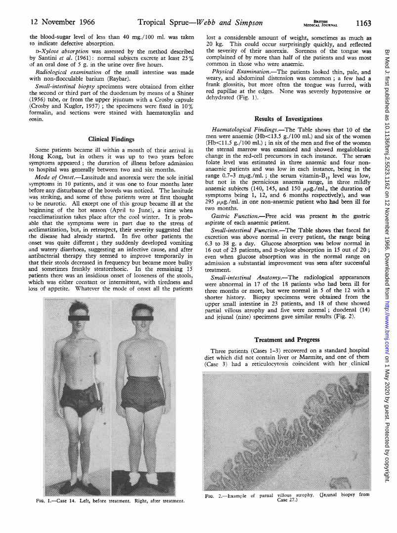
Physical Examination.-The patients looked thin, pale, At!weary, and abdominal distension was common; a few badifrank glossitis, but more often the tongue was furred, wi
red papillae at the edges. None was severely hypqtensive- t
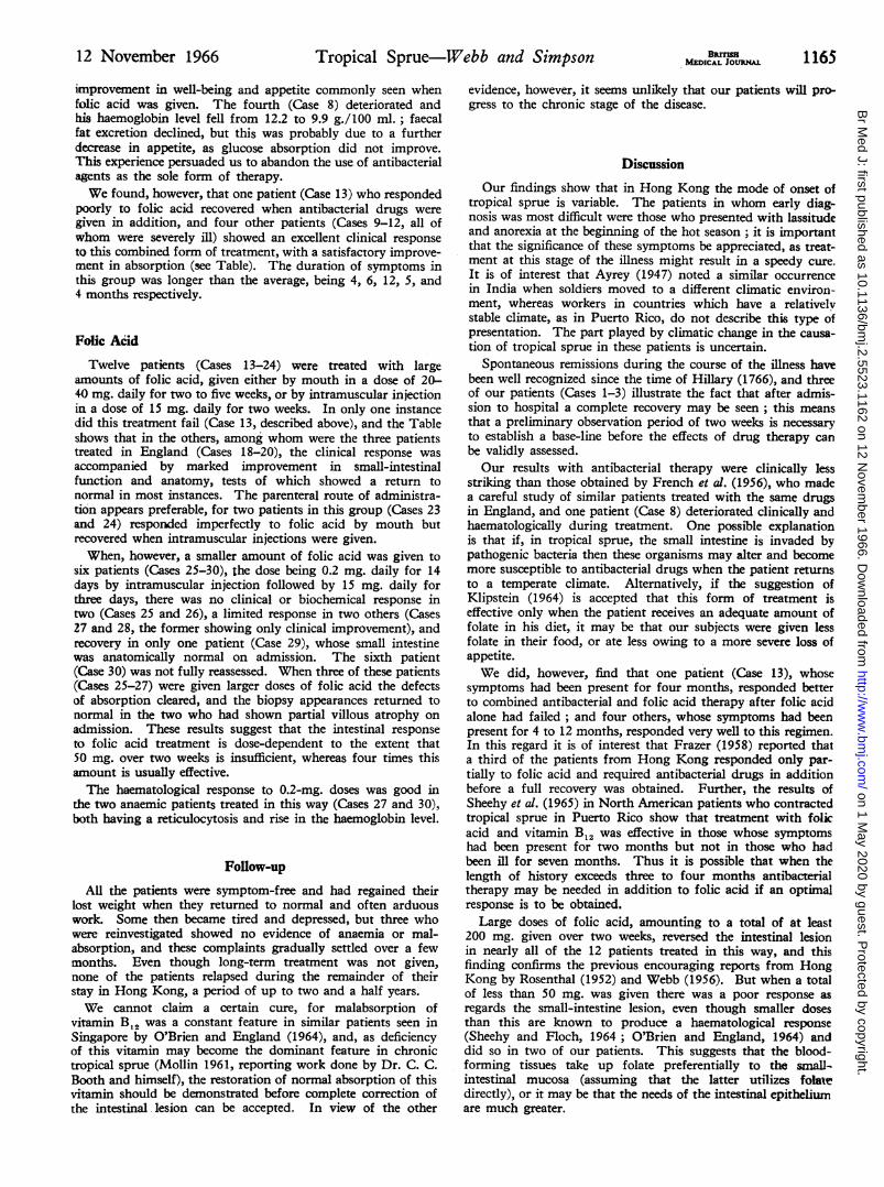
dehydrated (Fig. 1).4.
Results of Investigations
Haematologicpl Findings.-The Table shows that 10 of '.;'men were anaemic (Hb<13.5"g./100 ml.) and six of the woml2(Hb.<1 1.5 g./100 ml.); in six of the men and five of thewthe sternal marrow was examined and showedchange in the red-cell precursors in each instance. Th'e sefolate level was estimated in three anaemic and fouranaenic patients and was low in each instance, beingrange 0.7-3 kilug./ml.; the serum vitamin-B12 level was
but not in the pernicious anaemia range, in threeanaemic subjects (140, 145, and 150 1Atg./ml., the dusymptoms being 1, 12, and 6 months repectively), and295 ,puJg./ml. in one non-anaemic patient who had been ift
two months.Gastric Function.-Pree acid was present i the gaide
aspirate of each anaemic patient.Small-intestinl Function.-The Table shows that faecal i
excretion was above normal in every patient, the range being
6.3 to 38 g. a day. Glucose absorption was below nom id16 out of 23 patients, and D-xylose absorption in 15 but of2O: !<even when glucose absorption was in the normal range oAdadmission a substantial improvement was seen after suc&ssfuitreatment.,
Small-intestinal Anatomy.-The radiological appearanowere abnormal'in 17 of the 18 patients who had been illthree months or more, but were normal mi 5 of the 12 witli Xshorter history. Biopsy specimens were obtained from ifr-upper small intestine in 23 patients, and 18 of these showerIpartial villous atrophy and five were normal;'duodenal (14wand jejunal (nine) specimens gave similar results (Fig. 2).
Three patients (Cases 1-3) recovered on a standard os[diet which did not contain liver or Marmite, axid one of #1(Case 3) had a reticulocytosis coincident with her clii
FIG. 1.-Case 14. Left, before trEatment. Right, After treatment.FIG. 2.-Example of partial villous atrophy. (Jejunal biopsy fi
I Case 27.)
- ew I- "eI e;. -
on 1 May 2020 by guest. P
rotected by copyright.http://w
ww
.bmj.com
/B
r Med J: first published as 10.1136/bm
j.2.5523.1162 on 12 Novem
ber 1966. Dow
nloaded from
Tropical Sprue-Webb and Simpsonimprovement ; one still had mild steatorrhoea (see Table) oneand a half months later, but none required specific therapy.Signs of recovery were apparent within two weeks of theiradmission to hospital, whereas four other patients who wereobserved for over one month showed no similar improvement ;we therefore kept all patients under observation for two weeksbefore beginning specific therapy.
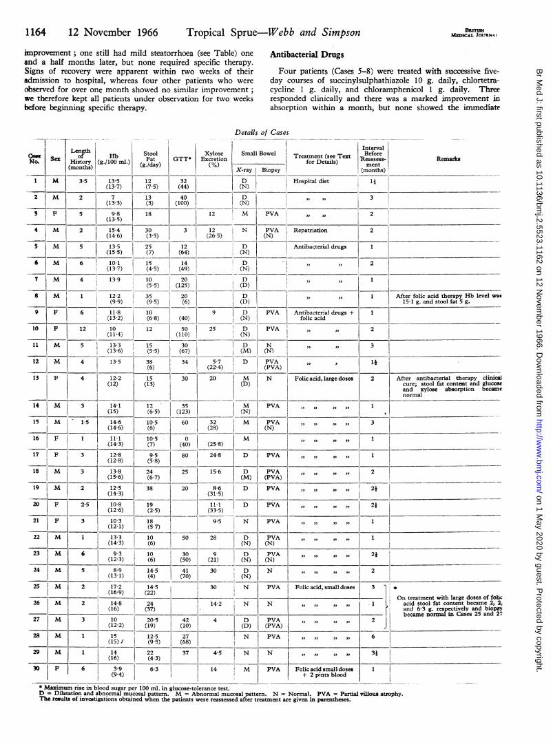
I
2
oeISex
M
M
Lengthof
History(months)
3.5
2
Hb(g.J100 ml.)I
13-5(13-7)
7(13-3)
StoolFat
(g./day)
12(7-5)13(3)
GTT*
32(44)40
(100) ~
A4EDICAL JOUaNA
Antibacterial Drugs
Four patients (Cases 5-8) were treated with successive five-
day courses of succinylsulphathiazole 10 g. daily, chlortetra-
cycline 1 g. daily, and chloramphenicol 1 g. daily. Three
responded clinically and there was a marked improvement in
absorption within a month, but none showed the immediate
Details of Cases
Xylose Small Bowel Treatment (see TextExcretion _________ for Details)
MX-ray Biopay
(N
(N)
Hospital diet
IntervalIBefore
"Reassess-ment
(months)
Remarks
3
S F ~~~~~59-8 18 12 M PVA , ,,2(13-5)
41M 2 115-4 30 3 12 N PVA Repatriation 2-____________ ~(14-6) (3-5) (26-5)(N
5 5 13-5 25 12 D Antibaqterial drugs 1____________ ~(15-5) (7) (64) (N)6 6 loll~~~~~~~1.1 115 14 D ,, ,
(13-7) (4-5) (49)__ (N
7 M 4 13-9 10 20 D,, , 1____________________ ___________________ (5-5) (125) __ _ _ _ _ (D) _ _ _ _ _ _ _ _ _ _ _ _ _ _ _ _ _ _ _ _ _ _ _ _ _ _ _ _ _
8 1 12-2 35 20 D I, , After folic acid therapy Hb level was- , I~~~~~~~~~~99(9-5) (6) (D) 15-1 g. and stool fat 5 g.
9 F 6 11-8 10 9 D PVA Antibacterial drugs + 1(13-2) (6-8) (40) (N) folic acid
to F 12 10 12 50 25 D IPVA ,, , 2(11-4) (110) (N )
11 M 5 13-3 15 30 -~D N ,, ,, 3(136) (5-5) (67) j(M) (N) _ _ _ _ _ _ _ _ _ _ _ _ _ _ _ _ _ _ _ _ _ _ _ _ _ _ _ _ _ _ _ _
12 M 4 13-5 38 34 5-7 D PVA ,, 14~~(6) (22-4)(PV
13 F 412-2 15 30 20 M N Folic acid, large doses 2 After antibacterial therapy clinical(12) (13) (D) cure; stool fat content and glucose
and xylose absorption becamenormal
14 M 3 14-1 12 35 M PVA ,,,,_____________ ~~~~~(15) (65) (123) (N)15 M 1.5 14-6 10-5 60 32 M PVA1 ,,,, 3
~~(14-6) (6) (28) (N )
16 F 1~~~~~11.1 10-5 0 M ,,,,,16__ F__ ____ (14-3) (7) (40) (25-8)17 F 3 12-8 9.5 80 24-8 D PVA ,,,,, 1
(12-8) (5-8)_
18 M 3 13-8 24 25 15-6 D PVA ,,, ,,, 2~~(15-6) (6-7) (M ) (PV A)
19 M 2 12-5 38 20 8-6 D PVA ,,,,,,,,2I 2t(14-3) (31-5)
20 F 2-5 10-8 19 11.1 -~D PVA J,,,,3 , 24(12-6) (2-5) (33-5)
21 F 3 10-3 18 9-5 N PVA S,,,,,,,3 1______ ____ ________ (12-1) (5-7) _ _ _ _ _ _ _ _ _ _ _ _ _ _ _ _ _ _ _ _ _
22 M 1 13-3 10 50 28 D PVA , ,, 1~(14-3) (6) (N ) (N )
23 M 6 9-3 10 30 9 D PVA ,,,,,,,,J 24(12-3) (6) (50) (21) (N)(N)___
24 M 5 8-9 14-5 41 30 D N ,,,3,,,, 2~~(13-1) (4) (70) (N )
25 l*M 2 17-2 14-5 30 N PVA Folic acid, small doses(169) (22)__________- ~~~~~~~~~~~~~~Ontreatment with large doses of folic
26 M 2 14-8 2414-2 N N M cdsolftcnetbcm 32(16) (37) I 1and 6-3 g. respectively and biopsy
27 M 310205 42 4 D PVA 2 ~~~~~~~~~~~~~~~~~becamenormal in Cases 25 and ~7______________12 _2 (19) (10) (D ) (PV A ) ___
28 M 1 15 12-5 27 N PVA ,,,,,,,3,2. 6~~~~~~(15)1 (9-5) (68)__
29 M 1 14 22 37 4-5 N N .,,,,,,,,S 31(16) (4-3) _____ ___________ ______1
30 F 3.9(9-4)
6-3 14 M PVA Folic: acid small doses+ 2 pints blood
1I1
* Maximum rise in blood sugar per 100 ml. in glucose-tolerance test.D = Dilatation and abnormal mucosal pattern. M = Abnormal mucosal pattern. N = Normal. PVA = Partial vilous atrophy.The results of investigations obtained when the patients were reassessed after treatment are given in parentheses.
1164 12 November 1966
1..
-..l 1..l1..
6
on 1 May 2020 by guest. P
rotected by copyright.http://w
ww
.bmj.com
/B
r Med J: first published as 10.1136/bm
j.2.5523.1162 on 12 Novem
ber 1966. Dow
nloaded from
improvement in well-being and appetite commonly seen whenfolic acid was given. The fourth (Case 8) deteriorated andhis haemoglobin level fell from 12.2 to 9.9 g./100 ml.; faecalfat excretion declined, but this was probably due to a furtherdecrease in appetite, as glucose absorption did not improve.This experience persuaded us to abandon the use of antibacterialagents as the sole form of therapy.We found, however, that one patient (Case 13) who responded
poorly to folic acid recovered when antibacterial drugs were
given in addition, and four other patients (Cases 9-12, all ofwhom were severely ill) showed an excellent clinical responseto this combined form of treatment, with a satisfactory improve-ment in absorption (see Table). The duration of symptoms inthis group was longer than the average, being 4, 6, 12, 5, and4 months respectively.
Folic Acid
Twelve patients (Cases 13-24) were treated with largeamounts of folic acid, given either by mouth in a dose of 20-40 mg. daily for two to five weeks, or by intramuscular injectionin a dose of 15 mg. daily for two weeks. In only one instancedid this treatment fail (Case 13, described above), and the Tableshows that in the others, among whom were the three patientstreated in England (Cases 18-20), the clinical response was
accompanied by marked improvement in small-intestinalfunction and anatomy, tests of which showed a return tonormal in most instances. The parenteral route of administra-tion appears preferable, for two patients in this group (Cases 23and 24) responded imperfectly to folic acid by mouth butrecovered when intramuscular injections were given.When, however, a smaller amount of folic acid was given to
six patients (Cases 25-30), the dose being 0.2 mg. daily for 14days by intramuscular injection followed by 15 mg. daily forthree days, there was no clinical or biochemical response intwo (Cases 25 and 26), a limited response in two others (Cases27 and 28, the former showing only clinical improvement), andrecovery in only one patient (Case 29), whose small intestinewas anatomically normal on admission. The sixth patient(Case 30) was not fully reassessed. When three of these patients(Cases 25-27) were given larger doses of folic acid the defectsof absorption cleared, and the biopsy appearances returned tonormal in the two who had shown partial villous atrophy on
admission. These results suggest that the intestinal responseto folic acid treatment is dose-dependent to the extent that50 mg. over two weeks is insufficient, whereas four times thisamount is usually effective.The haematological response to 0.2-mg. doses was good in
the two anaemic patients treated in this way (Cases 27 and 30),both having a reticulocytosis and rise in the haemoglobin level.
Follow-up
All the patients were symptom-free and had regained theirlost weight when they returned to normal and often arduouswork. Some then became tired and depressed, but three whowere reinvestigated showed no evidence of anaemia or mal-absorption, and these complaints gradually settled over a fewmonths. Even though long-term treatment was not given,none of the patients relapsed during the remainder of theirstay in Hong Kong, a period of up to two and a half years.
We cannot claim a certain cure, for malabsorption ofvitamin B, was a constant feature in similar patients seen inSingapore by O'Brien and England (1964), and, as deficiencyof this vitamin may become the dominant feature in chronictropical sprue (Mollin 1961, reporting work done by Dr. C. C.Booth and himself), the restoration of normal absorption of thisvitamin should be demonstrated before complete correction ofthe intestinal lesion can be accepted. In view of the other
B enMEDICAL JOURNAL 1165
evidence, however, it seems unlikely that our patients will pro-gress to the chronic stage of the disease.
DiscussionOur findings show that in Hong Kong the mode of onset of
tropical sprue is variable. The patients in whom early diag-nosis was most difficult were those who presented with lassitudeand anorexia at the beginning of the hot season; it is importantthat the significance of these symptoms be appreciated, as treat-ment at this stage of the illness might result in a speedy cure.
It is of interest that Ayrey (1947) noted a similar occurrence
in India when soldiers moved to a different climatic environ-ment, whereas workers in countries which have a relativelystable climate, as in Puerto Rico, do not describe this type ofpresentation. The part played by climatic change in the causa-tion of tropical sprue in these patients is uncertain.
Spontaneous remissions during the course of the illness havebeen well recognized since the time of Hillary (1766), and threeof our patients (Cases 1-3) illustrate the fact that after admis-sion to hospital a complete recovery may be seen; this meansthat a preliminary observation period of two weeks is necessaryto establish a base-line before the effects of drug therapy can
be validly assessed.Our results with antibacterial therapy were clinically less
striking than those obtained by French et al. (1956), who madea careful study of similar patients treated with the same drugsin England, and one patient (Case 8) deteriorated clinically andhaematologically during treatment. One possible explanationis that if, in tropical sprue, the small intestine is invaded bypathogenic bacteria then these organisms may alter and becomemore susceptible to antibacterial drugs when the patient returnsto a temperate climate. Alternatively, if the suggestion ofKlipstein (1964) is accepted that this form of treatment iseffective only when the patient receives an adequate amount offolate in his diet, it may be that our subjects were given lessfolate in their food, or ate less owing to a more severe loss ofappetite.We did, however, find that one patient (Case 13), whose
symptoms had been present for four months, responded betterto combined antibacterial and folic acid therapy after folic acidalone had failed; and four others, whose symptoms had beenpresent for 4 to 12 months, responded very well to this regimen.In this regard it is of interest that Frazer (1958) reported thata third of the patients from Hong Kong responded only par-tially to folic acid and required antibacterial drugs in additionbefore a full recovery was obtained. Further, the results ofSheehy et al. (1965) in North American patients who contractedtropical sprue in Puerto Rico show that treatment with folicacid and vitamin B12 was effective in those whose symptomshad been present for two months but not in those who hadbeen ill for seven months. Thus it is possible that when thelength of history exceeds three to four months antibacterialtherapy may be needed in addition to folic acid if an optimalresponse is to be obtained.
Large doses of folic acid, amounting to a total of at least200 mg. given over two weeks, reversed the intestinal lesionin nearly all of the 12 patients treated in this way, and thisfinding confirms the previous encouraging reports from HongKong by Rosenthal (1952) and Webb (1956). But when a totalof less than 50 mg. was given there was a poor response asregards the small-intestine lesion, even though smaller dosesthan this are known to produce a haematological response(Sheehy and Floch, 1964; O'Brien and England, 1964) anddid so in two of our patients. This suggests that the blood-forming tissues take up folate preferentially to the small-intestinal mucosa (assuming that the latter utilizes folatedirectly), or it may be that the needs of the intestinal epitheliumare much greater.
12 November 1966 Tropical Sprue-Webb and Simpson
on 1 May 2020 by guest. P
rotected by copyright.http://w
ww
.bmj.com
/B
r Med J: first published as 10.1136/bm
j.2.5523.1162 on 12 Novem
ber 1966. Dow
nloaded from
1166 12 November 1966 Tropical Sprue-Webb and Simpson BRourOur results show that the prognosis of expatriate patients
treated in Hong Kong and remaining there is excellent for atleast two and a half years. Long-term follow-up studies intropical sprue are rare. Hazari and Woodruff (1958) reportedthat 17 patients who had contracted tropical sprue in India10 years previously and had been evacuated to Britain were all;at full work, although some third of these had mild and inter-xmittent symptoms but were not disabled by them. As moreAeffective forms of treatment were available to our patients theiroutlook should be at least as good, but this point will need tobe established.
SummaryThirty patients who contracted tropical sprue in Hong
Kong are described. Nine of them became ill at the beginningof the hot season with initial complaints of lassitude andanorexia without distiurbance of the bowels, and the significanceof these early symptoms is stressed.Treatment with antibacterial drugs was not as effective as
treatment with large doses of folic acid, but it is suggested thatsome patients who have been ill for more than three monthsmay require both forms of therapy before an optimal responseis obtained.
It was confirmed that folic acid usually reverses the intestinallesion in these patients provided an adequate amount of thedrug is given; a total of less than 50 mg. was generallyineffective.
Twenty-six patients remained in Hong Kong for periods ofup to two and a half years after treatment, and none relapsed.
We are deeply indebted to our many medical and nursingcolleagues who worked with us; to Professor A. C. Frazer, whoadvised on sprue research in Hong Kong and who has permitted us
to include data on four cases which were transferred to his unit inBirmingham; and to Dr. A. C. Hobson (then Lieutenant-ColonelR.A.M.C.), who handed on the sprue project in Hong Kong to oneof us (J. F. W.). We are also grateful to Lieutenant-Colonels S. A.Biggart and C. B. F. Downie, R.A.M.C., who were in charge ofeight of the patients investigated. Dr. B. C. Morson and Lieutenant-Colonel N. W. J. England, R.A.M.C., have been most helpful inreviewing the small-intestinal biopsy specimens, and Professor D. L.Mollin and Colonel W. O'Brien, late R.A.M.C., have given usinvaluable advice and helpful criticism throughout the preparationof this paper. Finally, we would like to thank the Director-GeneralArmy Medical Services for permission to use case notes and recordsfor this publication.
REFERENCESAyrey, F. (1947). Trans. roy. Soc. trop. Med. Hyg., 41, 377.Crosby, W. H., and Kugler, H. W. (1957). Amer. 7. dig. Dis., 2, 236.Dacie, J. V. (1956). Practical Haematology, 2nd ed. Churchill, London.Frazer, A. C. (1958). Proceedings of World Congress of Gastroentero-
logy. Williams and Wilkins, Baltimore.French, J. M., Gaddie, R., and Smith, N. M. (1956). Quart. 7. Med., 25,
333.Gardner, F. H. (1956). Arch. intern. Med., 98, 44.Hazari, 0. K., and Woodruff, A. W. (1958). Brit. med. 7., 2, 344.Hillary, W. (1766). Observations on the changes of the Air and Con-
cormtant Epidemical Diseases in the Island of Barbados, edited byL. Hawes, W. Clarke, and R. Collins, 2nd ed. London.
Hutner, S. H., Bach, M. K., and Ross, G. I. M. (1956). 7. Protozool.,3, 101.
Kay, A. W. (1953). Brit. med. 7., 2, 77.Klipstein, F. A. (1964). Gastreenterology, 47, 457.Mollin, D. L. (1961). Trans. med. Soc. Lond., 78, 51.O'Brien, W., and England, N. W. J. (1964). Brit. med. 7., 2, 1573.Rosenthal, F. D. (1952). M.D. Thesis, University of London.Santini, R., Sheehy, T. W., and Martinez de Jesus, J. (1961). Gastro-
ent-erology, 40, 772.Sheehy, T. W., Cohen, W. C., Wallace, D. K., and Legters, L. J. (1965).
7. Amer. med. Ass., 194, 1069.- and Floch, M. H. (1964). The Small Intestine: its function and
diseases. Hoeber, New York.Shiner, M. (1956). Lancet, 1, 85.van de Kamer, J. H., Huinink, H. ten B., and Weyers, H. A. (1949). R.
biol. Chem., 177, 347.Waters, A. H., and Mollin, D. L. (1961). 7. clin. Path., 14, 335.Webb, J. F. (1956). M.D. Thesis, Dunelm.
Hypoproteinaemia in Anaphylactoid Purpura
N. F. JONES,* M.A., M.B., M.R.C.P.; B. CREAMERt M.D., F.R.C.P.; T. M. D. GIMLETTE, M.D., M.R.C
Brit. med.JY., 1966, 2, 1166-1168
Anaphylactoid (Henoch-Schonlein) purpura may produce anephrotic syndrome and thus cause hypoalbuminaemia. How-ever, some years ago we observed a patient with this disease inwhom the serum albumin level was reduced at a time whenurinary losses of protein were insignificant. Evidence of liverdysfunction was absent, but gastrointestinal complications ofthe disease were prominent. It was therefore suspected that thehypoalbuminaemia was due to abnormal losses of protein intothe bowel. We here record observations on five patients withanaphylactoid purpura which suggest that protein-losingtenteropathy may complicate this disease. This association doesinot seem to have been reported to date (Waldmann, 1966).
Methods
Los of protein via the gut was assessed by measuring theradioactivity present in a five-day collection of faeces after theintravenous injection of 13 'I-labelled polyvidone (P.V.P.) or51CGlc3. The former was used according to the method of
* Senior Lecturer, Department of Medicine, St. Thomas's HospitalMedical Srhool, London.
4 Physician, St. Thomas's Hospital, London.$ Chief Clinical Assistant, Radioisotope Department, St. Thomas's Hos-
pital, London.
Gordon (1959) with '311-P.V.P. obtained from the Radiochemi-cal Centre, Amersham. The use of 'ICrCl3 followed the methodof Rubini and Sheehy (1961).Normal values obtained in this hospital for faecal excretion
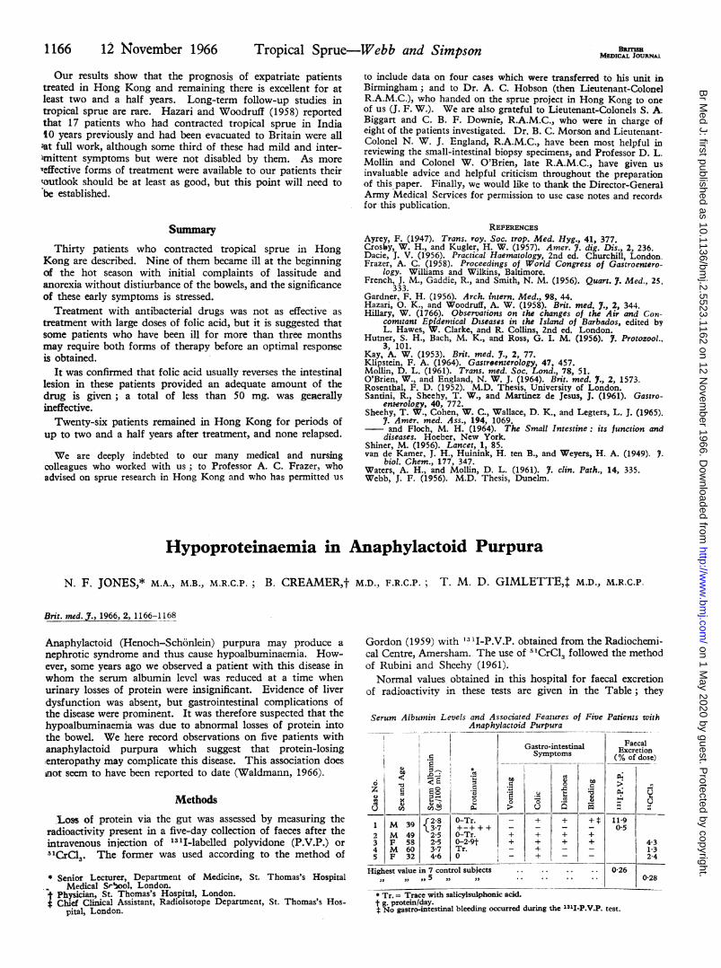
of radioactivity in these tests are given in the Table; they
Serum Albumin Levels and Associated Features of Five Patients withAnaphylactoid Purpura
I ~~~~~~~Gastro-intestinal FaecalExcretionSymptoms (%odse
.~~~~~~~~ o~~~~~( of dose
4 0 6 0 3 7 T-r2.t43
6 324- 6 0,__ 4
,, ,, ,, , ,. ..41. .02
Ut"~~~~ p~~ ~~. U U
1 9{2-8 O-Tr. - + + +* 11-9I 9 3-7 + ++ - - -- 0-5
2 M 49 2-5 O-Tr. + + + +3 F 58 2-5 0-2-9t1 + + + + 4.34 M 60 3-7 Tr. - + - - 1-35 F32 4-6 0 - + - - 2-4
Highest value in 7 control subjects.025 0-28
* Tr. = Trace with salicylsulphonic acid.t g. protein/day.$ No gastro-intestinal bleeding occurred during the 13I1-P.V.P. test.
on 1 May 2020 by guest. P
rotected by copyright.http://w
ww
.bmj.com
/B
r Med J: first published as 10.1136/bm
j.2.5523.1162 on 12 Novem
ber 1966. Dow
nloaded from