Embed Size (px)
Citation preview

Thorax (1967), 22,444.
Tridimensional photographic reconstruction in a
study of the pathogenesis of honeycomb lungJ. CORTEZ PIMENTEL
From l.A.N.T. (Department of Pathology, Sanatorium D, Carlos 1) and the Institute of Pathology,Faculty of Medicine, University of Lisbon, Portugal
Tridimensional photographic reconstruction of the lesions found in honeycomb lung in 10different types of pulmonary disease was made. The pathological picture was characteristic andwell defined by tridimensional microscopy and quite independent of the accompanying disease.The lesions responsible for its appearance involved the whole lobular bronchiolar system but were
most marked in the terminal and respiratory bronchioli. The changes were fundamentally diffuse,saccular, and cystic bronchiolectasis. Other bronchiolar lesions were found such as changes indirection and mode of division, amputations, and anastomoses between bronchioles and cystsbelonging to anatomically independent airways. There was some evidence that the bullous aspectsof honeycomb lung were due to multiple valvular arrangements which let the air in but do notlet it out. Honeycomb lung was, in the majority of cases, a complication of interstitial pulmonaryfibrosis. Honeycombing was usually due to marked changes in the lobular bronchioles caused bythe obliteration or rigidity of alveolar ducts and the corresponding alveoli and even by localizationof the interstitial fibrosis in the bronchiolar wall.
The term 'honeycomb lung' (Hyde, Hyde, andPokorny, 1951; Heppleston, 1956) has beenapplied to a pulmonary lesion characterized by thepresence of multiple cystic cavities with averagediameters between 1 and 10 mm. These cysts,microscopically, are bronchiolar dilatations with awell-defined wall made up of scar tissue or of agranulomatous process of varied aetiology(Heppleston, 1956).
This lesion can be diffuse or localized and isusually found in certain diseases that predomin-antly involve the alveolar septa: sarcoidosis(Nickerson, 1937), berylliosis (Dutra, 1948), asbes-tosis (Telischi and Rubenstone, 1961), eosinophilicgranuloma of the lung (Auld, 1957), Abt-Letterer-Siwe disease (van Creveld and ter Poorten, 1935),Hand-Schuller-Christian disease (Keats andCrane, 1954), diffuse scleroderma (Mahrer, Evans,and Steinberg, 1954), rheumatoid arthritis(Rubin, 1955), dermatomyositis (Mills andMathews, 1956), some cases of progressive pulmo-nary fibrosis-Hamman-Rich syndrome (Pea-body, Buechner, and Anderson, 1953), and tuber-culosis (Buechner and Anderson, 1955). Someauthors consider it a consequence of interstitialpulmonary fibrosis (Anderson and Foraker, 1960;Pimentel, 1962). Honeycomb lung is also part of
the morphological picture of a rare pulmonarydisease known as cystic cirrhosis of the lung (vonBuhl, 1872), pulmonary muscular hyperplasia(Rubenstein, Gutstein, and Lepow, 1955), bron-chiolar emphysema (Siebert and Fisher, 1957), anddiffuse pulmonary leiomyomatosis (Delarue,Paillas, Paley, Daumet, and Daussy, 1960).Numerous theories have been proposed to
explain the genesis of honeycomb lung. Hepple-ston (1956), using microdissection and the studyof serial histological sections, thought thatobliteration or dilatation of the respiratorybronchioles was the essential change in thedisease. According to this author, unaffectedadjacent bronchioles underwent a compensatorydilatation, thus forming cystic cavities near con-solidated and fibrotic areas. Heppleston alsobelieved that the progressive enlargement of thecysts was probably due to secondary valvularobstruction of the bronchioles that communicatedwith them.
Siebert and Fisher (1957), on the other hand,believed that a 'congenital hypoplasia of the distalsegment of the respiratory unit' was the cause ofthe honeycomb lesions in 'pulmonary muscularcirrhosis'. Gonzalez-Licea and Perez-Tamayo(1963) disagreed with Siebert and Fisher's theory
444

Tridimensional photographic reconstruiction in a study of the pathogenesis of honevcomb lung 445
as they found it to be based on erroneous data-improper fixation of the lungs and difficulty indistinguishing respiratory bronchioles, bronchiolaralveoli, alveolar ducts, and ductal alveoli in theseseverely altered lungs. These authors thought thatin muscular cirrhosis of the lung the lesionsacquired diffuse bronchiolectasis secondary tochronic infection, and that Heppleston's theorywas still the one that best explained honeycomb-ing.The methods used up to now, such as micro-
dissection and serial sections, do not permit anyfurther advances in our knowledge of the patho-genesis of honeycomb lung. The 'bronchovascularcast' technique which we tried out gave no betterresults, so we experimented with tridimensionalreconstructions from photographic enlargementsof serial sections of the lesions. In this paperpossible applications of this method in the studyof honeycomb lung are presented.
MATERIAL AND METHODS
Models were constructed of the honeycomblesions found in the following diseases: diffusescleroderma, dermatomyositis, Abt-Letterer-Siweand Hand-Schuller-Christian diseases, progressivepulmonary fibrosis (Hamman-Rich syndrome),muscular cirrhosis of the lung, sarcoidosis, anddiffuse fibrotic tuberculosis. Honeycombing of apurely local character, formed near tuberculomasand foci of endogenous lipoid pneumonia, wasstudied by the same method, as well as somelobular bronchiolar systems of three normal lungs.In eight cases the reconstructions were made fromnecropsy material, and in three from surgicalspecimens. The lungs were fixed by low-pressureintrabronchial instillations of a 10% aqueous solu-tion of formalin until a uniform distension of thepleura was obtained.One case of each of the diseases mentioned was
studied. From each case three of the most repre-sentative lesions were chosen, with a surface areaof about 5 x 3 x 2 cm. These were embedded inparaffin, and serial sections were made which werethen stained with haematoxylin and eosin. Thenumber of sections studied from each block wasbetween 100 and 250.
Reconstruction was made from microphoto-graphs of the serial sections using enlargementsof x 125, x 170, or x 250, according to the needfor more or less detail. Kodak 0 250 RapidOrthochromatic metalographic plates and Geva-tone k3, grade 2, 30 x 40 cm. Gevaert paper wereused. A green filter was used to improve the con-trast of the red stains. The photographs were
stuck on compressed cardboard of a thicknessrelated to the enlargement.Of each group of photographs corresponding to
a certain lesion, models in 'positive' and 'negative'were constructed so that not only the bronchiolar-cystic connexions could be noted but also thetopography of the bronchiolar lesion. The irregu-lar staircase effect caused by the superimpositionof the different layers was reduced by applying athin coat of synthetic filler.
RESULTS
Tridimensional reconstruction of the bronchiolesaccording to the method described made it rela-tively easy to study their course, calibre, andmanner of division in the diseased areas, and alsoto appreciate their intimate relations with thecysts. The results obtained with this method in thestudy of honeycomb lung seemed superior tothose so far obtained in alveolar emphysema,probably because the size of the air spaces andthe histological structure of their walls are funda-mentally different in the two conditions.
It has been stated that in severely damagedlungs, such as in honeycomb lungs with extensivefibrosis, it is difficult to distinguish respiratoryfrom non-respiratory bronchioles. Although itmay be difficult, usually it is not so if one keepsin mind the classical bronchiolar distribution(Miller, 1947; von Hayek, 1960), uses large sec-tions and adequate photographic enlargements,and compares them with reconstructions ofnormal bronchioles. The data obtained from tri-dimensional reconstruction of the honeycomblesions in all the types of disease studied wereidentical and as such will be described together.The bronchiolar changes were particularlyobvious in the fibrocystic areas, even though theymight have been found outside them. They in-volved the lobular bronchioles electively. Thechanges are described at three different levels:non-respiratory bronchioles, respiratory bron-chioles, and the alveolar ducts and alveoli.
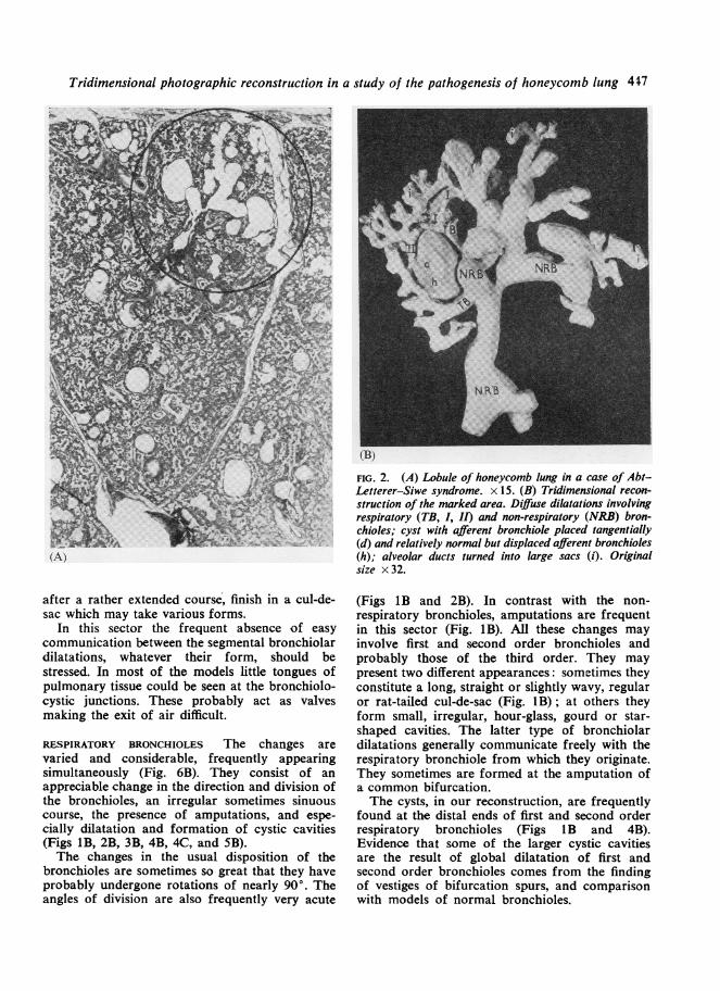
NON-RESPIRATORY BRONCHIOLES The changesgenerally involve most of the non-respiratoryportion of the lobular bronchiolar system, but areespecially found in the distal divisions of thelobular bronchiole and in the terminal bronchioles.These changes consist of diffuse and segmental(frequently cystic) dilatation, bronchiolar irregu-larities, changes in the usual bifurcation anglesand, more rarely, amputations (Figs lB and 2B).In the latter case the non-respiratory bronchioles.

FIG. 1. (A) Part of a honeycomb lung in a case ofHamman-Rich syndrome. x 16. (B) 'Negative' tridimen-sional reconstruction of the area marked by the circle(original x25). The non-respiratory bronchioles showeddilatations (a), amputations (b), and angulations (c); inthe respiratory bronchioles, the oblique disposition of theafferent bronchiole (d), the circular disposition of thebronchioles around the cyst (e), and various anastomoses(f, fl, f2) are shown. TB, terminal bronchiole; I, II and III,first, second, and third respiratory bronchioles. (C) 'Posi-tive' reconstruction ofthe opening of the afferent bronchiole(probable valve mechanism marked with an arrow). Originalsize x75.
(B)
Wip
Tridimensional photographic reconstruction in a study of the pathogenesis of honeycomb lung 417
FIG. 2. (A) Lobule of honeycomb lung in a case of Abt-Letterer-Siwe syndrome. x 15. (B) Tridimensional recon-struction of the marked area. Diffuse dilatations involvingrespiratory (TB, I, II) and non-respiratory (NRB) bron-chioles; cyst with afferent bronchiole placed tangentially(d) and relatively normal but displaced afferent bronchioles(h); alveolar ducts turned into large sacs (i). Originalsize x32.
(A)
after a rather extended course, finish in a cul-de-sac which may take various forms.
In this sector the frequent absence of easycommunication between the segmental bronchiolardilatations, whatever their form, should bestressed. In most of the models little tongues ofpulmonary tissue could be seen at the bronchiolo-cystic junctions. These probably act as valvesmaking the exit of air difficult.
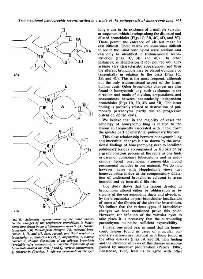
RESPIRATORY BRONCHIOLES The changes arevaried and considerable, frequently appearingsimultaneously (Fig. 6B). They consist of anappreciable change in the direction and division ofthe bronchioles, an irregular sometimes sinuouscourse, the presence of amputations, and espe-cially dilatation and formation of cystic cavities(Figs 1B, 2B, 3B, 4B, 4C, and 5B).The changes in the usual disposition of the
bronchioles are sometimes so great that they haveprobably undergone rotations of nearly 900. Theangles of division are also frequently very acute
(Figs lB and 2B). In contrast with the non-respiratory bronchioles, amputations are frequentin this sector (Fig. 1B). All these changes mayinvolve first and second order bronchioles andprobably those of the third order. They maypresent two different appearances: sometimes theyconstitute a long, straight or slightly wavy, regularor rat-tailed cul-de-sac (Fig. 1B); at others theyform small, irregular, hour-glass, gourd or star-shaped cavities. The latter type of bronchiolardilatations generally communicate freely with therespiratory bronchiole from which they originate.They sometimes are formed at the amputation ofa common bifurcation.The cysts, in our reconstruction, are frequently
found at the distal ends of first and second orderrespiratory bronchioles (Figs lB and 4B).Evidence that some of the larger cystic cavitiesare the result of global dilatation of first andsecond order bronchioles comes from the findingof vestiges of bifurcation spurs, and comparisonwith models of normal bronchioles.
J. Cortez Pimentel
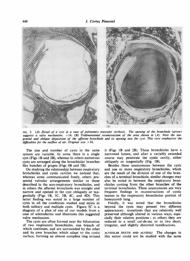
FIG.3.A)etal o a cst n acas ofpulmonary muscular cirrhosis. The opening of the bronchioke (arrow)
sugssa valve mechanism. x 14. (B) Tridimensional reconstruction of the area shown in (A). Note the tan-
gential and oblique disposition of the affierent bronchiole and its opening into the cyst. This view emphasizes the
diJtlculties for the outflow of air. Original size x 16.
The size and number of cysts in the same
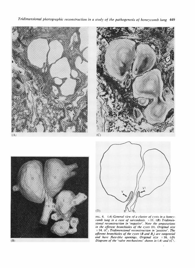
system are variable. In some there is a singlecyst (Figs 1B and 2B), whereas in others numerouscysts are arranged along the bronchiolar brancheslike bunches of grapes (Figs 4B and 5B).On studying the relationship between respiratory
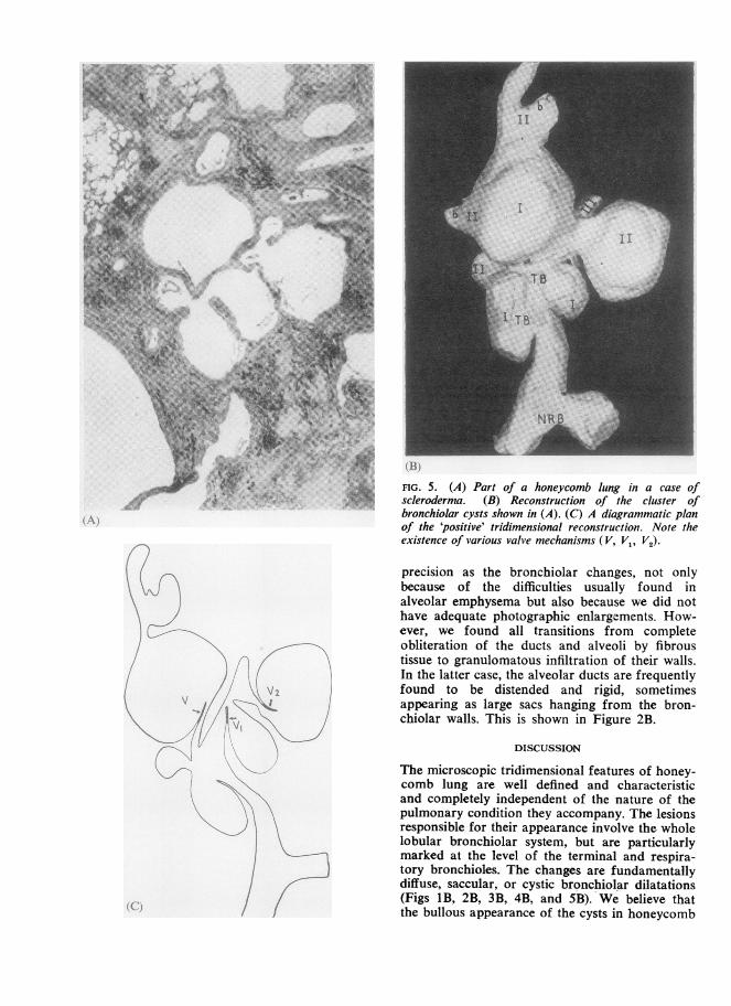
bronchioles and cystic cavities we noticed that,whereas some communicated freely, others pre-sented valvular arrangements similar to thosedescribed in the non-respiratory bronchioles, andin others the afferent bronchiole was straight andnarrow and opened in the cyst obliquely or tan-gentially (Figs 1B, IC, 3B, 4C, and 4D). Thislatter finding was noted in a large number ofcysts in all the conditions studied and exists inboth solitary and multiple cysts. Figure 5C is a
diagram of a plan of one of our models from a
case of scleroderma and illustrates this suggestedvalve mechanism.The cysts are often formed near the bifurcation
of two respiratory bronchioles, one branch ofwhich continues, and are surrounded by the otherand its own branches which adapt to the cysticsurface, forming an almost complete ring around
it (Figs I B and 2B). These bronchioles have anarrowed lumen, and after a variably extendedcourse may penetrate the cystic cavity, eitherobliquely or tangentially (Fig. iB).
Besides these anastomoses between the cystsand one or more respiratory bronchioles, whichare the result of the division of one of the bran-ches of a terminal bronchiole, similar changes mayalso be noted in between the respiratory bron-chioles coming from the other branches of theterminal bronchioles. These anastomoses are veryfrequent findings in reconstructions of cysticlesions in the respiratory bronchiolar portion ofhoneycomb lung.
Finally, it was noted that the bronchiolesbeyond the cysts may present two differentappearances: sometimes they are relatively wellpreserved although altered in various ways, espe-cially their relative positions; at others they arereduced to a small cul-de-sac or to two short.irregular, and slightly distorted ramifications.
ALVEOLAR DUCTS AND ALVEOLI The changes inthis sector could not be studied with the same
448
Triditnensional photographic reconistruction in a stuidy of the pathogenesis of honeycomb lung 449
(c)
(D) BB NJ/B,FiG. 4. (A) General view of a cluster of cysts in a honey-comb lung in a case of sarcoidosis. x 11. (B) Tridimen-sional reconstruction in 'negative'. Note the amputationsin the efferent bronchioles of the cysts (b). Original sizex 14. (C, Tridimensional reconstruction in 'positive'. Theafferent bronchioles of the cysts (B and B1) are tangentialand have flute-like openings. Original size x 16. (D)Diagram of the 'valve mechanisms' shown in (A) and (C'.
(A)
(By
BI
FIG. 5. (A) Part of a honeycomb lung in a case of-v. t -t -> ; scleroderma. (B) Reconstruction of the cluster of
bronchiolar cysts shown in (A). (C) A diagrammatic planof the 'positive' tridimensional reconstruction. Note theexistence of various valve mechanisms (V, V1, V2).
precision as the bronchiolar changes, not onlybecause of the difficulties usually found inalveolar emphysema but also because we did nothave adequate photographic enlargements. How-ever, we found all transitions from completeobliteration of the ducts and alveoli by fibroustissue to granulomatous infiltration of their walls.In the latter case, the alveolar ducts are frequentlyfound to be distended and rigid, sometimesappearing as large sacs hanging from the bron-chiolar walls. This is shown in Figure 2B.
DISCUSSION
The microscopic tridimensional features of honey-comb lung are well defined and characteristicand completely independent of the nature of thepulmonary condition they accompany. The lesionsresponsible for their appearance involve the wholelobular bronchiolar system, but are particularlymarked at the level of the terminal and respira-tory bronchioles. The changes are fundamentallydiffuse, saccular, or cystic bronchiolar dilatations(Figs 1B, 2B, 3B, 4B, and 5B). We believe thatthe bullous appearance of the cysts in honeycomb
Tridimensional photographic reconstruction in a study of the pathogenesis of honeycomb lung 451
(A)
TI
El
B
(B) A t ~
FIG. 6. Schematic representation of the more charac-teristic changes of the respiratory bronchioles in honey-comb lung found in our tridimensional models. (A) Normalbronchiole. (B) Pathological changes. TB, terminal bron-chiole; I, II, and III, first, second, and third respiratorybronchioles; a, dilatation (cyst); b, amputation; c, sinuouscourse; d, oblique disposition of the afferent bronchiole(probable valve mechanism); e, circular disposition of thebronchiole around the cyst; fand fi, various anastomoses;g, changes in direction; h, efferent bronchiole of the cyst.
lung is due to the existence of a multiple valvulararrangement which develops along the distorted anddilated bronchioles (Figs IC, 3B, 4C, 4D, and 5C).These permit the entrance of air but make itsexit difficult. These valves are sometimes difficultto see in the usual histological serial sections andcan only be identified in tridimensional recon-struction (Figs 1C, 3B, and 4C). In otherinstances, as Heppleston (1956) pointed out, theyassume very characteristic appearances, and thenthe afferent bronchiole may be placed obliquely ortangentially in relation to the cysts (Figs IC,3B, and 4C). This is the most frequent, althoughnot the only tridimensional aspect of the largerbullous cysts. Other bronchiolar changes are alsofound in honeycomb lung, such as changes in thedirection and mode of division, amputations, andanastomoses between anatomically independentbronchioles (Figs 1B, 2B, 4B, and 5B). The latterfinding is probably related to destruction of pul-monary parenchyma partly due to progressivedistension of the cysts.We believe that in the majority of cases the
aetiology of honeycomb lung is related to thelesions so frequently associated with it that formthe greater part of interstitial pulmonary fibrosis.
This close relationship between honeycomb lungand interstitial changes is also shown by the occa-sional findings of honeycombing near to localizedpulmonary lesions accompanied by fibrosis or bya granulomatous process of the septa as one findsin cases of pulmonary tuberculoma and in endo-genous lipoid pneumonia (tumour-like lipoidpneumonia) included in our material. We do not,however, agree with Heppleston's views thathoneycombing is due to the compensatory dilata-tion of undiseased bronchioles adjacent to areasimmobilized by interstitial fibrosis.Our study shows that the lesions develop in
bronchioles altered either by obliteration or byrigidity of the corresponding ducts and alveoli, orby the bronchiolar or peri-bronchiolar localizationof some of the fibrosis of the alveolar interstitium.We believe that the various types of bronchiolarchanges we have mentioned prove this point.However, for inflation of the valvular cysts totake place it is necessary that the surroundingparenchyma maintains sufficient expansibility.
Finally, one must bear in mind that the honey-comb lesions found in cases of muscular pul-monary cirrhosis are identical with those found inthe other diseases (Figs 3A and B). This findingand the existence of cases of this disease unaccom-panied by muscular proliferation (Pepere, 1906;Loeschcke, 1926) lead us to agree with other

J. Cortez Pimentel
authors (Siebert and Fisher, 1957), who believethat this disease is only another form of pro-gressive pulmonary fibrosis in which the prolifera-tion of smooth muscle is especially well developedfor reasons that we do not understand.
REFERENCES
Anderson, A. E., Jr., and Foraker, A. G. (1960). Morphologicalaspects of interstitial pulmonary fibrosis. Arch. Path., 70, 79.
Auld, D. (1957). Pathology of eosinophilic granuloma of the lung.Ibid., 63, 113.
Buechner, H. A., and Anderson, A. E. (1955). Diffuse indolent pul-monary tuberculosis. Amer. Rev. Tuberc., 71, 503.
von Buhl, L. (1872). Lungenentzundung, Tuberkulose und Schwindsucht.Oldenbourg, Munich.
Delarue, J., Paillas, J., Paley, P., Daumet, Ph., and Daussy, M. (1960).Insuffisance respiratoire progressive, emphyseme bulleux etleiomyomatose pulmonaire diffuse. J. franr. Med. Chir. thor.,14, 761.
Dutra, F. R. (1948). The pneumonitis and granulomatosis peculiar toberyllium workers. Amer. J. Path., 24, 1137.
Gonzalez-Licea, A., and Perez-Tamayo, R. (1963). Idiopathic diffusebronchiolectasis-so-called bronchiolar emphysema. Amer. J.clin. Path., 40, 157.
von Hayek, H. (1960). The Human Lung, trans. V. E. Krahl. Hafner,New York.
Heppleston, A. G. (1956). The pathology of honeycomb lung. Thorax,11, 77.
Hyde, L., Hyde, B., and Pokorny, C. (1951). Diffuse fibrocysticdisease of lungs (honey-comb lungs). Dis. Chest, 19, 190.
Keats, T. E., and Crane, J. F. (1954). Cystic changes of the lungs inhistiocytosis. Amer. J. Dis. Child., 88, 764.
Loeschcke, H. (1926). Emphysema bronchiolektaticum und prta-terminale Bronchiektasen als Systemerkrankungen isolierterAbschnitte des Bronchialbaumes. Verh. dsch. path. Ges., 21, 242.
Mahrer, P. R., Evans, J. A., and Steinberg, 1. (1954). Scleroderma:relation of pulmonary changes to esophageal disease. Ann.intern. Med., 40, 92.
Miller, W. S. (1947). The Lung, 2nd ed. Thomas, Springfield, Illinois.Mills, E. S., and Mathews, W. H. (1956). Interstitial pneumonitis in
dermatomyositis. J. Anmer. med. Ass., 160, 1467.Nickerson, D. A. (1937). Boeck's sarcoid. Report of six cases in E hich
autopsies were made. Arch. Path., 24, 19.Peabody, J. W., Jr., Buechner, H. A., and Anderson, A. E. (1953).
Hamman-Rich syndrome. Arch. intern. Med., 92, 806.Pepere, A. (1906). Della degenerazione cistica del polmone. Speri-
mentale, 60, 171.Pimentel, J. C. (1962). Aspectos morfologicos da 'fibrose pulmonar-.
J. Med. (P6rto), 49, 823.Rubenstein, L., Gutstein, W. H., and Lepow, H. (1955). Pulmonary
muscular hyperplasia (muscular cirrhosis of the lungs). Ann.intern. Med., 42, 36.
Rubin, E. H. (1955). Pulmonary lesions in 'rheumatoid disease' withremarks on diffuse interstitial pulmonary fibrosis. 4mer. J. Med..19, 569.
Siebert, F. T., and Fisher, E. R. (1957). Bronchiolar emphysema. So-called muscular cirrhosis of the lungs. Anmer. J. Path., 33, 1137.
Telischi, M., and Rubenstone, A. 1. (1961). Pulmonary asbestosis.Arch. Path., 72, 234.
van Creveld, S.. and ter Poorten, F. H. (1935). Infective reticulo-endotheliosis chiefly localized in lungs. bone marrow and thymus.Arch. Dis. Childh., 10, 125.
452