Embed Size (px)
Citation preview
Treatment protocol for skeletal Class III malocclusion in growing patients
Prof. Abdolreza Jamilian, Fellow of Orthognathic Surgery, Department of Orthodontics, Tehran Dental Branch, Islamic Azad University, Tehran, Iran. Email: [email protected]
Mr Saeed Khosravi, MSc, Tehran University of Medical Sciences, Tehran, Iran.
Dr. Alireza Darnahal, D.D.S, Tehran Dental Branch, Islamic Azad University, Tehran, Iran.
1. Introduction:
Skeletal class III malocclusion is characterized by mandibular prognathism, maxillary
deficiency or some combination of these two features. The prevalence of Class III
malocclusion varies among different ethnic groups, depending on the method of
studying and age group of the patients. The prevalence in Caucasians ranges
between 1% and 4%. A high prevalence has been reported in Asian population.
Various studies have reported that 4% to 12% of Chinese and 9% to 19% of Koreans
suffer from Class III malocclusion which is relatively higher than 0.6% to 1.2%
reported for African Americans and 6% reported for Swedish people.1
Approximately half of all skeletal class III malocclusions are reported to result from
maxillary deficiency. More precisely, the incidence of Class III malocclusions
suffering from maxillary deficiency was reported to be 65% to 67%.2 If the mandible
of patients is markedly affected then the most common treatment would be
orthodontics in combination with orthognathic surgery. In this chapter, the main focus
of attention will be on maxillary deficiency in growing patients.
In view of the high frequency of maxillary deficiency, maxillary advancement by
orthopedic forces, is considered to be a viable treatment option in growing patients.3,4
A number of techniques have been described, including the use of a facemask,5-7
reverse chin cup,8 direct force application through implants placed in the zygomatic
processes.9 It was also suggested that intentionally ankylosed teeth may be used as
abutments for extra-oral traction in patients with a severe disturbance in maxillary
growth. 10 Miniscrew implants and miniplates have also been used to provide the
necessary orthodontic anchorage in these cases.11-14 The tongue plate and tongue
appliance have also been used for the correction of maxillary deficiency in growing
patients.15-17 The mechanism of action associated with these appliances relies upon
forward pressure from the tongue, which is transmitted via the appliance to the
maxillary dentition and maxilla.

Treatment of maxillary deficiency in growing patients:
Growing patients with skeletal Class III malocclusion characterized by maxillary
deficiency can be treated by either extra oral or intra oral appliances. extra oral
appliances include facemask, reverse chin cup, reverse headgear, and protraction
head gear and intra oral appliances include tongue appliance, fixed tongue
appliance, tongue Plate, Frankel III, miniplate in combination with Class III elastics,
miniscrew in combination with Class III elastics.
1. Extra-oral Appliances:
1.1 Facemask
Face mask therapy has become a common technique used to correct the developing
Class III malocclusion. A search in literature will reveal copious researches about
face masks and their effects on nasomaxillary complex. In addition, the experimental
studies constantly demonstrate pronounced forward movement of the maxilla due to
heavy and continuous protraction forces of face masks.18
Face masks were first described more than a century ago.19 Delaire et al’s19
facemask promotes midface orthopedic expansion with slight inferior and anterior
movement of the maxilla. The protraction face mask provides a direct constant
anterior force to the maxilla with downward and backward rotation of the mandible.20
Nanda introduced a modified protraction headgear that aimed to control the point
and direction of force application.21 (Figure 01)
Similar appliances to facemask have been proposed by various clinicians which vary
slightly from each other but their mechanisms are almost the same. Some of these
appliances are reverse headgear, front pull headgear, protraction headgear and so
on.

Figure 1: facemask in situ including: Forehead and chin pads, main bar, cross bar,
and elastics connecting the cross bar to the maxilla.
Limitations:
However, one of the problems with face masks is their bulky size and shape, which
make it a discouraging choice for children. Especially patients who wear glasses will
be more susceptible to discomfort. This discomfort along with the embarrassment
caused by the large size for children, especially at school in front of other peers,
might reduce patient compliance. In facemask, the forehead and the chin are used
as areas of support. Nanda reported that in facemask therapy although maxilla would
translate forward; downward and backward rotations of mandible are unavoidable.21
The backward and downward rotation of the mandible is unfavorable in vertical
growth pattern patients. On the contrary, this effect would be favorable in patients
with horizontal growth pattern. Facemask would also cause forward movement of
the maxillary dentition and lingual movement of the mandibular incisors.8

1.2 Reverse chin cup
Reverse chic cup is an extra oral appliance first introduced by Showkatbaksh et al.8,22
Reverse chin cup is composed of an upper removable appliance and a custom made
porous acrylic chin cup with two vertical arms. The upper removable appliance
consist two Adams clasps on the permanent first molars, two C clasps on the primary
canines and two C clasps on the permanent central incisors. If necessary, the
number of C clasps and Adams clasps can be increased for anchorage
reinforcement. The end of each arm of the chin cup is bent to form a hook. Two
orthodontic latex elastics (recommended: 5/16, heavy) connect the hooks of the
palatal canine area of the upper removable appliance to the hooks of reverse chin
cup in order to deliver approximately 500 g of force on each side. A high pull head
cup is used to hold the reverse chin cup. The patients are instructed to wear the
appliance full-time except for eating, contact sports and tooth brushing. (Figure 2)
Figure 2: A 6 year old patient in the mixed dentition with Class III malocclusion and
maxillary deficiency. Concave profile was obvious in her. She had a reverse overjet,
and underbite. She was treated by reverse chin cup. After 18 months of treatment
her profile was improved and a positive overjet was achieved.
Reverse chin cup is very similar to facemask and is able to produce forward
movement of the maxilla in growing patients; however, chin cup may be more
favorable for patients due to its smaller size.
Limitations:
Similar to facemasks reverse chin cup is also associated with lingual tipping of the
lower incisors and labial tipping of the uppers. Another drawback of reverse chic
cup is causing backward and downward rotation of the mandible.

2. Intra-oral Appliances:
2.1 Removable tongue appliance
The tongue appliance is a habit breaker which is constructed by Adams clasps in the
first upper molars and C clasps in the anterior teeth in order to increase retention.
Three to five tongue separate cribs are placed in the palatal area between canine to
canine. These cribs are long enough to cage the tongue and are adjusted to avoid
traumatizing the floor of the mouth. A screw is mounted in midpalatal area to correct
bilateral posterior cross bite. The patients are instructed to tighten the screw once
per week.15 (Figure 3)
Figure 3: Tongue appliance
When the tongue appliance is in mouth, a considerable pressure is transmitted to the
deficient maxilla. The mechanism of this force is provided by two following ways:
1- The pressure of the tongue during swallowing is estimated to be about 5 pounds
in each swallowing. The frequency of swallowing is about 500 to 1200 times in 24
hours. This intermittent force is transferred through the tongue appliance to the
deficient nasomaxillary complex.

2- Tongue transmits considerable pressure to the appliance while it is in the rest
position. This continuous force of the tongue pushes the maxilla into a forward
position.
Physiological position and functional activity of tongue generate these available
forces. These forces are transmitted by tongue through the palatal cribs and finally
to the nasomaxillary complex. The more anterior the tongue is, the greater the force
will be. The more posterior the crib is, the greater the force will be. (Figure 4)
Unlike extra oral appliances such as facemasks and reverse chin cup, the
removable tongue appliance has no adverse effects on the mandible and would not
cause its backward and downward rotation. Another advantage of this appliance
over the other extra oral appliances is that it’s less conspicuous and needs less
patient compliance.
Limitations:
Removable tongue appliance will lingualize the lower incisors due to elimination of
tongue pressure on them. In other words, after discontinuing the appliance the IMPA
will be increased and the over jet will be decreased.23 Another disadvantage of
removable tongue appliance is the need for patient cooperation and compliance lack
of which would have negative effects on the final results.
2.2 Fixed tongue appliance
In order to remove the need for patient’s compliance in removable tongue appliance,
Showkatbakhsh et al designed a new appliance called “fixed tongue appliance”.24
Fixed tongue appliance consists of a hyrax® mounted on first maxillary molars and
premolars a few curved cribs soldered to the anterior side of the hyrax®. (Figure 04)
The patient are instructed to activate the screw of the hyrax® by making 1/4 turn at
the beginning of each week. Fixed tongue appliance is a habit breaker used in
conjunction with Hyrax for a different purpose other than its common application.
Hyrax® screw is for the purpose of loosening the maxillary sutures and extending
the width of the maxillary arch and thus creating a better intermaxillary relationship.
This expansion facilitates anterior displacement of the maxilla. When the fixed
tongue appliance is in the mouth, a considerable pressure is transmitted to the
deficient maxilla through the cribs of the appliance. The mechanism of this force is
similar to removable tongue appliance. (Figures 05-06) Fixed tongue appliance is
used for correction of skeletal problems and further treatment by fixed orthodontics
would be required for dental problems. (Figure 07)

Figure 04: Fixed tongue appliance in situ.
Figure 05: A 12.1 year-old girl with maxillary deficiency in the late mixed dentition
and class III molar and canine relationship. Skeletal problems of the patient were
corrected by means of fixed tongue appliance.

Figure 06: Pre and post treatment lateral cephalograms of the same patient
Figure 07: The dentition of the same patient treated by fixed orthodotnics
One of the advantages of fixed tongue appliance is that patient’s cooperation is not
needed. The vertical length of the cribs should be designed and adjusted in a way
to avoid traumatizing the floor of the mouth. The main advantage of fixed tongue

appliance over facemask is that fixed tongue appliance does not cause backward
rotation of the mandible; thus, it can be used in long face patients. While, the cup of
facemask results in backward rotation of the mandible and can have unfavorable
effects of long face patients.25
Limitations:
Fixed tongue appliance has one disadvantage. It will lingualize the lower incisors
due to elimination of tongue pressure on them. However, removal of the fixed tongue
appliance will restore the pressure of the tongue on the lower incisors and will
consequently result in increase of IMPA.
2.3 Tongue Plate
Tongue plate is a tightly fitting and well-retained upper removable appliance
fabricated with Adams clasps on the upper first permanent molars and C clasps
placed on the upper primary canines.17 Additional C clasps can be added if more
retention is needed. An acrylic plate was mounted posterior to the upper incisors.
The patients were instructed to wear the appliance full-time except for eating, contact
sports and tooth brushing. (Figure 08)
Figure 08: Tongue plate in situ
Mechanism of action of the tongue plate is very similar to the fixed and removable
tongue appliance. The force of tongue during swallowing and resting posture is
transferred through the tongue plate to the deficient nasomaxillary complex. The
considerable force of the tongue which is caged behind the acrylic plate moves the
maxilla in a forward position. The rounded surface of the plate and its softened edges

make it undamaging for the tongue. In addition, it is designed and adjusted in a way
to avoid traumatizing the floor of the mouth.
Limitations:
The disadvantages of tongue plate are similar to those of fixed and removable
tongue appliances in that it also lingualizes the lower incisors.
2.4 Frankel III
Frankel III appliance is a removable appliance used to stimulate the growth of the
upper jaw and move it forward. The appliance was first designed by Prof Frankel and
is composed of wire and four acrylic parts: two vestibular shields and two upper labial
pads.26 The vestibular shields extend from the depth of the mandibular vestibule to
the height of the maxillary vestibule. These shields act to remove the restrictive
forces created by the buccinator and associated facial muscles against the lateral
surfaces of the alveoli and the associated buccal dentition. The appliance allows the
maxillary molars to erupt and move mesially while holding the lower molars in place
vertically and anteroposteriorly tips the maxillary anterior teeth facially and retracts
the mandibular anterior teeth. Vertical movement of maxillary molar will help in
rotating the chin down, back, and improve facial appearance.
Limitations:
Frankel III requires a long treatment time and excellent patient cooperation.
3. Skeletal Anchorage:
Recently dental implants, miniplates, and modified fixation screws have become
popular as bone anchorage in orthodontic treatment. These temporary skeletal
anchorage devices (TAD) are smaller than extra oral appliances and require short
healing periods.27 Various techniques have been developed to use miniplates and
miniscrews as temporary anchorage devices. DeClerck et al treated a series of
Class III cases with orthopedic traction on miniplates.12
3.1 Miniplate in combination with Class III elastics
Showkatbakhsh et al13 used Class III elastics connected from two mandibular
miniplates to an upper removable appliance to treat an 11 year-old boy with maxillary
deficiency. Plates for Orthodontic Anchorage were placed under local anaesthesia
in the canine areas of the mandible by a maxillofacial surgeon. The ideal position for
miniplates insertion was evaluated by using a panoramic radiograph in order to avoid
damage to the roots of the adjacent teeth and mental foramen. A tightly fitting and

well-retained upper removable appliance was fabricated with two Adams clasps on
the upper first permanent molars. Each of the Adams clasps had a loop which was
used for retaining the elastics. A labial bow was also used on the anterior teeth for
retention. A maxillary posterior bite plate was used to disocclude the upper and lower
jaws. Orthodontic latex elastics (3/16” heavy size) were connected from the hooks
of the miniplates to the Adams clasps of the removable appliance to generate
approximately 500 g of anterior retraction. The patient was instructed to wear the
appliance full-time except for eating, contact sports, and tooth brushing; he was also
told to change the elastics every day. (Figure 09) After 10 months of active treatment
a positive overjet and Class I buccal segments were achieved and the anterior cross
bite of the patient was corrected. (Figures 10 and 11)
Figure 09: Miniplate in situ
Figure 10: Pretreatment photos of an 11 year old boy with maxillary deficiency

Figure 11: Post treatment photos of the same patient treated by miniplates and class
III elastics
Limitations:
The need for minor surgery for inserting and removing the miniplates is their biggest
disadvantage. Moreover, since the surgery involves flap elevation it must be done
by a maxillofacial surgeon under local anesthesia. Difficult oral hygiene around the
appliance is another disadvantage of miniplates.
3.2 Miniscrew in combination with Class III elastics
Ease of placement, often by orthodontists themselves has made miniscrews very
popular. When used as orthodontic anchorage they also have the advantage of
fewer adverse effects and lower operational costs than tooth implants.
Recently, Jamilian et al used titanium alloy miniscrews along with Class III elastics
for forward positioning of the maxilla of a patient with maxillary deficiency. In order
to do so, self-drilling titanium alloy JeilTM miniscrews (Jeil Medical Corp., Seoul,
Korea; 1.6 mm diameter, 8 mm length) were placed under local anesthesia into the
buccal alveolar bone between the mandibular canine and first premolar roots on both
sides. The ideal position for screw insertion was evaluated by using a panoramic
radiograph in order to avoid damage to the roots of the adjacent teeth and mental
foramen. A tightly fitting and well retained upper removable appliance was fabricated
with Adams clasps on the upper first permanent molars and premolars. C clasps
were placed on the upper permanent canines and central incisors. Orthodontic latex
elastics (5/16” medium size) were connected from the miniscrews to the Adams
clasps of the removable appliance to generate about 450 g of anterior retraction. The
patient was instructed to wear the elastics all the time, except for eating and to
change the elastics every day. In order to retain these elastics, the Adams clasps on
the molars and premolars were bent to form four loops; however in order to achieve
optimal traction the elastics were only connected to the loops adjacent to the molars
(Figure 12). An expansion screw was placed in the midpalatal area of the upper
removable appliance and the patient was instructed to turn the screw once a week
in order to correct the posterior cross-bites. Two Z-springs were inserted in the upper
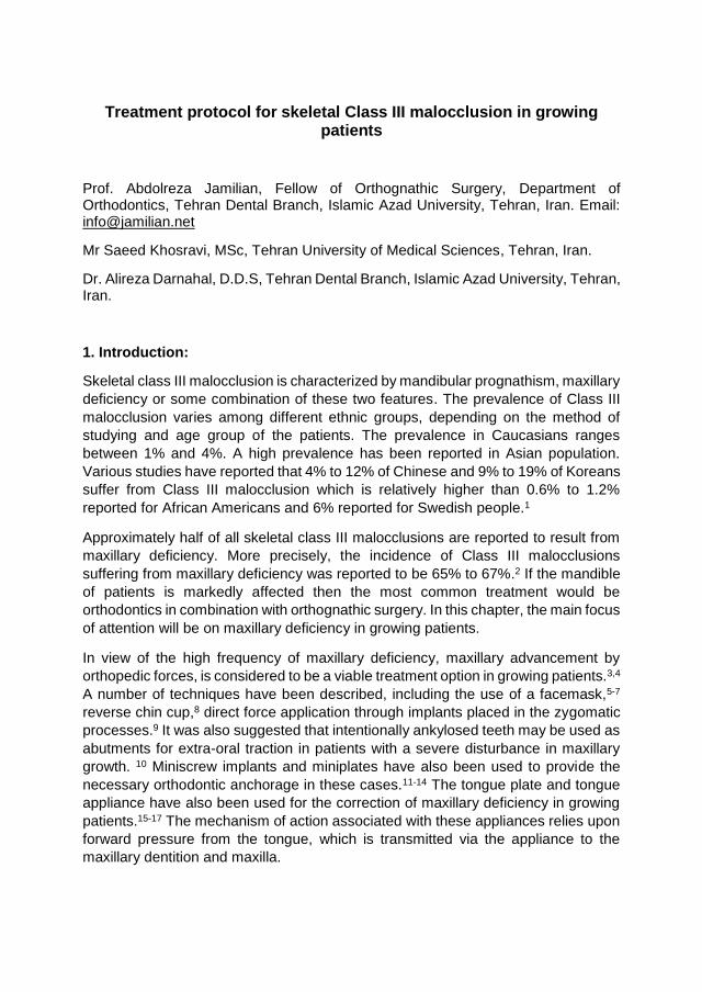
removable appliance to correct the cross-bite on the lateral incisors. (Figure 13) After
8 months of active treatment a positive overjet and Class I buccal segments were
achieved and the cross-bites were corrected. (Figures 14 and 15)
Figure 12: Miniscrews and Class III elastics
Figure 13: Expansion of the maxillary arch

Figure 14: Pretreatment photographs of a 12-year-old boy with maxillary deficiency
Figure 15: Post treatment photographs of the same patient
Limitations:
The limitations of miniscrews include a high risk of failure when placed in unattached
gingiva, screw loosening, root injury when placed in keratinized mucosa, and limited
amount and direction of tooth movement depending on the position of the
miniscrews.

References:
1. Ngan PW, Sung J-H. Chapter 14 - Treatment Strategies for Developing and Nondeveloping Class III Malocclusions. In: Nanda R, editor. Esthetics and Biomechanics in Orthodontics (Second Edition). St. Louis: W.B. Saunders; 2015. p. 246-293. 2. Ellis E, 3rd, McNamara JA, Jr. Components of adult Class III open-bite malocclusion. Am J Orthod 1984;86:277-290. 3. Arman A, Toygar TU, Abuhijleh E. Profile changes associated with different orthopedic treatment approaches in Class III malocclusions. Angle Orthod 2004;74:733-740. 4. Maspero C, Galbiati G, Perillo L, Favero L, Giannini L. Orthopaedic treatment efficiency in skeletal Class III malocclusions in young patients: RME-face mask versus TSME. Eur J Paediatr Dent 2012;13:225-230. 5. Delaire J, Verdon P. [The use of heavy postero-anterior extraoral forces by an orthopedic mask in the treatment of dentomaxillary sequellae of labiomaxillopalatal clefts]. Chir Pediatr 1983;24:315-322. 6. Jamilian A, Showkatbakhsh R, Taban T. The effects of fixed and removable face masks on maxillary deficiencies in growing patients. Orthodontics (Chic.) 2012;13:e37-43. 7. Perillo L, Vitale M, Masucci C, D'Apuzzo F, Cozza P, Franchi L. Comparisons of two protocols for the early treatment of Class III dentoskeletal disharmony. Eur J Orthod 2015. 8. Showkatbakhsh R, Jamilian A, Ghassemi M, Ghassemi A, Taban T, Imani Z. The effects of facemask and reverse chin cup on maxillary deficient patients. J Orthod 2012;39:95-101. 9. Singer SL, Henry PJ, Rosenberg I. Osseointegrated implants as an adjunct to facemask therapy: a case report. Angle Orthod 2000;70:253-262. 10. Kokich VG, Shapiro PA, Oswald R, Koskinen-Moffett L, Clarren SK. Ankylosed teeth as abutments for maxillary protraction: a case report. Am J Orthod 1985;88:303-307. 11. Jamilian A, Showkatbakhsh R. Treatment of maxillary deficiency by miniscrew implants--a case report. J Orthod 2010;37:56-61. 12. De Clerck HJ, Cornelis MA, Cevidanes LH, Heymann GC, Tulloch CJ. Orthopedic traction of the maxilla with miniplates: a new perspective for treatment of midface deficiency. J Oral Maxillofac Surg 2009;67:2123-2129. 13. Showkatbakhsh R, Jamilian A, Behnaz M. Treatment of Maxillary Deficiency by Miniplates: A Case Report. ISRN Surgery 2011;2011.

14. Jamilian A, Haraji A, Showkatbakhsh R, valaee N. The Effects of Miniscrew with Class III Traction in Growing Patients with Maxillary Deficiency. int J Orthod Milwaukee 2011;22:25-30. 15. Jamilian A, Showkatbakhsh R. The effect of tongue appliance on the maxilla in Class III malocclusion due to maxillary deficiency. Int J Orthod Milwaukee 2009;20:11-14. 16. Showkatbakhsh R, Jamilian A. A novel method of maxillary deficiency treatment by tongue plate--a case report. Int J Orthod Milwaukee 2011;22:31-34. 17. Showkatbakhsh R, Toumarian L, Jamilian A, Sheibaninia A, Mirkarimi M, Taban T. The effects of face mask and tongue plate on maxillary deficiency in growing patients: a randomized clinical trial. J Orthod 2013;40:130-136. 18. Cha KS. Skeletal changes of maxillary protraction in patients exhibiting skeletal class III malocclusion: a comparison of three skeletal maturation groups. Angle Orthod 2003;73:26-35. 19. Delaire J, Verdon P, Lumineau JP, Cherga-Negrea A, Talmant J, Boisson M. [Some results of extra-oral tractions with front-chin rest in the orthodontic treatment of class 3 maxillomandibular malformations and of bony sequelae of cleft lip and palate]. Rev Stomatol Chir Maxillofac 1972;73:633-642. 20. Roberts CA, Subtelny JD. An American Board of Orthodontics case report. Use of the face mask in the treatment of maxillary skeletal retrusion. Am J Orthod Dentofacial Orthop 1988;93:388-394. 21. Nanda R. Biomechanical and clinical considerations of a modified protraction headgear. Am J Orthod 1980;78:125-139. 22. Showkatbakhsh R, Jamilian A. A novel approach in treatment of maxillary deficiency by reverse chin cup. Int J Orthod Milwaukee 2010;21:27-31. 23. Showkatbakhsh R, Jamilian A, Taban T, Golrokh M. The effects of Face mask and Tongue Appliance on Maxillary Deficiency in growing patients: A randomized clinical trial. Prog Orthod 2012;13:266-272. 24. Showkatbakhsh R, Jamilian A, Ghassemi M, Ghassemi A, Shayan A. Maxillary deficiency treatment by fixed tongue appliance--a case report. Int J Orthod Milwaukee 2013;24:31-34. 25. Showkatbakhsh R, Jamilian A, Behnaz M, Ghassemi M, Ghassemi A. The short-term effects of face mask and fixed tongue appliance on maxillary deficiency in growing patients--a randomized clinical trial. Int J Orthod Milwaukee 2015;26:33-38. 26. McNamara JA, Jr., Huge SA. The functional regulator (FR-3) of Frankel. Am J Orthod 1985;88:409-424.

27. Cevidanes L, Baccetti T, Franchi L, McNamara JA, Jr., De Clerck H. Comparison of two protocols for maxillary protraction: bone anchors versus face mask with rapid maxillary expansion. Angle Orthod 2010;80:799-806.


![Class III Malocclusion: Missense Mutations in DUSP6 Gene · Caucasians have a skeletal origin and were a result of mandibular prognathism or macrognathia [6]. The prevalence of Class](https://img.pdfslide.us/doc/110x75/60fefd32deaac774f5094328/class-iii-malocclusion-missense-mutations-in-dusp6-gene-caucasians-have-a-skeletal.jpg)