Embed Size (px)
Citation preview
188 casE rEport
Received for publication 11/05/2018 - Accepted for publication 18/12/2018.The authors declare no conflicts of interests.
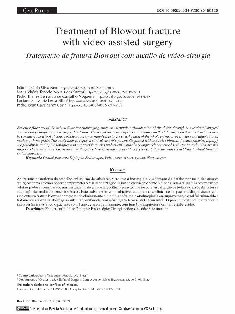
Treatment of Blowout fracture with video-assisted surgery
Tratamento de fratura Blowout com auxílio de vídeo-cirurgia
AbstrAct
Posterior fractures of the orbital floor are challenging, since an incomplete visualization of the defect through conventional surgical accesses may compromise the surgical outcome. The use of the endoscope as an auxiliary method during orbital reconstructions may be considered as a tool of considerable importance, mainly due to the visualization of the whole extension of fracture and adaptation of meshes or bone grafts. This study aims to report a clinical case of a patient diagnosed with extensive blowout fracture showing diplopy, enophthalmos, and ophthalmoplegia in supraversion, who underwent a subciliary approach combined with transantral video assisted surgery. There were no intercurrences on the procedure. Currently, patient has 1 year of follow up, with reestablished orbital function and architecture.
Keywords: Orbital fractures; Diplopia; Endoscopes; Video-assisted surgery; Maxillary antrum
Resumo
As fraturas posteriores do assoalho orbital são desafiadoras, visto que a incompleta visualização do defeito por meio dos acessos cirúrgicos convencionais poderá comprometer o resultado cirúrgico. O uso do endoscópio como método auxiliar durante as reconstruções orbitais pode ser considerado uma ferramenta de grande importância principalmente para visualização de toda a extensão da fratura e adaptação das malhas ou enxertos ósseos. Este trabalho tem como objetivo relatar um caso clínico de um paciente diagnosticado com uma extensa fratura blowout apresentando clinicamente diplopia, enoftalmo e oftalmoplegia em supraversão, o qual foi submetido a tratamento através da abordagem subciliar combinada com a cirurgia vídeo-assistida transantral. O procedimento foi realizado sem intercorrências, estando o paciente com 1 ano de acompanhamento, com função e arquitetura orbital restabelecidos.
Descritores: Fraturas orbitárias; Diplopia; Endoscópio; Cirurgia vídeo-assistida; Seio maxilar
1 Centro Universitário Tiradentes, Maceió, AL, Brazil. 2 Department of Oral and Maxillofacial Surgery, Centro Universitário Tiradentes, Maceió, AL, Brazil.
João de Sá da Silva Neto1 https://orcid.org/0000-0002-2396-9805Maria Vitória Tenório Novais dos Santos1 https://orcid.org/0000-0002-2319-2733Pedro Thalles Bernardo de Carvalho Nogueira2 https://orcid.org/0000-0002-1085-438XLuciano Schwartz Lessa Filho2 https://orcid.org/0000-0001-6077-9333Pedro Jorge Cavalcante Costa2 https://orcid.org/0000-0002-5248-6132
Rev Bras Oftalmol. 2019; 78 (3): 188-91
DOI 10.5935/0034-7280.20190126
189
An open surgical treatment was proposed due to fracture pattern in order to repair the bone defect with video assistance under general anesthesia. Complementary biochemical blood exams and a chest radiograph were requested, which did not show alterations that could contraindicate the procedure. Surgery began with a subciliary approach (Figure 4) in addition to a Caldwell Luc (Figure 5) access to introduce and adapt a 30º endoscope (Karl Storz, Tuttlingen, Germany). Then, the orbital content was released (Figure 6), and the effectiveness of the releasement was verified through a forced duction test.
IntRoductIon
Facial fractures usually occur due to traumas of moderate and high energy, such as aggressions, car accidents, sports accidents, and fallings.(1) When midface is involved, orbital
fractures represent 40% of these injuries, (2) with predominance of the male gender when compared to the female gender in a proportion of 5:1.(3,4)
Among the orbital walls, the floor is considered as a fragile region, which is susceptible to fractures due to the absence of support in the center of its surface and to its thin thickness of approximately 0,27mm.(4) The herniation of the orbital content into the maxillary sinus is of common occurrence in this type of fracture, which characterizes a blowout fracture.(5) Furthermore, diplopy, enophthalmos, ophthalmoplegia, and decreased visual acuity may be also present, thus constituting indications for surgical treatment.(2,4,6)
The main goal of the surgical approach in these fractures is the tridimensional reconstruction of the lost bone anatomy through the reestablishment of the orbital volume.(7) The subciliary, infra orbital, and transconjunctival incisions are the most used surgical accesses for this end.(8) However, some level of perioperative difficultness to view the whole defect is expected in extensive posterior bone defects, which can lead to an inadequate implant placement.(6) Herewith, the combination of an endoscopic approach via maxillary sinus allows excellent assessment and visualization of the defect, providing thus a more effective reconstruction through the complete adaptation of the titanium mesh on the bone defect.(4,9)
The video assisted reconstruction of the orbital floor presents a few limitations and disadvantages.(2) Sinus-related complications that occur due to the passage of the endoscope are rare and on most cases are associated to an unexperienced surgeon.(10) The aim of the present study is to report a clinical case of a patient diagnosed with a blowout fracture that underwent orbital floor reconstruction through a subcilliary approach combined with an endoscopic technique due to the location and extension of the defect.
case RePoRt
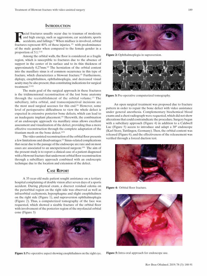
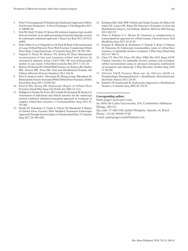
A 35-year-old male patient sought assistance on a tertiary hospital complaining of double vision after seven days of a sports accident. During physical exam, a discreet residual edema on the periorbital region on the right side was observed as well as infraorbital ecchymosis, hyposphagma and slight enophthalmos on the right side (Figure 1), and supraversion ophthalmoplegia (Figure 2). Thus, a computerized tomography of the face was requested, which showed a sizable fracture of the orbital floor with involvement of the posterior region of the myofascial orbital cone (Figure 3)
Treatment of Blowout fracture with video-assisted surgery
Rev Bras Oftalmol. 2019; 78 (3): 188-91
Figure 3: Pre-operative computerized tomography.
Figure 1: Pre-operative aspect showing enophthalmos on the right eye.
Figure 2: Ophthalmoplegia in supraversion.
Figure 4: Orbital floor fracture.
Figure 5: Intra-oral approach for endoscope use.

190 Silva Neto JS, Santos MVTN, Nogueira PTBC, Lessa Filho LS, Costa PJC
Rev Bras Oftalmol. 2019; 78 (3): 188-91
The orbital floor reconstruction was performed with a titanium mesh of the 1.5 system (OsteoMed, Dallas, USA), which was initially introduced via subciliary approach; however, the final adaptation of the implant on the posterior region was performed with a Freer elevator via maxillary sinus, assisted with the endoscope. During this phase, the importance of the indirect assessment via video of the titanium mesh involving the whole bone defect was verified and adapted on the healthy bone wall; such vision was not achieved via subciliary incision. Finally, hemostasis was reviewed, and the surgical accesses were sutured by layers.
With the patient hospitalized, a computerized tomography was requested for immediate post-operative control (Figure 7), which showed an excellent adaptation of the placed mesh, thus reconstructing the bone defect in its whole extension and reestablishing the orbital content. Patient is now on his twelfth month of post-operative follow up with reestablished ocular projection, absence of diplopy signs, and with no restrictions of ocular movements (Figure 8).
Figure 6: Endoscope indirect view showing adaptation of the titanium mesh.
Figure 8: Post-operative tomographic control after one year.
Figure 7: Absence of ocular movement restriction in supraversion.
dIscussIon
The treatment goal for orbital fractures is to reestablish anatomy, volume, and function.(2,4,6) Nevertheless, the difficulty to expose the surgical field for direct visualization is considered limited in extensive posterior fractures of the orbital floor.(11) The type of surgical access for orbital reconstructions will depend mainly on the location and size of fracture,(12) being the subciliary, infraorbital, and transconjunctival accesses the most used ones.(13,14) Currently, the endoscope use has been increasingly popularized. (15)
Chen (2018) recommends the use of an endoscope as auxiliary method on posterior fractures due to its safety to instantly determine the anatomical location and proximity with critical structures, besides defining important landmarks for reconstruction.(16) Furthermore, the method is also indicated when is difficult to release the orbital content on the interior of the maxillary sinus via direct access and on cases of prolapse greater than 1 cm.(11,17) Herewith, despite not presenting perioperative difficultness during the releasement, the endoscope use was indicated due to the posterior extension of fracture to the myofascial orbital cone, facilitating thus the adaptation of the titanium mesh. Nevertheless, the main disadvantages involve the maintenance of the patient on the surgical room and therefore more drugs are required to keep him anesthetized.(18) Still, on the present case we chose to perform this technique with the aim to reduce the chances to undergo the patient to a new surgery due to a bad adaptation of the titanium mesh.
Fortunately, the complications of endoscopic surgeries are rare; however, they occur in a few cases and are reported mostly by unexperienced operators and when the fracture involves more than one orbital wall. (2) According to Suzuki (2017), this approach may present some complications due to the antrostomy, such as oroantral fistulae, exposure of mucolabial wound, facial edema, numbness of teeth, and recurring sinusitis, as well as sensitive disturbances on the region of the superior alveolar nerve.(12) On the present case report, the patient has one year of postoperative follow up with no evidence of complications.
Alternatively to the endoscopic method, the possibility of a perioperative computerized tomography is a reality on important surgery centers, allowing visualization and tridimensional reformatting of the fracture in real time. (18) However, one must consider the quantity of radiation exposure and have the good sense that this method is not available in all surgery centers.(16) On the present case, the perioperative tomography was not performed due to the absence of equipment. Thus, video surgery combined with subciliary access represents an effective possibility on the reported case, allowing complete visualization of the bone defect, which consequently increases outcome predictability. Thus, it becomes necessary to disseminate this method among maxillofacial surgeons.
RefeRences
1. Zamboni RA, Wagner JCB, Volkweis MR, Gerhardt EL, Buchmann EM, Bavaresco CS. Levantamento epidemiológico das fraturas de face do Serviço de Cirurgia e Traumatologia Bucomaxilofacial da Santa Casa de Misericórdia de Porto Alegre – RS. Rev Col Bras Cir 2017; 44: 491-97.
2. Moura LB, Gabrielli MAC, Gabrielli MFR, Pereira-Filho VA. Reconstruction of orbital floor defects assisted by transantral endoscopy. Oral Maxillofac Surg 2017; 21: 65-68.
3. Ellis E. Orbital trauma. Oral Maxillofac Surg Clin North Am 2012; 24: 629-48.
191
Corresponding author: Pedro Jorge Cavalcante Costa Av. Hélio de Castro Vasconcelos, S/N, Condomínio Aldebaran Ômega, QD N5, Zip code: 57.080-548, Jardim Petrópolis, Maceió, Al, Brazil. Phone: +55-82-99999-9749 E-mail: [email protected]
4. Felix V, Veerasigamani N. Endoscopic Endonasal Approach of Blow Out Fracture Reduction – A Novel Technique. J Clin Diagn Res 2017; 11: MD06–08.
5. Noh WJ, Park TJ, Kim JY, Kwon JH. Isolated trapdoor-type medial blowout fracture in an adult presenting horizontal diplopia treated by endoscopic endonasal approach. J Surg Case Rep 2013; 2013(3): rjt009.
6. Park J, Huh J, Lee J, Chang M, Lee H, Park M, Baek S. Reconstruction of Large Orbital Posterior Floor Wall Fracture Considering Orbital Floor Slope Using Endoscope. J Craniofac Surg 2017; 28: 947–950.
7. Vignesh U, Divya M, Dichen, VA, Debraj H. Three dimensional reconstruction of late post traumatic orbital wall defects by customized implants using CAD-CAM, 3D stereolithographic models: A case report. J Oral Biol Craniofac Res 2017; 7: 212–18.
8. Morris CD, Tiwana PS. Orbital Wall Fractures. In: Fonseca RJ,č Barber HD,č Powers MP,č Frost DE. Oral and Maxillofacial Trauma. 4th Edition. Missouri: Elsevier Saunders; 2013: 434-36.
9. Patel S, Andrecovich C, Silverman M, Zhang Liying, Shkoukani M. Biomechanic Factors Associated With Orbital Floor Fractures. JAMA Facial Plast Surg 2017; 19:298-302.
10. Farwell DG, Strong EB. Endoscopic Repair of Orbital Floor Fractures. Facial Plast Surg Clin North Am 2006; 14: 11-6.
11. Polligkeit J, Grimm M, Peters JP, Cetindis M, Krimmel M, Reinert S. Assessment of indications and clinical outcome for the endoscopy assisted combined subciliary/transantral approach in treatment of complex orbital floor fractures. J Craniomaxillofac Surg 2013; 41: 797-802.
12. Suzuki M, Nakamura Y, Ozaki S, Yokota M, Murakami S. Repair of Orbital Floor Fracture With Modified Transnasal Endoscopic Approach Through Anterior Space to Nasolacrimal Duct. J Craniofac Surg 2017; 28: 998-1002.
13. Krishnan DG, Ochs MW. Orbital and Ocular Trauma. In: Miloro M, Ghali GE, Larsen PE, Waite PD. Peterson’s Principles of Oral and Maxillofacial Surgery, 3rd Edition. Shelton: McGraw-Hill Europe 2012: 483-517.
14. Olate S, Palmeri Jr C, Morais M. Chemosis as complications in transconjuntival approach for orbital trauma. J Korean Assoc Oral Maxillofac Surg 2017; 43: 42-45.
15. Soejima K, Shimoda K, Kashimura T, Yamaki T, Kono T, Sakurai H, Nakazawa H. Endoscopic transmaxillary repair of orbital floor fractures: A minimally invasive treatment. J Plast Surg Hand Surg 2013; 47: 368-73.
16. Chen CT, Pan CH, Chen CH, Shyu VBH, Wu JCH, Kang GCW. Clinical outcomes for minimally invasive primary and secondary orbital reconstruction using an advanced synergistic combination of navigation and endoscopy. J Plast Reconstr Aesthet Surg 2018; 71: 90-100.
17. Oliveira JAGP. Fraturas Blow-out. In: Oliveira JAGP, ed. Traumatologia Bucomaxilofacial e Reabilitação Morfofuncional. São Paulo: Santos; 2012: 229-45.
18. Schubert W, Jenabzadeh K. Endoscopic Approach to Maxillofacial Trauma. J Craniofac Surg 2009; 20: 154-56.
Rev Bras Oftalmol. 2019; 78 (3): 188-91
Treatment of Blowout fracture with video-assisted surgery