Embed Size (px)
Citation preview

Translating the hemodynamic response:why focused interdisciplinary integrationshould matter for the future of functionalneuroimagingSigita Cinciute
Institute of Biosciences, Life Sciences Center, Vilnius University, Vilnius, Lithuania
ABSTRACTThe amount of information acquired with functional neuroimaging techniques,particularly fNIRS and fMRI, is rapidly growing and has enormous potentialfor studying human brain functioning. Therefore, many scientists focus on solvingcomputational neuroimaging and Big Data issues to advance the discipline. However,the main obstacle—the accurate translation of the hemodynamic response (HR)by the investigation of a physiological phenomenon called neurovascular coupling—is still not fully overcome and, more importantly, often overlooked in this context.This article provides a brief and critical overview of significant findings fromcellular biology and in vivo brain physiology with a focus on advancing existingHR modelling paradigms. A brief historical timeline of these disciplines ofneuroscience is presented for readers to grasp the concept better, and somepossible solutions for further scientific discussion are provided.
Subjects Biophysics, Cell Biology, Molecular Biology, NeuroscienceKeywords Cerebrovascular regulation, Healthcare, Hemodynamic response, Neuroscience, Brain,Computational modelling, Functional near-infrared spectroscopy, Neurovascular coupling,Functional magnetic resonance imaging
INTRODUCTIONModern functional neuroimaging methods cover broad spatial and temporal scales(Pouratian et al., 2003) and facilitate the important exploration of the functionalorganisation of the human brain in health and disease (Liu et al., 2015). Numerousstatistical or methodological challenges are addressed with this complexity. However,some threats arise from fundamental conceptual challenges that remain widelyunderappreciated within the clinical and neuroimaging communities (Poldrack & Yarkoni,2016). Current trends in neuroimaging and computational neuroscience promote theadvanced mathematical modelling of human brain function based on neuroimaging, andthe implications associated with the use of Big Data (Hansen et al., 2014) in scientificresearch and healthcare innovations. These multidisciplinary interactions betweendifferent branches of science are vital for overall scientific progress. However, some mainconceptual challenges may remain shadowed by massive trends and become a barrierto progress. For example, the ability to assess neural activity in a non-invasive way by
How to cite this article Cinciute S. 2019. Translating the hemodynamic response: why focused interdisciplinary integration should matterfor the future of functional neuroimaging. PeerJ 7:e6621 DOI 10.7717/peerj.6621
Submitted 11 October 2018Accepted 14 February 2019Published 25 March 2019
Corresponding authorSigita Cinciute,[email protected]
Academic editorJafri Abdullah
Additional Information andDeclarations can be found onpage 15
DOI 10.7717/peerj.6621
Copyright2019 Cinciute
Distributed underCreative Commons CC-BY 4.0

measuring the brain’s circulation of blood has revolutionised neuroscience. As a result, weare witnesses to enormous growth in the field of human brain research (Raichle, 2009;Toga, 2015). Each of the functional neuroimaging techniques used today, such asfunctional near-infrared spectroscopy (fNIRS), functional magnetic resonance imaging(fMRI), positron emission tomography (PET) or single photon emission computedtomography (SPECT) explore different metabolic or particular physiological events, but allare based on the physiological principles of neurovascular coupling (NVC). NVC isthe process by which active brain regions induce a local increase in blood flow tomatch their energy demands via the dilation of capillaries and arterioles through variouscellular signalling paths (Mishra et al., 2016). Capillary dilation generates a significantportion of the blood flow increase, evoked by neuronal activity (Hall et al., 2014), and isexpected to contribute substantially to the observed hemodynamic response (HR)(Lindauer et al., 2010). Nevertheless, our understanding of NVC in humans despite itsimportance is still incomplete due to a lack of appropriate and consistent analysis strategiesand stimulation paradigms (Hillman, 2014; Phillips et al., 2016). Despite theirtechnological differences, widely used functional neuroimaging techniques, such asfMRI and less known but prominent fNIRS, are mainly based on the common underlyingphenomenon of NVC (Hillman, 2014; Huneau, Benali & Chabriat, 2015; Iadecola,2017; Phillips et al., 2016). It makes some results of current experimental methodsambiguous compared to the more in-depth fundamental perspective (Lindauer et al., 2010;Phillips et al., 2016; Sotero & Trujillo-Barreto, 2007).
The scientific knowledge is always limited to some extent and arguably with each newfinding. However, the real concerns occur summarising that at the moment, a big part ofscientific and clinical research production is based on sophisticated mathematicalmanipulations of neuroimaging data which was derived from the observations of NVC,referred to as a HR. A generalisation like this emphasises the need of tighter and focusedinterdisciplinary integration within particular neuroscience fields to improve thetranslation of physiological signal into neuroimaging data, which later is processed withsophisticated mathematical and statistical methods. The goal of this article was to fillthe gap between the critical and brief overviews of one of the under-appreciatedneuroimaging challenge: accurate translation of the HR to scientific and clinical findings.The next objective was to provide the scientific reader with a summary of key aspectsand analyse why this problem is relevant to those that are interested in or directlyinvolved in human cognition and behaviour neuroscience or clinical research. To servethis purpose, this article conceptualises the current knowledge of NVC from severalperspectives, mainly cellular and molecular neuroscience (CMN), and functionalneuroimaging (particularly fNIRS and fMRI as they are closely related).
SURVEY METHODOLOGYThe review was designed with a focus on the existing scientific paradigm in human brainfunctional neuroimaging research, mainly addressing the absence of necessary interactionsbetween different multidisciplinary branches of neuroscience such as cellular biology,human brain physiology and computational modelling (CM). The articles that were
Cinciute (2019), PeerJ, DOI 10.7717/peerj.6621 2/21
reviewed in this paper were identified in databases (e.g. Google Scholar, PubMed,ScienceDirect) and subject-specific professional journals and websites (e.g. PLOS,PeerJ, Frontiers, Journal of Cerebral Blood Flow & Metabolism). The literature reviewwas assured to be unbiased and comprehensive by narrowing down the exploration bysearching for original research articles and reviews that discuss (i) cerebrovascularregulation; functional hyperaemia; NVC; astroglial network; the origin of the HR signal infNIRS; the origin of the blood-oxygen-level-dependent (BOLD) signal in fMRI; or(ii) the biophysical model of fNIRS and fMRI signals; the CM methods used in fNIRSand fMRI; and (iii) studies that compare both methods or combine them in humans.The author also searched for articles in cellular and molecular biology, animalstudies, subsequently regarding studies in health and disease. However, it was done only incombination with the previously mentioned search criteria to identify relevantpublications. The other inclusion criteria for selected articles required that articles wouldbe directly related to the topic and would not exhaustively cover unrelated materialsuch as other neuroscience methods if the results were not directly comparable withfunctional neuroimaging.
WHAT IS A HEMODYNAMIC RESPONSE?The human brain represents only 2% of the total body mass. About 25% of the oxygenand from 20% to 70% of the glucose consumed by the human body is dedicated tocerebral functions (Herculano-Houzel, 2011). The maintenance and restoration of the iongradients dissipated by signalling processes such as post-synaptic and action potentials,as well as the uptake and recycling of neurotransmitters are the primary processescontributing to the high brain’s energy needs (Attwell et al., 2010). The brain bloodcirculation system actively regulates the constant demand and supply. However, activebrain regions are often provided more than they require. One of the researchers poeticallyillustrated it as ‘watering the garden for the sake of a single thirsty flower’ (Malonek &Grinvald, 1996). This overcompensation or functional hyperaemia is a fundamentalphenomenon in normal brain function. It was first confirmed by (Roy & Sherrington, 1890)and defines the dilation of arterioles and capillaries of a brain region in response to alocal episode of high neuronal activity. Functional hyperaemia is a generalised term for theoutcome of a complex cerebrovascular regulation mechanism which will be brieflydiscussed in this section. At this point, the term HR is associated with the quantitativemeasures of functional hyperaemia using fNIRS and fMRI. In fMRI, it is better known asthe HR function (HRF) to imply its mathematical properties. Further, in the text,only the HR term will be used, as it describes an observation of a physiological NVCevent common for both techniques.
Functional magnetic resonance imaging and fNIRS have different capacities toexplore human brain functions. As was introduced, both methods, despite their dataacquisition differences, are based on a common underlying phenomenon termed NVC.However, fMRI is more common in general, due to its broad application possibilities andhistorical background, especially in clinical practice (Glover, 2011), while fNIRS wasprimarily used for the bedside monitoring of infants, and other fields where fMRI were not
Cinciute (2019), PeerJ, DOI 10.7717/peerj.6621 3/21
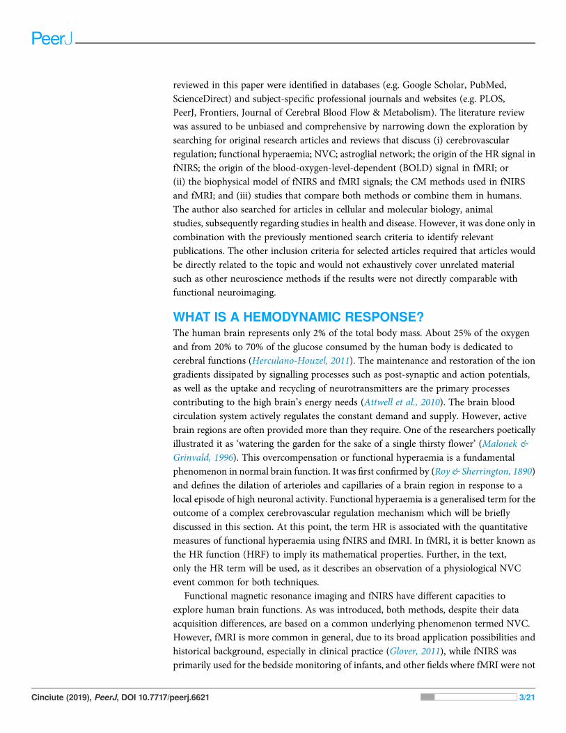
applicable. Thus only quite recently with technical progress fNIRS became an equivalentmethod for investigating human cognitive brain functions (Boas et al., 2014). Despite thecommon underlying phenomenon (Hillman, 2014; Huneau, Benali & Chabriat, 2015;Iadecola, 2017; Phillips et al., 2016) both methods have their own strengths and limitationsassociated with biophysical and physiological signal origins (Kim & Ogawa, 2012;Scarapicchia et al., 2017). A comparison of how the same physiological process of NVCoriginates as a HR measured using fNIRS (A) and HR function measured using fMRI (B)can be found in Fig. 1. The example of HR (A), is based on measuring the composition ofthe total cerebral blood volume in the particular brain area. These measures can be directlydone in vivo, and both oxygenated haemoglobin D(HbO2) and deoxygenated haemoglobinD(Hb) concentration changes are observed simultaneously. In contrast, the BOLD signal infMRI is based on the paramagnetic deoxyhemoglobin decrease in T2� contrast, relative to thesituation with diamagnetic oxyhemoglobin (Bandettini &Wong, 1995; Chavhan et al., 2009).Thus, the D(Hb) curve in Fig. 1A could be seen as a BOLD signal representation, eventhough it is not straightforward due to other physiological contributors captured in theBOLD response (such as regional cerebral blood flow and volume), (Arthurs & Boniface,2002; Kim & Ogawa, 2012).
With technological progress, the number of combined functional fMRI-fNIRS studieshas begun to increase slowly. According to the PubMed database search for the terms‘fMRI’ and ‘fNIRS’ in the title/abstract field and restricted to the results of the articlesand review papers published between January 1, 1977, and June 6, 2018, a total of
Figure 1 Examples of canonical hemodynamic response (A), and hemodynamic response function(B). A neural activity from 0 to 5 s (grey bar) causes neurometabolic and later neurovascular cou-pling, which can be seen as a delay of response (around 2 s). Box in hemodynamic response (A) indicates(a) small inflow of D(HbO2), when the total blood volume is still relatively unchanged (due to increasedcerebral blood flow), and later (b) D(HbO2) increases rapidly due to functional hyperemia caused byvasodilatation. The small increase of D(Hb) occurs due to insufficient washout when the cellular oxygendemand exceeds current supply in a tissue. The canonical example of a hemodynamic response is basedon measuring the composition of cerebral blood volume via chromophore concentration changes(oxy-Hb and deoxy-Hb). fNIRS studies can directly measure both oxy-Hb and deoxy-Hb), (Venclove,Daktariunas & Ruksenas, 2015). In contrast, the canonical hemodynamic response function (HRF)from the Blood Oxygenation Level Dependent (BOLD) method represents the magnetic field change inresponse to the D(Hb) curve (B) and is relative to the baseline.
Full-size DOI: 10.7717/peerj.6621/fig-1
Cinciute (2019), PeerJ, DOI 10.7717/peerj.6621 4/21
752 documents with human participants were identified. After additional restriction tosearch for these terms only in the title field, a total of 11 documents remained (Anwar et al.,2013; Cui et al., 2011; Frederick, Nickerson & Tong, 2012; Gagnon et al., 2012;Maggioni et al., 2013; Okamoto et al., 2004; Sasai et al., 2012; Sato et al., 2013; Strangmanet al., 2002; Tong & Frederick, 2010; Toyoda et al., 2008). A few of them were comparisons,which have been conducted for a variety of cognitive tasks to illustrate similaritiesand differences between fNIRS and fMRI capabilities (Cui et al., 2011; Steinbrink et al.,2006). Overall, the described PubMed search serves as an illustrative example fortwo points: (a) number of multimodal, cross-validation, or comparison studies withineven closely related techniques such as fMRI, and fNIRS is still insufficient; and (b) thatintegration of more distinct branches of neuroscience might be even more daring, but aswill be discussed in the further section, might also add compelling value to the field offunctional human brain research.
Hemodynamic response and classical approach in neuroimagingThe fundamentals of coupling between brain electrical activity, metabolism, and theobserved HR are incredibly complex. Classical functional neuroimaging approaches forsimplicity assume that the vascular response induced by neural activity is a nearlylinear function of blood volume increase. This approach is convenient to model andreconstruct the possible neural activity from the HR but is not entirely accurate.Nonlinearities are believed to arise from both nonlinearities in the vascular responseand neuronal activity, as several studies have demonstrated (Birn et al., 2013; Birn &Bandettini, 2005; Hillman, 2014; Wager et al., 2005). Many more studies may bediscussed depending on the reader’s point of view, but the two following studies werechosen subjectively to provide a short illustrative introduction to this topic fornon-experts. Wager et al. (2005) an empirical study with fMRI, where they attemptto characterise nonlinear effects in visual and motor cortex in 12 human participants,presents finding that these nonlinear effects are relatively consistent throughout the testedbrain areas. Additionally, a more recent and exciting multimodal study by Fabianiet al. (2014) published in 2014 is recommended. In this multimodal study, 19 young and44 older healthy adults were examined to address physical fitness and age effects onNVC in the primary visual cortex and show the quadratic relationship between neuralactivity and blood flow. The overall results indicate that nonlinearity in NVC hasmore than one aspect to be considered, and the classical neuroimaging approach is notsufficient to explain it.
What is the classical explanation of the origin of the HR? Over a decade, the understandingof NVC initiation and its overall role for HR formation dramatically changed (Attwellet al., 2010). From a CMN perspective, for a long time, researchers favoured the idea thatblood flow is locally controlled by a passive negative-feedback system, where neuralactivity leads to a local substrate demand. It was thought that the metabolic signal inducingHR could be a lack of O2 or glucose, or the local rise of CO2, ADP or lactate. However, thisnegative-feedback hypothesis failed to adequately explain the experimental findings ofNVC in animal models (Attwell et al., 2010;Walsh, 2016). Manipulations of O2 and glucose
Cinciute (2019), PeerJ, DOI 10.7717/peerj.6621 5/21
did not regulate blood flow as expected, and CO2, ADP together with lactate showed onlypartial effects (Attwell et al., 2010). More novel, feed-forwards neurotransmitter-mediatedmechanisms suggest active control of the vascular energy supply in the brain(Attwell et al., 2010). In this process, the neurones either signal or activate astrocytes torelease vasoactive messengers onto vessels. According to this hypothesis, astrocytes areanatomical intermediaries between neurones and blood vessels (Attwell et al., 2010;Petzold & Murthy, 2011). However, is it all about astrocytes or other glial cells involved aswell? Recent studies have begun to challenge the astrocytes role as the main drivers ofNVC due to inconsistencies between spatiotemporal properties of vasodilatation, and thestructure-functional properties and distribution of astrocytes in the cortical volume(Iadecola, 2017; McCaslin et al., 2011). In the following sub-section, the three examplesof recent in vivo experiments will be discussed, to better illustrate the importance ofnon-neuronal HR origin.
What induces a hemodynamic response and why does it matter?From a historical perspective 100 and 50 years ago the neuroglial was thought to be onlya connective material in the brain and was given an entirely passive supportive role(Kettenmann & Verkhratsky, 2008). Since then, a substantial amount of researchhas been published supporting the idea that the previous ‘neuron-centric’ perspectiveof neuroscience is not accurate. Today, it is evident that glial cells are integral to thedevelopment and maintenance of the healthy central nervous system and play a vital rolein the pathogenesis of many brain disorders (Liddelow & Barres, 2017).
The particular scientific focus was first given on astrocytes, as they are the mostabundant population of glia in the mammalian brain (Azevedo et al., 2009; Liddelow &Barres, 2017; Von Bartheld, Bahney & Herculano-Houzel, 2016). It was proved thatastrocytes are not only responsible for physical brain structuring but also are (a) criticalhomeostatic cellular elements that are capable of gluconeogenesis, provide neuroneswith lactate, and control over glucose levels (Bélanger, Allaman & Magistretti, 2011;Brooks, 2009;Magistretti, 2006); (b) form a tripartite synapse with neurones and modulatesynaptic activity via ion and neurotransmitters concentrations in the extracellular space(Allen & Eroglu, 2017; Haydon & Carmignoto, 2006; Krencik, Van Asperen & Ullian,2017; Perea & Araque, 2005; Perea, Navarrete & Araque, 2009); (c) they are responsible forsome immune activity, promote neuronal survival and enable re-myelination withinthe brain (Ayaz et al., 2008; Liddelow & Barres, 2017; Von Bernhardi, 2016); and (d) act asdirect and indirect modulators of cerebrovascular tone (Ayata & Lauritzen, 2015;Bazargani & Attwell, 2016; Filosa et al., 2016; Gratton, Chiarelli & Fabiani, 2017; Iadecola,2017; Mishra, 2017). Moreover, they form an equivalent to neurones astroglial network(Attwell et al., 2010; Giaume et al., 2010; Scemes & Spray, 2003).
After all, one may ask how these new cellular findings translate into functionalneuroimaging. For example, the study of in vivo animal models (cat and rat)for a single-vessel hemodynamic demonstrated that pial surface arteries in the cat’s visualcortex (as well as neurones) show orientation responsiveness (in contrast to rats,where orientation maps are not shown in general), meaning that propagation of vascular
Cinciute (2019), PeerJ, DOI 10.7717/peerj.6621 6/21

dilation between neighbouring columns in the brain needs to be accounted for whendecoding hemodynamic signals (O’Herron et al., 2016).
Another in vivo study of animal models (rat and mice) show that when the sensoryinput increases, blood flow capillaries dilate before arterioles and are estimated to produce84% of the blood flow increase (Hall et al., 2014). Previously, it was thought thatcapillaries usually do not significantly contribute. Moreover, the study identifies pericytesas significant regulators of cerebral blood flow as they are the first vascular elementsto dilate during neuronal activity, and, in turn, initiate hyperaemia. It also unexpectedlyshowed that vasodilators released from active neurones, interneurones and astrocytes(Hamel, 2006; Miller & Halpern, 2014) are not the only essential players in functionalimaging. In fact, the role of pericytes in CNS is as diverse as it was previously describedwith astrocytes: pericytes integrate, coordinate and process signals from their neighbouringcells to generate diverse functional responses that are critical for CNS functions inhealth and disease, including (a) regulation of the blood-brain barrier (BBB) permeability;(b) angiogenesis; (c) clearance of toxic metabolites; (d) neuroinflammation and stemcell activity; and finally (e) initiating capillary HRs (Hall et al., 2014; Iadecola, 2017;Kisler et al., 2017; Sweeney, Ayyadurai & Zlokovic, 2016).
Also, another non-neuronal cell type crucial for inducing HR was recently identified—vascular endothelium. Several kinds of research demonstrated that vascular endotheliumcould propagate upstream dilations of cerebral vessels (Andresen, Shafi & Bryan,2006; Chen et al., 2011; Hannah et al., 2011; Iadecola, 2017; Rosenblum, 1986). The in vivostudy by Chen et al. (2014), demonstrated that spatially selective endothelial disruptionwith light-dye treatment in rats somatosensory cortex significantly attenuated theHR by blocking the retrograde dilation. The early stage and the peak of hyperaemia wereaffected the most, meaning that neurones, astrocytes, pericytes and endothelial cells are allinvolved in forming HR detected by functional neuroimaging.
There are many other scientific sources regarding cellular, molecular biology and NVCthat could be discussed. However, even with the given three in vivo examples, it is evidentthat the main drivers of NVC, and temporal properties of HR associated with it,depends on the spatial location along the cerebral vasculature (Iadecola, 2017). These newfindings allow re-evaluating, how spatiotemporal specificity may be improved alongsidethe technological progress of fNIRS and fMRI. Because ultimately, the non-invasiveuse of HR is one of the most powerful tools at our disposal to explore human cognitionin health and disease.
ACCOUNTING FOR THE COMPLEXITY OFCEREBROVASCULAR REGULATIONThe HR describes the empirical observation of a physiological NVC event. It may bedetected as an amplitude changes in light absorption (fNIRS) or as a magnetic signalvariation (fMRI). In other words, a HR is a spatiotemporal picture of underlying NVC andcerebrovascular regulation at large. To better understand this, some structural andfunctional properties of cerebrovasculature must be explained.
Cinciute (2019), PeerJ, DOI 10.7717/peerj.6621 7/21

Neurovascular unitThe concept of the neurovascular unit (NVU) emerged around 2001 (Iadecola, 2017).The whole mechanism of cerebrovascular regulation can generally be decomposed intoseveral stages (Hamel, 2006; Phillips et al., 2016;Walsh, 2016). The most explored cerebralmicrocirculation is provided by the structural and functional derivative called NVU,(Attwell et al., 2010; Leybaert, 2005). The neurovascular unit represents the interfacebetween the vascular and neural compartments in the brain and is composed of vascular,glial and neuronal cells such as neurones, astrocytes, endothelial cells and pericytes(Hawkins & Davis, 2005; Sweeney, Ayyadurai & Zlokovic, 2016; Sweeney, Walker-Samuel &Shipley, 2018), (Fig. 2). The NVU is an essential structure for several main processes:formation of neurometabolic coupling (NMC), NVC and formation of the BBB,(Leybaert, 2005; Leybaert et al., 2007; Petzold &Murthy, 2011). NVUmay vary in structureand function depending on its location in the brain (Iadecola, 2017; Kowia�nski et al., 2013;Petzold & Murthy, 2011), thus emphasising the complexity of numerous processesthat are involved in maintaining adequate blood flow in the healthy human brain.Neuronal activity in the brain causes two cerebrovascular regulation processes: NMC,which undergoes a substrate exchange between a neurone and an astrocyte and laterinitiates (but not necessarily) NVC. NVU also supports the BBB coupling but is believed tobe unbundled from the already mentioned NBC and NVC, as it regulates the integrityand functions of the BBB (Leybaert, 2005; Sweeney, Ayyadurai & Zlokovic, 2016).New pieces of evidence suggest that another critical component of the NVU are theinterneurones that transduce signals from perivascular nerves (Hamel, 2006;Walsh, 2016),(Fig. 2). The crucial role of the perivascular nerves is to regulate the cerebrovascular toneinfluencing the overall brain perfusion. NVC is then determined by the chemical
Figure 2 Schematic representation of the Neurovascular Unit (NVU).Full-size DOI: 10.7717/peerj.6621/fig-2
Cinciute (2019), PeerJ, DOI 10.7717/peerj.6621 8/21
signals released from the activated perivascular nerves and astrocytes, and together alterthe vascular tone to adjust local perfusion in accordance with the brain activity (Hamel,2006; Walsh, 2016).
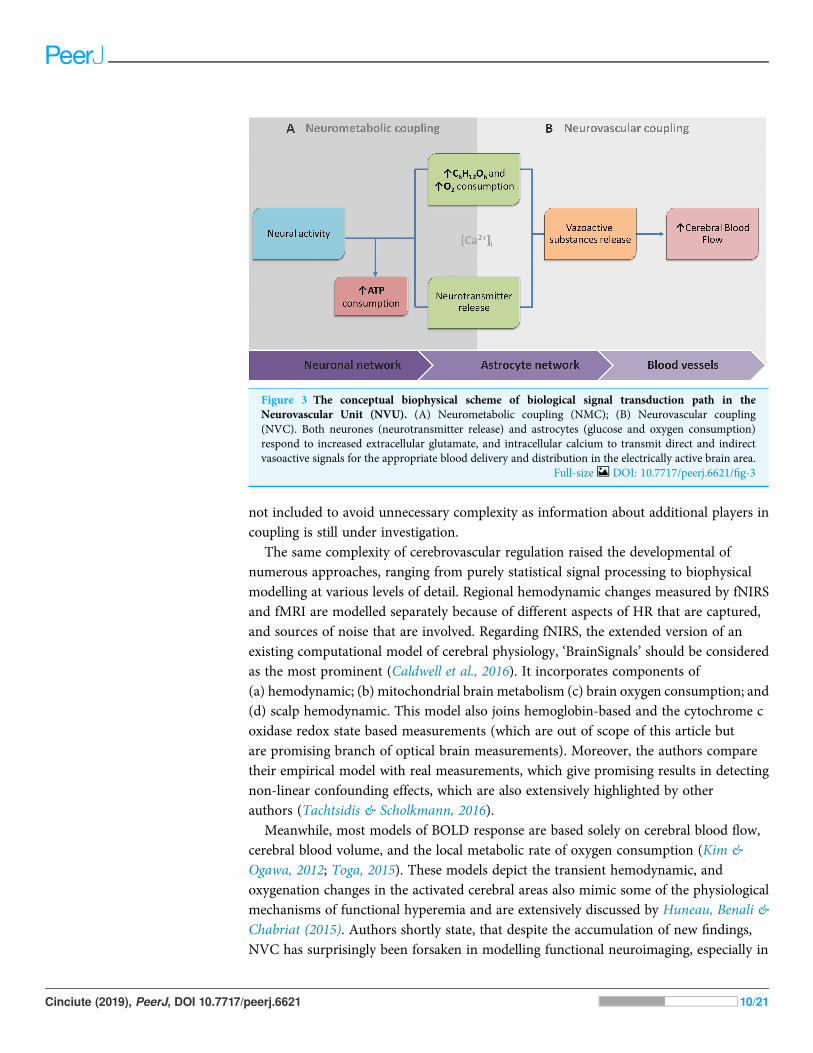
Signal transduction path in the neurovascular unitWith progress in molecular and cellular biology, a conceptual shift in our understanding ofcerebral blood flow occurred where astrocytes, previously believed to be passive supportingcells, had been shown to actively participate in many other physiological processes,as well as creating equivalent to neural to the astroglial network, and directly modulatingneural activity (Giaume et al., 2010; Kowia�nski et al., 2013; Scemes & Spray, 2003). For awhile, the idea that elevations of calcium concentration in the astrocytes may releasetransmitters that regulate neuronal and vascular functions was controversial (Barres, 2008;Bezzi et al., 2004; Fiacco, Agulhon & McCarthy, 2009). This changed when numerouscontradictions were reported between different studies and had been resolved(Bazargani & Attwell, 2016). Shortly after the discovery that glutamate triggers an increaseof intracellular calcium concentration ([Ca2+]i), it was suggested that there might be amechanism by which calcium signalling propagates towards astrocyte’s endfeet andstimulates the release of vasoactive messengers (Attwell et al., 2010). Vasoactivemessengers can cause vasodilatation (most of the neurotransmitters; nitric oxide;prostaglandins; epoxyeicosatrienoic acids; lactate; adenosine etc.) or vasoconstriction(norepinephrine; 20-Hydroxyeicosatetraenoic acid etc.) of arterioles (Giaume et al., 2010;Kowia�nski et al., 2013; Leybaert, 2005; Petzold & Murthy, 2011; Scemes & Spray, 2003).Current understanding suggests that astrocytes, as well as neurones, should bedivided into three spatial compartments, such as processes, soma and endfeet (Bazargani &Attwell, 2016). In this way, the release of specific vasoactive messengers in theendfeet is explained by an overall summation of ([Ca2+]i) in the soma and processes.The response may differ in terms of frequency, kinetics, spatial spreads and interactionwith other cellular messengers (Bazargani & Attwell, 2016). As was previously discussed,astrocytes are not the only cells that are involved in HR, but they continue to beconsidered the main drivers of NVC.
Because of this, the article proposes the conceptual biophysical scheme of the biologicalsignal transduction path in the neurovascular unit (Fig. 3), as a brief adaptation of theclassical approach of NVU. Figure 3 summarises how metabolic and physiologicalevents (NMC and NVC accordingly) via calcium concentration elevation cause the HR.This, in turn, can be observed with functional neuroimaging. Note that the conceptualbiophysical scheme inevitably assumes neural activity-derived NVC. Nonetheless,recent findings show that astroglial metabolic networks may sustain or suppress neuronalactivity (Giaume et al., 2010). For simplicity, this article does not account for it or discuss itthoroughly, as more studies have to be done to generalise new findings (Giaume et al.,2010;Walsh, 2016). However, the idea should be kept in mind for the critical evaluation ofcurrent functional neuroimaging methods, and the proposed scheme should be usedas a tool for a brief explanation of how signal transduction for cerebrovascular regulationoccurs in NVU. Previously mentioned evidence of spatiotemporal specificity of NVU are
Cinciute (2019), PeerJ, DOI 10.7717/peerj.6621 9/21
not included to avoid unnecessary complexity as information about additional players incoupling is still under investigation.
The same complexity of cerebrovascular regulation raised the developmental ofnumerous approaches, ranging from purely statistical signal processing to biophysicalmodelling at various levels of detail. Regional hemodynamic changes measured by fNIRSand fMRI are modelled separately because of different aspects of HR that are captured,and sources of noise that are involved. Regarding fNIRS, the extended version of anexisting computational model of cerebral physiology, ‘BrainSignals’ should be consideredas the most prominent (Caldwell et al., 2016). It incorporates components of(a) hemodynamic; (b) mitochondrial brain metabolism (c) brain oxygen consumption; and(d) scalp hemodynamic. This model also joins hemoglobin-based and the cytochrome coxidase redox state based measurements (which are out of scope of this article butare promising branch of optical brain measurements). Moreover, the authors comparetheir empirical model with real measurements, which give promising results in detectingnon-linear confounding effects, which are also extensively highlighted by otherauthors (Tachtsidis & Scholkmann, 2016).
Meanwhile, most models of BOLD response are based solely on cerebral blood flow,cerebral blood volume, and the local metabolic rate of oxygen consumption (Kim &Ogawa, 2012; Toga, 2015). These models depict the transient hemodynamic, andoxygenation changes in the activated cerebral areas also mimic some of the physiologicalmechanisms of functional hyperemia and are extensively discussed by Huneau, Benali &Chabriat (2015). Authors shortly state, that despite the accumulation of new findings,NVC has surprisingly been forsaken in modelling functional neuroimaging, especially in
Figure 3 The conceptual biophysical scheme of biological signal transduction path in theNeurovascular Unit (NVU). (A) Neurometabolic coupling (NMC); (B) Neurovascular coupling(NVC). Both neurones (neurotransmitter release) and astrocytes (glucose and oxygen consumption)respond to increased extracellular glutamate, and intracellular calcium to transmit direct and indirectvasoactive signals for the appropriate blood delivery and distribution in the electrically active brain area.
Full-size DOI: 10.7717/peerj.6621/fig-3
Cinciute (2019), PeerJ, DOI 10.7717/peerj.6621 10/21

humans. On the other hand, the field of mathematical modelling of BOLD reachedsome significant consensus across variables that should be involved in a generativehemodynamic model (using dynamic causal modelling approach), (Havlicek et al., 2015).It involves several different models, such as (a) neuronal; (b) NVC; (c) hemodynamic;and (d) BOLD in a joint model. The approach reflects experimental observations ofunderlying physiological processes and corresponds well with multimodal experimentaldatasets (Havlicek et al., 2017).
Meanwhile, technological improvement of neuroimaging techniques allows creatingnew and more specific models for investigating NVC. For example, a novel in vivo studycombining imaging of cortical microvascular and mural cell architecture togetherwith mathematical modelling of blood flow and oxygen transport provided new insights onseemingly paradoxical observations in the literature around reduced blood velocity inresponse to arteriolar constrictions, and found that it might be caused by propagation ofconstrictions to upstream penetrating arterioles (Sweeney, Walker-Samuel & Shipley,2018). A similar investigation of cerebral blood flow (CBF) regulation would beinaccessible in a conventional experimental context. In this study, results were achieved byusing in vivo collected information for in silico experimentation.
WHY FOCUSED INTERDISCIPLINARY INTEGRATIONSHOULD MATTERHow does one determine whether HR under neurological or psychiatric conditions reflectsunderlying neural activity rather than altered NVC? Does it mean that in many casesadditional validation studies linking neuronal activity with NVC might be needed to relyon cognitive inferences derived from functional neuroimaging entirely? A growingbody of evidence from animal studies suggests it (Lindauer et al., 2010; O’Herron et al.,2016). Other questions like (i) how new findings of non-neural cell interactions change theinterpretation of neural activity derived from previous functional neuroimaging, and(ii) how to distinguish between neural-activity-derived and only glia-activity-derivedhemodynamic events, remain open.
Neuroscience is a multidisciplinary branch of biology, and its scope has broadened overtime to include a lot of new and different approaches in many aspects of the nervoussystem. As a result, neuroscience exploded in a number of interdisciplinary fields such asneurophysiology, cognitive and behavioural neuroscience, computational neuroscienceand translational neuroscience research. Somehow, common researchers’ knowledgeassumes that in such a broad community of neuroscientists and clinicians, there must beenough professionals dedicating their time and effort to some issues and that necessaryintegration will occur naturally at the particular threshold. However, some mainconceptual challenges may remain daunting due to an unfocused, one-side-driveninterdisciplinary integration. Illustrating it with terms of symbiosis: when relationshipsand interactions between different branches of neuroscience are based more oncommensalism (when part A benefits from part B, but B remain unaffected), rather thanmutualism (when part A benefits from part B, and vice versa). Of course, interdisciplinaryintegrations are way more complicated, but some relevant patterns may be observed.
Cinciute (2019), PeerJ, DOI 10.7717/peerj.6621 11/21

For example, a few historical moments of fMRI, fNIRS and CMN are given in a singletimeline (Fig. 4). As can be seen from Fig. 4, some significant milestones, such as a burst offunctional neuroimaging studies using fMRI and fNIRS were achieved simultaneouslyaround 1992. While others, conceptually very important, such as the concepts of thetripartite synapse and neurovascular unit, emerged only around 2000. There is no surprisein the different timing between different neuroscience branch achievements in general.However, even after more than a decade following seminal research in 2000, the NVCis still surprisingly overlooked in functional neuroimaging modelling, especially inhumans. This means that current functional neuroimaging inference guidelines are poorlyaddressing even already known findings of underlying physiological mechanisms ofNVC. This renders further interpretations of the HR based functional neuroimagingresults ambiguous and entirely reliable only with support from anatomical andelectrophysiological studies. As is often the case, the primary concern is not the validity ofprevious and current studies using functional neuroimaging, but advancements andinnovation in the existing paradigm. Ultimately, the non-invasive use of HR is one of themost powerful tools at our disposal to explore human cognition in health and disease,and thus the focused interdisciplinary attention on its accuracy should matter to anyoneworking or with interests in the field of human brain research.
CONCLUSIONS AND FUTURE PERSPECTIVESThe significant part of scientific and clinical research production is based on complexmathematical manipulations of neuroimaging data derived from NVC. To improve theoverall quality of this production, a complete interpretation of HR should become anumber one concern in the field, as it is the primary information source of underlyinghuman brain neurophysiology. However, the amount of available information is growingexponentially, and due to this information overload, researchers’ attention span may be
Figure 4 A timeline of magnetic resonance imaging (MRI), functional near-infrared spectroscopy(fNIRS), and cellular and molecular neuroscience (CMN) milestones.
Full-size DOI: 10.7717/peerj.6621/fig-4
Cinciute (2019), PeerJ, DOI 10.7717/peerj.6621 12/21
naturally decreased, thus making it a definite obstacle. On the other hand, as was illustratedwith the PubMed database search, even within closely related techniques such as fMRI,and fNIRS, there is evident lack of close integration. Moreover, significant resultsfrom even more distinct branches of neuroscience such as cellular biology and in vivo brainphysiology are instead suggested for consideration than provided for implementing inexisting CM of human brain function.
One way to overcome it is to stress the concern and make it easy to perceive for broaderscientific communities. In accordance, this article fills the gap of a critical view on HRtranslation into scientific findings and expresses the need for more similarly focusedinterdisciplinary reviews, as numerous aspects cannot be thoroughly generalised at once.Also, it addresses the need to integrate neurophysiology and computational neurosciencefields to stimulate innovations in neuroimaging by improving an accurate translationof physiological brain signals. At this point, another possible suggestion would be toimplement existing machine learning (ML) algorithms for data mining. It would allowmeticulous comparison of existing data in CMN studies of NVC, and functionalneuroimaging.
In contrast to existing approaches (which use sophisticated algorithms to performlarge-scale medical data analysis to search for patterns and predictions in certain braindiseases), attention could be focused on the problem of translating complex physiologicalphenomena (NVC) to functional neuroimaging (brain maps of HR). This analyticalapproach in biomedicine is successfully implemented elsewhere (Cao et al., 2018;Ching et al., 2018). In fact, according to latest report of artificial intelligence (AI) use(Shoham et al., 2018), the significant portion of AI-technology-based papers in the USAand Europe tend to be focused on the humanities, and medical and health sciences.Unfortunately, no such attempt was found in the current literature concerning the HR.The existing approaches, from previous (Banaji et al., 2008; Buxton, Wong & Frank, 1998;Caldwell et al., 2016; Friston et al., 2000; Huneau, Benali & Chabriat, 2015; Sotero &Trujillo-Barreto, 2007, 2008), to more recent (Havlicek et al., 2017; Sweeney,Walker-Samuel &Shipley, 2018) models are exploratory, meaning that they try to determine, whether what isbeing observed might be explained by a currently existing theory. Further, an analyticalapproach with ML algorithms could be used for patterning and prediction in aconceptually different manner. Despite the notable advantages, it is important to note thatapplying it would be inevitably challenging; mostly because of data properties, as severaldifferent approaches might be needed at once (Cao et al., 2018; Ching et al., 2018).Meanwhile, the author suggests a few general points to discuss on how necessaryintegration could be initiated:
(a) Systematic reviews and meta-analyses of previous research studies could be performedselectively on different aspects of NVC and HR (i.e. modality used to investigate,species of a subject, spatial location of interest in the brain volume, goals of researchand employed pharmacological agents). The literature search could be expanded withAI algorithms dedicated for the search of relevant scientific content with extensivevocabulary from different neuroscience fields to avoid losing information, when
Cinciute (2019), PeerJ, DOI 10.7717/peerj.6621 13/21

publications conceptually are about the same physiological phenomenon, but due tohistorical context, or other reasons, is described differently (like terms HR and HRfunction). This would ensure unbiased (by the investigators’ prior knowledge)collection of relevant publications. As an existing equivalent could be considered AI2system by the Allen Institute, called ‘Semantic Scholar’, dedicated to finding peer-reviewed research from only trusted, verified sources (https://www.semanticscholar.org). Another example—Elsevier Fingerprint Engine,—the same systems that wereapplied to explore previously mentioned AI tendency to focus on healthcare. It used aprimary set of about 800 keywords relevant to AI (Shoham et al., 2018). Other enginesthat are not mentioned in this publication may also be used directly or as a prototype.After the initial search on particular aspects of NCV and HR (mostly to make easier thequality check before further analysis), the data could be combined and sliced by anyrelevant dimension. With a sufficient quantity of information, several different datamining approaches could be possibly applied.
(b) The initial collection for systematic reviews and meta-analyses could be reused tobuild a database (or a platform with some user interface) and a unified template for anew data entry could be created, as a suggestion what metadata file that could beassociated with a new publication. It would make it easier to import new data and thisin turn would increase publication’s impact and visibility. Some concepts of similarsystems could also be borrowed and implemented from already existing projects suchas the Human Brain Project (https://www.humanbrainproject.eu), or maybe evenbranch out as a separate compartment of an already existing platform.
(c) This newly created database could also serve as an information source for any level ofcomputational insight: CM, deep learning, ML, or AI. The database could provide someguidelines for the researchers when searching, or preparing training data for their insilico experimentation. The purpose of mentioned algorithms may vary from anautomated classification of inputs from living cell microarrays (Jonczyk et al., 2016) tosophisticated machine learning algorithms searching for discrepancies acrossmultimodal studies as in previously given examples.
To sum up, most of the perspective tools are already available and needs only to beimplemented with a particular problem. A broader and multi-disciplinary appreciationof NVC could further boost basic and clinical neuroscience. Thus, a reason why it is stillnot in the frontlines of functional neuroimaging remains debatable. On another hand,this gap in neuroscience requires state-of-the-art scientific research. Because of this,the author invites scientific researches to respond in comments or with a follow-uppublication and propose tools or strategies that could be implemented towards accuratetranslation of the HR.
ACKNOWLEDGEMENTSThe author would like to thank colleagues A. Daktariunas and O. Ruksenas forcontributing with helpful ideas. The author wishes to thank the editor and the reviewersfor their contribution.
Cinciute (2019), PeerJ, DOI 10.7717/peerj.6621 14/21
ADDITIONAL INFORMATION AND DECLARATIONS
FundingThe author received no funding for this work.
Competing InterestsThe author declare having no competing interests.
Author Contributions� Sigita Cinciute conceived and designed the experiments, performed the experiments,analysed the data, contributed reagents/materials/analysis tools, prepared figures and/ortables, authored or reviewed drafts of the paper, approved the final draft.
Data AvailabilityThe following information was supplied regarding data availability:
The research in this article did not generate any data or code (review article).
REFERENCESAllen NJ, Eroglu C. 2017. Cell biology of astrocyte-synapse interactions. Neuron 96(3):697–708
DOI 10.1016/j.neuron.2017.09.056.
Andresen J, Shafi NI, Bryan RM. 2006. Endothelial influences on cerebrovascular tone.Journal of Applied Physiology 100(1):318–327 DOI 10.1152/japplphysiol.00937.2005.
Anwar AR, Muthalib M, Perrey S, Galka A, Granert O,Wolff S, Deuschl G, Raethjen J, Heute U,Muthuraman M. 2013. Comparison of causality analysis on simultaneously measured fMRIand NIRS signals during motor tasks. In: Proceedings of the Annual International Conference ofthe IEEE Engineering in Medicine and Biology Society. Piscataway: IEEE, 2628–2631.
Arthurs OJ, Boniface S. 2002. How well do we understand the neural origins of the fMRIBOLD signal? Trends in Neurosciences 25(1):27–31 DOI 10.1016/S0166-2236(00)01995-0.
Attwell D, Buchan AMA, Charpak S, LauritzenM,MacVicar BBA, Newman EEA. 2010.Glial andneuronal control of brain blood flow. Nature 468(7321):232–243 DOI 10.1038/nature09613.
Ayata C, Lauritzen M. 2015. Spreading depression, spreading depolarizations, and the cerebralvasculature. Physiological Reviews 95(3):953–993 DOI 10.1152/physrev.00027.2014.
Ayaz H, Allen SL, Platek SM, Onaral B. 2008. Maze Suite 1.0: a complete set of tools to prepare,present, and analyze navigational and spatial cognitive neuroscience experiments.Behavior Research Methods 40(1):353–359 DOI 10.3758/BRM.40.1.353.
Azevedo FAC, Carvalho LRB, Grinberg LT, Farfel JM, Ferretti REL, Leite REP, Filho WJ,Lent R, Herculano-Houzel S. 2009. Equal numbers of neuronal and nonneuronal cells makethe human brain an isometrically scaled-up primate brain. Journal of Comparative Neurology513(5):532–541 DOI 10.1002/cne.21974.
Banaji M, Mallet A, Elwell CE, Nicholls P, Cooper CE. 2008. A model of brain circulation andmetabolism: NIRS signal changes during physiological challenges. PLOS Computational Biology4(11):e1000212 DOI 10.1371/journal.pcbi.1000212.
Bandettini PA, Wong EC. 1995. Effects of biophysical and physiologic parameters on brainactivation-induced R2� and R2 changes: simulations using a deterministic diffusion model.International Journal of Imaging Systems and Technology 6(2–3):133–152DOI 10.1002/ima.1850060203.
Cinciute (2019), PeerJ, DOI 10.7717/peerj.6621 15/21
Barres BA. 2008. The mystery and magic of glia: a perspective on their roles in health anddisease. Neuron 60(3):430–440 DOI 10.1016/j.neuron.2008.10.013.
Bazargani N, Attwell D. 2016. Astrocyte calcium signaling: the third wave. Nature Neuroscience19(2):182–189 DOI 10.1038/nn.4201.
Bélanger M, Allaman I, Magistretti PJ. 2011. Brain energy metabolism: focus on astrocyte-neuronmetabolic cooperation. Cell Metabolism 14(6):724–738 DOI 10.1016/j.cmet.2011.08.016.
Bezzi P, Gundersen V, Galbete JL, Seifert G, Steinhäuser C, Pilati E, Volterra A. 2004.Astrocytes contain a vesicular compartment that is competent for regulated exocytosis ofglutamate. Nature Neuroscience 7(6):613–620 DOI 10.1038/nn1246.
Birn RM, Bandettini PA. 2005. The effect of stimulus duty cycle and “off” duration on BOLDresponse linearity. NeuroImage 27(1):70–82 DOI 10.1016/j.neuroimage.2005.03.040.
Birn RM, Molloy EK, Patriat R, Parker T, Meier TB, Kirk GR, Nair VA, Elizabeth Meyerand M,Prabhakaran V. 2013. The effect of scan length on the reliability of resting-state fMRIconnectivity estimates. NeuroImage 83:550–558 DOI 10.1016/j.neuroimage.2013.05.099.
Boas DA, Elwell CE, Ferrari M, Taga G. 2014. Twenty years of functional near-infraredspectroscopy: introduction for the special issue. NeuroImage 85:1–5DOI 10.1016/j.neuroimage.2013.11.033.
Brooks GA. 2009. Cell-cell and intracellular lactate shuttles. Journal of Physiology587(23):5591–5600 DOI 10.1113/jphysiol.2009.178350.
Buxton RB, Wong EC, Frank LR. 1998. Dynamics of blood flow and oxygenation changesduring brain activation: the balloon model. Magnetic Resonance in Medicine 39(6):855–864DOI 10.1002/mrm.1910390602.
Caldwell M, Scholkmann F, Wolf U, Wolf M, Elwell C, Tachtsidis I. 2016. Modellingconfounding effects from extracerebral contamination and systemic factors on functionalnear-infrared spectroscopy. NeuroImage 143:91–105 DOI 10.1016/j.neuroimage.2016.08.058.
Cao C, Liu F, Tan H, Song D, Shu W, Li W, Zhou Y, Bo X, Xie Z. 2018. Deep learning and itsapplications in biomedicine. Genomics, Proteomics & Bioinformatics 16(1):17–32DOI 10.1016/j.gpb.2017.07.003.
Chavhan GB, Babyn PS, Thomas B, Shroff MM, Haacke EM. 2009. Principles, techniques,and applications of T2�-based MR imaging and its special applications. RadioGraphics29(5):1433–1449 DOI 10.1148/rg.295095034.
Chen BR, Bouchard MB, McCaslin AFH, Burgess SA, Hillman EMC. 2011. High-speed vasculardynamics of the hemodynamic response. NeuroImage 54(2):1021–1030DOI 10.1016/j.neuroimage.2010.09.036.
Chen BR, Kozberg MG, Bouchard MB, Shaik MA, Hillman EMC. 2014. A critical role for thevascular endothelium in functional neurovascular coupling in the brain. Journal of theAmerican Heart Association 3(3):e000787 DOI 10.1161/JAHA.114.000787.
Ching T, Himmelstein DS, Beaulieu-Jones BK, Kalinin AA, Do BT, Way GP, Ferrero E,Agapow P-M, Zietz M, Hoffman MM, Xie W, Rosen GL, Lengerich BJ, Israeli J,Lanchantin J, Woloszynek S, Carpenter AE, Shrikumar A, Xu J, Cofer EM, Lavender CA,Turaga SC, Alexandari AM, Lu Z, Harris DJ, DeCaprio D, Qi Y, Kundaje A, Peng Y,Wiley LK, Segler MHS, Boca SM, Joshua Swamidass S, Huang A, Gitter A, Greene CS. 2018.Opportunities and obstacles for deep learning in biology and medicine. Journal of the RoyalSociety Interface 15(141):20170387 DOI 10.1098/rsif.2017.0387.
Cui X, Bray S, Bryant DM, Glover GH, Reiss AL. 2011. A quantitative comparison ofNIRS and fMRI across multiple cognitive tasks. NeuroImage 54(4):2808–2821DOI 10.1016/j.neuroimage.2010.10.069.
Cinciute (2019), PeerJ, DOI 10.7717/peerj.6621 16/21
Fabiani M, Gordon BA, Maclin EL, Pearson MA, Brumback-Peltz CR, Low KA, McAuley E,Sutton BP, Kramer AF, Gratton G. 2014. Neurovascular coupling in normal aging:a combined optical, ERP and fMRI study. NeuroImage 85(5):592–607DOI 10.1016/j.neuroimage.2013.04.113.
Fiacco TA, Agulhon C, McCarthy KD. 2009. Sorting out astrocyte physiology frompharmacology. Annual Review of Pharmacology and Toxicology 49(1):151–174DOI 10.1146/annurev.pharmtox.011008.145602.
Filosa JA, Morrison HW, Iddings JA, Du W, Kim KJ. 2016. Beyond neurovascular coupling,role of astrocytes in the regulation of vascular tone. Neuroscience 323(5):96–109DOI 10.1016/j.neuroscience.2015.03.064.
Frederick B, Nickerson LD, Tong Y. 2012. Physiological denoising of BOLD fMRI data usingregressor interpolation at progressive time delays (RIPTiDe) processing of concurrentfMRI and near-infrared spectroscopy (NIRS). NeuroImage 60(3):1913–1923DOI 10.1016/j.neuroimage.2012.01.140.
Friston KJ, Mechelli A, Turner R, Price CJ. 2000. Nonlinear responses in fMRI: the balloonmodel, Volterra Kernels, and other Hemodynamics. NeuroImage 12(4):466–477DOI 10.1006/nimg.2000.0630.
Gagnon L, Yücel MA, Dehaes M, Cooper RJ, Perdue KL, Selb J, Huppert TJ, Hoge RD,Boas DA. 2012. Quantification of the cortical contribution to the NIRS signal over themotor cortex using concurrent NIRS-fMRI measurements. NeuroImage 59(4):3933–3940DOI 10.1016/j.neuroimage.2011.10.054.
Giaume C, Koulakoff A, Roux L, Holcman D, Rouach N. 2010. Astroglial networks: a step furtherin neuroglial and gliovascular interactions. Nature Reviews Neuroscience 11(2):87–99DOI 10.1038/nrn2757.
Glover GH. 2011. Overview of functional magnetic resonance imaging. Neurosurgery Clinics ofNorth America 22(2):133–139 DOI 10.1016/j.nec.2010.11.001.
Gratton G, Chiarelli AM, Fabiani M. 2017. From brain to blood vessels and back: a noninvasiveoptical imaging approach. Neurophotonics 4(3):031208 DOI 10.1117/1.NPh.4.3.031208.
Hall CN, Reynell C, Gesslein B, Hamilton NB, Mishra A, Sutherland BA, O’Farrell FM,Buchan AM, Lauritzen M, Attwell D. 2014. Capillary pericytes regulate cerebral blood flow inhealth and disease. Nature 508(7494):55–60 DOI 10.1038/nature13165.
Hamel E. 2006. Perivascular nerves and the regulation of cerebrovascular tone. Journal ofApplied Physiology 100(3):1059–1064 DOI 10.1152/japplphysiol.00954.2005.
Hannah RM, Dunn KM, Bonev AD, Nelson MT. 2011. Endothelial SKCa and IKCa Channelsregulate brain parenchymal arteriolar diameter and cortical cerebral blood flow.Journal of Cerebral Blood Flow & Metabolism 31(5):1175–1186 DOI 10.1038/jcbfm.2010.214.
Hansen MM, Miron-Shatz T, Lau AYS, Paton C. 2014. Big data in science and healthcare:a review of recent literature and perspectives. Yearbook of Medical Informatics 23(1):21–26DOI 10.15265/IY-2014-0004.
Havlicek M, Ivanov D, Roebroeck A, Uludağ K. 2017. Determining excitatory and inhibitoryneuronal activity from multimodal fMRI data using a generative hemodynamic model.Frontiers in Neuroscience 11:1–20 DOI 10.3389/fnins.2017.00616.
Havlicek M, Roebroeck A, Friston K, Gardumi A, Ivanov D, Uludag K. 2015.Physiologically informed dynamic causal modeling of fMRI data. NeuroImage 122:355–372DOI 10.1016/j.neuroimage.2015.07.078.
Hawkins B, Davis T. 2005. The blood-brain barrier/neurovascular unit in health and disease.Pharmacological Reviews 57(2):173–185 DOI 10.1124/pr.57.2.4.
Cinciute (2019), PeerJ, DOI 10.7717/peerj.6621 17/21

Haydon PGP, Carmignoto G. 2006. Astrocyte control of synaptic transmission and neurovascularcoupling. Physiological Reviews 86(3):1009–1031 DOI 10.1152/physrev.00049.2005.
Herculano-Houzel S. 2011. Scaling of brain metabolism with a fixed energy budget perneuron: implications for neuronal activity, plasticity and evolution. PLOS ONE 6(3):e17514DOI 10.1371/journal.pone.0017514.
Hillman EMCC. 2014. Coupling mechanism and significance of the BOLD signal: a status report.Annual Review of Neuroscience 37(1):161–181 DOI 10.1146/annurev-neuro-071013-014111.
Huneau C, Benali H, Chabriat H. 2015. Investigating human neurovascular coupling usingfunctional neuroimaging: a critical review of dynamic models. Frontiers in Neuroscience 9:467DOI 10.3389/fnins.2015.00467.
Iadecola C. 2017. The neurovascular unit coming of age: a journey through neurovascular couplingin health and disease. Neuron 96(1):17–42 DOI 10.1016/j.neuron.2017.07.030.
Jonczyk R, Kurth T, Lavrentieva A, Walter J-G, Scheper T, Stahl F. 2016. Living cell microarrays:an overview of concepts. Microarrays 5(2):11 DOI 10.3390/microarrays5020011.
Kettenmann H, Verkhratsky A. 2008. Neuroglia: the 150 years after. Trends in Neurosciences31(12):653–659 DOI 10.1016/j.tins.2008.09.003.
Kim S-G, Ogawa S. 2012. Biophysical and physiological origins of blood oxygenationlevel-dependent fMRI signals. Journal of Cerebral Blood Flow & Metabolism 32(7):1188–1206DOI 10.1038/jcbfm.2012.23.
Kisler K, Nelson AR, Rege SV, Ramanathan A, Wang Y, Ahuja A, Lazic D, Tsai PS, Zhao Z,Zhou Y, Boas DA, Sakadži�c S, Zlokovic BV. 2017. Pericyte degeneration leads to neurovascularuncoupling and limits oxygen supply to brain. Nature Neuroscience 20(3):406–416DOI 10.1038/nn.4489.
Kowia�nski P, Lietzau G, Steliga A, Waśkow M, Moryś J. 2013. The astrocytic contributionto neurovascular coupling—still more questions than answers? Neuroscience Research75(3):171–183 DOI 10.1016/j.neures.2013.01.014.
Krencik R, Van Asperen JV, Ullian EM. 2017. Human astrocytes are distinct contributorsto the complexity of synaptic function. Brain Research Bulletin 129:66–73DOI 10.1016/j.brainresbull.2016.08.012.
Leybaert L. 2005. Neurobarrier coupling in the brain: a partner of neurovascular andneurometabolic coupling? Journal of Cerebral Blood Flow & Metabolism 25(1):2–16DOI 10.1038/sj.jcbfm.9600001.
Leybaert L, De Bock M, Van Moorhem M, Decrock E, De Vuyst E. 2007. Neurobarriercoupling in the brain: adjusting glucose entry with demand. Journal of Neuroscience Research85(15):3213–3220 DOI 10.1002/jnr.21189.
Liddelow SA, Barres BA. 2017. Reactive astrocytes: production, function, and therapeuticpotential. Immunity 46(6):957–967 DOI 10.1016/j.immuni.2017.06.006.
Lindauer U, Dirnagl U, Füchtemeier M, Böttiger C, Offenhauser N, Leithner C, Royl G. 2010.Pathophysiological interference with neurovascular coupling—when imagingbased on hemoglobin might go blind. Frontiers in Neuroenergetics 2:25DOI 10.3389/fnene.2010.00025.
Liu S, Cai W, Liu S, Zhang F, Fulham M, Feng D, Pujol S, Kikinis R. 2015. Multimodalneuroimaging computing: a review of the applications in neuropsychiatric disorders.Brain Informatics 2(3):167–180 DOI 10.1007/s40708-015-0019-x.
Maggioni E, Molteni E, Arrigoni F, Zucca C, Reni G, Triulzi FM, Bianchi AM. 2013. Coupling offMRI and NIRS measurements in the study of negative BOLD response to intermittent photic
Cinciute (2019), PeerJ, DOI 10.7717/peerj.6621 18/21
stimulation. In: Proceedings of the Annual International Conference of the IEEE Engineering inMedicine and Biology Society. Piscataway: IEEE, 1378–1381.
Magistretti PJ. 2006. Neuron-glia metabolic coupling and plasticity. Journal of ExperimentalBiology 209(12):2304–2311 DOI 10.1242/jeb.02208.
Malonek D, Grinvald A. 1996. Interactions between electrical activity and corticalmicrocirculation revealed by imaging spectroscopy: implications for functional brain mapping.Science 272(5261):551–554 DOI 10.1126/science.272.5261.551.
McCaslin AFH, Chen BR, Radosevich AJ, Cauli B, Hillman EMC. 2011. In vivo 3D morphologyof astrocyte–vasculature interactions in the somatosensory cortex: implications forneurovascular coupling. Journal of Cerebral Blood Flow & Metabolism 31(3):795–806DOI 10.1038/jcbfm.2010.204.
Miller DI, Halpern DF. 2014. The new science of cognitive sex differences. Trends in CognitiveSciences 18(1):37–45 DOI 10.1016/j.tics.2013.10.011.
Mishra A. 2017. Binaural blood flow control by astrocytes: listening to synapses and thevasculature. Journal of Physiology 595(6):1885–1902 DOI 10.1113/JP270979.
Mishra A, Reynolds JP, Chen Y, Gourine AV, Rusakov DA, Attwell D. 2016. Astrocytes mediateneurovascular signaling to capillary pericytes but not to arterioles. Nature Neuroscience19(12):1619–1627 DOI 10.1038/nn.4428.
O’Herron P, Chhatbar PY, Levy M, Shen Z, Schramm AE, Lu Z, Kara P. 2016. Neural correlatesof single-vessel haemodynamic responses in vivo. Nature 534(7607):378–382DOI 10.1038/nature17965.
Okamoto M, Dan H, Shimizu K, Takeo K, Amita T, Oda I, Konishi I, Sakamoto K, Isobe S,Suzuki T, Kohyama K, Dan I. 2004. Multimodal assessment of cortical activation duringapple peeling by NIRS and fMRI. NeuroImage 21(4):1275–1288DOI 10.1016/j.neuroimage.2003.12.003.
Perea G, Araque A. 2005. Glial calcium signaling and neuron–glia communication. Cell Calcium38(3–4):375–382 DOI 10.1016/j.ceca.2005.06.015.
Perea G, Navarrete M, Araque A. 2009. Tripartite synapses: astrocytes process and controlsynaptic information. Trends in Neurosciences 32(8):421–431 DOI 10.1016/j.tins.2009.05.001.
Petzold GC, Murthy VN. 2011. Role of astrocytes in neurovascular coupling. Neuron71(5):782–797 DOI 10.1016/j.neuron.2011.08.009.
Phillips AA, Chan FH, Zheng MMZ, Krassioukov AV, Ainslie PN. 2016.Neurovascular couplingin humans: physiology, methodological advances and clinical implications. Journal of CerebralBlood Flow & Metabolism 36(4):647–664 DOI 10.1177/0271678X15617954.
Poldrack RA, Yarkoni T. 2016. From brain maps to cognitive ontologies: informatics and thesearch for mental structure. Annual Review of Psychology 67(1):587–612DOI 10.1146/annurev-psych-122414-033729.
Pouratian N, Sheth SA, Martin NA, Toga AW. 2003. Shedding light on brain mapping:advances in human optical imaging. Trends in Neurosciences 26(5):277–282DOI 10.1016/S0166-2236(03)00070-5.
Raichle ME. 2009. A brief history of human brain mapping. Trends in Neurosciences32(2):118–126 DOI 10.1016/j.tins.2008.11.001.
Rosenblum WI. 1986. Endothelial dependent relaxation demonstrated in vivo in cerebralarterioles. Stroke 17(3):494–497 DOI 10.1161/01.STR.17.3.494.
Roy C, Sherrington C. 1890. On the regulation of the blood-supply of the brain.Journal of Physiology 11(1–2):85–158 DOI 10.1113/jphysiol.1890.sp000321.
Cinciute (2019), PeerJ, DOI 10.7717/peerj.6621 19/21

Sasai S, Homae F, Watanabe H, Sasaki AT, Tanabe HC, Sadato N, Taga G. 2012.A NIRS–fMRI study of resting state network. NeuroImage 63(1):179–193DOI 10.1016/j.neuroimage.2012.06.011.
Sato H, Yahata N, Funane T, Takizawa R, Katura T, Atsumori H, Nishimura Y, Kinoshita A,Kiguchi M, Koizumi H, Fukuda M, Kasai K. 2013. A NIRS–fMRI investigation ofprefrontal cortex activity during a working memory task. NeuroImage 83:158–173DOI 10.1016/j.neuroimage.2013.06.043.
Scarapicchia V, Brown C, Mayo C, Gawryluk JR. 2017. Functional magnetic resonance imagingand functional near-infrared spectroscopy: insights from combined recording studies.Frontiers in Human Neuroscience 11:419 DOI 10.3389/fnhum.2017.00419.
Scemes E, Spray DC. 2003. The astrocytic syncytium. Advances in Molecular and Cell Biology31:165–179 DOI 10.1016/S1569-2558(03)31007-0.
Shoham Y, Perrault R, Brynjolfsson E, Clark J, Manyika J, Niebles JC, Lyons T,Etchemendy J, Bauer Z. 2018. The AI Index 2018 Annual Report. Stanford.Available at http://cdn.aiindex.org/2018/AI%20Index%202018%20Annual%20Report.pdf.
Sotero RC, Trujillo-Barreto NJ. 2007. Modelling the role of excitatory and inhibitoryneuronal activity in the generation of the BOLD signal. NeuroImage 35(1):149–165DOI 10.1016/j.neuroimage.2006.10.027.
Sotero RC, Trujillo-Barreto NJ. 2008. Biophysical model for integrating neuronalactivity, EEG, fMRI and metabolism. NeuroImage 39(1):290–309DOI 10.1016/j.neuroimage.2007.08.001.
Steinbrink J, Villringer A, Kempf F, Haux D, Boden S, Obrig H. 2006. Illuminating theBOLD signal: combined fMRI–fNIRS studies. Magnetic Resonance Imaging 24(4):495–505DOI 10.1016/j.mri.2005.12.034.
Strangman G, Culver JP, Thompson JH, Boas DA. 2002. A quantitative comparison ofsimultaneous BOLD fMRI and NIRS recordings during functional brain activation. NeuroImage17(2):719–731 DOI 10.1016/S1053-8119(02)91227-9.
Sweeney MD, Ayyadurai S, Zlokovic BV. 2016. Pericytes of the neurovascular unit: key functionsand signaling pathways. Nature Neuroscience 19(6):771–783 DOI 10.1038/nn.4288.
Sweeney PW, Walker-Samuel S, Shipley RJ. 2018. Insights into cerebral haemodynamics andoxygenation utilising in vivo mural cell imaging and mathematical modelling. Scientific Reports8(1):1373 DOI 10.1038/s41598-017-19086-z.
Tachtsidis I, Scholkmann F. 2016. False positives and false negatives in functional near-infraredspectroscopy: issues, challenges, and the way forward. Neurophotonics 3(3):030401.
Toga AW. 2015. Brain mapping: an Encyclopedic reference. First Edition. Vol. 1–3. Waltham:Elsevier.
Tong Y, Frederick BD. 2010. Time lag dependent multimodal processing of concurrent fMRI andnear-infrared spectroscopy (NIRS) data suggests a global circulatory origin for low-frequencyoscillation signals in human brain. NeuroImage 53(2):553–564DOI 10.1016/j.neuroimage.2010.06.049.
Toyoda H, Kashikura K, Okada T, Nakashita S, Honda M, Yonekura Y, Kawaguchi H,Maki A, Sadato N. 2008. Source of nonlinearity of the BOLD response revealed bysimultaneous fMRI and NIRS. NeuroImage 39(3):997–1013DOI 10.1016/j.neuroimage.2007.09.053.
Venclove S, Daktariunas A, Ruksenas O. 2015. Functional near-infrared spectroscopy: acontinuous wave type based system for human frontal lobe studies. EXCLI Journal14:1145–1152.
Cinciute (2019), PeerJ, DOI 10.7717/peerj.6621 20/21

Von Bartheld CS, Bahney J, Herculano-Houzel S. 2016. The search for true numbers of neuronsand glial cells in the human brain: a review of 150 years of cell counting. Journal of ComparativeNeurology 524(18):3865–3895 DOI 10.1002/cne.24040.
Von Bernhardi R. 2016. Glial cells in health and disease of the CNS. In: Von Bernhardi R, ed.Vol. 949. Cham: Springer International Publishing, Available at https://link.springer.com/book/10.1007%2F978-3-319-40764-7.
Wager TD, Vazquez A, Hernandez L, Noll DC. 2005. Accounting for nonlinear BOLD effectsin fMRI: parameter estimates and a model for prediction in rapid event-related studies.NeuroImage 25(1):206–218 DOI 10.1016/j.neuroimage.2004.11.008.
Walsh V. ed. 2016. New horizons in neurovascular coupling: a bridge between brain circulationand neural plasticity. First edition. Amsterdam: Elsevier.
Cinciute (2019), PeerJ, DOI 10.7717/peerj.6621 21/21