Embed Size (px)
Citation preview
Running head: TRPA1 receptor activation mediates MSU inflammation
Title: TRPA1 receptor stimulation by hydrogen peroxide is critical to trigger pain
during MSU-induced inflammation
Gabriela Trevisana (PhD Student), Carin Hoffmeisterb (PhD Student), Mateus F. Rossatoa
(PhD Student), Sara M. Oliveiraa (PhD), Mariane A. Silvaa (PhD Student), Rafael P. Ineub
(PhD), Gustavo P. Guerrac (PhD), Serena Materazzid (PhD), Camilla Fusid (PhD Student),
Romina Nassinid (PhD), Pierangelo Geppettid (MD), Juliano Ferreiraa,b,e,* (PhD).
aGraduate Program in Biological Sciences: Toxicological Biochemistry; bGraduate Program
in Pharmacology, Federal University of Santa Maria (UFSM), Santa Maria, RS, Brazil.
cFederal University of Technology of Paraná, Medianeira Campus, Medianeira, PR, Brazil.
dDepartment of Health Sciences, Clinical Pharmacology Unit, University of Florence,
Florence, Italy. eDepartment of Pharmacology, Federal University of Santa Catarina
(UFSC), Florianópolis, SC, Brazil.
*Corresponding author: Juliano Ferreira, Department of Pharmacology, Biological
Sciences Centre, Block "D"/CCB, Federal University of Santa Catarina, Trindade, Zip
code: 88040-900, Florianópolis, SC, Brazil, Phone: +55 48 3721 9491, FAX: +55 48 3337
5479, email: [email protected].
Financial support: This study was supported by Conselho Nacional de Desenvolvimento
Científico (CNPq) (Brazil) to J.F. and in part by Ente Cassa di Risparmio di Firenze (Italy)
to S.M. The authors thank the fellowships from CNPq and Coordenação de
Aperfeiçoamento de Pessoal de Nível Superior (CAPES).
Full Length Arthritis & RheumatismDOI 10.1002/art.38112
This article has been accepted for publication and undergone full peer review but has not beenthrough the copyediting, typesetting, pagination and proofreading process which may lead todifferences between this version and the Version of Record. Please cite this article as an‘Accepted Article’, doi: 10.1002/art.38112© 2013 American College of RheumatologyReceived: Jun 13, 2013; Accepted: Jul 25, 2013
2
Conflict-of-interest disclosure: The authors declare no competing financial interests.
P.G. is a member of the editorial boards of Physiological Reviews, Pain and Molecular
Pain, and receives research support from Chiesi Farmaceutici, Merck Sharp & Dohme,
Italian Institute of Technology, Regione Toscana, Italian Ministry of University and
Research, and Ente Cassa di Risparmio di Firenze.
Page 2 of 36
John Wiley & Sons
Arthritis & Rheumatism
3
Abstract
Objective: Gout is a common cause of inflammatory arthritis provoked by the
accumulation of monosodium urate (MSU) crystals, but the underlying mechanisms of the
pain in acute gout attacks are poorly understood. The aim of the present study was to
evaluate the role of transient receptor potential ankyrin 1 (TRPA1) and TRPA1 stimulants,
such as hydrogen peroxide (H2O2), in the MSU-induced inflammation model in rodents.
Methods: MSU or H2O2 were injected into the hind paw of rodents, or applied in cultured
sensory neurons and intracellular calcium in vitro and inflammatory or nociceptive
responses in vivo were evaluated. Pharmacological, genetic or biochemical tools and
methods have been used.
Results: We found that TRPA1 antagonism, TRPA1 gene deletion or defunctionalization
by capsaicin pretreatment of peptidergic TRP-expressing primary sensory neurons
markedly decreased MSU-induced nociception and edema. In addition to these
neurogenic effects, MSU increased H2O2 levels in the injected tissue, an effect that was
abolished by the H2O2-detoxifing enzyme, catalase. H2O2, but not MSU, directly stimulated
sensory neurons through the activation of TRPA1. Nociceptive responses evoked by MSU
or H2O2 injection were attenuated by catalase, the reducing agent dithiothreitol. In addition,
MSU injection increased the expression of TRPA1 and TRPV1 as well as enhanced
cellular infiltration and IL-1β levels, which were blocked by TRPA1 antagonism.
Conclusion: Our results suggest that MSU-injection increases tissue H2O2 thereby
stimulating TRPA1 on sensory nerve endings to produce inflammation and nociception.
TRPV1, by a hitherto unknown mechanism, also contributes to these responses.
Page 3 of 36
John Wiley & Sons
Arthritis & Rheumatism
4
Introduction
Gout is the most common cause of painful inflammatory arthritis among men and
postmenopausal women and, mainly due to an aging population and lifestyle changes, its
incidence and prevalence are steadily increasing (1,2). Poorly controlled gout leads to the
limitation of activities and significant decrease in health-related quality of life (3). Signs and
symptoms of gout are caused by soft tissue deposits of monosodium urate (MSU) crystals,
which trigger intense bouts of articular and periarticular inflammation and excruciating pain
(1,4). However, the underlying mechanism that from the gout inflammatory process results
in sensory symptoms and pain is poorly understood, and accordingly, patients who suffer
acute gout attacks are undertreated (1,2).
Some members of the transient receptor potential (TRP) family of ion channels
expressed on primary sensory neurons, including the ankyrin 1 (TRPA1) and the vanilloid
1 (TRPV1), have been labeled as thermo-TRP because of their ability to sense changes in
temperature (5,6). TRPA1 expressing neurons also contain the neuropeptides, substance
P (SP) and neurokinin A (NKA), and the calcitonin gene-related peptide (CGRP), which
when released from peripheral terminals cause neurogenic vasodilatation and edema
(5,7). We previously demonstrated that TRPV1, TRPV1-positive sensory neurons, and
mast cell degranulation contribute to nociceptive and edematogenic responses in
experimental animals evoked by MSU in rodents (8).
The observation that high levels of oxidative stress byproducts are found in patients
with gout (9), and are produced endogenously after MSU challenge in experimental
animals (10-12) suggests a role of oxidative stress in these conditions. In addition to a
number of food ingredients (allyl isothiocyanate, found in mustard oil), environmental
irritants (acrolein, a volatile and irritant agent present in vehicle exhaust fumes and tear
gas), TRPA1 is activated by an unprecedented series of endogenous compounds
generated by oxidative stress. These include, hydrogen peroxide (H2O2), 4-
Page 4 of 36
John Wiley & Sons
Arthritis & Rheumatism
5
hydroxynonenal, 4-oxononenal and other compounds (7,13,14), which qualify TRPA1 as a
sensor of oxidative stress. There is a large body of evidence indicating that TRPA1
receptor causes inflammatory responses, as well as cold and mechanical hypersensitivity,
in models of inflammatory and neuropathic pain (14,15). Thus, the first aim of the present
study was to evaluate the contribution of TRPA1 and its activation and sensitization by
reactive oxygen species (ROS) in a model of MSU-induced inflammation in rodents.
TRPA1 is usually co-expressed with TRPV1 in a subset of nociceptive neurons, and
several studies have described the synergic action of the two channels in different pain
conditions (16-18). TRPV1 has already been shown to contribute to hypersensitivity and
edema evoked by MSU injection in rodents (8). Thus, the second aim of this study was to
explore the cooperation of TRPA1 and TRPV1 in the mechanism of pain-related behavior
and inflammation in an MSU-evoked model of inflammation.
Page 5 of 36
John Wiley & Sons
Arthritis & Rheumatism
6
Materials and Methods
Animals
Adult male Wistar rats (200-300 g) bred in-house, and wild-type (Trpa1+/+) or
TRPA1 deficient mice (Trpa1-/-) (20-30 g, C57BL/6 background) (19) were used. All
protocols employed have been approved by the Ethics Committee of the Federal
University of Santa Maria (23081.003640/2009-61) or by the University of Florence
(204/2012-B). In addition, the number of animals and intensity of noxious stimuli used
were the minimum necessary to demonstrate the consistent effects of the drug treatments
in accordance with current ethical guidelines for the investigation of experimental pain in
conscious animals (20). Experimenters were blinded to treatment conditions.
Drugs
If not otherwise indicated, all reagents were from Sigma (Sigma, St Louis, USA) and
were dissolved in the appropriate vehicle solutions. HC-030031 was synthesized as
previously described (21).
Preparation and administration of MSU crystals
The synthetic MSU crystals were prepared as previously described (8). The crystals
were characterized by polarizing light microscopy and showed clinical morphologic
characteristics with a mean length of 12±2 µm, as described previously (8). The
preparation was endotoxin free, as determined by the Limulus amebocyte cell lysate assay
(Thermo, Rockford, USA). MSU crystals were suspended in sterile phosphate-buffered
saline (PBS) before injection. MSU suspension (0.25 mg/paw) was administered
subcutaneously (s.c.) in the plantar surface of the right hind paw of unanesthetized rats
(100 µL) or mice (20 µL) as described (8).
Page 6 of 36
John Wiley & Sons
Arthritis & Rheumatism
7
Evaluation of nociceptive response
To observe the ongoing nociception, animals were individually placed in transparent
glass chambers. After the acclimation period (20 minutes), the amount of time spent
flinching or licking the injected paw was timed with a chronometer following s.c. injection,
and was used as a measure of ongoing nociception (8,22). Moreover, cold-evoked
nociception (cold allodynia) was determined using the acetone evaporative cooling test
(23,24), using the following nociceptive scores: (0) no response, (1) quick withdrawal, (2)
prolonged withdrawal or repeated flicking of the paw, and (3) repeated flicking and licking
of the paw.
Determination of inflammatory response
Edema formation was observed as an increase in paw thickness measured by a
digital caliper and calculated as the difference between the basal and the test value (8).
Myeloperoxidase (MPO) activity was determined as described before (8). Interleukin-1
beta (IL-1β) content was assessed using ELISA kit (PeproTech, Rocky Hill, USA).
Haematoxylin-eosin (H&E) staining and histological evaluation of inflammatory infiltrated
cells (polymorphonuclear leukocytes, PMN) was carried out in a representative area
randomly selected by light microscopic analysis with a 20x objective (25).
Treatment protocols
HC-030031 (TRPA1 selective antagonist, 30-300 nmol/paw), camphor (TRPA1
poorly-selective antagonist, 150 nmol/paw), SB-366791 (TRPV1 selective antagonist,
10 nmol/paw), indomethacin (cyclooxygenase inhibitor, 280 nmol/paw) or their vehicle
solution (0.1% DMSO in PBS, 100 µL/paw,) were s.c. co-injected with MSU (0.25
mg/paw), its vehicle (PBS, 100 µL), or the TRPA1 agonists allyl isothiocyanate (AITC, 1
nmol/paw) and H2O2 (3 µmol/paw). In another set of experiments, we have orally (p.o.)
Page 7 of 36
John Wiley & Sons
Arthritis & Rheumatism
8
administrated the HC-030031 (300 µmol/kg) or its vehicle (1% DMSO in PBS, 1 mL/kg)
1 hour before the s.c. injection of MSU (0.25 mg/paw) or its vehicle (PBS, 100 µL).
Moreover, we have co-injected the SB-366791 (0.1 nmol/paw) plus HC-030031 (30
nmol/paw) or their vehicle (100 µL/paw, 0.1% DMSO in PBS) with MSU (0.25 mg/paw),
H2O2 (3 µmol/paw), or its vehicle (PBS, 100 µL). In a different set of experiments,
catalase from bovine liver (300 UI/paw), DTT (20 nmol/paw), or their vehicle (PBS, 100
µL/paw) were co-injected with MSU (0.25 mg/paw), H2O2 (3 µmol/paw), AITC (1
nmol/paw, co-injected only with catalase or vehicle) or its vehicle (PBS, 100 µL). DTT
(20 nmol/paw) was injected 5 minutes before the H2O2 to prevent any ongoing reaction
with H2O2. External hind paw temperature was measured before and 10 minutes after
the i.pl. injection of H2O2 (3 µmol/paw) as described previously (26).
TRPV1 and TRPA1 positive sensory fibers were ablated as described previously
(8,27). Briefly, anesthetized animals were desensitized using a perineural injection of
capsaicin (2%, 10 µL) or its vehicle (10% ethanol, 10% Tween 80 in PBS) into the nerve
sheath using a microsyringe. Seven days later, MSU (0.25 mg/paw), AITC (positive
control, 1 nmol/paw), or their vehicle (PBS, 100 µL/paw) was s.c. injected. The
treatment time and drug doses were chosen on data from previous literature as well as
pilot experiments using positive controls (data not shown).
Western blot analysis
After 7 days of the desensitization protocol or 0.5 or 6 hours of MSU injection (0.5 or
6 hours), rats were euthanized, and the right sciatic nerves or skin hind paw, respectively,
were quickly isolated and homogenized in lysis buffer containing protease inhibitors. After
centrifugation (3,000 xg, 30 minutes, 4ºC), the supernatant was collected. The protein
content was determined using BSA as a standard (28). Then, samples protein (30 µg) was
mixed with loading buffer and boiled for 10 minutes (22,30). The proteins were separated
Page 8 of 36
John Wiley & Sons
Arthritis & Rheumatism
9
by electrophoresis on 10% SDS-polyacrylamide gels and transferred to polyvinylidene
difluoride membranes. The proteins on the membrane were stained with a solution (0.5%
ponceau+1% glacial acetic acid in water), as loading control (22,29,30). After staining, the
membranes were dried, scanned, and quantified. Membranes were then processed using
the SNAP system (Millipore, USA), blocked with 1% BSA, incubated for 10 minutes with an
anti-TRPV1 or anti-TRPA1 antibody (1:150, Santa Cruz Biotechnology, Santa Cruz, USA),
washed three times, incubated with an alkaline phosphatase-coupled secondary antibody
(1:3000) and visualized with a 5-bromo-4-chloro-3-indolyl phosphate/p-nitro blue
tetrazolium system. The membranes were dried, scanned and quantified with the Scion
Image PC version of NIH Image. The results were normalized by arbitrarily setting the
densitometry of the control group.
Calcium influx in rat dorsal root ganglia (DRG) neurons
Rat DRG neurons were cultured as previously described (23). Intracellular calcium
fluorescence was measured in neurons, as previously reported (21,23). Neurons were
exposed to uric acid (100-300 µM), MSU crystals (0.003-0.100 g/L), H2O2 (10-5000 µM),
acrolein (30 µM), capsaicin (0.1 µM), or their vehicles (buffer solution). HC-030031 and
SB-366791 vehicles (used in all the in vitro experiments) were both 1% DMSO. Results
are expressed as the increase of Ratio340/380 over the baseline, normalized to the
maximum effect induced by ionomycin (5 µM) added at the end of the experiment (%
Change R340/380).
H2O2 production assay
To determine the H2O2 levels in paw skin after MSU s.c. injection, we performed a
protocol using the phenol red-HRPO method (31). Briefly, 0.25 to 48 hours after MSU
(0.25 mg/paw) or vehicle (PBS, 100 µL), and 0.25 hours after MSU or vehicle plus HC-
Page 9 of 36
John Wiley & Sons
Arthritis & Rheumatism
10
030031 (300 nmol/paw), SB-366791 (10 nmol/paw), or catalase (300 UI/paw) injection,
rats were euthanized and hind paw skins were removed. Basal values were assessed in
rats not injected. The samples were homogenized in 50 mM phosphate buffer (pH 7.4)
containing 5 mM of sodium azide at 4°C for 60 seconds, and the homogenate was
centrifuged (12,000 xg, 20 minutes, 4°C. The supernatant obtained was used to determine
the H2O2 levels (31). The H2O2 levels were expressed as µmol of H2O2 on the basis of a
standard curve of HRPO-mediated oxidation of phenol red by H2O2, corrected by protein
content (in mg) of the paw skin sample analyzed.
Statistical analysis
All values are expressed as mean ± S.E.M. ED50 values (i.e., the necessary dose of
H2O2 to elicit 50% of the response relative to the control value), which are reported as
geometric means accompanied by their respective 95% confidence limits. The
percentages of inhibition are reported as the mean ± SEM and calculated with the
maximum developed responses obtained after injection of MSU, AITC or H2O2 when
compared to vehicle-treated animals (control). The statistical significance between groups
was assessed by the Student’s “t” test (to evaluate statistical significance between 2
groups), in addition to 1- (to assess statistical significance among more than 2 groups) or
2- (to evaluate statistical significance among 2 or more groups in time-course curves) way
analysis of variance (ANOVA) when appropriate. Bonferroni’s post hoc test was conducted
when 1- or 2-way ANOVA was used. P values lower than 0.05 (P< 0.05) were considered
to be significant. The ED50 values were determined by nonlinear regression analysis with a
sigmoid dose-response equation using GraphPad Software version 5.0 (GraphPad
Software Inc, La Jolla, CA, USA).
Page 10 of 36
John Wiley & Sons
Arthritis & Rheumatism
11
Results
TRPA1 activation mediates pain-related behaviors and edema induced by MSU
MSU injection (0.25 mg/paw, s.c.) into the rat paw caused a short-lasting ongoing
nociception (from 0 to 15 min), a prolonged cold-evoked allodynia (from 0.25 to 4 hours),
and paw edema (from 0.5 to 48 hours) (Figure 1A). According to the time course of the
effects produced by MSU administration, time intervals of 0-10 minutes for ongoing
nociception, 15 minutes for cold allodynia, and 30 minutes for edema were chosen to
investigate their respective mechanisms.
Administration of poorly-selective and selective TRPA1 antagonists, camphor, and
HC-030031, respectively, decreased the nociceptive and edematogenic responses evoked
by MSU. Local co-administration of HC-030031 (300 nmol/paw) or indomethacin (280
nmol/paw) also markedly inhibited MSU-induced ongoing nociception (84 and 86%
inhibition), cold allodynia (100 and 100% inhibition at 0.25 hour), and edema (93 and 87%
inhibition at 0.5 hour) at all time points evaluated (Figure 1A and B). The local co-
administration of camphor (150 nmol/paw) reduced by 84%, 100%, and 80% of ongoing
nociception, cold allodynia, and edema caused by MSU, respectively (Figure 1B). Similar
to MSU, s.c. injection of the TRPA1 agonist, AITC, into the rat paw induced ongoing
nociception, cold allodynia, and paw edema, all events that were prevented by co-
administration with either HC-030031 (300 nmol/paw) or camphor (150 nmol/paw) (Table
1). Neither HC-030031 (300 nmol/paw), camphor (150 nmol/paw, s.c.), nor indomethacin
(280 nmol/paw, s.c.) induced nociceptive or edematogenic response per se (Table 1).
When orally administered, HC-030031 (300 µmol/kg, p.o.) was also very effective in
preventing MSU-evoked ongoing nociception, cold allodynia and paw edema (93%, 100%,
and 75% inhibition, respectively) (Figure 1C). The effects produced by MSU s.c. injection
in wild-type mice were markedly reduced in TRPA1-deleted mice (Trpa1-/-) (ongoing
Page 11 of 36
John Wiley & Sons
Arthritis & Rheumatism
12
nociception by 79%; cold allodynia by 100%; edema by 95%), further supporting a major
role of TRPA1 channel (Figure 1D).
As the MSU i.pl. was able to increase the TRPA1 and TRPV1 expression 6 h, but
not 0.5 h after injection (Figure 2A), we then explored the role of TRPV1- and TRPA1-
expressing sensory fibers in pain-related behaviors and edema evoked by MSU. The
ability of perineural injection of capsaicin to deplete nociceptive fibers was confirmed by
the marked reduction in the density of TRPV1- and TRPA1-positive nerve fibers in the
sciatic nerve 7 days after treatment (Figure 2A). Capsaicin pre-treatment practically
abolished ongoing nociception, cold allodynia, and edema induced by AITC and MSU
(Figure 2B). These results further support the key role of TRPA1 channels, expressed by
TRPV1-positive sensory neurons, in MSU-induced nociception and edema.
TRPV1 and TRPA1 receptor possess synergic action in MSU-mediated nociception
and edema
As previously observed (8), local injection of SB-366791 (10 nmol/site) significantly
reduced MSU-elicited ongoing nociception (98% inhibition) and edema (88% inhibition),
but not cold allodynia (Table 1). Furthermore, low doses of HC-030031 (30 nmol/paw) or
SB-366791 (0.1 nmol/paw) did not affect MSU-induced nociception or edema when
injected alone. However, their combination markedly reduced MSU-induced ongoing
nociception (78% inhibition), cold allodynia (82% inhibition), and edema (73% inhibition) in
rats (Figure 2C).
Inflammatory responses induced by MSU injection were reduced by TRPA1
blockage
The MPO levels were only increased 6 hours after the MSU s.c. injection, This
response was reduced by the co-administration of HC-030031 (300 nmol/paw) or
Page 12 of 36
John Wiley & Sons
Arthritis & Rheumatism
13
indomethacin (280 nmol/paw) (Figure 3A). Similar results were observed in the histological
analysis, The number of inflammatory infiltrated cells (PMN) was increased 6 hours after
MSU challenge and it was reduced by the co-administration of HC-030031 (300 nmol/paw)
or indomethacin (280 nmol/paw) (Figure 3B). In addition, the injection of MSU enhanced
the levels of IL-1β (6 h after treatment) and this effect was reduced by the co-treatment
with HC-030031 (300 nmol/paw) or indomethacin (280 nmol/paw) (Figure 3C).
Neither MSU crystals nor uric acid were able to directly activate TRPA1 receptor
Because of the primary role of TRPA1 in the nociceptive and edematogenic
responses by MSU in vivo, we asked whether MSU crystals or uric acid could promote
calcium influx in rat sensory neurons by TRPA1 activation. Both MSU crystals and uric
acid failed to evoke any significant calcium response in 83 of the 134 capsaicin-sensitive
DRG neurons which responded to the TRPA1 agonist, acrolein (33±3% change R340/380)
(Figure 3D). This finding argues against a direct action of MSU on TRPA1 channel
expressed in sensory neurons.
MSU induces H2O2 production to stimulate TRPA1 and trigger nociception and
edema
Since MSU stimulates ROS production and TRPA1 is a sensor of oxidative stress
(6, 32), we tested whether ROS were involved in TRPA1-medited responses evoked by
MSU. As catalase decomposes H2O2 to H2O2 and O2 (33), we co-injected the enzyme with
MSU. Catalase (300 UI/paw) abolished the development of ongoing nociception (100%
inhibition), cold allodynia (100% inhibition) and edema (95% inhibition) (Figure 4A).
However, the ongoing nociception, cold allodynia, and edema induced by AITC s.c. paw
injection were not reduced by catalase co-administration (data not shown). Next, we found
an increase in H2O2 production in the injected tissue 0.25 to 6 hours after MSU
Page 13 of 36
John Wiley & Sons
Arthritis & Rheumatism
14
administration (Figure 4B). The H2O2 concentration at 0.25 hour was about 7 times greater
than baseline values or values measured in vehicle-treated animal tissues (Figure 4B).
Moreover, the administration of catalase in a dose that produces antinociceptive and
antiedematogenic effects (300 UI/paw), but not HC-030031 (300 nmol/paw) or SB-366791
(10 nmol/paw) prevented the increase in H2O2 levels evoked by MSU. Thus, MSU-induced
H2O2 production seems to be independent of TRPA1 or TRPV1 stimulation (Figure 4C).
As already reported (32,34), H2O2 (10-5000 µM) produced a concentration-related
(EC50 of 566 µM and Emax of 47±4% change R340/380) calcium influx in sensory neurons
which responded to TRPA1 agonists (Figure 3D). H2O2 (500 µM) evoked a robust calcium
influx (in 59 of 112 capsaicin-sensitive DRG neurons), an effect that was significantly
prevented by incubation with HC-030031 (30 µM), but not by SB-366791 (3 µM) (Figure
3D). HC-030031 and SB-366791 reduced the calcium response, evoked by selective
agonists of TRPA1 and TRPV1 receptors, respectively (Figure 3D). Thus, the H2O2
generated by MSU may act on sensory neurons mainly activating TRPA1 receptors,
thereby causing nociception and edema. In line with this hypothesis, paw injection of H2O2
(3 µmol/paw, s.c.) produced a transient ongoing nociceptive response and prolonged cold
allodynia and edema in rats, with estimated ED50 values of 2.8 (2.1-3.9), 4.7 (2.6-8.7) and
1.2 (0.8-1.8) µmol/paw, respectively (Figure 5A and B). Both HC-030031 (300 nmol/paw)
and camphor (150 nmol/paw) markedly inhibited H2O2-evoked ongoing nociception (71%
and 75% inhibition), cold allodynia (both 100% inhibition), and edema (96% and 94%
inhibition) (Figure 5C). However, the TRPV1 antagonist SB-366791 (10 nmol/site) only
decreased ongoing nociception (89% inhibition), without altering cold allodynia or edema
induced by H2O2 (Figure 5C).
Similar to data obtained with MSU, low doses of HC-030031 (30 nmol/paw) or SB-
366791 (0.1 nmol/paw) were unable to alter H2O2-induced nociception or edema when
injected alone. In contrast, their combination markedly reduced H2O2-evoked ongoing
Page 14 of 36
John Wiley & Sons
Arthritis & Rheumatism
15
nociception (81% inhibition), cold allodynia (100% inhibition), and edema (100% inhibition)
(Figure 5D). Moreover, H2O2 (s.c. paw injection) increased external paw skin temperature
(from 28±1ºC before treatment to 32±0.8ºC 10 minutes after H2O2 injection, P< 0.05, n = 5-
6), an effect that could contribute to TRPV1 activation/sensitization (27,35).
It has been demonstrated that reactive TRPA1 agonists bind to intracellular cysteine
residues to activate the channel, an effect prevented by the reducing agent DTT, which
reverses cysteine disulfide formation and nitrosylation or oxidization of cysteine sulfhydryls
(36,37). Local pre-treatment with DTT (20 nmol/paw) decreased both MSU and H2O2-
induced ongoing nociception (71% and 100% inhibition), cold allodynia (100% and 100%
inhibition), and edema (90% and 100% inhibition) (Figure 4D), thus supporting the
participation of cysteine residues of TRPA1 receptor in this phenomenon.
Page 15 of 36
John Wiley & Sons
Arthritis & Rheumatism
16
Discussion
Gout is a recurrent cause of acute inflammatory arthritis, which considerably
worsens the patient’s quality of life (1,2). Despite the vast amount of information about the
disease, only limited knowledge on the underlying mechanism of gout pain is available,
thus producing an unfavorable impact on current treatments (1,38). In the present study,
we obtained biochemical, pharmacological, and genetic data, which suggest a key role of
TRPA1 and oxidative stress in pain-like behaviors and edema in a rodent model of MSU-
induced inflammation. Recent evidence has underlined the role of TRPA1 in different
rodent models of neuropathic and inflammatory pain (7,14,23). Here, we extend the
previous findings, showing that both pharmacological inhibition and genetic ablation of the
TRPA1 channel abrogate MSU-induced nociception and edema in rodents.
The acute gout flare usually presents as a painful condition associated with the
development of cold allodynia and burning pain (3,39), implying the involvement of
thermoreceptors found in sensory neurons. Accordingly, we previously identified the
contribution of TRPV1 receptor expressed by a subset of primary sensory neurons in
ongoing nociception response and edema induced by MSU in rats (8). TRPA1 receptor is
co-expressed in about 30% of TRPV1-positive sensory neurons (40). TRPA1-positive
neurons also contain neuropeptides, which, upon release from peripheral terminals,
mediate neurogenic inflammatory responses. According to these previous findings, we
found that ablation of TRPV1-positive sensory fibers by capsaicin treatment markedly
reduced the expression of TRPA1-positive nerve fibers and inhibited MSU-induced
ongoing nociception, cold allodynia, and edema. Although the initial proposal of TRPA1 as
a sensor of cold temperature has been questioned, several recent studies have proposed
the contribution of TRPA1 in cold allodynia in a wide range of experimental conditions
(15,23,41). In agreement with these conclusions, we found that TRPA1, but not TRPV1,
antagonism reduced MSU-induced cold allodynia.
Page 16 of 36
John Wiley & Sons
Arthritis & Rheumatism
17
Six hours after injection, MSU induced the local infiltration of PMN leukocytes and
the increase of IL-1β levels, two hallmarks of acute gouty attacks (1,42). Moreover, it also
enhanced the expression of TRPV1 and TRPA1 receptors. Of note, it has been recently
demonstrated that IL-1 is able to increase the expression of TRPA1 cultured synoviocytes
(43). Thus, PMN infiltration, IL-1β production, and TRPA1 increased expression induced
by MSU seem to be related with edema formation (that was greater at this moment), but
not with nociception (that was intense in earlier time points). Furthermore, TRPA1 receptor
activation was also important for leukocyte infiltration and cytokine production induced by
MSU. Since the blockade of IL-1β has been proposed to be a reliable treatment for acute
gouty attacks (42), the reduction of IL-1β by TRPA1 antagonism is a relevant issue.
It has been demonstrated that MSU crystals or uric acid may directly activate
different host cell types, in some cases in a manner independent of crystal phagocytosis
(10,44,45). The hypothesis that uric acid or MSU crystals directly activate sensory neurons
by TRPA1 targeting was excluded by their failure to produce any calcium mobilization in
cultured primary sensory neurons. The alternative possibility that uric acid or MSU crystals
activate TRPA1 and sensory neurons via indirect mechanisms is suggested by the kinetic
of the response to MSU. In fact, uric acid or MSU crystals produced a delayed ongoing
nociception, which appeared 5 minutes after stimulus administration, while an almost
instantaneous response was observed after the injection of AITC or capsaicin (data not
shown).
Stimulation of resident or infiltrating proinflammatory cells by MSU crystals and uric
acid is known to generate ROS (10-12). We found that MSU injection concomitantly to the
appearance of nociception and edema induced a remarkable increase in H2O2 levels within
the injected tissue. We have detected that the increase of MSU-induced at the H2O2
concentration peaked at 0.25 hour and it was still significantly different from vehicle up to 6
hours, but in lower levels. Thus, H2O2 levels seem to be pivotal to the early nociception
Page 17 of 36
John Wiley & Sons
Arthritis & Rheumatism
18
development, but accessory to the late edema maintenance, which must involve other pro-
inflammatory mediators. H2O2 has been identified as an endogenous TRPA1 agonist
(32,34,46). Thus, it is possible that, following exposure to uric acid or MSU crystals,
neighboring cells produce H2O2 which, targeting TRPA1 on peptidergic nerve terminals,
produces nociceptive and inflammatory responses. While the TRPA1 expressed in
neuronal cells seems to be predominant in the MSU-induced responses, non-neuronal
cells expressing TRPA1 such as endothelial cells (47) could account, at least in part, for
the effects of MSU.
Similarly to direct TRPA1 agonists, H2O2 injection provoked an ongoing nociception,
cold allodynia, and edema, all phenomena that were observed much earlier than the
delayed effects produced by MSU. To further support the role of H2O2 we proved the ability
of the cell-permeable reducing agent DTT, which, by binding to the cysteine residues,
inhibits channel activation (32,36) to protect against the TRPA1-mediated pro-nociceptive
and inflammatory responses evoked by MSU. It is worth noting that gout patients have
been described as having an increased content of oxidative substances (9).
We previously demonstrated that TRPV1 contributes to nociception and
inflammation in a model of acute gout (8). However, present data provide robust evidence
that TRPA1 also plays a major role in this process. A combination of low doses of TRPA1
or TRPV1 antagonists which, if administered alone, had no effect, abolished MSU-induced
cold allodynia, ongoing nociception, and edema. Previous studies demonstrated that H2O2
in mice caused nociception and edema in a manner that is dependent on both TRPA1 and
TRPV1 (32,46). In accordance with these findings, in the present study we found that
H2O2-elicited nociception and edema were inhibited by a high dose of a TRPA1 antagonist
or by the combination of low doses of TRPA1 and TRPV1 antagonists. TRPV1 does not
seem directly activated by H2O2 (48). However, it is possible that in vivo TRPV1
activation/sensitization is produced by mediators/effects evoked by H2O2 or TRPA1. This
Page 18 of 36
John Wiley & Sons
Arthritis & Rheumatism
19
hypothesis is supported by the finding that H2O2 injection increased paw temperature by
about 4ºC, a phenomenon which, in turn, could lead to gate TRPV1 (27,35). TRPV1
sensitization by H2O2 (48) and the ensuing enhanced stimulation by heat might also
exaggerate TRPA1 activation, as observed in previous (27,35,49) and present studies.
Thus, it is possible that, it has been shown in other experimental conditions of
inflammatory pain (16-18), both TRPV1 and TRPA1 contribute synergistically to the
development of inflammatory painful responses evoked by MSU.
In conclusion, H2O2 production by resident cells and the consequent activation of
TRPA1 receptor in sensory neurons seem to start the process that generates MSU-
induced pain and inflammation. From this initial event, additional mechanisms contributing
to the overall inflammatory and sensory response are progressively recruited, in a time-
dependent manner. Accordingly, early blockade of TRPA1 in gout might be a reliable
pharmacological choice to completely suppress inflammation and pain in acute gout
attacks.
Page 19 of 36
John Wiley & Sons
Arthritis & Rheumatism
20
References
1. Terkeltaub R. Update on gout: new therapeutic strategies and options. Nat Rev
Rheumatol 2010;6:30-8.
2. Richette P, Bardin T. Gout. Lancet 2010;375:318-28.
3. Lindsay K, Gow P, Vanderpyl J, Logo P, Dalbeth N. The experience and impact of
living with gout: a study of men with chronic gout using a qualitative grounded theory
approach. J Clin Rheumatol 2011;17:1-6.
4. Busso N, So A. Mechanisms of inflammation in gout. Arthritis Res Ther
2010;12:206.
5. Moran MM, McAlexander MA, Biro T, Szallasi A. Transient receptor potential
channels as therapeutic targets. Nat Rev Drug Discov 2011;10:601-20.
6. Nilius B, Prenen J, Owsianik G. Irritating channels: the case of TRPA1. J Physiol
2011;589:1543-9.
7. Baraldi PG, Preti D, Materazzi S, Geppetti P. Transient receptor potential ankyrin 1
(TRPA1) channel as emerging target for novel analgesics and anti-inflammatory agents. J
Med Chem 2010;53:5085-107.
8. Hoffmeister C, Trevisan G, Rossato MF, de Oliveira SM, Gomez MV, Ferreira J.
Role of TRPV1 in nociception and edema induced by monosodium urate crystals in rats.
Pain 2011;152:1777-88.
9. Krishnan E. Inflammation, oxidative stress and lipids: the risk triad for
atherosclerosis in gout. Rheumatology (Oxford) 2010;49:1229-38.
10. Falasca GF, Ramachandrula A, Kelley KA, O'Connor CR, Reginato AJ. Superoxide
anion production and phagocytosis of crystals by cultured endothelial cells. Arthritis
Rheum 1993;36:105-16.
11. Abramson S, Hoffstein ST, Weissmann G. Superoxide anion generation by human
neutrophils exposed to monosodium urate. Arthritis Rheum 1982;25:174-80.
Page 20 of 36
John Wiley & Sons
Arthritis & Rheumatism
21
12. Thomas MJ. Urate causes the human polymorphonuclear leukocyte to secrete
superoxide. Free Radic Biol Med 1992;12:89-91.
13. Trevisani M, Siemens J, Materazzi S, Bautista DM, Nassini R, Campi B, et al. 4-
Hydroxynonenal, an endogenous aldehyde, causes pain and neurogenic inflammation
through activation of the irritant receptor TRPA1. Proc Natl Acad Sci U S A
2007;104:13519-24.
14. Andrade EL, Meotti FC, Calixto JB. TRPA1 antagonists as potential analgesic
drugs. Pharmacol Ther 2012;133:189-204.
15. del Camino D, Murphy S, Heiry M, Barrett LB, Earley TJ, Cook CA, et al. TRPA1
contributes to cold hypersensitivity. J Neurosci 2010;30:15165-74.
16. Schwartz ES, Christianson JA, Chen X, La JH, Davis BM, Albers KM, et al.
Synergistic role of TRPV1 and TRPA1 in pancreatic pain and inflammation.
Gastroenterology 2011;140:1283-1291 e1-2.
17. Salas MM, Hargreaves KM, Akopian AN. TRPA1-mediated responses in trigeminal
sensory neurons: interaction between TRPA1 and TRPV1. Eur J Neurosci 2009;29:1568-
78.
18. Miyamoto T, Dubin AE, Petrus MJ, Patapoutian A. TRPV1 and TRPA1 mediate
peripheral nitric oxide-induced nociception in mice. PLoS One 2009;4:e7596.
19. Bautista DM, Jordt SE, Nikai T, Tsuruda PR, Read AJ, Poblete J, et al. TRPA1
mediates the inflammatory actions of environmental irritants and proalgesic agents. Cell
2006;124:1269-82.
20. Zimmermann M. Ethical guidelines for investigations of experimental pain in
conscious animals. Pain 1983;16:109-10.
21. Andre E, Campi B, Materazzi S, Trevisani M, Amadesi S, Massi D, et al. Cigarette
smoke-induced neurogenic inflammation is mediated by alpha,beta-unsaturated aldehydes
and the TRPA1 receptor in rodents. J Clin Invest 2008;118:2574-82.
Page 21 of 36
John Wiley & Sons
Arthritis & Rheumatism
22
22. Andrade EL, Luiz AP, Ferreira J, Calixto JB. Pronociceptive response elicited by
TRPA1 receptor activation in mice. Neuroscience 2008;152:511-20.
23. Nassini R, Gees M, Harrison S, De Siena G, Materazzi S, Moretto N, et al.
Oxaliplatin elicits mechanical and cold allodynia in rodents via TRPA1 receptor stimulation.
Pain 2011;152:1621-31.
24. Flatters SJ, Bennett GJ. Ethosuximide reverses paclitaxel- and vincristine-induced
painful peripheral neuropathy. Pain 2004;109:150-61.
25. Nassini R, Materazzi S, Andre E, Sartiani L, Aldini G, Trevisani M, et al.
Acetaminophen, via its reactive metabolite N-acetyl-p-benzo-quinoneimine and transient
receptor potential ankyrin-1 stimulation, causes neurogenic inflammation in the airways
and other tissues in rodents. Faseb J 2010;24:4904-16.
26. Ferreira J, da Silva GL, Calixto JB. Contribution of vanilloid receptors to the overt
nociception induced by B2 kinin receptor activation in mice. Br J Pharmacol 2004;141:787-
94.
27. Moriyama T, Higashi T, Togashi K, Iida T, Segi E, Sugimoto Y, et al. Sensitization of
TRPV1 by EP1 and IP reveals peripheral nociceptive mechanism of prostaglandins. Mol
Pain 2005;1:3.
28. Bradford MM. A rapid and sensitive method for the quantitation of microgram
quantities of protein utilizing the principle of protein-dye binding. Anal Biochem
1976;72:248-54.
29. Romero-Calvo I, Ocon B, Martinez-Moya P, Suarez MD, Zarzuelo A, Martinez-
Augustin O, et al. Reversible Ponceau staining as a loading control alternative to actin in
Western blots. Anal Biochem 2010;401:318-20.
30. Andre E, Ferreira J, Malheiros A, Yunes RA, Calixto JB. Evidence for the
involvement of vanilloid receptor in the antinociception produced by the dialdeydes
Page 22 of 36
John Wiley & Sons
Arthritis & Rheumatism
23
unsaturated sesquiterpenes polygodial and drimanial in rats. Neuropharmacology
2004;46:590-7.
31. Nakamura Y, Murakami A, Ohto Y, Torikai K, Tanaka T, Ohigashi H. Suppression of
tumor promoter-induced oxidative stress and inflammatory responses in mouse skin by a
superoxide generation inhibitor 1'-acetoxychavicol acetate. Cancer Res 1998;58:4832-9.
32. Andersson DA, Gentry C, Moss S, Bevan S. Transient receptor potential A1 is a
sensory receptor for multiple products of oxidative stress. J Neurosci 2008;28:2485-94.
33. Goyal MM, Basak A. Human catalase: looking for complete identity. Protein Cell
2011;1:888-97.
34. Sawada Y, Hosokawa H, Matsumura K, Kobayashi S. Activation of transient
receptor potential ankyrin 1 by hydrogen peroxide. Eur J Neurosci 2008;27:1131-42.
35. Dai Y, Moriyama T, Higashi T, Togashi K, Kobayashi K, Yamanaka H, et al.
Proteinase-activated receptor 2-mediated potentiation of transient receptor potential
vanilloid subfamily 1 activity reveals a mechanism for proteinase-induced inflammatory
pain. J Neurosci 2004;24:4293-9.
36. Takahashi N, Mizuno Y, Kozai D, Yamamoto S, Kiyonaka S, Shibata T, et al.
Molecular characterization of TRPA1 channel activation by cysteine-reactive inflammatory
mediators. Channels (Austin) 2008;2:287-98.
37. Macpherson LJ, Dubin AE, Evans MJ, Marr F, Schultz PG, Cravatt BF, et al.
Noxious compounds activate TRPA1 ion channels through covalent modification of
cysteines. Nature 2007;445:541-5.
38. Malawista SE, de Boisfleury AC, Naccache PH. Inflammatory gout: observations
over a half-century. Faseb J 2011;25:4073-8.
39. Dorwart BB. Thomas sydenham (1624-1689), on gout: 1717. J Clin Rheumatol
2004;10:227.
Page 23 of 36
John Wiley & Sons
Arthritis & Rheumatism
24
40. Story GM, Peier AM, Reeve AJ, Eid SR, Mosbacher J, Hricik TR, et al. ANKTM1, a
TRP-like channel expressed in nociceptive neurons, is activated by cold temperatures.
Cell 2003;112:819-29.
41. da Costa DS, Meotti FC, Andrade EL, Leal PC, Motta EM, Calixto JB. The
involvement of the transient receptor potential A1 (TRPA1) in the maintenance of
mechanical and cold hyperalgesia in persistent inflammation. Pain 2010;148:431-7.
42. Torres R, Macdonald L, Croll SD, Reinhardt J, Dore A, Stevens S, et al.
Hyperalgesia, synovitis and multiple biomarkers of inflammation are suppressed by
interleukin 1 inhibition in a novel animal model of gouty arthritis. Ann Rheum Dis
2009;68:1602-8.
43. Hatano N, Itoh Y, Suzuki H, Muraki Y, Hayashi H, Onozaki K, et al. Hypoxia-
inducible factor-1alpha (HIF1alpha) switches on transient receptor potential ankyrin repeat
1 (TRPA1) gene expression via a hypoxia response element-like motif to modulate
cytokine release. J Biol Chem 2012;287:31962-72.
44. Ng G, Sharma K, Ward SM, Desrosiers MD, Stephens LA, Schoel WM, et al.
Receptor-independent, direct membrane binding leads to cell-surface lipid sorting and Syk
kinase activation in dendritic cells. Immunity 2008;29:807-18.
45. Rock KL, Kataoka H, Lai JJ. Uric acid as a danger signal in gout and its
comorbidities. Nat Rev Rheumatol 2012;9:13-23.
46. Keeble JE, Bodkin JV, Liang L, Wodarski R, Davies M, Fernandes ES, et al.
Hydrogen peroxide is a novel mediator of inflammatory hyperalgesia, acting via transient
receptor potential vanilloid 1-dependent and independent mechanisms. Pain
2009;141:135-42.
47. Fernandes ES, Fernandes MA, Keeble JE. The functions of TRPA1 and TRPV1:
moving away from sensory nerves. Br J Pharmacol 2012;166:510-21.
Page 24 of 36
John Wiley & Sons
Arthritis & Rheumatism
25
48. Chuang HH, Lin S. Oxidative challenges sensitize the capsaicin receptor by
covalent cysteine modification. Proc Natl Acad Sci U S A 2009;106:20097-102.
49. Patil MJ, Jeske NA, Akopian AN. Transient receptor potential V1 regulates
activation and modulation of transient receptor potential A1 by Ca2+. Neuroscience
2010;171:1109-19.
Page 25 of 36
John Wiley & Sons
Arthritis & Rheumatism
26
Acknowledgements
Master and doctoral fellowships from Conselho Nacional de Desenvolvimento
Científico (CNPq), Coordenação de Aperfeiçoamento de Pessoal de Nível Superior
(CAPES), Programa de Apoio aos Núcleos de Excelência (PRONEX) and Fundação de
Amparo à Pesquisa do Estado do Rio Grande do Sul (FAPERGS) (Brazil) are
acknowledged. Guerra G.P. also acknowledged the PRODOC/CAPES fellowship. We are
grateful to Prof. David Julius (UCSF, CA USA) for the kind gift of the TRPA1 deficient mice
and Dr. Delia Preti (University of Ferrara, Italy) for providing HC-030031.
Author Contributions
All authors were involved in drafting the article or revising it critically for important
intellectual content, and all authors approved the final version to be published.
Juliano Ferreira had full access to all of the data in the study and takes
responsibility for the integrity of the data and the accuracy of the data analysis.
Study conception and design: Gabriela Trevisan, Carin Hoffmeister, Mateus Fortes
Rossato, Pierangelo Geppetti, Juliano Ferreira.
Acquisition of data: Gabriela Trevisan, Carin Hoffmeister, Mateus Fortes Rossato,
Sara Marchesan Oliveira, Mariane Arnoldi da Silva, Romina Nassini, Serena Materazzi,
Camila Fusi, Gustavo Petri Guerra, Rafael Porto Ineu.
Analysis and interpretation of data: Gabriela Trevisan, Carin Hoffmeister, Mateus
Fortes Rossato, Sara Marchesan Oliveira, Mariane Arnoldi da Silva, Romina Nassini,
Serena Materazzi, Camila Fusi, Gustavo Petri Guerra, Rafael Porto Ineu, Pierangelo
Geppetti, Juliano Ferreira.
Page 26 of 36
John Wiley & Sons
Arthritis & Rheumatism
27
Figure legends
Figure 1. TRPA1 activation mediates monosodium sodium (MSU) crystals-induced
ongoing nociception, cold allodynia, and edema in rodents. (A) MSU (0.25 mg/paw)-
induced responses were largely reduced by local administration of the TRPA1 receptor
selective antagonist HC-030031 (HC, 300 nmol/paw) or indomethacin (INDO,
cyclooxygenase inhibitor, 280 nmol/paw). (B) The MSU-induced ongoing nociception, cold
allodynia, and edema were reduced by local administration of HC (30 or 300 nmol/paw);
and the non-selective TRPA1 antagonist camphor (Cam, 150 nmol/paw). (C) The HC (300
µmol/kg, oral route) systemically administered diminished the ongoing nociception, cold
allodynia, and edema elicited by MSU (0.25 mg/paw). (D) TRPA1-deficient mice (Trpa1-/-)
presented a diminished MSU-induced ongoing nociceptive response, cold allodynia, and
edema. Ongoing nociception, cold allodynia, and edema were measure (in B, C, and D)
from 0-10 minutes, 0.25 hour, and 0.5 hour after injection, respectively. Each column
represents the mean ± S.E.M. of five to seven rats or six to nine mice. The asterisks
denote the significance levels. *P< 0.05, **P< 0.01, ***P< 0.001 in comparison to vehicle
(Veh) treated group (Trpa1+/+); or #P< 0.05, ##P< 0.01, or ###P< 0.001 difference in
comparison to MSU treated group (Trpa1+/+); 1 or 2-way ANOVA followed by Bonferroni’s
post hoc test.
Figure 2. Monosodium sodium (MSU) crystals injection increased TRPA1 and TRPV1
expression, and ablation of positive TRPA1 and TRPV1 fibers diminished the MSU-elicited
responses. Western blot (inset) showing TRPA1 or TRPV1 immunoreactivity in (A) skin
hindpaw samples 6 hours after the injection of MSU (0.25 mg/paw, s.c.) or vehicle (Veh) or
in (B) right sciatic nerve samples after the injection of capsaicin (CPS, 2%) or Veh. (C)
After seven days of intraneural injection of vehicle or CPS; MSU (0.25 mg/paw), AITC (1
nmol/paw), or vehicle were s.c. paw injected. (D) MSU (0.25 mg/paw)-induced responses
Page 27 of 36
John Wiley & Sons
Arthritis & Rheumatism
28
were reduced by local administration of HC-030031 (HC, 30 nmol/paw) jointly to SB-
366791 (SB, 0.1 nmol/site) in a subdose manner. Ongoing nociception, cold allodynia, and
edema were measure from 0-10 minutes, 0.25 hour, and 0.5 hour after injection,
respectively. Each column represents the mean ± S.E.M. of three to four samples or six to
nine rats. *P< 0.05, ***P< 0.001 in comparison to Veh treated group (pre-treated with Veh);
or #P< 0.05, ###P< 0.001 difference in comparison to MSU or AITC treated group (pre-
treated with Veh); Student's “t” test (in A and B), and 1-way ANOVA followed by
Bonferroni’s post hoc test (in C and D).
Figure 3. Monosodium urate (MSU) crystals-induced inflammatory responses were
reduced by TRPA1 antagonism. (A) Myeloperoxidase (MPO) level, (B) IL-1β content, or
(C) number of inflammatory infiltrated cells per high power fields (HPF, 20x) was increased
6 hours after MSU (0.25 mg/paw) injection, and these effects were reduced by the co-
administration of HC-030031 (HC, 300 nmol/paw) or indomethacin (INDO, 280 nmol/paw).
(D) Typical traces and pooled data showing that neither MSU (0.1 mg/ml) or Uric Acid (300
µM) produced a calcium influx in rat capsaicin-sensitive DRG neurons that normally
respond to ACR or H2O2. HC-030031 (HC, 30 µM) significantly reduced the effect evoked
by ACR (30 µM) or H2O2 (500 µM). Veh is the vehicle of the various agonist (buffer), Veh1
is the vehicle of the antagonists (1% DMSO). N.D. (not detectable). Each column
represents the mean ± S.E.M. of five to seven samples or value of at least 25 neurons.The
asterisks denote the significance levels. ***P< 0.001 in comparison to vehicle treated
group (pre-treated with vehicle) or vehicle (Veh or Veh1); or ##P< 0.01, ###P< 0.001
difference in comparison to MSU treated group (pre-treated with vehicle); 1-way ANOVA
followed by Bonferroni’s post hoc test.
Page 28 of 36
John Wiley & Sons
Arthritis & Rheumatism
29
Figure 4. MSU induces H2O2 production to stimulate TRPA1 and trigger nociception and
edema. (A) MSU (0.25 mg/paw)-induced responses were reduced by the catalase (300
UI/paw) s.c. paw administration. (B) The MSU (0.25 mg/paw) is able to induce the H2O2
production in skin paw at 0.25 to 6 hours after s.c. paw administration. (C) The injection of
catalase (300 UI/paw), but not HC-030031 (HC, 300 nmol/paw) or SB-366791 (SB, 10
nmol/paw), was able to reduced the H2O2 content (0.25 hour after injection). (D) MSU
(0.25 mg/paw) and H2O2 (3 µmol/paw) elicited ongoing nociception, cold allodynia, and
edema were reduced by the cell-permeable reducing agent dithiothreitol (DTT, 20
nmol/paw) s.c. paw injection. Ongoing nociception, cold allodynia, and edema were
measure from 0-10 minutes, 0.25 hour, and 0.5 hour after injection, respectively. Each
column represents the mean ± S.E.M. of five to seven rats. The asterisks denote the
significance levels. *P< 0.05, **P< 0.01, ***P< 0.001 in comparison to vehicle (Veh) treated
group; or #P< 0.05, ###P< 0.001 difference in comparison to MSU or H2O2 treated group; 1-
way ANOVA followed by Bonferroni’s post hoc test.
Figure 5. H2O2-mediated responses were dependent on TRPA1 activation. (A) Time
course of H2O2 (3 µmol/paw) elicited ongoing nociception, cold allodynia, and edema in
rats. (B) Dose-response curve of ongoing nociception, cold allodynia, and edema induce
by H2O2 (0.3, 1, 3, or 10 µmol/paw). (C) TRPA1 antagonists reduced the responses
induced by H2O2 s.c. paw injection. The selective antagonist HC-030031 (HC, 300
nmol/paw), or the non-selective TRPA1 antagonist camphor (Cam, 150 nmol/paw), the
selective TRPV1 antagonist SB-366791 (SB, 10 nmol/paw), or vehicle (Veh) were s.c. co-
injected with H2O2 (3 µmol/paw). (D) The ongoing nociceptive, cold allodynic, and
edematogenic responses elicited by H2O2 (3 µmol/paw) were largely reduced by local
administration of HC-030031 (HC, 30 nmol/paw) jointly to SB-366791 (SB, 0.1 nmol/site) in
a subdose manner. Ongoing nociception, cold allodynia, and edema were measure (in B,
Page 29 of 36
John Wiley & Sons
Arthritis & Rheumatism
30
C, and D) from 0-10 minutes, 0.25 hour, and 0.5 hour after injection, respectively. Each
column represents the mean ± S.E.M. of five to nine rats. The asterisks denote the
significance levels. *P< 0.05, **P< 0.01, ***P< 0.001 in comparison to vehicle treated
group; or #P<0.05, ###
P< 0.001 difference in comparison to H2O2 treated group; 1 or 2-way
ANOVA followed by Bonferroni’s post hoc test.
Page 30 of 36
John Wiley & Sons
Arthritis & Rheumatism
1
Tables
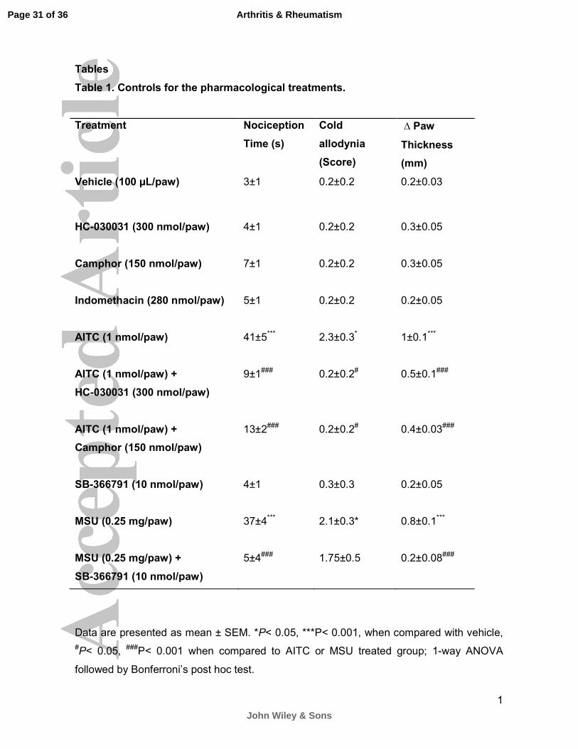
Table 1. Controls for the pharmacological treatments.
Treatment Nociception
Time (s)
Cold
allodynia
(Score)
∆ Paw
Thickness
(mm)
Vehicle (100 µL/paw) 3±1 0.2±0.2 0.2±0.03
HC-030031 (300 nmol/paw)
4±1
0.2±0.2
0.3±0.05
Camphor (150 nmol/paw)
7±1
0.2±0.2
0.3±0.05
Indomethacin (280 nmol/paw)
5±1
0.2±0.2
0.2±0.05
AITC (1 nmol/paw)
41±5***
2.3±0.3*
1±0.1***
AITC (1 nmol/paw) +
HC-030031 (300 nmol/paw)
9±1###
0.2±0.2#
0.5±0.1###
AITC (1 nmol/paw) +
Camphor (150 nmol/paw)
13±2###
0.2±0.2#
0.4±0.03###
SB-366791 (10 nmol/paw)
4±1
0.3±0.3
0.2±0.05
MSU (0.25 mg/paw)
37±4***
2.1±0.3*
0.8±0.1***
MSU (0.25 mg/paw) +
SB-366791 (10 nmol/paw)
5±4###
1.75±0.5
0.2±0.08###
Data are presented as mean ± SEM. *P< 0.05, ***P< 0.001, when compared with vehicle,
#P< 0.05, ###P< 0.001 when compared to AITC or MSU treated group; 1-way ANOVA
followed by Bonferroni’s post hoc test.
Page 31 of 36
John Wiley & Sons
Arthritis & Rheumatism
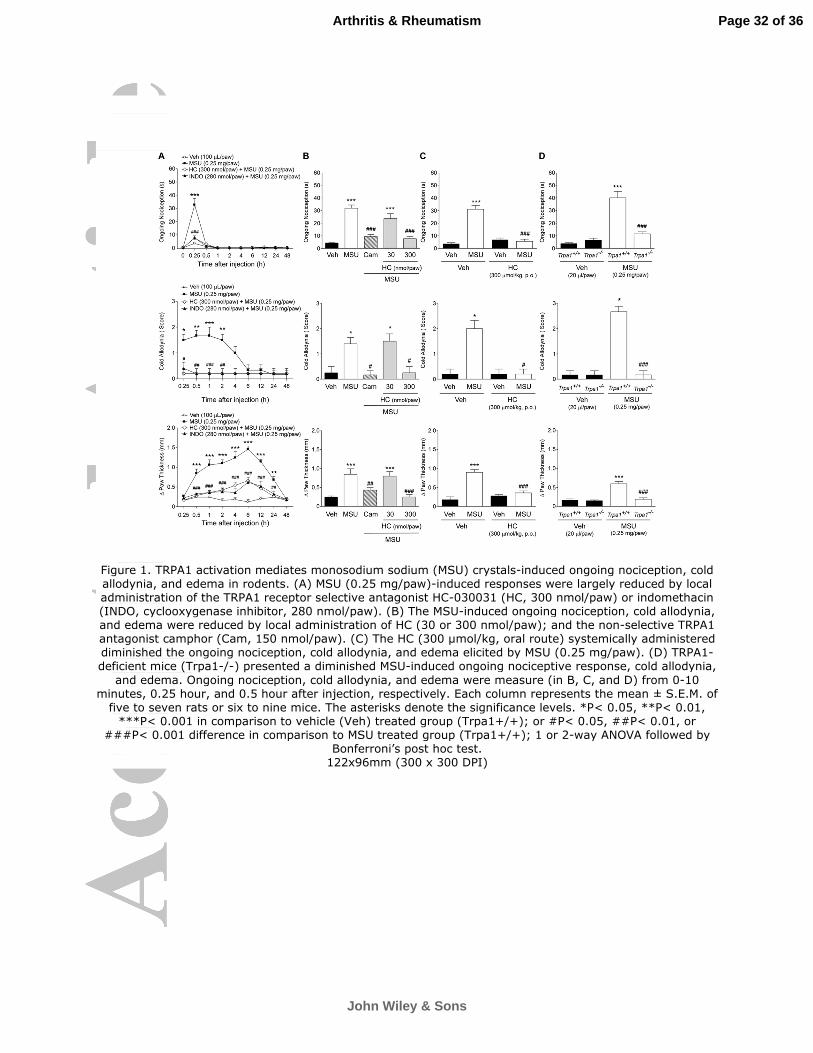
Figure 1. TRPA1 activation mediates monosodium sodium (MSU) crystals-induced ongoing nociception, cold allodynia, and edema in rodents. (A) MSU (0.25 mg/paw)-induced responses were largely reduced by local administration of the TRPA1 receptor selective antagonist HC-030031 (HC, 300 nmol/paw) or indomethacin (INDO, cyclooxygenase inhibitor, 280 nmol/paw). (B) The MSU-induced ongoing nociception, cold allodynia, and edema were reduced by local administration of HC (30 or 300 nmol/paw); and the non-selective TRPA1 antagonist camphor (Cam, 150 nmol/paw). (C) The HC (300 µmol/kg, oral route) systemically administered diminished the ongoing nociception, cold allodynia, and edema elicited by MSU (0.25 mg/paw). (D) TRPA1-deficient mice (Trpa1-/-) presented a diminished MSU-induced ongoing nociceptive response, cold allodynia,
and edema. Ongoing nociception, cold allodynia, and edema were measure (in B, C, and D) from 0-10 minutes, 0.25 hour, and 0.5 hour after injection, respectively. Each column represents the mean ± S.E.M. of
five to seven rats or six to nine mice. The asterisks denote the significance levels. *P< 0.05, **P< 0.01, ***P< 0.001 in comparison to vehicle (Veh) treated group (Trpa1+/+); or #P< 0.05, ##P< 0.01, or
###P< 0.001 difference in comparison to MSU treated group (Trpa1+/+); 1 or 2-way ANOVA followed by Bonferroni’s post hoc test.
122x96mm (300 x 300 DPI)
Page 32 of 36
John Wiley & Sons
Arthritis & Rheumatism
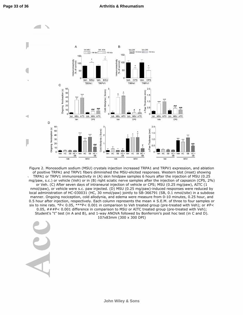
Figure 2. Monosodium sodium (MSU) crystals injection increased TRPA1 and TRPV1 expression, and ablation of positive TRPA1 and TRPV1 fibers diminished the MSU-elicited responses. Western blot (inset) showing TRPA1 or TRPV1 immunoreactivity in (A) skin hindpaw samples 6 hours after the injection of MSU (0.25
mg/paw, s.c.) or vehicle (Veh) or in (B) right sciatic nerve samples after the injection of capsaicin (CPS, 2%) or Veh. (C) After seven days of intraneural injection of vehicle or CPS; MSU (0.25 mg/paw), AITC (1
nmol/paw), or vehicle were s.c. paw injected. (D) MSU (0.25 mg/paw)-induced responses were reduced by local administration of HC-030031 (HC, 30 nmol/paw) jointly to SB-366791 (SB, 0.1 nmol/site) in a subdose manner. Ongoing nociception, cold allodynia, and edema were measure from 0-10 minutes, 0.25 hour, and
0.5 hour after injection, respectively. Each column represents the mean ± S.E.M. of three to four samples or six to nine rats. *P< 0.05, ***P< 0.001 in comparison to Veh treated group (pre-treated with Veh); or #P<
0.05, ###P< 0.001 difference in comparison to MSU or AITC treated group (pre-treated with Veh); Student's “t” test (in A and B), and 1-way ANOVA followed by Bonferroni’s post hoc test (in C and D).
107x83mm (300 x 300 DPI)
Page 33 of 36
John Wiley & Sons
Arthritis & Rheumatism
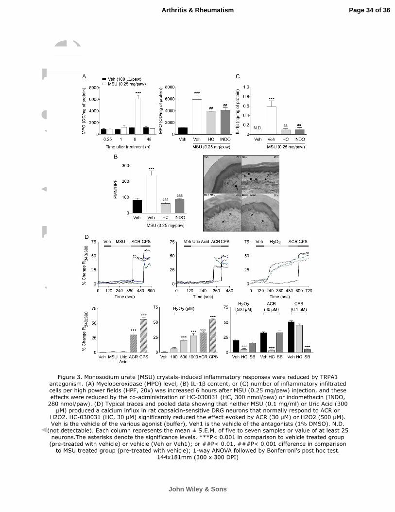
Figure 3. Monosodium urate (MSU) crystals-induced inflammatory responses were reduced by TRPA1 antagonism. (A) Myeloperoxidase (MPO) level, (B) IL-1β content, or (C) number of inflammatory infiltrated cells per high power fields (HPF, 20x) was increased 6 hours after MSU (0.25 mg/paw) injection, and these
effects were reduced by the co-administration of HC-030031 (HC, 300 nmol/paw) or indomethacin (INDO, 280 nmol/paw). (D) Typical traces and pooled data showing that neither MSU (0.1 mg/ml) or Uric Acid (300
µM) produced a calcium influx in rat capsaicin-sensitive DRG neurons that normally respond to ACR or H2O2. HC-030031 (HC, 30 µM) significantly reduced the effect evoked by ACR (30 µM) or H2O2 (500 µM). Veh is the vehicle of the various agonist (buffer), Veh1 is the vehicle of the antagonists (1% DMSO). N.D.
(not detectable). Each column represents the mean ± S.E.M. of five to seven samples or value of at least 25 neurons.The asterisks denote the significance levels. ***P< 0.001 in comparison to vehicle treated group
(pre-treated with vehicle) or vehicle (Veh or Veh1); or ##P< 0.01, ###P< 0.001 difference in comparison to MSU treated group (pre-treated with vehicle); 1-way ANOVA followed by Bonferroni’s post hoc test.
144x181mm (300 x 300 DPI)
Page 34 of 36
John Wiley & Sons
Arthritis & Rheumatism
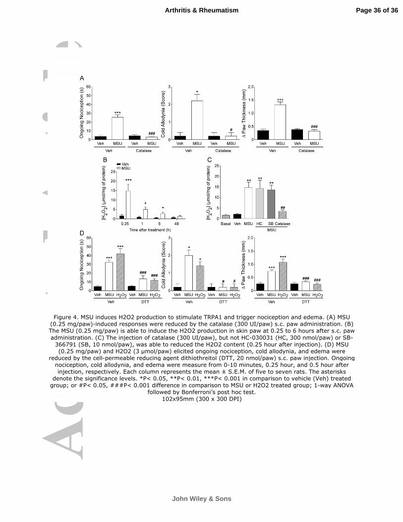
Figure 4. MSU induces H2O2 production to stimulate TRPA1 and trigger nociception and edema. (A) MSU (0.25 mg/paw)-induced responses were reduced by the catalase (300 UI/paw) s.c. paw administration. (B) The MSU (0.25 mg/paw) is able to induce the H2O2 production in skin paw at 0.25 to 6 hours after s.c. paw
administration. (C) The injection of catalase (300 UI/paw), but not HC-030031 (HC, 300 nmol/paw) or SB-366791 (SB, 10 nmol/paw), was able to reduced the H2O2 content (0.25 hour after injection). (D) MSU (0.25 mg/paw) and H2O2 (3 µmol/paw) elicited ongoing nociception, cold allodynia, and edema were
reduced by the cell-permeable reducing agent dithiothreitol (DTT, 20 nmol/paw) s.c. paw injection. Ongoing nociception, cold allodynia, and edema were measure from 0-10 minutes, 0.25 hour, and 0.5 hour after injection, respectively. Each column represents the mean ± S.E.M. of five to seven rats. The asterisks
denote the significance levels. *P< 0.05, **P< 0.01, ***P< 0.001 in comparison to vehicle (Veh) treated group; or #P< 0.05, ###P< 0.001 difference in comparison to MSU or H2O2 treated group; 1-way ANOVA
followed by Bonferroni’s post hoc test. 102x95mm (300 x 300 DPI)
Page 36 of 36
John Wiley & Sons
Arthritis & Rheumatism
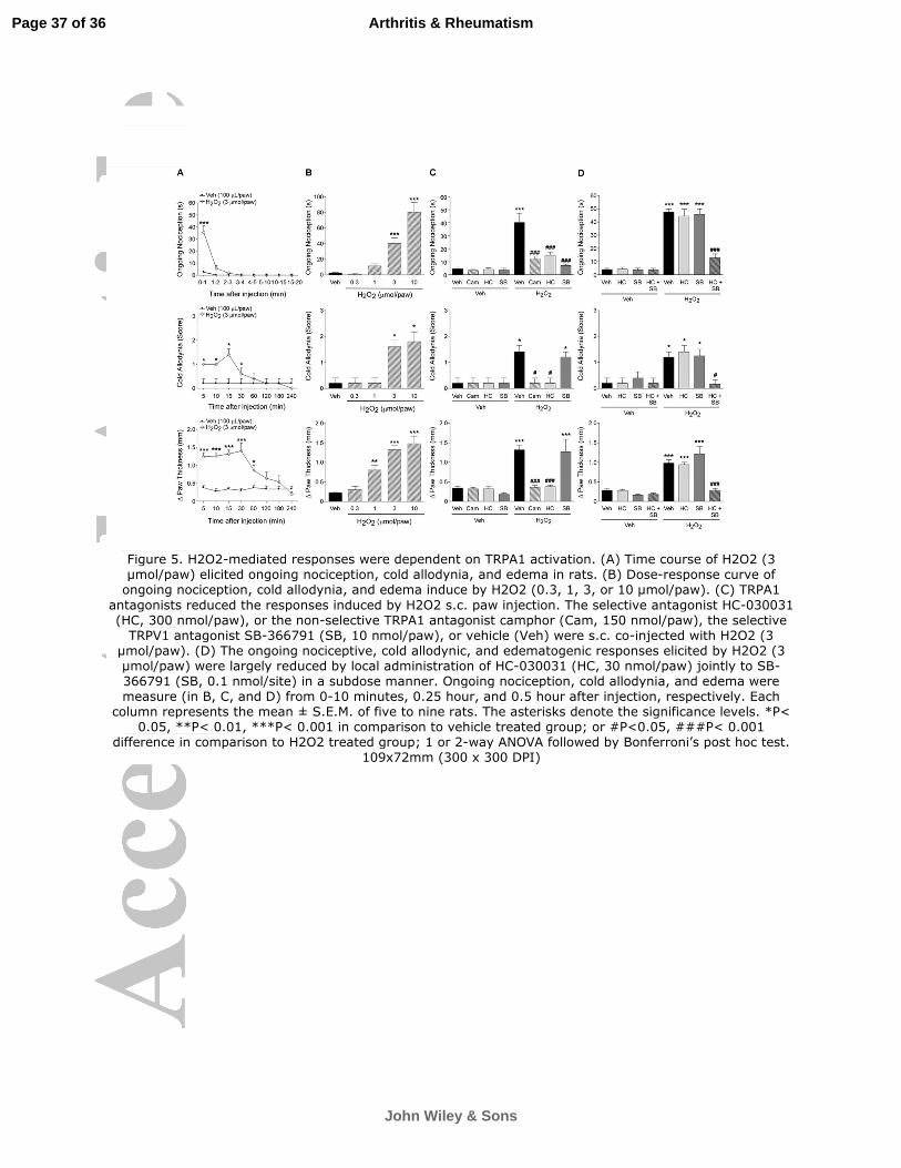
Figure 5. H2O2-mediated responses were dependent on TRPA1 activation. (A) Time course of H2O2 (3 µmol/paw) elicited ongoing nociception, cold allodynia, and edema in rats. (B) Dose-response curve of
ongoing nociception, cold allodynia, and edema induce by H2O2 (0.3, 1, 3, or 10 µmol/paw). (C) TRPA1
antagonists reduced the responses induced by H2O2 s.c. paw injection. The selective antagonist HC-030031 (HC, 300 nmol/paw), or the non-selective TRPA1 antagonist camphor (Cam, 150 nmol/paw), the selective
TRPV1 antagonist SB-366791 (SB, 10 nmol/paw), or vehicle (Veh) were s.c. co-injected with H2O2 (3 µmol/paw). (D) The ongoing nociceptive, cold allodynic, and edematogenic responses elicited by H2O2 (3 µmol/paw) were largely reduced by local administration of HC-030031 (HC, 30 nmol/paw) jointly to SB-366791 (SB, 0.1 nmol/site) in a subdose manner. Ongoing nociception, cold allodynia, and edema were measure (in B, C, and D) from 0-10 minutes, 0.25 hour, and 0.5 hour after injection, respectively. Each
column represents the mean ± S.E.M. of five to nine rats. The asterisks denote the significance levels. *P< 0.05, **P< 0.01, ***P< 0.001 in comparison to vehicle treated group; or #P<0.05, ###P< 0.001
difference in comparison to H2O2 treated group; 1 or 2-way ANOVA followed by Bonferroni’s post hoc test. 109x72mm (300 x 300 DPI)
Page 37 of 36
John Wiley & Sons
Arthritis & Rheumatism