Embed Size (px)
Citation preview
CLINICAL REPORT
Veterinary Research Forum. 2015; 6 (2) 177 - 180
Journal Homepage: vrf.iranjournals.ir
Visceral urate deposition in a little bittern (Ixobrychus minutus)
Morad Rahimi1*, Zahra Minoosh Siavosh Haghighi2
1 Department of Clinical Sciences, Faculty of Veterinary Medicine, Razi University, Kermanshah, Iran; 2 Department of Pathology, Faculty of Veterinary Medicine, Razi University, Kermanshah, Iran.
Article Info Abstract
Article history: Received: 08 October 2013 Accepted: 02 December 2013 Available online: 15 June 2015
Visceral urate deposition (visceral gout) is a common finding during post-mortem examination of poultry. Rare cases of visceral gout may occur in wild birds. A rare case of visceral urate deposition in a little bittern (Ixobrychus minutus) is reported here. In May 2013, carcass of a little bittern was submitted for necropsy to the Clinic of Poultry Diseases (Faculty of Veterinary Medicine, Razi University) by local authorities of Iran Department of Environment. At necropsy, white chalky deposits were observed on the heart and thoracic air sacs of the bird. To confirm the presence of urates, chalky deposits were collected from pericardium and tested by muerxide test. Heart and kidneys were sampled, preserved in 10% neutral-buffered formalin solution and submitted to laboratory for histopathology. Murexide test was positive for presence of uric acid in chalky deposits collected from pericardium. Light microscopy of affected organs confirmed the condition as visceral urate deposition. To the best of our knowledge, this is the first report on the occurrence of visceral urate deposition in a little bittern.
© 2015 Urmia University. All rights reserved.
Key words: Bittern Ixobrychus minutus Urate deposition Visceral gout
(مینوتوسایکسوبریکوس )رسوب احشایی اورات در یک بوتیمار کوچک
چکیده
بدهد. در اینجا مورد کمیابی از وقوع رسوب احشایی اورات )نقرس احشایی( یک یافته عادی در کالبدگشایی طیور است. موارد نادری از نقرس احشایی ممکن است در پرندگان وحشی رخ
، از طرف مسئولین محلی اداره کل حفاظت محیط زیست، لاشه یک بوتیمار 2931گزارش می شود. در اردیبهشت سال (مینوتوس)ایکسوبریکوس ار کوچک رسوب احشایی اورات در یک بوتیم
وی قلب و کیسه های هوایی سینه ای پرنده رنگی بر رکوچک برای کالبدگشایی به کلینیک طیور دانشکده دامپزشکی دانشگاه رازی کرمانشاه ارجاع داده شد. در کالبدگشایی، رسوب گچی سفید
درصد بافری قرار داده شد و به 21ه ها در فرمالین مشاهده شد. برای تأیید وجود اورات، آزمایش مورکساید بر روی رسوبات جمع آوری شده از ناحیه پریکارد انجام شد. برش هایی از قلب و کلی
مورکساید از نظر وجود اسید اوریک در رسوبات جمع آوری شده از ناحیه پریکارد مثبت بود. در بررسی میکروسکوپی اندام های درگیر، رسوب آزمایشگاه پاتولوژی ارسال گردید. نتیجه آزمایش
شایی اورات در بوتیمار کوچک است.رسوب احاحشایی اورات مورد تأیید قرار گرفت. بر اساس جستجو در پایگاه ها و منابع علمی و تا آنجایی که می دانیم، این اولین گزارش از رخداد
بوتیمار کوچک، رسوب اورات، نقرس احشایی، مینوتوسایکسوبریکوس واژه های کلیدی:
*Correspondence:
Morad Rahimi. DVM, PhD Department of Clinical Sciences, Faculty of Veterinary Medicine, Razi University, Kermanshah, Iran. E-mail: [email protected]
Veterinary Research
Forum
178
M. Rahimi and Z. Minoosh Siavosh Haghighi. Veterinary Research Forum. 2015; 6 (2) 177 - 180
Introduction
Gout is correctly used as a term in human medicine to describe an enzyme defect that causes an abnormal nitrogen metabolism resulting in excessive uric acid production. In avian medicine, “gout” is a historical misnomer whereas urate deposition is a more correct term.1 Visceral urate deposition (visceral gout) is defined as the accumulation of urates in kidneys, on serous surfaces of the heart, liver, mesenteries, and air sacs. In severe cases, surfaces of muscles and synovial sheaths of joints may be involved, and precipitation may occur within the liver, spleen, and other organs. The deposits on serous surfaces appear grossly as a white chalky coating, while those within visceral organs may only be recognized microscopically.2 The patho-genesis of visceral urate deposition is not completely clear, but it is generally associated with conditions that reduce uric acid excretion or increase uric acid production.3-5
Pathologic examination of gouty lesions confirms the diagnosis of visceral urate deposition by demonstrating urate tophi.6,7 Visceral urate deposition has been reported in various caged and aviary birds from different parts of the World. It is among the most commonly diagnosed causes of mortality in poultry.8,9 Rare cases of visceral urate deposition may occur in wild birds. A rare case of visceral urate deposition in a little bittern (Ixobrychus minutus) is reported here.
Case Description
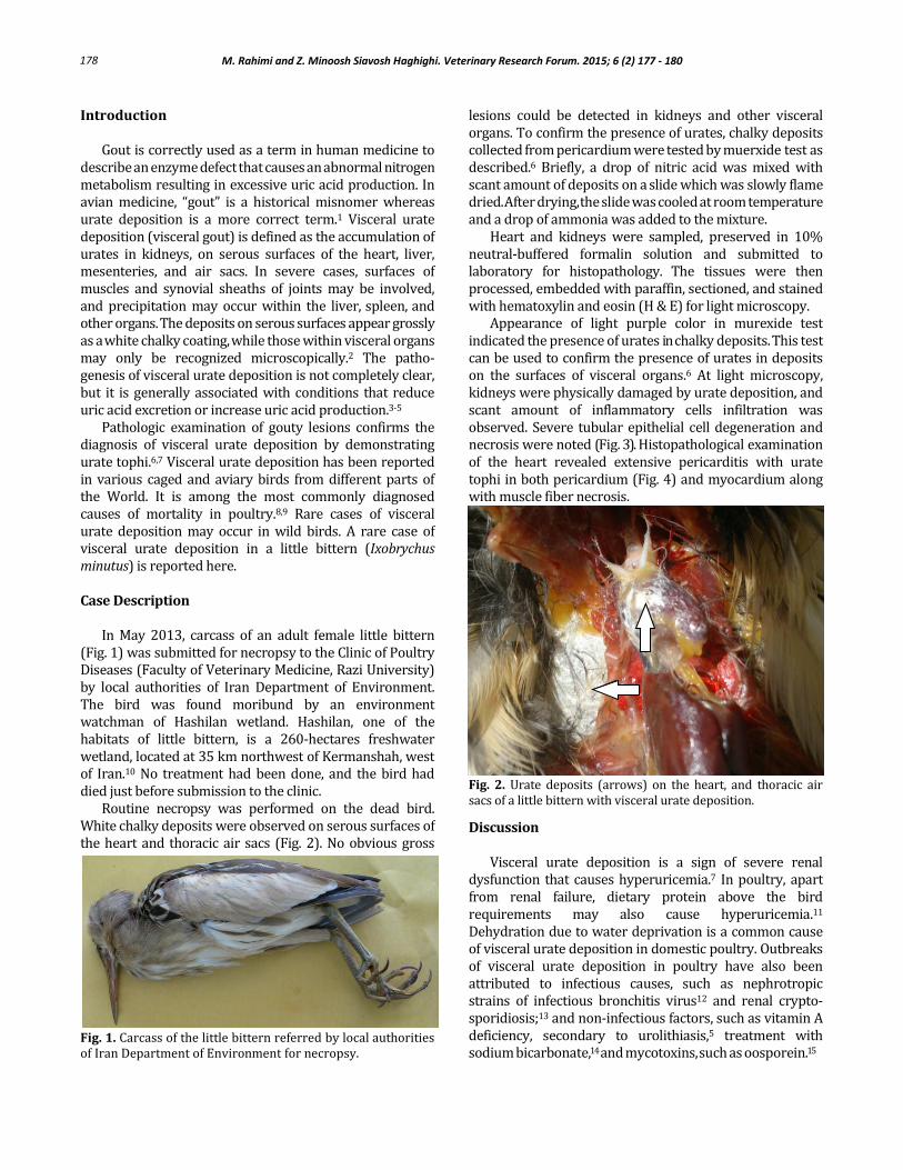
In May 2013, carcass of an adult female little bittern (Fig. 1) was submitted for necropsy to the Clinic of Poultry Diseases (Faculty of Veterinary Medicine, Razi University) by local authorities of Iran Department of Environment. The bird was found moribund by an environment watchman of Hashilan wetland. Hashilan, one of the habitats of little bittern, is a 260-hectares freshwater wetland, located at 35 km northwest of Kermanshah, west of Iran.10 No treatment had been done, and the bird had died just before submission to the clinic.
Routine necropsy was performed on the dead bird. White chalky deposits were observed on serous surfaces of the heart and thoracic air sacs (Fig. 2). No obvious gross
Fig. 1. Carcass of the little bittern referred by local authorities of Iran Department of Environment for necropsy.
lesions could be detected in kidneys and other visceral organs. To confirm the presence of urates, chalky deposits collected from pericardium were tested by muerxide test as described.6 Briefly, a drop of nitric acid was mixed with scant amount of deposits on a slide which was slowly flame dried. After drying, the slide was cooled at room temperature and a drop of ammonia was added to the mixture.
Heart and kidneys were sampled, preserved in 10% neutral-buffered formalin solution and submitted to laboratory for histopathology. The tissues were then processed, embedded with paraffin, sectioned, and stained with hematoxylin and eosin (H & E) for light microscopy.
Appearance of light purple color in murexide test indicated the presence of urates in chalky deposits. This test can be used to confirm the presence of urates in deposits on the surfaces of visceral organs.6 At light microscopy, kidneys were physically damaged by urate deposition, and scant amount of inflammatory cells infiltration was observed. Severe tubular epithelial cell degeneration and necrosis were noted (Fig. 3). Histopathological examination of the heart revealed extensive pericarditis with urate tophi in both pericardium (Fig. 4) and myocardium along with muscle fiber necrosis.
Fig. 2. Urate deposits (arrows) on the heart, and thoracic air sacs of a little bittern with visceral urate deposition.
Discussion
Visceral urate deposition is a sign of severe renal dysfunction that causes hyperuricemia.7 In poultry, apart from renal failure, dietary protein above the bird requirements may also cause hyperuricemia.11 Dehydration due to water deprivation is a common cause of visceral urate deposition in domestic poultry. Outbreaks of visceral urate deposition in poultry have also been attributed to infectious causes, such as nephrotropic strains of infectious bronchitis virus12 and renal crypto-sporidiosis;13 and non-infectious factors, such as vitamin A deficiency, secondary to urolithiasis,5 treatment with sodium bicarbonate,14 and mycotoxins, such as oosporein.15

179 M. Rahimi and Z. Minoosh Siavosh Haghighi. Veterinary Research Forum. 2015; 6 (2) 177 - 180
Fig. 3. Micrograph of kidney indicating severe tubular epithelial cell degeneration and necrosis in a little bittern with visceral urate deposition (H & E, 400×).
Fig. 4. Pericarditis with urate tophi (arrow) in a little bittern with visceral urate deposition (H & E, 400×).
Masses of fat deposits in peritoneal cavity of the little bittern and relatively good musculature showed that the condition had led to its death during an acute course. In acute renal failure visceral urate deposition might occur alone.11 Inflammatory reactions are not often detected as birds die rapidly. Hyperkalaemia can develop, and this, rather than the uric acid, might lead to cardiac arrest and the sudden death seen with visceral urate deposition.7 Factors, such as dehydration and vitamin A deficiency are common causes of visceral urate deposition in domestic poultry,5 but they could not cause visceral urate deposition in domestic poultry,5 but they could not cause visceral urate deposition and acute death in this case, since little bitterns had free access to water sources and variety of feeds.
Although it is still theorized that long term of high-protein feeding may induce hyperuricemia in granivorous or nectivorous birds,4,16 Guo et al. induced visceral urate deposition in growing layers fed high calcium and high protein diets.17 As little bitterns are essentially insectivorous
and take aquatic adult and larval insects, molluscs, crustaceans, fish, frogs, tadpoles, small reptiles and birds,18 they are adopted to high protein and high calcium diets. As a result, high protein and high calcium diets are unlikely to cause visceral urate deposition in little bitterns.
In conclusion, visceral urate deposition and acute death in this little bittern might be due to acute intoxication. Because of recent droughts in Iran, especially in western parts of the country, little bitterns are threatened by habitat degradation and loss through direct destruction, pollution and hydrological changes in rivers, lakes and wetlands. References
1. Crespo R, Shivaprasad HL. Developmental, metabolic,
and other non-infectious disorders. In: Saif YM, Fadly AM, Glisson JR, et al. (Eds.). Diseases of poultry. 12th ed. Iowa, USA: Iowa State University Press 2008; 149-195.
2. Phalen DN, Ambrus S, Graham DL. The avian urinary system: Form, function, diseases. In proceedings: Annual conference of association of avian veterinarians. Boca Raton, USA: 1990; 44-57.
3. Austic RE, Cole RK. Impaired renal clearance of uric acid in chickens having hyperuricemia and articular gout. Am J Physiol 1972; 223(3): 525-530.
4. Styles DK, Phalen DN. Clinical avian urology. Semin Avian Exot Pet 1998; 7(2): 104-113.
5. Siller WG. Renal pathology of the fowl - A review. Avian Pathol 1981; 10(3): 187-262.
6. Speer BL. Diseases of the urogenital system. In: Altman RB, Clubb SL, Dorrestein GM, et al. (Eds.). Avian medicine and surgery. Philadelphia, USA: WB Saunders, 1997; 625-644.
7. Lierz M. Avian renal disease: Pathogenesis, diagnosis, and therapy. Vet Clin Exot Anim 2003; 6(1): 29-55.
8. Riddell C. Urinary system. In: Avian histopathology. Philadelphia, USA: American association of avian pathologists 1987; 67-73.
9. Brown TP. Urinary system. In: Riddell C (Ed.). Avian histopathology. 2nd ed. Philadelphia, USA: American association of avian pathologists 1996; 167-181.
10. Karami M, Kasmaniand ME, Alamesh AA. Plants of Hashilan wetland, Kermanshah, Iran. J Sciences 2001; 12(3): 201-207.
11. Lumeij JT. Nephrology. In: Ritchie BW, Harrison GJ, Harrison LR (Eds.). Avian medicine, principles and application. Lake Worth, USA: Wingers Publishing Inc. 1994; 538-555.
12. Cumming RB. Infectious avian nephrosis (uremia) in Australia. Aust Vet J 1963; 39(4): 145-147.
13. Trampel DW, Pepper TM, Blagburn BL. Urinary tract cryptosporidiosis in commercial laying hens. Avian Dis 2000; 44(2): 479-484.
14. Davison S, Wideman RF. Excess sodium bicarbonate in the diet and its effect on Leghorn chickens. Br Poult Sci

180
M. Rahimi and Z. Minoosh Siavosh Haghighi. Veterinary Research Forum. 2015; 6 (2) 177 - 180
1992; 33(4): 859-870. 15. Pegram RA, Wyatt RD. Avian gout caused by oosporein,
a mycotoxin produced by Caetomium trilaterale. Poult Sci 1981; 60(11): 2429-2440.
16. Siller WG. Avian nephritis and visceral gout. Lab Invest 1959; 8(6): 1319-1357.
17. Guo X, Huang K, Tang J. Clinicopathology of gout in growing layers induced by high calcium and high protein diets. Br Poult Sci 2005; 46(5): 641-646.
18. Del Hoyo J, Elliot A, Sargatal J. Handbook of the birds of the world. Vol. 1: Ostrich to ducks. Barcelona, Spain: Lynx Edicions 1992; 376-403.