Embed Size (px)
Citation preview
Research Signpost 37/661 (2), Fort P.O., Trivandrum-695 023, Kerala, India
Recent Res. Devel. Food Biotechnology. Enzymes as Additives or Processing Aids
Transglutaminase in food biotechnology
Loredana Mariniello, Prospero Di Pierro, Concetta Valeria L. Giosafatto, Angela Sorrentino and Raffaele Porta Department of Food Science, University of Naples “Federico II”. Parco Gussone, Portici, Naples, Italy.

Loredana Mariniello et al. 186
Abstract Food industries worldwide are planning to develop new ingredients with novel physical and functional characteristics. The introduction of additional covalent cross-links by means of enzymes represents a promising tool to improve physicochemical properties, such as solubility, water-binding or emulsifying capacity, foaming, viscosity, elasticity and gelation, of proteins intended for human consumption where chemical reagents for modifications are not acceptable. Transglutaminases are cross-linking enzymes currently available for catalyzing covalent bond formation among protein molecules. In this chapter general characteristics of the enzyme family are described together with transglutaminase applications in several food industry sectors.
Correspondence/Reprint request: Prof. Raffaele Porta, Department of Food Science, University of Naples “Federico II”, P.co Gussone, 80055 Portici, Naples, Italy. E-mail: [email protected]
Introduction Research on the applications of transglutaminases (TGases) in protein-based food preparation started with the isolation of the enzymes from mammalian tissues and body fluids [1]. Guinea pig liver TGase was the only molecular form of the enzyme commercially available until the late 1980s. However, its scarce source, the extensive purification procedure, as well as the Ca2+-dependence activity which leads to protein precipitation in some food systems (i.e. casein, soybean globulin or myosin) [2], entailed extremely high prices on the market, resulting in a low attractiveness for potential industrial applications. Factor XIII, a TGase isoform isolated from blood, is not suitable in food industry as it requires thrombin for its activation. In 1989 Ando et al. [3] reported the isolation of TGase from the cultural broth of the microorganism Streptoverticillium S-8112, which has been identified as a variant of Streptoverticillium mobaraense, also known as Streptomyces mobaraensis [4]. In contrast to many other TGases, the microbial isoform (mTGase) is Ca2+-independent and is remarkably stable over a wide range of temperatures and pHs [5]. Such characteristics, including the higher reaction rate, the broad substrate specificity for the acyl donor and the low-cost mass production by traditional fermentation technology, make mTGase particularly useful for industrial and biotechnological applications [6] as a food-grade

Transglutaminase in food 187
additive capable of improving many important features of different protein-based foods.
2. Transglutaminases: a family of enzymes with diverse functions In 1957, introducing the term “transglutaminase” for describing the transamidating activity observed in guinea pig liver, Waelsch and coll. [7] probably did not image that, 50 years later, this class of enzymes would have been known as food modifying enzymes. In this chapter we are interested in elucidating the whys and wherefores TGase is useful in food biotechnology applications. In order to fulfil this task, it is advisable to spend some additional words about TGase and the relative catalyzed reactions. The complete name of TGase is R-glutaminyl-peptide: amine γ-glutamyltransferase (EC 2.3.2.13), indicating that the catalysis consists in the acyl transfer of γ-glutamyl residues, present in protein and peptide substrates (acyl donor or Q-donor), to an acyl acceptor substrate, resulting in a variety of different products depending on the involved molecules [1, 8]. The transamidation reaction occurs when the acyl acceptor is either the ε-amino group of an endoprotein lysine or a low molecular mass primary amine, thus generating ε-(γ-glutamyl)lysine cross-links in the first case, and protein-amine conjugates in the latter (Figure 1, a and b). Either water or alcohol molecules
a) transamidation
H2N CH2CH2CH2CH2
CH2CH2C NH2
O
+ NH3
TGase
b) amine incorporation
CH2CH2C NH2
O
H2N R
+ NH3
TGase
c) deamidation
CH2CH2C NH2
O
+ NH3
CH2CH2C OH
O
TGase
d) esterification
CH2CH2C NH2
O
HO R’
+ NH3CH2CH2C
OO R’
TGase
e) isopeptide cleavage
CH2CH2C OH
O
H2N CH2CH2CH2CH2
+
TGase
N CH2CH2CH2CH2CH2CH2C
O
H
CH2CH2C
ON R
H
H2OTGase
N CH2CH2CH2CH2CH2CH2C
O
H
H2O
a) transamidation
H2N CH2CH2CH2CH2
H2N CH2CH2CH2CH2
CH2CH2C NH2
OO
+ NH3
TGase
b) amine incorporation
CH2CH2C NH2
OO
H2N RH2N R
+ NH3
TGase
c) deamidation
CH2CH2C NH2
OO
+ NH3
CH2CH2C OH
OCH2CH2C OH
OO
TGase
d) esterification
CH2CH2C NH2
OO
HO R’HO R’
+ NH3CH2CH2C
OO R’CH2CH2C
OCH2CH2C
O OOO R’O R’
TGase
e) isopeptide cleavage
CH2CH2C OH
OCH2CH2C OH
OO
H2N CH2CH2CH2CH2
H2N CH2CH2CH2CH2
+
TGase
N CH2CH2CH2CH2CH2CH2C
O
H
N CH2CH2CH2CH2CH2CH2C
OO
HH
CH2CH2C
ON R
H
CH2CH2C
OCH2CH2C
O OON R
HH
H2OH2OTGase
N CH2CH2CH2CH2CH2CH2C
OO
HH
H2OH2O
Figure 1. Transglutaminase-catalyzed reactions.
Loredana Mariniello et al. 188
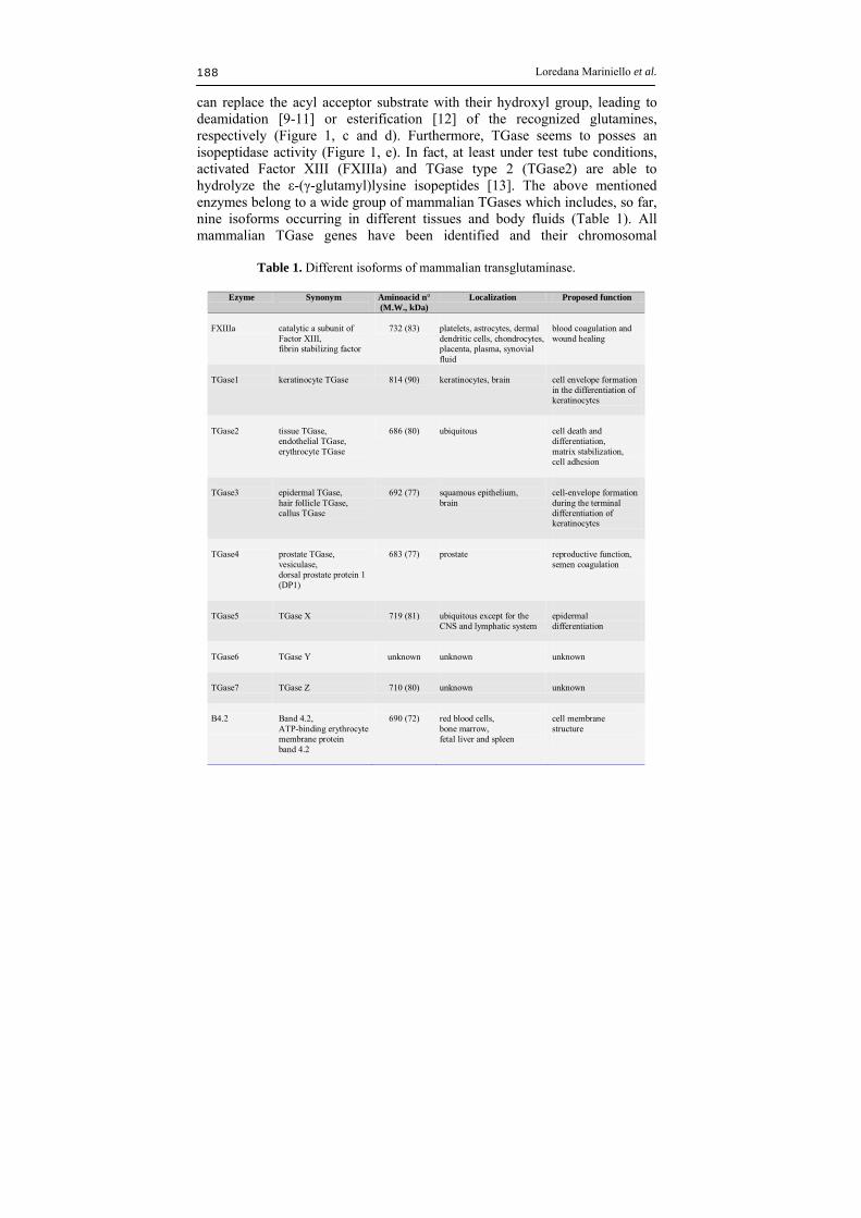
can replace the acyl acceptor substrate with their hydroxyl group, leading to deamidation [9-11] or esterification [12] of the recognized glutamines, respectively (Figure 1, c and d). Furthermore, TGase seems to posses an isopeptidase activity (Figure 1, e). In fact, at least under test tube conditions, activated Factor XIII (FXIIIa) and TGase type 2 (TGase2) are able to hydrolyze the ε-(γ-glutamyl)lysine isopeptides [13]. The above mentioned enzymes belong to a wide group of mammalian TGases which includes, so far, nine isoforms occurring in different tissues and body fluids (Table 1). All mammalian TGase genes have been identified and their chromosomal
Table 1. Different isoforms of mammalian transglutaminase.
Ezyme Synonym
Aminoacid n° (M.W., kDa)
Localization Proposed function
FXIIIa
catalytic a subunit of Factor XIII, fibrin stabilizing factor
732 (83)
platelets, astrocytes, dermal dendritic cells, chondrocytes, placenta, plasma, synovial fluid
blood coagulation and wound healing
TGase1
keratinocyte TGase
814 (90)
keratinocytes, brain
cell envelope formation in the differentiation of keratinocytes
TGase2
tissue TGase, endothelial TGase, erythrocyte TGase
686 (80)
ubiquitous
cell death and differentiation, matrix stabilization, cell adhesion
TGase3
epidermal TGase, hair follicle TGase, callus TGase
692 (77)
squamous epithelium, brain
cell-envelope formation during the terminal differentiation of keratinocytes
TGase4
prostate TGase, vesiculase, dorsal prostate protein 1 (DP1)
683 (77)
prostate
reproductive function, semen coagulation
TGase5
TGase X
719 (81)
ubiquitous except for the CNS and lymphatic system
epidermal differentiation
TGase6
TGase Y
unknown
unknown
unknown
TGase7
TGase Z
710 (80)
unknown
unknown
B4.2
Band 4.2, ATP-binding erythrocyte membrane protein band 4.2
690 (72)
red blood cells, bone marrow, fetal liver and spleen
cell membrane structure

Transglutaminase in food 189
positions have been mapped [8, 14]. Moreover, seven isoforms have been well characterized at protein level. Briefly, FXIII is a zymogen and becomes active by thrombin cleavage during the blood coagulation cascade, being involved in stabilization of fibrin clots and in wound healing [15, 16]. TGase1, TGase3 and TGase5 are required for skin maturation and integrity [8, 17]. Mutation in TGase1 gene is responsible for lamellar ichthyosis, a severe skin disease characterized by an abnormal epidermal differentiation and a reduced cutaneous barrier function [18]. Recently, also TGase5 has been correlated to a rare pathology named Acral Peeling Skin Syndrome (APSS) in which skin peeling is strictly limited to the dorsa of the hands and feet [19]. TGase2 is widespread into several tissues including liver, kidney, lung, spleen and brain [20]. Up to now, a specific biological role for this isoform is still debated since its activity seems to be related to several biological events. In fact, TGase2 was associated to cell proliferation, apoptosis, extracellular matrix stabilization and cell adhesion [21, 22]. Moreover, new evidences lead to consider the involvement of TGase2 in several pathologies, such as cataract formation, atherosclerosis, inflammation, fibrosis, diabetes, cancer metastases and autoimmune deseases [8, 14]. Finally, TGase2 was also suggested to be responsible for inducing celiac disease in genetically predisposed patients. In fact, gliadins are supposed to become allergenic after specific deamidation catalyzed by TGase2 occurring in the intestinal lumen [23] and this event seems to be triggered for an immuno–response in which TGase2 results to be the major autoantigen [24]. The biological role of TGase4 was suggested to be related to sperm maturation in human [25] and to immunogenicity suppression of spermatozoa during fertilization in rats [26, 27]. Nowadays, among mammalian TGases, only TGase6 and TGase7 remain without hypothesized functions, also because their catalytic activity has not yet demonstrated [8]. The Band 4.2 (B4.2) from erythrocytes, instead, is not enzymatically active. In fact, despite the remarkable sequence similarity with other mammalian TGases, B4.2 carries a CysAla substitution in the active site [28] that makes the protein unable to catalyze the reaction. However, this isoform plays a role as structural component of cytoskeletal network underlying the plasma membrane, being determinant in maintaining the integrity of erythrocyte membrane as its lack causes spherocytosis [29, 30]. In the course of decades, the family of TGases has been notably enlarged due to the discovery of novel isoforms in other vertebrates as well as in invertebrates, plants, fungi and micro organisms (see Table 2 for a rapid overview and for references). Thus, TGase reaction occurs in different fishes and the enzymatic activity was observed to vary in the different species [31, 32]. Experimental evidences suggested the presence of TGase also in amphibian eggs and skin, in turtle shell, and in chicken epidermis, erythrocytes

Loredana Mariniello et al. 190
and gizzard (Table 2). Up to ten TGases in various invertebrate species have so far been reported (see Table 2 for references) including those responsible for blood coagulation in crustaceans [33-35].
Table 2. Non-mammalian organisms expressing transglutaminase. Organism Vulgar name Species Localization References
vertebrates fishes mud carp Cirrhiana microlepis [149] red sea bream Pagrus major [150, 151] walleye pollack [152] tilapia Oreochromis niloticus [32] threadfin bream [153] amphibians frog and toad epidermis [154] egg [155] reptiles lizard [154] turtle [154] birds chicken gizzard
epidermis erythrocytes
[156] [157] [158]
invertebrates horse shoe Limulus hemocytes [159] nematode Brugia malayi
Caenorhabditis elegans [160]
[161] arthropods [162] scallop striated adductor muscle [163] japanese oyster Crassostrea gigas [164] shrimp Penaeus monodon
Marsupenaeus japonicus
[34, 35, 165, 166]
crayfish Pacifastacus leniusculus [33] starfish [167] plants Heliantus tuberosus chloroplasts [168] soybean Glycine max leaves [36] apple Malus domestica pollen [41] tobacco Nicotiana tabacum flowers [38] Arabidopsis thaliana [39] maize Zea mais chloroplasts [40] fungi Candida albicans [43] Phytophthora sp [44] Saccharomyces cerevisiae [45]
microorganisms Leishmania sp [169] Streptoverticillium sp [3, 46, 48, 51-54] Bacillus circulans [170] Bacillus subtilis [49-50]
Several TGases were detected in plants whose activities were suggested to be related to the organization of the cell wall in higher plants or to the apical growth of pollen [36-42]. In addition, TGase activity was found in Candida albicans (human pathogen, [43]) and the oomycete Phytophtora sojae (plant pathogen, [44]), as well as in Saccharomyces cerevisiae [45]. In all cases the enzyme was found localized at the cell wall level, suggesting its involvement

Transglutaminase in food 191
in the stabilization of cell wall and in the ifae extention process during mycelium growing. A similar role has been hypothesized for the microbial TGase isolated from Streptoverticillium spp. (Streptoverticillia are now unified with Streptomyces according to Witt and Stackebrandt, 1990 [4]), a microorganism that shows the ability of developing a mycelium under particular growth conditions. Indeed, some Streptomyces secrete large amounts of mTGase in the culture medium. Even though the precise role of this enzyme in the life-cycle of Streptomyces remains to be elucidated, it is possible that protein cross-linking fortifies the aerial cell wall and the spore envelope [46]. Since 1989, when the first mTGase was isolated from the culture supernatant of Streptoverticillium sp. strain S-8112 [3] several studies looking for TGase activity in microorganisms were carried out [47]. Enzyme activity has been found mainly in several strains of the genus Streptoverticillium spp. [3, 5, 48] and of the genus Bacillus spp. [49]. While TGase from Streptoverticillium spp. was found extracellularly, the Bacillus spp. enzyme is localized on spores. However, the Gram-positive bacterium B. subtilis does not exhibit the multicellular morphology of Streptomyces and produces mTGase intracellularly during the late stage of spore maturation [50]. Recently, about 300 Actinomycete strains have been screened for TGase and a strain of Streptomyces platensis M5218 with high enzyme activity was isolated [51]. The enzymes from Streptoverticillium sp. S-8112 [3, 52], Sv. mobaraense DSMZ strain [53], Sv. ladakanum [54], Sv. cinnamoneum CBS 683.68 [48], and Bacillus subtilis [50] have been purified and characterized. The protein structure of the known bacterial TGase is quite different from that of the mammalian enzymes. The bacterial enzyme has smaller molecular mass (about 40 kDa despite 80-100 kDa of mammalian TGases) and are Ca2+-independent while the enzymatic reaction catalyzed by TGases from vertebrates and some invertebrates requires Ca2+ in order to allow the enzyme to expose a cysteine residue in its active site domain [55]. In fact, mammalian TGases possess a catalytic triad of aminoacids, namely Cys-His-Asp or Cys-His-Asn and a crucial Trp residue located 36 residues upstream the Cys occurring in the active site [8]. The reaction proceeds via the formation of a -glutamylthiolester bond between the glutamyl residue and the active site Cys producing an acyl-enzyme intermediate [56, 57] (Figure 2). mTGase follows the same mechanism, even though it possesses an atypical catalytic triad and a very low sequence identity with other TGases [52, 58]. However, the hydrophobic environment of the catalytic site, including a single Cys residue, is similar to those of other TGases while no sequence identity with the calcium-binding domain was found [52]. Crystallographic studies have underlined differences between the three-dimensional structure of mTGase
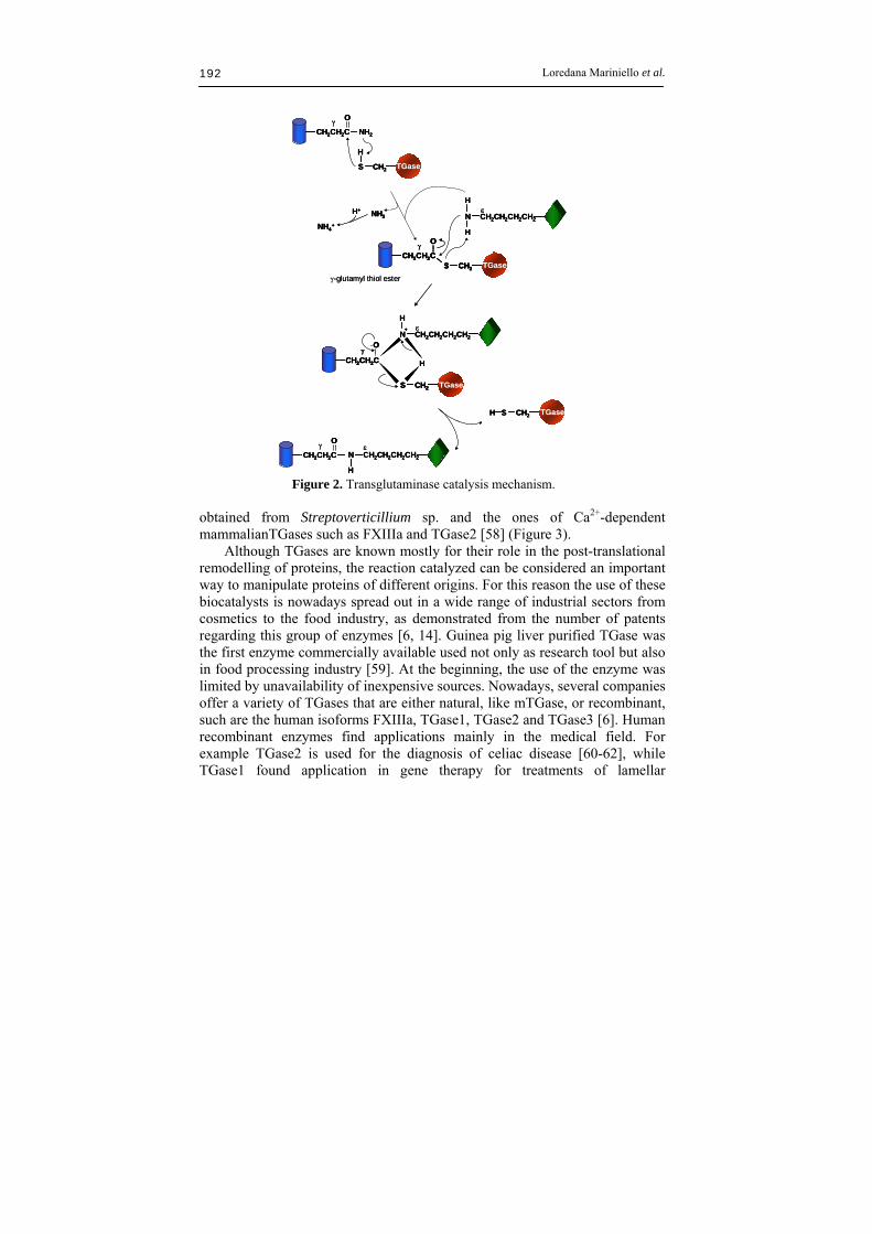
Loredana Mariniello et al. 192
-glutamyl thiol ester
CH2CH2C NH2
O
S CH2 TGase
H
S CH2
CH2CH2C
O
TGase
H
H
N CH2CH2CH2CH2
H+
NH4+
NH3
S CH2 TGase
CH2CH2C
_O
H
+
H
N CH2CH2CH2CH2
H S CH2 TGase
N CH2CH2CH2CH2CH2CH2C
O
H
-glutamyl thiol ester
CH2CH2C NH2
O
S CH2 TGase
H
CH2CH2C NH2
OO
S CH2 TGase
H
S CH2
CH2CH2C
O
TGase
H
H
N CH2CH2CH2CH2
S CH2
CH2CH2C
O
TGaseS CH2
CH2CH2C
OO
TGase
H
H
N CH2CH2CH2CH2
H+
NH4+
NH3H+
NH4+
NH3NH3
S CH2 TGase
CH2CH2C
_O
H
+
H
N CH2CH2CH2CH2
S CH2 TGaseS CH2 TGase
CH2CH2C
_O
CH2CH2C
_O
H
+
H
N CH2CH2CH2CH2
H S CH2 TGaseH S CH2 TGase
N CH2CH2CH2CH2CH2CH2C
O
H
N CH2CH2CH2CH2CH2CH2C
OO
HH Figure 2. Transglutaminase catalysis mechanism.
obtained from Streptoverticillium sp. and the ones of Ca2+-dependent mammalianTGases such as FXIIIa and TGase2 [58] (Figure 3). Although TGases are known mostly for their role in the post-translational remodelling of proteins, the reaction catalyzed can be considered an important way to manipulate proteins of different origins. For this reason the use of these biocatalysts is nowadays spread out in a wide range of industrial sectors from cosmetics to the food industry, as demonstrated from the number of patents regarding this group of enzymes [6, 14]. Guinea pig liver purified TGase was the first enzyme commercially available used not only as research tool but also in food processing industry [59]. At the beginning, the use of the enzyme was limited by unavailability of inexpensive sources. Nowadays, several companies offer a variety of TGases that are either natural, like mTGase, or recombinant, such are the human isoforms FXIIIa, TGase1, TGase2 and TGase3 [6]. Human recombinant enzymes find applications mainly in the medical field. For example TGase2 is used for the diagnosis of celiac disease [60-62], while TGase1 found application in gene therapy for treatments of lamellar

Transglutaminase in food 193
a
b c
a
b c
Figure 3. Three-dimensional structures of a) TGase from Streptoverticillium mobarense (PDB: 1iu4C), b) human Factor XIIIa (PDB: 1ex0), c) human tissue TGase (PDB: 1kv3). ichthyosis, even though still at the experimental level [63-64]. In the food field, as previously stated, the microbial isoform is largely utilized. The reason is related to some advantages offered by mTGase, like its low cost and ease of use. In fact, mTGase can be obtained in large quantities through microbial fermentation, is stable at high temperatures, and is active in a wide pH range. In the following paragraphs the use of TGase in food related products will be described and, mostly, we will linger over the microbial isoform which employment in processing foods is much more diffused that one can imagine.
Loredana Mariniello et al. 194
3. Employment of transglutaminase in the food industry TGases have been used in the last two decades in several studies to assess their capability in modifying proteins of food interest. Thus, many kinds of proteins have been tested as potential substrates for the enzyme. The main aim of these researches was to improve texture, mechanical properties and emulsifying characteristics of food proteins for their employment as ingredients of complex foods with enhanced quality [65]. Investigations have been carried out using proteins of different origins, like those extracted from both plants [66, 67] and animals, as fish or other ingredients useful to prepare sea food–based products [68]. Moreover, many studies have been performed using milk derived proteins, such as caseins and whey proteins, since it is known that they are TGase substrates endowed with both glutamine and lysine reactive residues [69-71]. Further researches have assessed that the enzyme is an useful tool to prepare a variety of foods that are very popular and spread out in Asian countries, as Japan and China [68], as well as for preparing other worldwide consumed foods. In the following first paragraph foods prepared using TGase in the flow process will be issued, while the second paragraph is addressed to proteins coming from different processes as by-products potentially interested to be utilized as TGase substrates for their valorization in the food industry. The third paragraph will deal with a novel use of the enzyme as cross-linking agent to reticulate the protein component of hydrocolloid-based edible films. Finally, the bioavailability of TGase-cross-linked proteins and the use of TGase for stabilization of different proteins will be reported. 3.1 Improvement of foods by transglutaminase addition The use of TGase in the food industry started with the manufacturing surimi (fish paste)-based products in Japan [65, 72-74]. Surimi gel preparation consists of a two-step process. The first step, also called “setting”, occurs at temperatures below 40 °C and leads to the formation of a translucent and elastic gel. The second step, occurring at high temperatures (>80 °C), is required to make gels stronger. The setting is caused by myosin cross-linking induced by an endogenous Ca2+-dependent TGase occurring in species of Pollack [75], a fish used for surimi preparation. It has been proved that the addition of exogenous TGases, either the blood isoform or the microbial one, during the setting process is advantageous in order to obtain a fish paste with enhanced elasticity and tensile strength [76-79]. Furthermore, all fish–based products restructured with the aid of mTGase show high whiteness, which is considered an

Transglutaminase in food 195
important attribute for consumers [80]. Thus, accordingly to the scheme illustrated in the panel A of Figure 4, the use of TGase is advisable to obtain efficiently restructured surimi-based products. Another important TGase application, in particular of the microbial isoform, is the yogurt production [81, 87]. The enzyme can be added before the fermentation process or simultaneously to it [88, 89]. Its use represents an effective methodology to improve texture properties of the final product, without or by reducing dry matter enrichment, leading to an increase in gel strength, viscosity and firmness as well as to a decrease in syneresis [82]. Furthermore, recent studies stated that the enzymatic cross-linking of milk proteins by mTGase improves consistence and microstructure of the goat milk yogurt gel [90]. Interestingly, microbiological analyses have shown that the enzyme modification of goat milk proteins plays a positive role in the survival of the cultured probiotic bacteria used in yogurt production [90]. Another type of dairy product where the use of TGase has a great potential is the cheese-making. Improved water-holding capacity and texture in both cottage and ripened cheese is considered a potential advantage of TGase treatment. It was demonstrated that [65, 91] curd yield increases following to the protein cross-linking while, by adding mTGase during the manufacturing process, it is possible to obtain a new protein-enriched cheese endowed with an enhanced firmness [92, 93]. An additional food, mainly consumed in Asia, prepared in the presence of TGase is tofu. Tofu is constituted by soybeans which are reach in proteins able to act as TGase substrates [66, 94-98]. The first step in making tofu is milk extraction from the soybeans and its coagulation. Traditionally the coagulant used is nigari, a sea water precipitate rich in minerals like magnesium and calcium chlorides. Modern manufacturers use TGase together with nigari coagulant (see the scheme illustrated in the panel B of Figure 4) to make tofu with a smoother and firmer texture (silken tofu). The addition of TGase before nigari coagulant makes the coagulation process more moderate and easier to control. The same effect is provided by adding glucono--lactone, even though its use confers an undesirable acid taste to the final product. In Japan TGase is also used to manufacture noodles and pasta. In fact gluten, despite its low lysine content in contrast to the high glutamine content, is rich of proteins able to act as effective TGase substrates [99]. There are many reports concerning the use of TGase to modify gluten proteins. For example, Porta et al. [100] found that wheat globulins, glutenins and gliadins were more effective in being modified by tissue TGase than prolamines from oat, maize and rice, while Wang et al. [101] showed that the gelation behaviour and gel properties of wheat gluten were markedly improved by heat treatment followed by mTGase catalyzed cross-

Loredana Mariniello et al. 196
linking reaction. The ability of these proteins to be modified by TGase has been exploited by the food industries to enhance the functional and texture properties of several products [102]. Nowadays, the microbial isoform of the enzyme is widely used in the production of noodles
Soybean milk
CoagulationCoagulation
TGaseCoagulant (nigari)
SILKEN TOFU
AgingAging
CuttingCutting
Cooling in waterCooling in water
Fish
(Pollack)
TGase
Cutting with saltCutting with salt
< 40 °C
> 80 °C
FINAL PRODUCT
-KAMABOKO-
Setting (suwari)Setting (suwari)
CookingCooking
CoolingCooling
Surimi (fish paste)Surimi (fish paste)
A BSoybean milk
CoagulationCoagulation
TGaseCoagulant (nigari)
SILKEN TOFU
AgingAging
CuttingCutting
Cooling in waterCooling in water
Fish
(Pollack)
TGase
Cutting with saltCutting with salt
< 40 °C
> 80 °C
FINAL PRODUCT
-KAMABOKO-
Setting (suwari)Setting (suwari)
CookingCooking
CoolingCooling
Surimi (fish paste)Surimi (fish paste)
A B
Figure 4. General scheme for the production of both surimi-based food “kamaboko” (A) and silken-tofu (B). and pasta in Japan [103]. In fact, pasta treated with mTGase has a higherfirmness with respect to untreated pasta while, in the case of noodles, the breaking energy increases with the amount of the enzyme added [65]. mTGase-catalyzed reactions were demonstrated to be effective also on the functional properties of baked goods via protein aggregation and polymerization [104-109], leading to a polypeptide network with the viscoelastic properties required for breadmaking. Rosell and Collar report in this book several studies on the physical properties of TGase-treated bakery products, as well as on the great potential of TGase also on modifying rice proteins.

Transglutaminase in food 197
3.2 Transglutaminase for food by-product valorization Food processing leads to large amounts of wastes that, most of the times, contain valuable by-products such as proteins, polysaccharides, fats, antioxidants etc. Increasing industrial processes are devoted to by-product valorization to obtain important ingredients for food industry and many technologies are now available to reach this aim. One example is represented by lactose recovered by the ultrafiltration of whey coming from cheese production. Another one is the production of essential oils achieved by distillation. Among the techniques utilized for food waste valorization there are some involving enzymes. The most common enzymatic treatments are related to the use of hydrolases, like proteinases and carbohydrases [110]. Proteinases are used to produce protein hydrolysates from fish or meat by-products, while carbohydrases, essentially cellulases, are used to treat vegetable wastes to give rise to molecules to be fermented and serve as biofuel source. Moreover, carbohydrases can be used to degrade plant cell walls to facilitate the extraction of valuable compounds from seeds and peels. Recently, also TGase has been proposed as enzymatic tool for the valorization and the recycle of protein-based wastes coming from different food sectors. In this respect, many reports suggest the use of TGase to cross-link proteins occurring in wastes derived from meat and fish industries, as well as from soy-based product manufacturing [5]. In the same manner, many researches have been carried out using proteins generated from cheese-making to obtain foods with an high nutritional value. The leading idea is to provide the market with products with improved texture and emulsifying properties and/or to produce food items useful for both human and animal nutrition. Thus, TGase has been utilized to cross-link caseomacropeptide (CMP), a small peptide derived by rennet degradation of k-casein, to whey proteins [111]. The covalently linked CMP aggregates can be removed by microfiltration or diafiltration and utilized as by-products in food or feed production. Furthermore, the use of TGase as “biological glue” for restructuring fish or meat products from small pieces derived from industrial processing has also been proposed. Baxter et al. [112] suggested TGase as a tool to transform the meat derived from the Jonah crab processing into value-added products. The ability of meat proteins to act as TGase substrates was preliminarily investigated by using the blood coagulation FXIII, but the most recent studies were carried out with the microbial isoform of the enzyme. The effectiveness of mTGase to improve texture is well documented for both raw and cooked meat, as well as for restructured meat. Pietrasik and Li-Chan [113, 114] reported that beef gel samples containing mTGase exhibited greater hardness and cohesiveness than those prepared

Loredana Mariniello et al. 198
without mTGase. Moreover, it has been shown that raw sausage batters made in the presence of mTGase had greater elasticity, resulting in a finer network structure [115]. Recently, Lantto et al. [116] have reported that an apple powder containing TGase was able to improve gel hardness and cohesion of unheated meat homogenates. These findings are of potential interest as TGase activity has been reported to be present in several plants [42], even though an industrial exploitation of plant TGases has not yet been reported. The combination of mTGase and non-muscle protein ingredients, such as soy or whey protein concentrates, casein and egg white, was demonstrated to be an alternative procedure for restructuring fresh meat [117, 118]. In fact, in this way, small pieces of meat can be bound together to form larger pieces with an increased cohesiveness and springiness. In particular, Carballo et al. [118] suggested the use of mTGase and caseinate as cold set binders in order to improve the water binding and textural properties, such as hardness and chewiness, of meat from chicken breast or lamb and pork legs. Finally, the use of mTGase has been proposed to partially mimic the effects of salts to improve the water holding capacity, the binding consistence and overall texture of the meat products [65, 119]. The formation of ε-(γ-glutamyl)lysil cross-links was shown to be related to strengthening restructured minced meats or meat gels produced with or without lowered concentration of NaCl and/or phosphates. As a matter of fact, Tseng et al. [120] found that the gel strength of low-salt chicken meat balls was higher by increasing mTGase levels up to 1%. 3.3 Transglutaminase as cross-linking agent for edible film preparation Lately, edible films became of great interest for both consumers and food technology enterprises [121]. Their use has been proposed as an alternative to chemically synthesized polymeric films that have been assessed not safe for both human health and the environment. In fact, pvc-based wrappers, widely used to protect foods, contain phthalates that are harmful for endocrine system, especially in children. Moreover, petrol chemical-based plastics are not biodegradable and, thus, are strongly pollutant. Edible films, as they are made of natural molecules, offer the double advantage to be adequate for food packaging as well as environmentally friendly. Their efficiency is related to the nature of their components that can be hydrocolloids and/or lipids [122, 123]. Typical hydrocolloids for edible film production are proteins and polysaccharides, both highly represented in food processing by-products. Films made of these macromolecules, while possess good gas barrier properties towards oxygen and carbon dioxide, exhibit poor mechanical resistance and, due to

Transglutaminase in food 199
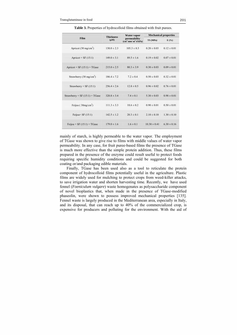
their hydrophilic features, are somewhat too water-sensitive for applications in foods with high and intermediate moisture [122, 124]. These disadvantages can be resolved by the preparation of multicomponent films, as protein-polysaccharide films added with lipids. Another strategy to improve hydrocolloid films containing proteins is to create a net structure introducing covalent cross-links. In this regard, many chemical cross-linkers have been used, such as formaldehyde, glutaraldehyde, glyoxal and genipin [125-127]. However, due to their toxicity, the addition of these molecules is not advisable. On the contrary, a great interest is devoted to enzymes able to covalently bind proteins. Among these, TGase is the most efficient in catalyzing protein cross-linking. Mahmoud and Savello [69, 70] were the first to have utilized TGase as cross-linker to produce homo and hetero-protein based edible films. The films obtained with guinea pig liver TGase cross-linked whey proteins showed a decrease in water vapor permeability of about 60% with respect to controls. Their results showed also that both mechanical properties and solubility were improved in TGase cross-linked films. Yildirim and Hettiarachchy [71] used the same isoform of TGase to produce films from whey protein isolates, soybean 11S globulin and their mixture (1:1 w/w). All the obtained cross-linked films showed a tensile strength two fold greater than that of the homologous controls. Solubility at different pHs and in denaturant environment resulted lower than that of control films, while the degree of hydrolysis by trypsin and -chymotrypsin of both TGase cross-linked and control films was similar after 24 h of incubation. The use of the guinea pig liver derived TGase was replaced in the last decade with the microbial isoform of the enzyme, today commercially available from Ajinomoto Inc. (Kawasaki, Japan) at a price that makes this enzyme feasible for edible film production in a large scale. By using mTGase many authors have produced films with different proteins. Lim et al. [128] have produced cross-linked egg protein-based films, Lerré et al. [99] prepared deamidated gluten films in the presence or absence of diamines, Oh et al. [129] incorporated zein hydrolysates into whey protein or casein films. All these films exhibited higher elongation to break values, but lower tensile strength, without any significant variation of the water vapor permeability. Chambi & Grosso [130] used mTGase as cross-linker in gelatin-casein blend edible films. Only for the casein-gelatin formulation (75:25) TGase improved both the elongation and the water vapor permeability. Mariniello et al. [66] have utilized mTGase as cross-linker to produce a multicomponent edible film containing commercial soy flour (SF) and pectin (ratio 1:2 w/w) respectively. In the cross-linked films the tensile strength was observed to increase from 6.8 MPa to 12.4 MPa, while the

Loredana Mariniello et al. 200
elongation to break was reduced from 11.6% to 7.2%. Same films [131] showed higher barrier properties to both CO2 and O2 as well as to the water vapor. Also the solubility at different pHs and denaturant agents resulted improved. Other multicomponent mTGase cross-linked edible films have been made by mixing either chitosan and whey proteins [132] or chitosan and ovalbumin [133]. All the films prepared in the presence of TGase showed an effective improvement both in mechanical and barrier properties. Since the potential supply of the produced fruit exceeds the market, considerable interest exists to find out new uses for fruit purees. Pectic and cellulosic substances are the most abundant polysaccharides in fruit and vegetables. Therefore, the incorporation of them into edible films and coatings deserves to be investigated. McHugh et al. [134] proposed the utilization of peach puree for edible film production. The obtained films showed a good oxygen barrier property but, as expected, they were ineffective in providing a barrier to moisture. With the aim to obtain fruit puree-based films with acceptable moisture barrier properties, strawberry, apricot and feijoa homogenates were investigated in our laboratories. Strawberry and apricot are widely cultivated in Italy for both fresh consumption and industrial processing. Feijoa (Feijoa sellowiana), belonging to Mirtaceae family, is an autochthonous subtropical plant in Southern America even though it is currently present also in Northern America (California and Florida) and Southern Europe (Italy). Despite its nutritional value, due to the presence of iodine and vitamin C, feijoa is still not commercialized and its employment in some industrial processes can be suggested. Therefore, different films made of different fruit purees were cast in the presence of un-modified and TGase-modified soy proteins and then characterized for their thickness, mechanical properties and moisture barrier properties (Table 3, unpublished results). The presence of the enzyme always influenced thickness of the films, which increased probably because of the presence of TGase-catalyzed isopeptide bonds [131-132]. Only in the feijoa–SF films the TGase was able to significantly reduce water permeability and influence the mechanical properties measured as tensile strength and elongation to break. To assess the role played by TGase in influencing the water vapor barrier property, which is a particularly important feature in the food sector, edible films prepared in the presence of the enzyme were compared to the conventional mineral oil-based polymers and to the commercially available biobased materials (i.e. Mater-Bi, Ecoflex). Figure 5 reported data from the literature and those obtained in our laboratories expressed in logarithmic scale. As well known, Low Density Polyethylene (LDPE) and High Density Polyethylene (HDPE) show the lowest barrier capacity, while Mater-Bi, a biobased material made
Transglutaminase in food 201
Table 3. Properties of hydrocolloid films obtained with fruit purees.
Mechanical properties Film Thickness
(µM)
Water vapor permeability
(cm3 mm/ m2 d kPa) TS (MPa) E (%)
Apricot (30 mg/cm2)
130.0 ± 2.3 105.3 ± 8.5 0.20 ± 0.03 0.12 ± 0.01
Apricot + SF (15:1)
149.0 ± 3.1 89.5 ± 1.6 0.19 ± 0.02 0.07 ± 0.01
Apricot + SF (15:1) + TGase
215.0 ± 2.5 80.3 ± 3.9 0.30 ± 0.03 0.09 ± 0.01
Strawberry (30 mg/cm2)
186.4 ± 7.2 7.2 ± 0.4 0.50 ± 0.03 0.32 ± 0.01
Strawberry + SF (15:1)
256.4 ± 2.6 12.8 ± 0.5 0.96 ± 0.02 0.76 ± 0.01
Strawberry + SF (15:1) + TGase
320.8 ± 3.4 7.4 ± 0.1 5.30 ± 0.03 0.98 ± 0.01
Feijoa ( 30mg/cm2)
111.3 ± 3.3 18.6 ± 0.2 0.90 ± 0.01 0.50 ± 0.01
Feijoa+ SF (15:1)
142.5 ± 1.2 20.3 ± 0.1 2.10 ± 0.10 1.30 ± 0.10
Feijoa + SF (15:1) + TGase
179.0 ± 1.6 1.6 ± 0.1 10.30 ± 0.41 6.30 ± 0.16
mainly of starch, is highly permeable to the water vapor. The employment of TGase was shown to give rise to films with middle values of water vapor permeability. In any case, for fruit puree-based films the presence of TGase is much more effective than the simple protein addition. Thus, these films prepared in the presence of the enzyme could result useful to protect foods requiring specific humidity conditions and could be suggested for both coating or/and packaging edible materials. Finally, TGase has been used also as a tool to reticulate the protein component of hydrocolloid films potentially useful in the agriculture. Plastic films are widely used for mulching to protect crops from weed-killer attacks, to save irrigation water and shorten harvesting time. Recently, we have used fennel (Foeniculum vulgare) waste homogenates as polysaccharide component of novel bioplastics that, when made in the presence of TGase-modified phaseolin, were shown to possess improved mechanical properties [135]. Fennel waste is largely produced in the Mediterranean area, especially in Italy, and its disposal, that can reach up to 40% of the commercialized crop, is expensive for producers and polluting for the environment. With the aid of

Loredana Mariniello et al. 202
TGase, therefore, this waste could represent an useful raw material for the production of environmentally friendly bioplastics.
Figure 5. Water vapor permeability (WVP) properties of biobased materials compared to the ones of the conventional mineral oil-based materials. Values of materials marked with “*“ were measured by gravimetric test at 25°C, 90/0% RH. WVP values of the other materials derive from the literature and were measured under the same conditions (from “a” to “e” see reference 122, 131, 132, 133, 172, respectively) SF: Soy flour; WP: Whey proteins; OVA: Ovalbumin; EVOH: Ethyl Vinyl Alcohol; LDPE: Low Density Polyethylene; HDPE. High Density Polyethylene.
3.4 Nutritional aspects of transglutaminase cross-linked proteins Many investigations have been performed to determine the effect of glutamine-lysine isopeptide bonds produced by TGase on the nutritional value of cross-linked proteins. In this respect, safety of TGase-modified proteins has been deduced from different experimental approaches. First of all it has been established that many foods, both in raw or processed forms, contain a certain amount of proteins or peptides in which glutamine-lysine isopeptide bonds occur. Mainly meat and fish based foods present this characteristic due to the activity of endogenous TGases [136]. During cooking such enzymes can still give rise to cross-linked proteins increasing the content of isopeptide bonds, as reported by Sakamoto et al. [137]. Thus, the mankind has been probably ingesting molecules containing glutamine-lysine isopeptide bonds before the discovery of fire and cooking, suggesting that their introduction in the human body does not represent an health risk.
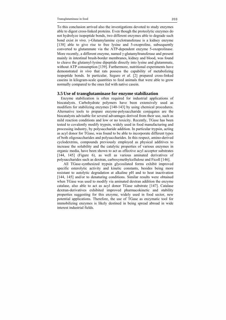
Transglutaminase in food 203
To this conclusion arrived also the investigations devoted to study enzymes able to digest cross-linked proteins. Even though the proteolytic enzymes do not hydrolyze isopeptide bonds, two different enzymes able to degrade such bond exist in vivo. -Glutamylamine cyclotransferase is a kidney enzyme [138] able to give rise to free lysine and 5-oxoproline, subsequently converted to glutammate via the ATP-dependent enzyme 5-oxoprolinase. More recently, a different enzyme, named -glutamyltransferase and present mainly in intestinal brush-border membranes, kidney and blood, was found to cleave the glutamyl-lysine dipeptide directly into lysine and glutammate, without ATP consumption [139]. Furthermore, nutritional experiments have demonstrated in vivo that rats possess the capability of metabolizing isopeptide bonds. In particular, Seguro et al. [2] prepared cross-linked caseins in kilogram-scale quantities to feed animals that were able to grow normally compared to the ones fed with native casein. 3.5 Use of transglutaminase for enzyme stabilization Enzyme stabilization is often required for industrial applications of biocatalysts. Carbohydrate polymers have been extensively used as modifiers for stabilizing enzymes [140-143] by using chemical procedures. Alternative tools to prepare enzyme-polysaccharide conjugates are the biocatalysts advisable for several advantages derived from their use, such as mild reaction conditions and low or no toxicity. Recently, TGase has been tested to covalently modify trypsin, widely used in food manufacturing and processing industry, by polysaccharide addition. In particular trypsin, acting as acyl donor for TGase, was found to be able to incorporate different types of both oligosaccharides and polysaccharides. In this respect, amino-derived cyclodextrins, compounds previously employed as physical additives to increase the solubility and the catalytic properties of various enzymes in organic media, have been shown to act as effective acyl acceptor substrates [144, 145] (Figure 6), as well as various aminated derivatives of polysaccharides such as dextran, carboxymethylcellulose and Ficoll [146]. All TGase-synthesized trypsin glycosilated forms exhibit improved specific esterolytic activity and kinetic constants, besides being more resistant to autolytic degradation at alkaline pH and to heat inactivation [144, 145] and/or to denaturing conditions. Similar results were obtained when TGase was used to modify via aminated dextran addition the enzyme catalase, also able to act as acyl donor TGase substrate [147]. Catalase dextran-derivatives exhibited improved pharmacokinetic and stability properties suggesting for this enzyme, widely used in food sector, new potential applications. Therefore, the use of TGase as enzymatic tool for immobilizing enzymes is likely destined in being spread abroad in wide interest industrial fields.

Loredana Mariniello et al. 204
Figure 6. Transglutaminase-catalyzed synthesis of trypsin-cyclodextrin (CD) conjugates. 2<n<6.
Conclusions As reported in the present chapter, TGase is an enzyme of wide interest in the food industry even though its effective use has been made only lately. Its employment regards mainly the microbial isoform identified by Ando et al. in 1989 [3]. Since then, many investigations were devoted to characterization studies on this new member of TGase family, as well as to various applications in different industrial sectors [6]. Several of the obtained results indicated the effectiveness of mTGase in being employed as texturing agent in food industry. The commercialized mTGase derives from S. mobaraensis and is distributed mainly by Ajinomoto with the name of ACTIVA in a powder form which contain about 1% of the active enzyme. More recently, a Chinese company, named Yiming Fine Chemicals Corporation, has commercialized further TGase-containing preparations to be used as novel food additive. In Europe the use of the enzyme is allowed as processing aid and, thus, according to the Directive 89/107/EC, its presence does not need to be indicated in the final product. The use of TGase as processing aid is allowed because of the Food and Drug Administration (FDA) decision to accept Ajinomoto request to consider TGase-containing preparations as GRAS (Generally Recognized As Safe). Even though FDA accepted Ajinomoto conclusions on TGase safety without making its own determinations regarding the GRAS status of the enzyme, it must be outlined that Ajinomoto GRAS panel based its

Transglutaminase in food 205
evaluation on studies devoted to assess the potential allergenicity of mTGase by using the “2001 FAO/WHO Decision Tree” [148]. These studies have established that no safety concerns have been identified [149]. Thus, the use of TGase in food industry seems to be destined in becoming more and more diffused in all protein containing foods.
References 1. Folk, J.E., and Finlayson, J.S. 1977, Adv. Prot. Chem., 31, 1. 2. Ando, H., Adachi, M., Umeda, K., Matsumura, A., Nonaka, M., Uchio, R.
1989, Agr. Biol. Chem., 53, 2613. 3. Seguro, K., Nio, N., and Motoki, M. 1996, Macromolecular Interactions in
Food Technology, Parris, N., Kato, A., Creamer, L.K., and Pearce, J. (Eds), ACS Symposium Series 650, Am. Chem. Soc., Columbus, OH. p. 271.
4. Witt, D., and Stackebrandt, E. 1990, Syst. Appl. Microbiol., 13, 361. 5. Yokoyama, K., Nio, N., and Kikuchi, Y. 2004, Appl. Microbiol. Biotech.,
64, 447. 6. Mariniello, L., and Porta, R. 2005, Transglutaminases, Family of Enzyme
with Diverse Functions, Metha, K., and Eckert, R. (Eds), Karger Press, Basel, vol. 38, p. 174.
7. Sarkar, N.K., Clarke, D.D., and Waelsch, H. 1957, Biochim. Biophys. Acta, 25, 451.
8. Lorand, L., and Graham, R.M. 2003, Nat. Rev. Mol. Cell Biol., 4, 140. 9. Shan, L., Molberg, O., Parrot, I., Hausch, F., Filiz, F., Gray, G.M., Sollid,
L.M., and Khosla, C. 2002, Science, 297, 2275. 10. Schmidt, G., Selzer, J, Lerm, M, and Aktories, K. 1998, J. Biol. Chem.,
273, 13669. 11. Aktories, K., and Schmidt, G. 2003, Trends Microbiol., 11(4), 152. 12. Nemes, Z., Marekov, L.N., Fesus, L. and Steinert, P.M. 1999, Proc. Natl.
Acad. Sci. USA, 96, 8402. 13. Parameswaran, K.N., Cheng, X.-F., Chen, E.C., Velasco, P.T., Wilson J.H.,
and Lorand, L. 1997, J. Biol. Chem., 272, 10311. 14. Griffin, M., Casadio, R., and Bergamini, C. M. 2002, Biochem. J., 368, 377. 15. Tamaki, T., and Aoki, N. 1982, J. Biol. Chem., 257, 14767. 16. Hornyak, T.J., and Shafer, J.A. 1992, Biochemistry, 31, 423. 17. Candi E., Schmidt R., and Melino G. 2005, Nat. Rev. Mol. Cell Biol., 6,
328. 18. Huber, M., Rettler, I., Bernascono, K., Frenck, E., Lavrijsen, S.P., Ponec,
M., Bon, A., and Lautenchlager, S. 1995, Science, 267, 525. 19. Cassidy, A.J., van Steensel, M.A.M., Steijlen, P.M., van Geel, M., van der
Velden, J., Morley, S.M., Terrinoni, A., Melino, G., Candi, E., McLean, W.H.I. 2005, J. Hum. Genet., 77, 909.
20. Fesus, L., and Piacentini, M. 2002, Trends Biochem. Sci., 27, 534. 21. Aeschlimann, D., and Thomazy, V. 2000, Connect. Tissue Res., 41, 1. 22. Piacentini, M., and Coalizzi, V. 1999, Immunol. Today, 20, 130.

Loredana Mariniello et al. 206
23. Skovbjerg, H., Koch, C., Anthonsen, D. M., and Sjostrom, H. 2004, Biochim. Biophys. Acta, 1690, 220.
24. Dieterich, W., Ehnis, T., Bauer, M., Donner, P., Volta, U., Riecken, E.O., and Schuppan, D. 1997, Nat. Med., 3(7), 797.
25. Porta, R., Esposito, C., De Santis, A., Fusco, A., Iannone, M., and Metafora, S. 1986, Biology of eproduction,Biol. Reprod., 35, 965.
26. Paonessa, G., Metafora, S., Tajana, G., Abrescia, P., De Santis, A., Gentile, V., and Porta, R. 1984, Science, 226, 852.
27. Porta, R., Esposito, C., Gentile, V., Mariniello, L., Peluso, G., and Metafora, S. 1990, Int. J. Pept. Prot. Res., 35, 117.
28. Korsgren, C., Lawler, J., Lambert, S., Speiche, D., and Choen, C.M. 1990, Biochemistry, 87, 613.
29. Ideguchi, H., Nishimura, J., Nawata, H., and Hamasaki, N. 1990, Br. J. Hematol., 74, 347.
30. Matsuki, M., Yamashita, F., Ishida-Yamamoto, A., Yamada, K., Kinoshita, C., Fushiki, S., Ueda, E., Morishima, Y., Tabata, K., Yasuno, H., Hashida, M., Iizuka, H., Ikawa, M., Okabe, M., Kondoh, G., Kinoshita, T., Takeda, J., and Yamanishi K. 1998, Proc.P. Natl. Acad. Sci. USA, 95, 1044.
31. Worratao, A., and Yongsawatdigul, J. 2003, J. Food Biochem., 27, 35. 32. Worratao, A, and Yongsawatdigul, J. 2005, Food Chem., 93(4), 651. 33. Wang, R., Liang, Z., Hall, M., and Soderhall, K.A. 2001, Fish Shellfish
Immun., 11, 623. 34. Chen, M.-Y., Hu, K.-Y., Huang, C.-C., and Song, Y.-L. 2005, Dev. Comp.
Immunol., 29, 1003. 35. Yeh, M.S., Kao, L.R., Huang, C.J., and Tsai, I.H. 2006, Biochim. Biophys.
Acta, 1764, 1167. 36. Kang, H., and Cho, Y.D. 1996, Biochem. Biophys. Res. Com., 223, 288. 37. Lilley, G., Skill, J., Griffin, M., and Bonner, P.L.R. 1998, Plant Physiol.,
117, 1115. 38. Serafini-Fracassini, D., Del Duca, S., Monti, F., Poli, F., Sacchetti, G.,
Bregoli, A.M., Biondi, S., and Della Mea, M. 2002, Cell Death Differ., 9(3), 309.
39. Della Mea, M., Caparrós-Ruiz, D., Claparols, I., Serafini-Fracassini, D., and Rigau, J. 2004, Plant Physiol., 135, 2046.
40. Villalobos, E., Santos, M., Talavera, D., Rodríguez-Flacón, M., and Torné, J.M. 2004, Gene, 336, 93.
41. Del Duca, S., Bregoli, A.M., Bergamini, C., and Serafini-Fracassini, D. 1997, Sex. Plant Reprod., 10, 89.
42. Del Duca, S., and Serafini-Fracassini, D. 2005, Transglutaminases, Family of Enzyme with Diverse Functions, Metha, K., and Eckert, R. (Eds), Karger Press, Basel, vol. 38, p. 223.
43. Ruiz-Herrera, J., Iranzo, M., Elorza, M.V., Sentandreu, R., and Mormeneo, S. 1995, Arch. Microbiol., 164, 186.
44. Brunner, F., Rosahl, S., Lee, J., Rudd, J.J., Geiler, C., Kauppinen, S., Rasmussen, G., Scheel, D., Nurnberger T. 2002, EMBO J., 21, 6681.
45. Iranzo, M., Aguado, C., Pallotti, C., Canizares, J.V., and Mormeneo, S. 2002, Microbiology, 148, 1329.
Transglutaminase in food 207
46. Pasternack, R., Dorsch, S., Otterbach, J.T., Robenek, I.R., Wolf, S. and Fuchsbauer, H.L. 1998, Eur. J. Biochem., 257, 570.
47. Wu, J.-W., Tsai, G.-J., and Jiang, S.-T. 1996, J. Chin. Agr. Chem. Soc., 34, 228.
48. Duran, R., Junqua, M., Schmitter, J.M., Gancet, C., and Goulas, P. 1998, Biochimie, 80, 313.
49. Kobayashi, K., Suzuki, S.I., Izawa, Y., Miwa, K., and Yamanaka, S. 1998, J. Gen. Appl. Microbiol., 44, 85.
50. Suzuki, S., Izawa, Y., Kobayashi, K., Eto, Y., Yamanaka, S., Kubota, K., and Yokozeki, K. 2000, Biosci. Biotech. Bioch., 64, 2344.
51. Lin, Y.S., Chao, M.L., Liu, C.H., Tseng, M., and Chu, W.S. 2006, Process Biochem., 41, 519.
52. Kanaji, T., Ozaki, H., Takao, T., Kawajiri, H., Ide, H., Motoki, M., and Shimonishi, Y. 1993, J. Biol. Chem., 268, 11565.
53. Gerber, U., Jucknischke, U., Putzien, S., and Fuchsbauer, H.L. 1994, Biochem. J., 299, 825.
54. Tsai, G.-J., Lin, S.-M., and Jiang, S.-T. 1996, J. Food Sci., 61, 1234. 55. Casadio, R., Polverini, E., Mariani, P., Spinozzi, F., Carsughi, F., Fontana,
A., Polverino de Laureto P., Matteucci G., Bergamini C. M. 1999, Eur. J. Biochem., 262, 672.
56. Hettasch, J.M., and Greenberg, C.S. 1994, J. Biol. Chem., 269, 28309. 57. Micanovic, R., Procyk, R., Lin, W., and Matsueda, C.R. 1994, J. Biol.
Chem., 269, 9190. 58. Kashiwagi, T., Yokoyama, K., Ishikawa, K., Ono, K., Ejima, D., Matsui,
H., and Suzuki, E. 2002, J. Biol. Chem., 277, 44252. 59. Matheis, G., and Whitaker, J.R. 1987, J. Food Biochem. 11, 309. 60. Sorrentino, A., Schillberg, S., Fischer, R., Rao, R., Porta, R., and
Mariniello, L. 2005, Int. J. Biochem. Cell Biol., 37, 842. 61. Osman, A.A., Richter, T., Stern, M., Conrad, K., Henker, J., Brandsch, C.,
Zimmer, K.P., Mothes, T. 2002, Eur. J. Gastroenterol. Hepatol., 14, 1217. 62. Sblattero, D., Berti, I., Trevisol, C., Marzari, R., Tommasini, A., Bradbury,
A., Fasano, A., Ventura, A., Not, T. 2000, Am. J. Gastroenterol., 95, 1253. 63. Choate, K.A., Kinsella, T.M., Williams, M.I., Nolan, G.P., and Khavari,
P.A. 1996, Hum. Gene Ther., 7, 2247. 64. Choate, K.A., and Khavari, P.A. 1997, Hum. Gene Ther., 8, 1659. 65. Kuraishi, C., Yamazaki, K., and Susa, Y. 2001, Food Rev. Int., 17, 221. 66. Mariniello, L., Di Pierro, P., Esposito, C., Sorrentino, A., Masi, P., and
Porta, R. 2003, J. Biotechnol., 102, 191. 67. Mariniello L., Giosafatto C.V.L., Di Pierro P., Sorrentino A., and Porta R.
2007, J. Agr. Food Chem. 55, 4717. 68. Motoki, M. and Seguro, K. 1998, Trends Food Sci. Tech., 9, 204. 69. Mahmoud, R., and Savello, P.A. 1992, J. Dairy Sci., 75(4), 942. 70. Mahmoud, R., and Savello, P.A. 1993, J. Dairy Sci., 76(1), 29. 71. Yildirim M., and Hettiarachchy N.S. 1998, J. Food Sci., 63, 248. 72. Nielsen, P.M. 1995, Food Biotech., 9(3), 119. 73. Nakagoshi, H., and Ishida, R. 2007, Patent US2007054346. 74. Shah H.N., Marthi, G.B., Prasad, J.M. 2001, Patent WO0162888.

Loredana Mariniello et al. 208
75. Kumazawa, Y., Seguro, M., Tamakura, M. and Motoki, M. 1993, J. Food Sci., 58(5), 1062.
76. Dondero, M., Curotto, E., and Figueroa, V. 2002, Food Sci. Tech. Int., 8(1), 49.
77. Uresti, M.R., Tèllez-Luis, S., Ramírez, J.A., and Vàzquez, M. 2004, Food Chem., 86, 257.
78. Uresti, M.R., Velazquez, G., Vazquez, M., Ramírez, J.A., and Torres, J.A. 2006, Food Chem., 94, 202.
79. Borderias-Juarez, A.J., Montero-Garcia, P., and Gomez-Guillen, M.D.C. 2001, Patent WO0193702.
80. Ramírez, J.A., Del Ángel A., Velazquez, G., and Vàzquez, M. 2006, Eur. Food Res. Tech., 223, 341.
81. Lauber, S., Klostermeyer, H., and Henle, T. 1999, Lebensmittelchemie, 53, 128.
82. Jaros, D., Partchefeld, C., Henle, T., and Rohm, H. 2006, J. Texture Stud., 37, 113.
83. Ishii, C., Soeda, T; and Yamazaki, K. 1994, Patent EP0610649. 84. Kuraishi, C., Sakamoto, J., and Soeda, T. 1999, Patent EP0711504. 85. Kuraishi, C., Sakamoto, J., and Soeda, T. 1997, Patent US5681598. 86. Miwa, N., Kumazawa, Y., Nagakoshi, H., and Sakagushi, S. 2004, Patent
US6716461. 87. Bönish, M.P, and Kulozik, U. 2007, Patent EP1749447. 88. Lorenzen, P.C., Neve, H., Mautner, A., and Schlimme, E. 2002, Int. J.
Dairy Technol., 55, 152. 89. Bönish, M.P., Hiss, M., Lauber, S., Kulozik, U. 2007, Food Hydrocolloids,
21, 585. 90. Farnsworth, J.P.; Li, J., Hendricks, G.M., Guo, M.R. 2006, Small Ruminant
Res., 65, 113. 91. Kumazawa, Y., and Miwa, N. 2005, Patent US2005123645. 92. Cozzolino, A., Di Pierro, P., Mariniello, L., Sorrentino, A., Masi, P., Porta,
R. 2003, Biotechnol. Appl. Biochem., 38, 289. 93. Han, X.-Q., and Pfeifer, J.K. 2002, Patent EP1232692. 94. Ikura K., Kometani, T., Yoshikawa, M., Sasaki, R. and Chida, H. 1980,
Agric. Biol. Chem. 44, 1567. 95. Motoki, M., and Nio, N. 1983, J. Food Sci., 48, 561. 96. Nonaka, M., Tanaka, H., Okiyama, A., Motoki, M., Ando, H., Umeda, K.,
and Matsumura, A. 1989, Agr. Biol. Chem., 53, 2619. 97. Kang, I.J., Matsumura, Y., Ikura, K., Motoki, M., Sakamoto, H., and Mori
T. 1994, J. Agr. Food Chem., 42, 159. 98. Chanyongvorakul, Y., Matsumura, Y., Sakamoto H., Motoki, M., Ikura K.,
and Mori T. 1994, Biosci. Biotechnol. Biochem., 58 (5), 864. 99. Larré, C., Denery-Papini, S., Pompineau, y., Deshayes, G., Desserme, C.,
Lefebvre, J. 2000, Cereal Chem., 77, 32. 100. Porta, R., Gentile, V., Esposito, C., Mariniello, L., and Auricchio, S. 1990,
Phytochemistry, 29, 2801. 101. Wang, J.-S., Zhao, M.-M., Yang, X.-Q., Jiang, Y.-M., and Chun, C. 2007,
Food Hydrocolloids, 21, 174.

Transglutaminase in food 209
102. Caballero, P.A., Gòmez, M., Rosell, C.M. 2007, Eur. Food Res. Technol., 224, 525.
103. Yamazaki, K., Toba, S., and Soeda, T. 1994, JP 6014733. 104. Collar, C., and Bollaìn, C. 2004, Eur. Food Res. Technol., 218, 139. 105. Collar, C., and Bollaìn, C. 2005, Eur. Food Res. Technol., 221, 298. 106. Collar, C., Bollaìn, C., and Angioloni, A. 2005, J. Food Eng. 70, 479. 107. Moore, M.M., Heinbockel, M., Dochery, P., Ulmer, H.M., and Arendt, E.K.
2006, Cereal Chem., 83(1), 27. 108. Kang, D.O. 2006, Patent WO20066057520. 109. Molina-Rosell, P., Solis, N., and Daniel, R. 2004, Patent WO2004084638. 110. AWARENET, 2004, Handbook for the preservation and minimization of
waste and valorization of by-products in european agro-food industries. Ed. Agro-food wastes minimization and reduction network, AWARENET.
111. Tolkach, A., and Kulozik, U. 2005, J. Food Eng., 67(1-2), 13. 112. Baxter, S.R., and Skonberg, D.I. 2006, J. Food Sci., 71(8), C499. 113. Pietrasik, Z., and Li-Chan, E.C.Y. 2002, Food Res. Int., 35, 91. 114. Pietrasik, Z., and Li-Chan, E.C.Y. 2002, Food Res. Int., 35, 387. 115. Hammer, G.F. 1998, Fleischwirtschaft, 78, 1155-1156, 1159-1162, 1186. 116. Lantto, R., Plathin, P., Niemstö, M., Buchert, J., and Autio, K. 2006, LWT,
39, 1117. 117. Ramírez-Suárez, J.C., Addo, K., and Xiong, Y.L. 2005, Food Res. Int.,
38(10), 1143. 118. Carballo, J., Ayo, J., Colmenero, F.J. 2006, LWT, 39, 692. 119. Moller, A.J., and Nielsen, G. 1997,. Patent US5,686,124. 120. Tseng, T.F., Liu, D.C., and Chen, M.T. 2000, Meat Sci., 55, 427. 121. Haugaard, V.K., Udsen, A.M., Mortensen, G., Hoegh, L., Petersen, K.,
Monahan, F. 2001, Potential food applications of biobased materials. An EU-concerted action project. STARCH-STARKE 53 (5), 189.
122. Nisperos-Carriedo, M.O. 1994, Edible Coatings and Films to Improve Food Quality, Krochta, J.M., Baldwin E.A., and Nisperos-Carriedo, M., (Eds), Technomic, Lancaster, PA, USA, p. 305.
123. Krochta, J.M. 1997, Food Proteins and Their Applications in Foods, Damodaran, S., and Paraf, A. (Eds), Marcel Dekker Inc., New York, USA, cptr. 18, p. 529.
124. Krochta, J.M., and Mulder-Johnston, C.L.C. 1997, Food Technol., 51, 61. 125. Rhim, J.W., and Weller, C.L. 2000, Food Sci. Biotechnol., 9, 228. 126. Ghorpade, V.M., Li, H., Gennadios, A., and Hanna, M.A. 1995, T. ASAE,
38, 1805. 127. Marquié, C. 2001, J. Agr. Food Chem., 49, 4676. 128. Lim, L.-T., Mine, Y., and Tung, M. A. 1998, J. Agr. Food Chem., 46, 4022. 129. Oh, J.H., Wang, B., Field, P.D., and Aglan, H.A. 2004, Int. J. Food Sci.
Tech., 39(3), 287. 130. Chambi, H., and Grosso, C. 2006, Food Res. Int., 39, 458. 131. Di Pierro, P., Mariniello, L., Giosafatto, C.V.L., Masi, P., and Porta, R.
2005, Food Biotech., 19, 37. 132. Di Pierro, P., Chico, B., Villalonga, R., Mariniello, L., Damiao, A.E., Masi,
P., and Porta, R. 2006, Biomacromolecules, 7, 744.

Loredana Mariniello et al. 210
133. Di Pierro, P., Chico, B., Villalonga, R., Mariniello, L., Masi, P., and Porta, R. 2007, Enzyme Microb. Tech., 40, 437.
134. McHugh, T.H., Huxsoll, C.C., and. Krochta, J.M. 1996, J. Food Sci., 61, 88. 135. Mariniello, L., Giosafatto, C.V.L., Moschetti, G., Aponte, M., Masi, P.,
Sorrentino, A., and Porta, R. 2007, Biomacromolecules, in press. 136. Kumazawa, Y., Sakamoto, H., Kawajiri, H., Seguro, K. and Motoki M.
1996, Fish. Sci. 62, 331. 137. Sakamoto, H., Kumazawa, Y., Kawajiri, H., and Motoki, M. 1995, J. Food
Sci. 60, 416. 138. Fink, M.L., Chung, S.I., and Folk, J.E. 1980, Proc. Natl. Acad. Sci., 77,
4564. 139. Seguro, K., Kumazawa, Y., Ohtsuka, T., Ide, H., Nio, N., Motoki, M., and
Kubota, K. 1995, J. Agric. Food Chem., 43, 1977. 140. Sundaram, P.V., and Venkatesh, R. 1998, Protein Eng., 11, 691. 141. Villalonga, R., Villalonga, M.L., and Gómez, L. 2000, J. Mol. Catal. B-
Enzym., 10, 483. 142. Masárová, J., Mislovičová, D., Gemeiner, P., and Michailková, E. 2001,
Biothec. Appl. Biochem., 34, 127. 143. Villalonga, M.L., Fernández, M., Fragoso, A., Cao, R., and Villalonga, R.
2003, Prep. Biochem. Biotechnol., 33, 53. 144. Villalonga, R., Fernàndez, M., Fragoso, A., Cao, R., Di Pierro, P.,
Mariniello, L., and Porta, R. 2003, Biotechnol. Bioeng, 81, 732. 145. Villalonga, R., Fernàndez, M., Fragoso, A., Cao, R., Mariniello, L., and
Porta, R. 2003, Biotechnol. Appl. Biochem., 38, 53. 146. Villalonga, M.L., Villalonga, R., Mariniello, L., Gomez, L., Di Pierro, P.,
and Porta, P. 2006, World J. Microbiol. Biotech., 22, 595. 147. Valdivia, A., Villalonga, R., Di Pierro, P., Perez, Y., Mariniello, L., Gomez,
L., and Porta R. 2006, J. Biotechnol. 122, 326. 148. FAO & WHO, 2001, Evaluation of Allergenicity of Genetically Modified
Foods. Report of a Joint FAO/WHO Expert Consultation on Allergenicity of Foods Derived from Biotechnology. 22–25 January 2001, www.fao.org/es/ESN/food/pdf/allergygm.pdf
149. Pedersen, M.H., Hansen, T.K., Sten, E., Seguro, K., Ohtsuka, T., Morita, A., Bindslev-Jensen, C., and Poulsen, L.K. 2004, Mol. Nutr. Food Res., 48(6), 434.
150. Yongsawatdigul, J., Piyadhammaviboon, P., and Singchan, K. 2006, Eur. Food Res. Technol., 223(6), 769.
151. Yasueda, H., Kumazawa, Y., and Motoki, M. 1994, Biosci. Biotech. Biochem., 58(11), 2041.
152. Noguchi, K., Ishikawa, K., Yokoyama, K., Ohtsuka, T., Nio, N., and Suzuki, E. 2001, J. Biol. Chem., 276(15), 12055.
153. Kumazawa, Y., Nakanishi, K., Yasueda, H., and Motoki, M. 1996, Fish. Sci., 62, 959.
154. Yongsawatdigul, J., Worratao, A., and Park, J.W. 2002, J. Food Sci., 67(9), 3258.
155. Alibardi, L., and Toni, M. 2004, J. Exp. Zool. (Mol. Dev. Evol.), 302B, 526.

Transglutaminase in food 211
156. Zhang, J., and Maui, Y. 1997, Mol. Reprod. Dev., 47,302. 157. Puzkin, E.G., and Raghuraman, V. 1985, J. Biol. Chem., 260, 16012. 158. Bures, D.M., and Goldsmith, L.A. 1978, Arch. Dermatol. Res., 262(3), 329. 159. Weraarchakul-Boonmark, N., Jeong, J.-M., Murthy, S.N.P., Engel, J. D.,
and Lorand L. 1992, Proc. Natl. Acad. Sci., USA, 89, 9804. 160. Tokunaga, F., Muta, T., Iwanaga, S., Ichinose, A., Davie, E.W., Kuma, K.,
and Miyata, T. 1993, J. Biol. Chem., 268, 262. 161. Singh, R.N., and Metha, K. 1994, Eur. J. Biochem., 225, 625. 162. Mádi, A., Punyiczki, M., Di Rao, M., Piacentini, M., Fésüs, L. 1998, Eur. J.
Biochem., 253(3), 583. 163. Theopold, U., Schmidt, O., Söderhäll, K., and Dushay, M.S. 2004, Trends
Immunol., 25, 289. 164. Nozawa, H., and Seki, N. 2001, Fisheries Sci., 67(3), 493. 165. Kumazawa, Y., Nakanishi, K., Yasueda, H., and Motoki M. 1997, J. Agr.
Food Chem., 45, 604. 166. Yeh, M.S., Huang, C.J., Leu, J.H., Lee, Y.C., and Tsai, I.H. 1999, Eur. J.
Biochem., 266, 624. 167. Huang, C.C., Sritunyalucksana, K., Soderhall, K., and Song, Y.L. 2004,
Dev. Comp. Immunol., 28, 279. 168. Sugino, H., Terakawa, Y., Yamasaki, A., Nakamura, K., Higuchi, Y.,
Matsubara, J., Kunyoshi, H., and Ikegami, S. 2002, Eur. J. Biochem., 269, 1957.
169. Del Duca, S., Tidu, V., Bassi, R., Esposito, C., and Serafini-Fracassini, D. 1993, Planta, 193, 283.
170. Reynolds, K., Brobey, B., and Soong. L. 2006, Exp. Parasitol., 114, 94. 171. De Barros Soares, L.H., Assmann, F., and. Zàchia Ayub, M.A. 2003,
Biotechnol. Appl. Biochem., 37, 295. 172. Yamamoto, M., Witt, U., Skupin, G., Beimborn, D., Müller, R.J. 2003,
Biopolymers, Steinbüchel, A. (Ed), Weeley-VCH, Weinheim, Germany, vol. 4, p. 521.