Embed Size (px)
Citation preview
Transforming Growth Factor � (TGF�) Mediates Schwann CellDeath In Vitro and In Vivo: Examination of c-Jun Activation,Interactions with Survival Signals, and the Relationship ofTGF�-Mediated Death to Schwann Cell Differentiation
David B. Parkinson,1 Ziping Dong,4 Howard Bunting,1 Jonathan Whitfield,3 Carola Meier,5 Helene Marie,2Rhona Mirsky,1 and Kristjan R. Jessen1
1Departments of Anatomy and Developmental Biology and 2Physiology, and 3Eisai London Research, University CollegeLondon, London WC1E 6BT, United Kingdom, 4Reneuron Ltd., Denmark Hill, London SE5 8AF, United Kingdom, and5Institut fur Neuroanatomie, Med. Einrichtungen der Ruhr-Universitat, 44780 Bochum, Germany
In some situations, cell death in the nervous system is con-trolled by an interplay between survival factors and negativesurvival signals that actively induce apoptosis. The presentwork indicates that the survival of Schwann cells is regulated bysuch a dual mechanism involving the negative survival signaltransforming growth factor � (TGF�), a family of growth factorsthat is present in the Schwann cells themselves. We analyze theinteractions between this putative autocrine death signal andpreviously defined paracrine and autocrine survival signals andshow that expression of a dominant negative c-Jun inhibits
TGF�-induced apoptosis. This and other findings pinpoint ac-tivation of c-Jun as a key downstream event in TGF�-inducedSchwann cell death. The ability of TGF� to kill Schwann cells,like normal Schwann cell death in vivo, is under a strongdevelopmental regulation, and we show that the decreasingability of TGF� to kill older cells is attributable to a decreasingability of TGF� to phosphorylate c-Jun in more differentiatedcells.
Key words: autocrine signals; apoptosis; nerve development;peripheral nerve; nerve injury; nerve regeneration
It is likely that two sets of signals play a major role in promotingthe survival of developing Schwann cells. These are �-neuregulins(NRG�s) (Dong et al., 1995; Grinspan et al., 1996; Syroid et al.,1996; Trachtenberg and Thompson, 1996) and autocrine Schwanncell signals, which include a synergistic combination of insulin-like growth factor 2 (IGF-2), neurotrophin-3 (NT3), and platelet-derived growth factor-BB (PDGF-BB) (Meier et al., 1999) inaddition to leukemia-inhibitory factor (LIF) (Dowsing et al.,1999; Jessen and Mirsky, 1999). NRG� is provided mainly byaxons and is probably of paramount importance in embryonic andearly postnatal nerves, whereas the autocrine circuits are active inpostnatal cells and likely to be especially significant after injuryand consequent loss of axonal NRG�. It is possible to envisagethat Schwann cell survival is regulated exclusively by these andother positive survival factors, a view that would imply thatSchwann cell death, seen for example in normal and, especially,injured neonatal nerves, is caused by a limited availability of suchsignals, in line with the classical neurotrophic theory. More re-cently, an alternative view of why cells die has emerged (Cassacia-Bonnefil et al., 1999; Raoul et al., 2000). These experimentsindicate that cell death can be caused not only by the absence of
survival signals but also by the advent of active cell killingmediated by factors that trigger apoptosis. In the nervous system,nerve growth factor (NGF) is one of the factors that may act inthis way, both in retinal development and in Schwann cells (Xiaet al., 1995; Frade et al., 1996; Cassacia-Bonnefil et al., 1999;Frade and Barde, 1999; Soilu-Hanninen et al., 1999; Raoul et al.,2000).
Transforming growth factor �s (TGF�s) are expressed bySchwann cells and have various proliferative and phenotypiceffects on these cells (for review, see Mirsky and Jessen, 1996;Scherer and Salzer, 1996). In the present work we have exploredthe idea that TGF� might act as a death signal for Schwann cells.We find that TGF� induces Schwann cell apoptosis under anumber of different conditions in vitro. This effect is blocked bythe combined presence of NRG� and autocrine signals, and, inline with this, TGF� kills Schwann cells in the distal stump of cutneonatal nerves but not in normal nerves. We provide evidencethat TGF� induces apoptosis by activating c-Jun in Schwann cellsand that overexpression of a dominant negative c-Jun inhibitsTGF�-induced apoptosis in Schwann cells. A resistance to TGF�killing emerges in tandem with Schwann cell differentiation, andthis is related to a failure of TGF� to activate c-Jun in differen-tiated cells. We show that nerve transection leads to elevation ofTGF�1 mRNA and protein in the distal stump of neonatalanimals, in line with observations in the adult which suggest thatthis factor is involved in events that follow nerve damage. Takentogether, this information builds a case for TGF� as a negativeSchwann cell survival signal in perinatal nerves.
MATERIALS AND METHODSMaterials. OX7 hybridoma cell line secreting Ig recognizing Thy1.1 wasfrom the European Collection of Animal Cell Cultures (DERA, Wilt-
Received Jan. 30, 2001; revised Aug. 8, 2001; accepted Aug. 10, 2001.This work was supported by a Wellcome Trust Program grant and a European
Community (EC) Biomed 2 collaborative research grant (CT97/2069) to K.R.J. andR.M., an EC Training and Mobility Research fellowship (CT961028) to C.M., aWellcome Trust 4 year PhD fellowship to H.M., and a Wolfson Scholarship to H.B.We thank G. Evan and J. Ham for gifts of antibodies and D. Bartram for editing thismanuscript.
D.P. and Z.D. are joint first authors.Correspondence should be addressed to K. R. Jessen, Department of Anatomy
and Developmental Biology, University College London, Gower Street, LondonWC1E 6BT, UK. E-mail: [email protected] © 2001 Society for Neuroscience 0270-6474/01/218572-14$15.00/0
The Journal of Neuroscience, November 1, 2001, 21(21):8572–8585
shire, UK). Rabbit polyclonal antibody to S-100 was from Dakopatts(Copenhagen, Denmark), mouse monoclonal antibody to myelin basicprotein was from Roche Diagnostics (Lewes, UK), and goat anti-mouseIg and anti-rabbit Ig conjugated to fluorescein were from Cappel Labs(Durham, NC). Rabbit polyclonal antibody to c-Jun was a gift from G.Evan (University of California San Francisco). Antibody to LexA wasfrom Santa Cruz Biotechnology (Santa Cruz, CA). Monoclonal anti-FLAG antibody was from Sigma (Poole, UK). Recombinant humanTGF�1, pan-specific TGF�, and TGF�1 antibodies were from R & DSystems (Minneapolis, MN). Purified TGF�2 (porcine) was from BritishBiotechnology, IGF-1 was from PeproTech EC Ltd (London, UK),neuregulin �1 was from Amgen (Thousand Oaks, CA), and puromycinwas from Sigma. Monoclonal antibody SM 1.2 was from the Develop-mental Studies Hybridoma Bank (Iowa City, IA). Polyclonal antibodyCM1 specific for active cleaved caspase-3 was from BD PharMingen(Oxford, UK). Sources of other reagents used in immunocytochemistry,RT-PCR, Western blotting, and cell cultures have been detailed inprevious papers (Jessen et al., 1994; Morgan et al., 1994; Dong et al.,1995, 1999; Stewart, 1995; Blanchard et al., 1996).
Cell culture. Cultures of Schwann cells were prepared essentially asdescribed previously (Jessen et al., 1994; Gavrilovic et al., 1995; Meier etal., 1999). Sciatic nerves and brachial plexuses were removed fromnewborn, postnatal day (P) 4 and P8 and adult Sprague Dawley rats,desheathed, and treated either with a mixture of collagenase (4 mg/ml),hyaluronidase (1.2 mg/ml), and trypsin inhibitor (0.5 mg/ml) in calciumand magnesium-free DMEM at 37°C for 70–80 min or alternatively witha mixture of collagenase (2 mg/ml) and trypsin 1.25 mg/ml for 35 min(newborn, P4, P8) or twice for 1.5–2 hr in total (adult). The tissue wasthen gently dissociated through a plastic pipette tip, and cells werecentrifuged and then purified by negative immunopanning on dishescoated with Thy1.1 antibodies as described previously (Dong et al.,1997).
For the survival assays and tests of TGF�1-induced apoptosis, freshlyimmunopanned rat Schwann cells were plated on polyornithine or poly-L-lysine (PLL)/ laminin-coated coverslips (Meier et al., 1999) as indicatedin Results. To test survival in the absence of autocrine signals, cells wereplated at low density (300 cells per 20 �l per coverslip). To test survivalin the presence of autocrine survival support, cells were plated at highdensity (3000 cells per 10 �l per coverslip).
In most survival assays, the culture medium was a simple mediumcontaining only a 1:1 mixture of DMEM and Ham’s F12 plus BSA (0.3mg/ml final). In these experiments TGF� or NRG� was added 3 hr afterplating. In other experiments we used a supplemented defined mediumidentical to that used in previous work (Jessen et al., 1994), except thatdexamethasone and IGF-1 were left out. TGF� was added 16–18 hr afterplating. These experiments are specially indicated in the text. Nearly allthe survival assays lasted for 24 hr, timed from the addition of growthfactors. Experiments using longer survival times are indicated in the text.At 3 hr and at specified times, cells were fixed in 2% paraformaldehydein PBS for 20 min, immunolabeled with S100 antibodies, and mounted inCitifluor mounting medium containing 4 �g/ml Hoechst dye. The num-ber of living cells in this assay is expressed as survival percentage.Survival percentage is the number of living cells present at the end of theexperiment as a percentage of the number of cells that had platedsuccessfully at the beginning of the experiment, i.e., the number of cellsthat had attached and begun to flatten on the substrate 3 hr after plating.Routinely, dead cells were identified by observing Hoechst nuclearstaining and obvious morphological changes associated with death. Thuscells classified as dead showed either clearly elevated intensity of Hoechstnuclear labeling or nuclei that had fragmented, showing two or moreHoechst-labeled bodies per cell, and in addition had retracted processesand cytoplasm that by phase contrast appeared granulated and mostoften also vacuolated; the nucleus of these cells appeared condensedand/or fragmented by phase contrast. To validate the classification ofthese cells as dead, we examined cultures of dying cells that had beenlabeled with the terminal deoxynucleotidyl transferase-mediated biotin-ylated UTP nick end labeling (TUNEL) method. In addition, apoptosisin cells was confirmed using an antibody (CM1) specific for the activatedcleaved form of caspase 3. For the assay of cell death and measurementof TGF� mRNA or protein after axotomy, newborn rats were anesthe-tized, the left sciatic nerve was transected, and the proximal stump wasdissected and sutured to the muscle. For experiments measuring celldeath, the growth factor, TGF�1, control, or anti-TGF� antibodies orPBS were applied three times at 8 hourly intervals during 24 hr. TGF�1or antibodies diluted in PBS were injected into the relatively large
intermuscular space that surrounds the sciatic nerve in the mid-thighregion. For the first injection, a volume of 10 �l was used, followed by thetwo further injections in 10 �l. After the times indicated, the control ortransected sciatic nerves were removed, and cryostat sections were pre-pared for immunohistochemistry.
Immunocytochemistry. Immunolabeling for S100 and myelin basic pro-tein (MBP) was performed as follows. Cells were fixed in 2% parafor-maldehyde for 10 min (MBP) and 20 min (S100), washed in PBS, andthen treated with methanol (�20°C) for 10 min. After rinsing in PBS,cells were incubated in S100 (1:100) antibody or MBP (1:100) antibodyfor 30 min, washed, and incubated in anti-rabbit Ig fluorescein (S100) oranti-mouse Ig fluorescein (MBP) for 30 min, washed, and mounted inCitifluor anti-fade mounting medium. All antibodies were diluted in PBScontaining 0.1 M lysine, 0.2% sodium azide, and 10% calf serum. Immu-nolabeling with c-Jun antibody was performed exactly as described pre-viously (Stewart, 1995). For immunolabeling with antibodies to LexA,FLAG, or ser-63 phospho c-Jun, cells were fixed in 4% paraformalde-hyde in PBS for 15 min, then permeabilized in 0.5% Triton X-100/PBSfor 5 min. After a block of 50% goat serum/1% BSA in PBS, primary andsecondary antibodies were then applied in block solution for LexA,FLAG, or 1% BSA/PBS for ser-63 phospho-Jun. For labeling of cellswith CM1 antibody for active caspase 3, cells were fixed in 4% parafor-maldehyde for 20 min, followed by a block of 20% goat serum/0.4%Triton X-100/PBS for 30 min; primary and secondary antibodies wereapplied in this block solution. For immunolabeling of sections withTGF�1 antibody, sections were fixed in 4% paraformaldehyde for 20 minfollowed by a 10% goat serum/0.1% Tween 20/PBS block for 1 hr.Primary and secondary antibodies were applied in this block solution.
Adenoviral infection of Schwann cells. Adenoviral supernatants forrecombinant adenoviral constructs expressing either LacZ or the domi-nant negative c-Jun molecule FLAG�169-Jun in the adenoviral vectorpAdCMVpoly A were prepared and titered as described previously(Garnier et al., 1994; Berkner, 1998; J. Whitfield, unpublished observa-tions). Immunopanned Schwann cells from newborn rats were plated ata density of 3000 cells per 10 �l drop on laminin-coated glass coverslipsin supplemented defined medium. Approximately 16 hr after plating,adenoviral supernatant corresponding to a multiplicity of infection of�1500 was added to the cells. Twenty-four hours later the adenoviralsupernatant was removed, and the medium of the cells was changed intofresh supplemented defined medium. No toxic effects of addition of theadenoviral supernatant on the Schwann cells were observed. After anadditional 24 hr to allow expression of the lacZ or FLAG�169-Jun, thetime 0 controls were fixed, and the Schwann cells were changed to freshsupplemented defined medium alone or with increasing amounts ofTGF�1. Twenty-four hours later, the cells were fixed and stained withHoechst dye, and survival was assessed as described previously.
Infection of Schwann cells with retroviral constructs. For retroviralinfection experiments, Schwann cells from newborn nerves were purifiedby culture in DMEM and 10% calf serum containing 10 � 5 M cytosinearabinoside for 3 d as described previously (Morgan et al., 1991). ThecDNAs for the LexA and LexA-vJun (Struhl, 1988) were cloned into theretroviral plasmid vector pBABEpuro, and the GP�E ecotropic packag-ing cell line (Morgenstern and Land, 1990) was then stably transfectedwith the plasmid DNA. Retroviral supernatant from the GP�E cells wasthen used to infect rat Schwann cells, and puromycin-selected pools ofinfected Schwann cells were cultured and used for all experiments.Antibodies against the LexA portion of the fusion protein were used toconfirm LexA-vJun protein expression (data not shown).
Transient transfection of Schwann cells. Schwann cells for transfectionwere grown to semi-confluency on PLL-coated 90 mm tissue culturedishes in DMEM/10% FCS/4 �M forskolin. Just before transfection, themedium of the cells was changed into supplemented defined mediumcontaining 0.5% FCS. Schwann cells were transfected using 3 �g of theAP1-responsive collagenase I gene promoter, Coll(-514)-CAT (Bossy-Wetzel et al., 1997), together with 3 �g of the SV40-driven LacZ plasmidpCH110 (Amersham Pharmacia, St. Albans, UK) together with 18 �l ofFugene 6 transfection reagent (Roche Diagnostics) in 600 �l of DMEMper the manufacturer’s instructions and added to the Schwann cells.Twenty-four hours after addition of the transfection mix to the cells,TGF� 1 was added to a final concentration of 5 ng/ml. After 30 hr, lysateswere prepared from the cells and assayed for CAT activity; assay of LacZactivity was used to correct for transfection efficiency. The relative CATactivities shown represent data from duplicate transfections.
Western blotting. Protein extracts were prepared from control andtransected newborn rat nerves. Thirty micrograms of protein were elec-
Parkinson et al. • TGF� and Schwann Cell Death J. Neurosci., November 1, 2001, 21(21):8572–8585 8573
trophoresed on 12% SDS-polyacrylamide gels. Protein was transferredonto nitrocellulose membrane, blocked with 5% fat-free milk in PBS/0.1% Tween 20, and incubated with primary and secondary antibodiesdiluted in PBS/Tween 20. After washing, specific protein complexes wererevealed using ECL Plus chemiluminescent reagent (AmershamPharmacia).
Preparation of RNA, cDNA synthesis, and semiquantitative PCR analy-sis. Total RNA was isolated from freshly dissected tissue (sciatic nerveand brachial plexus) or cultured Schwann cells using Ultraspec RNAreagent (Biotecx Laboratories, Houston, TX) according to the manufac-turer’s instructions. For the preparation of RNA from transected nerve,newborn rats were anesthetized, the left sciatic nerve was transected, andthe proximal stump was dissected and sutured to the muscle. cDNA wasprepared from 500 ng of total RNA with random hexamer primers usingSuperscript II reverse transcriptase (Life Technologies) in a 50 �l reac-tion containing (in mM): 50 Tris-Cl, pH 7.3, 75 KCl, 3 MgCl2, 10 DTT,and 0.5 dNTPs. One microliter of cDNA, equivalent to 10 ng of totalRNA, was used for quantification of cDNA species using semiquantita-tive PCR analysis. The following primer pairs were used: c-Jun sense5�-CTGATCATCCAGTCCAGC-3�, antisense 5�-CGTAGAC CGGAG-GCTCAC-3�; ALK1 sense 5�-TTCTCCTCACGAGATGAGCAGTC-3�, antisense 5�-TCCCAGGTCTGCAATGCAAC-3�; ALK2 sense 5�-GCAGGGGAAGATGACGTGTAAGAC-3�, antisense 5�-CGACACA-CTCCAACAGGGTTATCTG-3�; ALK5 sense 5�-AGCTGTCAT-TGCTGGTCCAGTC-3�, antisense 5�-TCTGCCTCTCGGAACCAT-GAAC-3�; TGF�1 sense 5�-ACCTGCAAGACCATCGACATGG, anti-sense 3�-CGTCAAAAGACAGCCACTCAGG; TGF�2 sense 5�-GAA-TCTGGTGAAGGCAGAGTTCAG, antisense 3�-GCAACAACATT-
AGCAGGAGATGTG; TGF�3 sense 5�-GAGTTGCTGGAA-GAGATGCACG, antisense 3�-CAGAGTGGCTGTCCTTCGATGT;cyclin D1 sense 5�-GAAGTTGTGCATCTACACTGACAAC-3�, anti-sense 5�-CCGGGTCACACTTGATGACTCTGG-3�. 18S rRNA prim-ers were as described by Owens and Boyd (1991).
RESULTSTGF� induces cell death of Schwann cells in vitroBecause our preliminary observations in vitro suggested thatTGF� adversely affected Schwann cell numbers [our unpublishedresults; see also Cheng and Mudge (1996); Stewart et al.(1995a,b); Skoff et al. (1998)], we set out to analyze the effects ofTGF� on Schwann cell survival using immunopurified primarySchwann cells. First, we examined the effect of TGF� on cellsplated on polyornithine under conditions similar to those that wehave used previously to demonstrate the existence of autocrinesurvival loops in Schwann cells (Meier et al., 1999). Cell death inthese cultures was assayed in three ways: first by cell morphology,second by nuclear condensation as viewed by Hoechst stain, andthird by TUNEL analysis. In comparing these three methods forassessing cell death (Fig. 1A–C), we found an essentially completeoverlap between cells judged to be dead by morphology and cellswith condensed, strongly Hoechst-stained nuclei. Furthermore,
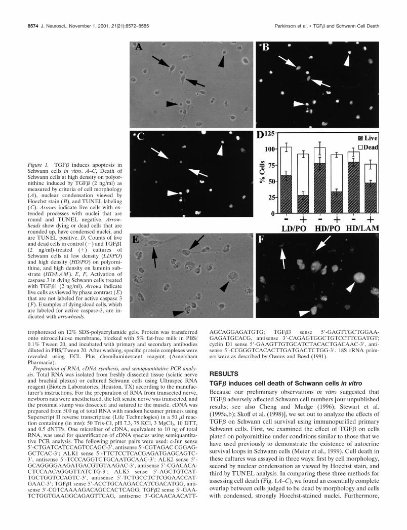
Figure 1. TGF� induces apoptosis inSchwann cells in vitro. A–C, Death ofSchwann cells at high density on polyor-nithine induced by TGF� (2 ng/ml) asmeasured by criteria of cell morphology(A), nuclear condensation viewed byHoechst stain (B), and TUNEL labeling(C). Arrows indicate live cells with ex-tended processes with nuclei that areround and TUNEL negative. Arrow-heads show dying or dead cells that arerounded up, have condensed nuclei, andare TUNEL positive. D, Counts of liveand dead cells in control (�) and TGF�1(2 ng/ml)-treated (�) cultures ofSchwann cells at low density (LD/PO)and high density (HD/PO) on polyorni-thine, and high density on laminin sub-strate (HD/LAM ). E, F, Activation ofcaspase 3 in dying Schwann cells treatedwith TGF�1 (2 ng/ml). Arrows indicatelive cells as viewed by phase contrast ( E)that are not labeled for active caspase 3(F). Examples of dying/dead cells, whichare labeled for active caspase-3, are in-dicated with arrowheads.
8574 J. Neurosci., November 1, 2001, 21(21):8572–8585 Parkinson et al. • TGF� and Schwann Cell Death
combined Hoechst /TUNEL analysis showed that all cells consid-ered to be alive on the basis of Hoechst staining were TUNELnegative, whereas �95% of the cells judged to be dead byHoechst staining were TUNEL positive. In control experimentsfor the TUNEL analysis in which the terminal transferase en-zyme was omitted from the reaction, no labeling of nuclei wasobserved (data not shown). Thus all three methods for assessingSchwann cell death gave similar results. Hoechst staining/mor-phological analysis was used in most of the following experi-ments, whereas in a number of instances, TUNEL staining oractive caspase-3 immunolabeling was used as well. These areindicated in the text.
In an initial set of experiments, immunopurified Schwann cellsfrom the sciatic nerve of newborn rats were exposed to TGF� (2ng/ml) for 1 d under three experimental conditions: high (3000cells per coverslip) density and low (300 cells per coverslip)density on a polyornithine substrate and high density on a lamininsubstrate. High and low cell density cultures were comparedbecause Schwann cell survival in vitro is subject to autocrine,density-dependent regulation, and polyornithine and lamininwere compared because laminin can support Schwann cell sur-vival (Meier et al., 1999). At the end of the experiment, theTGF�-treated cells and control untreated cultures were fixed,Hoechst stained, and TUNEL labeled. In addition, sister cover-slips were fixed and immunolabeled with antibodies to activecaspase-3, because caspase-3 activation is associated with apopto-tic cell death (Nicholson and Thornberry, 1997; Cryns and Yuan,1998). Under all three conditions TGF� caused significantSchwann cell death (Fig. 1D). On polyornithine, �90% of thecells could be accounted for throughout the assay because eventhe dead cells remained attached to the coverslip at the end of theexperiment, as seen previously (Meier et al., 1999). Because ofthis it could be seen unambiguously that TGF� killed Schwanncells, rather than causing living cells to detach from the coverslips.Even on laminin substrate, although there was slightly more lossof cells from the coverslips with TGF� treatment (24%), analysisof the supernatant of the tissue culture revealed cells with con-densed nuclei or just cellular debris, thereby excluding the possi-bility that TGF�1 was causing a significant number of live cells todetach under these conditions. To further characterize the cell
death caused by TGF�, we immunolabeled Schwann cells with anantibody specific for the active cleaved form of caspase-3. Weobserved that �80% of cells judged to be dead by morphologicalcriteria in TGF�-treated cultures were labeled for active caspase3 (Fig. 1E,F), indicating that TGF� is causing apoptotic death ofSchwann cells. Having shown that TGF� will cause cell death inSchwann cells, we next performed a series of experiments tofurther characterize this effect and the relationship betweenTGF� and positive survival signals for Schwann cells.
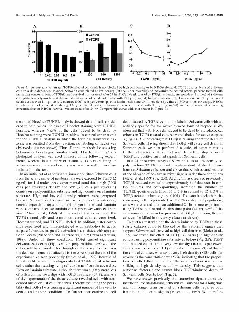
In a 24 hr survival assay of Schwann cells at low density onpolyornithine, TGF�1 induced dose-dependent cell death in new-born rat Schwann cells over and above that which occurs becauseof the absence of positive survival signals under these conditions(Meier et al., 1999) (Fig. 2A). At 2 ng/ml, as observed previously,TGF�1 reduced survival to approximately half that seen in con-trol cultures and correspondingly increased the number ofTUNEL-positive cells (from 35 � 7% in control to 62 � 3% inTGF�-treated cultures; p � 0.001). To examine whether theremaining cells represented a TGF�-resistant subpopulation,cells were counted after an additional 24 hr in one experimentusing TGF�1 at 5 ng/ml. At this time point (48 hr) �2% of thecells remained alive in the presence of TGF�, indicating that allcells can be killed in this assay (data not shown).
To further test whether the death induced by TGF� in thesesparse cultures could be blocked by the autocrine signals thatsupport Schwann cell survival at high cell densities (Meier et al.,1999), we tested the effect of TGF�1 (2 ng/ml) in high-densitycultures using polyornithine substrate as before (Fig. 2B). TGF�still induced cell death: at very low density (100 cells per cover-slip), survival of cells in TGF�-treated cultures was 59% of that inthe control cultures, whereas at very high density (8100 cells percoverslip) the same statistic was 57%, indicating that the propor-tion of cells killed in the TGF�1-treated cultures was just asstriking at high density as at low density. This suggests thatautocrine factors alone cannot block TGF�-induced death ofSchwann cells (see below) (Fig. 3).
We have shown previously that autocrine signals alone areinsufficient for maintaining Schwann cell survival for a long timeand that longer term survival of Schwann cells requires bothlaminin and autocrine signals (Meier et al., 1999). We therefore
Figure 2. In vitro survival assays. TGF�-induced cell death is not blocked by high cell density or by NRG� alone. A, TGF�1 causes death of Schwanncells in a dose-dependent manner. Schwann cells plated at low density (300 cells per coverslip) on polyornithine-coated coverslips were treated withincreasing concentrations of TGF�1, and survival was assessed after 24 hr. B, Cell death caused by TGF�1 is density independent. Survival of Schwanncells plated on polyornithine at different densities as indicated and treated with TGF�1 (2 ng/ml) for 24 hr is shown. C, Dose-dependent TGF�1-induceddeath occurs even in high-density cultures (3000 cells per coverslip) on a laminin substrate. D, In low-density cultures (300 cells per coverslip), NRG�is relatively ineffective at inhibiting TGF�1-induced death. Schwann cells were treated with TGF�1 (2 ng/ml) in the presence of increasingconcentrations of NRG�; survival was assessed after 24 hr. Compare this curve with that shown in Figure 3A.
Parkinson et al. • TGF� and Schwann Cell Death J. Neurosci., November 1, 2001, 21(21):8572–8585 8575
tested the effect of TGF� on Schwann cells plated at high densityon a laminin substrate (Fig. 2C). TGF�1 was equally effective ininducing death in these cultures after 24 hr as in cultures platedon polyornithine. In cell death assays of Schwann cells on lamininsubstrate treated with TGF�1, a higher proportion of dead cells(24% for 2 ng/ml TGF�1 after 24 hr) detached from coverslips.Analysis of cells still attached to the coverslip also revealedincreased apoptosis with TGF�1 treatment as measured by com-bined Hoechst /TUNEL staining (data not shown).
In addition to autocrine signals, NRG�s are likely to be a majorregulator of the survival of developing Schwann cells in vivo, andthey act as potent survival factors for cultured Schwann cells(Grinspan et al., 1996; Syroid et al., 1996; Trachtenberg andThompson, 1996). We therefore tested whether NRG� alone(i.e., in low-density cultures and the consequent absence of auto-crine signals) could block the death-promoting effects of TGF�.Sparse cultures were prepared on either polyornithine- orlaminin-coated coverslips and exposed to 2 ng/ml TGF�1 andvarying concentrations of NRG�. Under these conditions,NRG� was relatively ineffective at promoting Schwann cell sur-vival except at very high doses (Fig. 2D, compare with 3A).
TGF�1 and TGF�2 were equipotent in their ability to induceSchwann cell death, consistent with their indistinguishable effectsobserved in other in vitro assays (Ten Dijke et al., 1990). At 2ng/ml, a dose used frequently in this study, Schwann cell survivalaveraged 25 � 1.29% for TGF�1 (n 6) and 28 � 3.77% forTGF�2 (n 3) in an assay identical to that described in Figure2A. Each isoform was reconstituted in an acid–BSA mix. WhenTGF� was omitted from this mix and cells were treated with theappropriate volume of carrier solution, the percentage survivalwas not reduced from the level observed in control cultures,indicating that TGF� is the active killing ingredient. AlthoughSchwann cells in culture make TGF�, this is secreted in aninactive from (Stewart et al., 1995b). It is also important to notethat expansion of newborn Schwann cells with cAMP elevatingagents and growth factors is accompanied by a reduction inTGF�-mediated cell death (data not shown).
Together these experiments show that TGF� kills primarySchwann cells from nerves of newborn rats under various condi-tions in culture. Individually, neither of the two major signalslikely to totally regulate the survival of these cells in vivo, NRG�or autocrine signals, prevents TGF�-induced death.
TGF�-induced cell death is blocked by a combinationof NRG� and autocrine signalsHaving shown that neither NRG� nor autocrine survival signalscould completely prevent TGF�-induced death when present
separately (above), we now tested, in three different ways,whether the combination of these survival factors could block theeffect of TGF�.
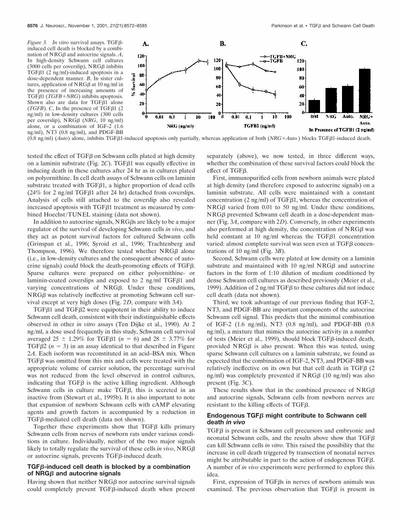
First, immunopurified cells from newborn animals were platedat high density (and therefore exposed to autocrine signals) on alaminin substrate. All cells were maintained with a constantconcentration (2 ng/ml) of TGF�1, whereas the concentration ofNRG� varied from 0.01 to 50 ng/ml. Under these conditions,NRG� prevented Schwann cell death in a dose-dependent man-ner (Fig. 3A, compare with 2D). Conversely, in other experimentsalso performed at high density, the concentration of NRG� washeld constant at 10 ng/ml whereas the TGF�1 concentrationvaried: almost complete survival was seen even at TGF� concen-trations of 10 ng/ml (Fig. 3B).
Second, Schwann cells were plated at low density on a lamininsubstrate and maintained with 10 ng/ml NRG� and autocrinefactors in the form of 1:10 dilution of medium conditioned bydense Schwann cell cultures as described previously (Meier et al.,1999). Addition of 2 ng/ml TGF� to these cultures did not inducecell death (data not shown).
Third, we took advantage of our previous finding that IGF-2,NT3, and PDGF-BB are important components of the autocrineSchwann cell signal. This predicts that the minimal combinationof IGF-2 (1.6 ng/ml), NT3 (0.8 ng/ml), and PDGF-BB (0.8ng/ml), a mixture that mimics the autocrine activity in a numberof tests (Meier et al., 1999), should block TGF�-induced death,provided NRG� is also present. When this was tested, usingsparse Schwann cell cultures on a laminin substrate, we found asexpected that the combination of IGF-2, NT3, and PDGF-BB wasrelatively ineffective on its own but that cell death in TGF� (2ng/ml) was completely prevented if NRG� (10 ng/ml) was alsopresent (Fig. 3C).
These results show that in the combined presence of NRG�and autocrine signals, Schwann cells from newborn nerves areresistant to the killing effects of TGF�.
Endogenous TGF� might contribute to Schwann celldeath in vivoTGF� is present in Schwann cell precursors and embryonic andneonatal Schwann cells, and the results above show that TGF�can kill Schwann cells in vitro. This raised the possibility that theincrease in cell death triggered by transection of neonatal nervesmight be attributable in part to the action of endogenous TGF�.A number of in vivo experiments were performed to explore thisidea.
First, expression of TGF�s in nerves of newborn animals wasexamined. The previous observation that TGF� is present in
Figure 3. In vitro survival assays. TGF�-induced cell death is blocked by a combi-nation of NRG� and autocrine signals. A,In high-density Schwann cell cultures(3000 cells per coverslip), NRG� inhibitsTGF�1 (2 ng/ml)-induced apoptosis in adose-dependent manner. B, In sister cul-tures, application of NRG� at 10 ng/ml inthe presence of increasing amounts ofTGF�1 (TGFB�NRG) inhibits apoptosis.Shown also are data for TGF�1 alone(TGFB). C, In the presence of TGF�1 (2ng/ml) in low-density cultures (300 cellsper coverslip), NRG� (NRG, 10 ng/ml)alone, or a combination of IGF-2 (1.6ng/ml), NT3 (0.8 ng/ml), and PDGF-BB(0.8 ng/ml) (Auto) alone, inhibits TGF�1-induced apoptosis only partially, whereas application of both (NRG�Auto.) blocks TGF�1-induced death.
8576 J. Neurosci., November 1, 2001, 21(21):8572–8585 Parkinson et al. • TGF� and Schwann Cell Death
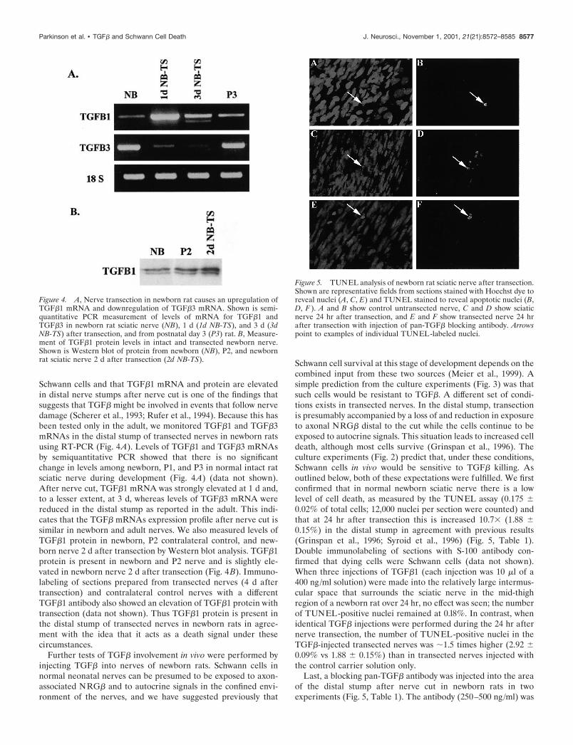
Schwann cells and that TGF�1 mRNA and protein are elevatedin distal nerve stumps after nerve cut is one of the findings thatsuggests that TGF� might be involved in events that follow nervedamage (Scherer et al., 1993; Rufer et al., 1994). Because this hasbeen tested only in the adult, we monitored TGF�1 and TGF�3mRNAs in the distal stump of transected nerves in newborn ratsusing RT-PCR (Fig. 4A). Levels of TGF�1 and TGF�3 mRNAsby semiquantitative PCR showed that there is no significantchange in levels among newborn, P1, and P3 in normal intact ratsciatic nerve during development (Fig. 4A) (data not shown).After nerve cut, TGF�1 mRNA was strongly elevated at 1 d and,to a lesser extent, at 3 d, whereas levels of TGF�3 mRNA werereduced in the distal stump as reported in the adult. This indi-cates that the TGF� mRNAs expression profile after nerve cut issimilar in newborn and adult nerves. We also measured levels ofTGF�1 protein in newborn, P2 contralateral control, and new-born nerve 2 d after transection by Western blot analysis. TGF�1protein is present in newborn and P2 nerve and is slightly ele-vated in newborn nerve 2 d after transection (Fig. 4B). Immuno-labeling of sections prepared from transected nerves (4 d aftertransection) and contralateral control nerves with a differentTGF�1 antibody also showed an elevation of TGF�1 protein withtransection (data not shown). Thus TGF�1 protein is present inthe distal stump of transected nerves in newborn rats in agree-ment with the idea that it acts as a death signal under thesecircumstances.
Further tests of TGF� involvement in vivo were performed byinjecting TGF� into nerves of newborn rats. Schwann cells innormal neonatal nerves can be presumed to be exposed to axon-associated NRG� and to autocrine signals in the confined envi-ronment of the nerves, and we have suggested previously that
Schwann cell survival at this stage of development depends on thecombined input from these two sources (Meier et al., 1999). Asimple prediction from the culture experiments (Fig. 3) was thatsuch cells would be resistant to TGF�. A different set of condi-tions exists in transected nerves. In the distal stump, transectionis presumably accompanied by a loss of and reduction in exposureto axonal NRG� distal to the cut while the cells continue to beexposed to autocrine signals. This situation leads to increased celldeath, although most cells survive (Grinspan et al., 1996). Theculture experiments (Fig. 2) predict that, under these conditions,Schwann cells in vivo would be sensitive to TGF� killing. Asoutlined below, both of these expectations were fulfilled. We firstconfirmed that in normal newborn sciatic nerve there is a lowlevel of cell death, as measured by the TUNEL assay (0.175 �0.02% of total cells; 12,000 nuclei per section were counted) andthat at 24 hr after transection this is increased 10.7 (1.88 �0.15%) in the distal stump in agreement with previous results(Grinspan et al., 1996; Syroid et al., 1996) (Fig. 5, Table 1).Double immunolabeling of sections with S-100 antibody con-firmed that dying cells were Schwann cells (data not shown).When three injections of TGF�1 (each injection was 10 �l of a400 ng/ml solution) were made into the relatively large intermus-cular space that surrounds the sciatic nerve in the mid-thighregion of a newborn rat over 24 hr, no effect was seen; the numberof TUNEL-positive nuclei remained at 0.l8%. In contrast, whenidentical TGF� injections were performed during the 24 hr afternerve transection, the number of TUNEL-positive nuclei in theTGF�-injected transected nerves was �1.5 times higher (2.92 �0.09% vs 1.88 � 0.15%) than in transected nerves injected withthe control carrier solution only.
Last, a blocking pan-TGF� antibody was injected into the areaof the distal stump after nerve cut in newborn rats in twoexperiments (Fig. 5, Table 1). The antibody (250–500 ng/ml) was
Figure 4. A, Nerve transection in newborn rat causes an upregulation ofTGF�1 mRNA and downregulation of TGF�3 mRNA. Shown is semi-quantitative PCR measurement of levels of mRNA for TGF�1 andTGF�3 in newborn rat sciatic nerve (NB), 1 d (1d NB-TS), and 3 d (3dNB-TS) after transection, and from postnatal day 3 (P3) rat. B, Measure-ment of TGF�1 protein levels in intact and transected newborn nerve.Shown is Western blot of protein from newborn (NB), P2, and newbornrat sciatic nerve 2 d after transection (2d NB-TS).
Figure 5. TUNEL analysis of newborn rat sciatic nerve after transection.Shown are representative fields from sections stained with Hoechst dye toreveal nuclei (A, C, E) and TUNEL stained to reveal apoptotic nuclei (B,D, F ). A and B show control untransected nerve, C and D show sciaticnerve 24 hr after transection, and E and F show transected nerve 24 hrafter transection with injection of pan-TGF� blocking antibody. Arrowspoint to examples of individual TUNEL-labeled nuclei.
Parkinson et al. • TGF� and Schwann Cell Death J. Neurosci., November 1, 2001, 21(21):8572–8585 8577
injected three times during a 24 hr period after nerve cut asdescribed above. Averaging the two experiments (two rats in eachexperiment) reveals that the blocking antibody reduced the num-ber of apoptotic nuclei in the nerve by 62%. Identical injections ofa control antibody SM 1.2 (280 ng/ml; see Materials and Meth-ods) resulted in an insignificant (5%) reduction in the number ofapoptotic nuclei (Table 1).
All of the above data are consistent with the possibility thatSchwann cell death in transected neonatal nerves is not causedsolely by loss of axon-associated NRG� but is caused also byTGF� present in the nerve.
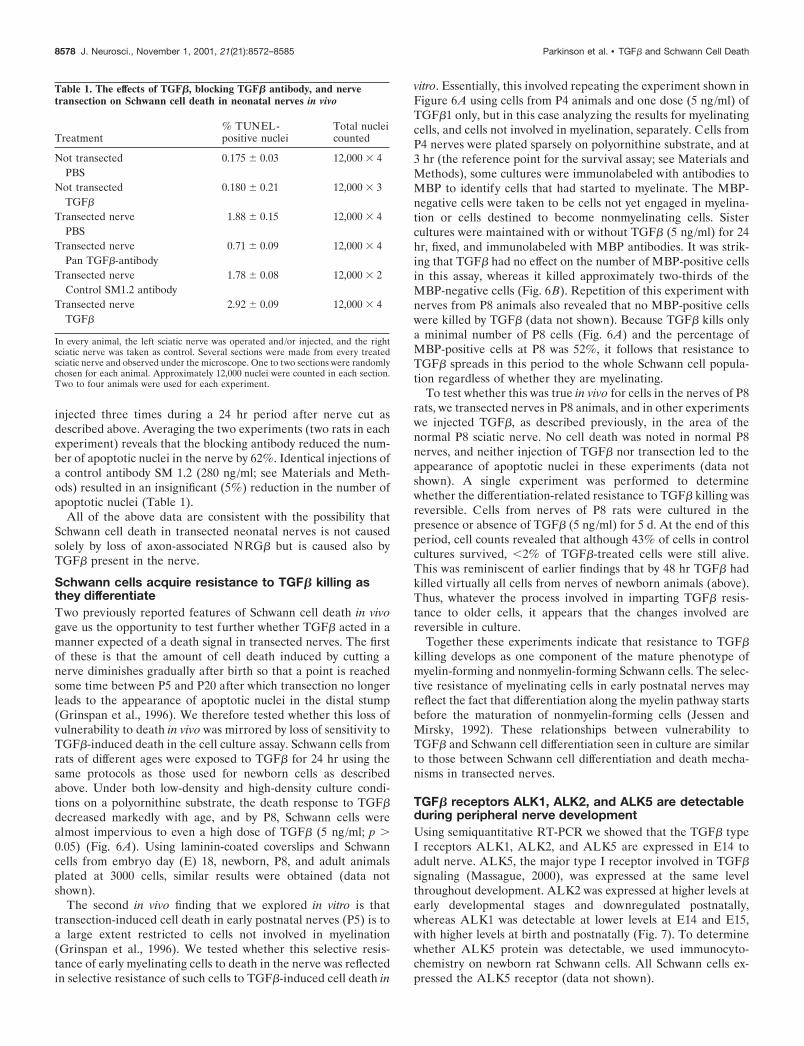
Schwann cells acquire resistance to TGF� killing asthey differentiateTwo previously reported features of Schwann cell death in vivogave us the opportunity to test further whether TGF� acted in amanner expected of a death signal in transected nerves. The firstof these is that the amount of cell death induced by cutting anerve diminishes gradually after birth so that a point is reachedsome time between P5 and P20 after which transection no longerleads to the appearance of apoptotic nuclei in the distal stump(Grinspan et al., 1996). We therefore tested whether this loss ofvulnerability to death in vivo was mirrored by loss of sensitivity toTGF�-induced death in the cell culture assay. Schwann cells fromrats of different ages were exposed to TGF� for 24 hr using thesame protocols as those used for newborn cells as describedabove. Under both low-density and high-density culture condi-tions on a polyornithine substrate, the death response to TGF�decreased markedly with age, and by P8, Schwann cells werealmost impervious to even a high dose of TGF� (5 ng/ml; p �0.05) (Fig. 6A). Using laminin-coated coverslips and Schwanncells from embryo day (E) 18, newborn, P8, and adult animalsplated at 3000 cells, similar results were obtained (data notshown).
The second in vivo finding that we explored in vitro is thattransection-induced cell death in early postnatal nerves (P5) is toa large extent restricted to cells not involved in myelination(Grinspan et al., 1996). We tested whether this selective resis-tance of early myelinating cells to death in the nerve was reflectedin selective resistance of such cells to TGF�-induced cell death in
vitro. Essentially, this involved repeating the experiment shown inFigure 6A using cells from P4 animals and one dose (5 ng/ml) ofTGF�1 only, but in this case analyzing the results for myelinatingcells, and cells not involved in myelination, separately. Cells fromP4 nerves were plated sparsely on polyornithine substrate, and at3 hr (the reference point for the survival assay; see Materials andMethods), some cultures were immunolabeled with antibodies toMBP to identify cells that had started to myelinate. The MBP-negative cells were taken to be cells not yet engaged in myelina-tion or cells destined to become nonmyelinating cells. Sistercultures were maintained with or without TGF� (5 ng/ml) for 24hr, fixed, and immunolabeled with MBP antibodies. It was strik-ing that TGF� had no effect on the number of MBP-positive cellsin this assay, whereas it killed approximately two-thirds of theMBP-negative cells (Fig. 6B). Repetition of this experiment withnerves from P8 animals also revealed that no MBP-positive cellswere killed by TGF� (data not shown). Because TGF� kills onlya minimal number of P8 cells (Fig. 6A) and the percentage ofMBP-positive cells at P8 was 52%, it follows that resistance toTGF� spreads in this period to the whole Schwann cell popula-tion regardless of whether they are myelinating.
To test whether this was true in vivo for cells in the nerves of P8rats, we transected nerves in P8 animals, and in other experimentswe injected TGF�, as described previously, in the area of thenormal P8 sciatic nerve. No cell death was noted in normal P8nerves, and neither injection of TGF� nor transection led to theappearance of apoptotic nuclei in these experiments (data notshown). A single experiment was performed to determinewhether the differentiation-related resistance to TGF� killing wasreversible. Cells from nerves of P8 rats were cultured in thepresence or absence of TGF� (5 ng/ml) for 5 d. At the end of thisperiod, cell counts revealed that although 43% of cells in controlcultures survived, �2% of TGF�-treated cells were still alive.This was reminiscent of earlier findings that by 48 hr TGF� hadkilled virtually all cells from nerves of newborn animals (above).Thus, whatever the process involved in imparting TGF� resis-tance to older cells, it appears that the changes involved arereversible in culture.
Together these experiments indicate that resistance to TGF�killing develops as one component of the mature phenotype ofmyelin-forming and nonmyelin-forming Schwann cells. The selec-tive resistance of myelinating cells in early postnatal nerves mayreflect the fact that differentiation along the myelin pathway startsbefore the maturation of nonmyelin-forming cells (Jessen andMirsky, 1992). These relationships between vulnerability toTGF� and Schwann cell differentiation seen in culture are similarto those between Schwann cell differentiation and death mecha-nisms in transected nerves.
TGF� receptors ALK1, ALK2, and ALK5 are detectableduring peripheral nerve developmentUsing semiquantitative RT-PCR we showed that the TGF� typeI receptors ALK1, ALK2, and ALK5 are expressed in E14 toadult nerve. ALK5, the major type I receptor involved in TGF�signaling (Massague, 2000), was expressed at the same levelthroughout development. ALK2 was expressed at higher levels atearly developmental stages and downregulated postnatally,whereas ALK1 was detectable at lower levels at E14 and E15,with higher levels at birth and postnatally (Fig. 7). To determinewhether ALK5 protein was detectable, we used immunocyto-chemistry on newborn rat Schwann cells. All Schwann cells ex-pressed the ALK5 receptor (data not shown).
Table 1. The effects of TGF�, blocking TGF� antibody, and nervetransection on Schwann cell death in neonatal nerves in vivo
Treatment% TUNEL-positive nuclei
Total nucleicounted
Not transected 0.175 � 0.03 12,000 4PBS
Not transected 0.180 � 0.21 12,000 3TGF�
Transected nerve 1.88 � 0.15 12,000 4PBS
Transected nerve 0.71 � 0.09 12,000 4Pan TGF�-antibody
Transected nerve 1.78 � 0.08 12,000 2Control SM1.2 antibody
Transected nerve 2.92 � 0.09 12,000 4TGF�
In every animal, the left sciatic nerve was operated and/or injected, and the rightsciatic nerve was taken as control. Several sections were made from every treatedsciatic nerve and observed under the microscope. One to two sections were randomlychosen for each animal. Approximately 12,000 nuclei were counted in each section.Two to four animals were used for each experiment.
8578 J. Neurosci., November 1, 2001, 21(21):8572–8585 Parkinson et al. • TGF� and Schwann Cell Death
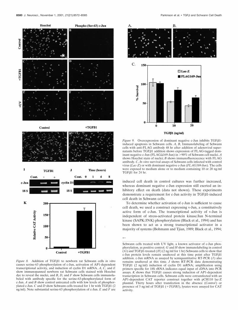
Treatment of Schwann cells with TGF�1 causesserine-63 phosphorylation of c-Jun and activation ofAP1-dependent transcriptionWe now examined the intracellular mechanisms by which TGF�induces cell death. Activation of the transcription factor c-Junand induction of AP1-dependent transcription has been shown tobe involved in apoptosis of several cell types, including fibroblasts(Bossy-Wetzel et al., 1997) and sympathetic neurons (Ham et al.,1995). Activation of c-Jun and AP1-dependent transcription re-quires phosphorylation of c-Jun on two N-terminal serine resi-dues, ser-63 and -73 (Smeal et al., 1991). We therefore examinedwhether activation of c-Jun occurred in response to application ofTGF�1 to freshly isolated Schwann cells using an antibody that isspecific for c-Jun when phosphorylated on the serine-63 residue(Watson et al., 1998). Schwann cells were plated at high densityon laminin-coated glass coverslips in supplemented defined me-dium (see Materials and Methods). As a positive control, identi-cal cultures were exposed to ultraviolet (UV) light, a procedurethat has been shown previously to result in phosphorylation andactivation of c-Jun (Devary et al., 1991; Derijard et al., 1994). Onehour after addition of TGF�1 (2 ng/ml), strong nuclear labelingwas seen (Fig. 8D), indicating phosphorylation of c-Jun at serine-63. Similar labeling was seen 1 hr after UV irradiation (Fig. 8F).To determine whether levels of c-Jun protein and mRNA wereunchanged during this time, we immunolabeled Schwann cellstreated with TGF�1 for 1 hr with antibodies to c-Jun. Levels ofc-Jun were unchanged during this time in response to TGF�1treatment using several different serial dilutions of antibody (Fig.
8G,H) (data not shown). These results were confirmed at themRNA level by semiquantitative RT-PCR of TGF�1-treated andcontrol cells (Fig. 8 I); however, induction of c-Jun mRNA wasobserved 8 hr after TGF�1 addition (data not shown), consistentwith the previously reported induction of c-Jun itself by activatedAP1 complexes (Stein et al., 1992; van Dam et al., 1995; Eilers etal., 1998). In support of activation of c-Jun in Schwann cells, wefound that TGF�1 caused an increase in cyclin D1 mRNA (Fig.8J), a known transcriptional target for c-Jun-dependent transcrip-tion (Albanese et al., 1995; Bakiri et al., 2000).
To test functionally whether TGF�1 activated c-Jun and stim-ulated the corresponding AP1-dependent transcriptional activity,we performed transient transfections into Schwann cells with anAP1-responsive CAT reporter construct (Bossy-Wetzel et al.,1997). Figure 8K shows that addition of TGF�1 to Schwann cellsresulted in a massive increase (�23-fold) in AP1-dependenttranscription in Schwann cells, further demonstrating the linkbetween TGF�1 and activation of c-Jun/AP1 in Schwann cells.
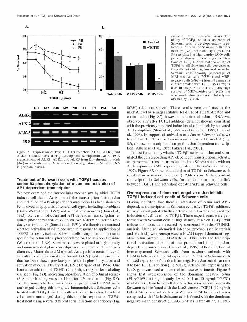
Overexpression of dominant negative c-Jun inhibitsTGF�1-induced cell death of Schwann cellsHaving identified that there is activation of c-Jun and AP1-dependent transcription in Schwann cells after TGF�1 addition,we next investigated whether activity of c-Jun was required forinduction of cell death by TGF�1. These experiments were per-formed with Schwann cells at high density at which TGF�1 willinduce apoptosis as measured by combined Hoechst /TUNELanalysis. Using an adenoviral infection protocol (see Materialsand Methods) we overexpressed a FLAG-tagged dominant neg-ative c-Jun protein, FLAG�169-Jun. This lacks the transcrip-tional activation domain of the protein and inhibits c-Jun-dependent transcription (Ham et al., 1995). After infection ofimmunopanned Schwann cells from newborn animals withFLAG�169-Jun adenoviral supernatant, �90% of Schwann cellsshowed expression of the dominant negative c-Jun protein at time0 before TGF�1 addition (Fig. 9A,B). Adenovirus expressing theLacZ gene was used as a control in these experiments. Figure 9shows that overexpression of the dominant negative c-Jun(FLAG169-Jun) significantly ( p � 0.01 at 10 ng/ml TGF�1)inhibits TGF�1-induced cell death in this assay as compared withSchwann cells infected with the LacZ control. TGF�1 (10 ng/ml)kills 46% of control cells (LacZ) over a 24 hr period whencompared with 15% in Schwann cells infected with the dominantnegative c-Jun construct (FLAG169-Jun). After 48 hr, TGF�1-
Figure 6. In vitro survival assays. Theability of TGF�1 to cause apoptosis ofSchwann cells is developmentally regu-lated. A, Survival of Schwann cells fromnewborn (NB), postnatal day 4 (P4 ), andP8 rats plated at high density (3000 cellsper coverslip) with increasing concentra-tions of TGF�1. Note that the ability ofTGF� to kill Schwann cells decreases asthe cells get older. B, Survival assay forSchwann cells showing percentage ofMBP-positive cells (MBP�) and MBP-negative cells (MBP�) from P4 animals incultures treated with TGF�1 (5 ng/ml) ina 24 hr assay. Note that the percentagesurvival of MBP-positive cells (cells thatwere myelinating in vivo) is relatively un-affected by TGF�1.
Figure 7. Expression of type I TGF� receptors ALK1, ALK2, andALK5 in sciatic nerve during development. Semiquantitative RT-PCRmeasurement of ALK1, ALK2, and ALK5 from E14 through to adult(Ad.) in rat sciatic nerve. Note marked downregulation of ALK2 mRNAin postnatal nerves.
Parkinson et al. • TGF� and Schwann Cell Death J. Neurosci., November 1, 2001, 21(21):8572–8585 8579
induced cell death in control cultures was further increased,whereas dominant negative c-Jun expression still exerted an in-hibitory effect on death (data not shown). These experimentsdemonstrate a requirement for c-Jun activity in TGF�1-inducedcell death in Schwann cells.
To determine whether activation of c-Jun is sufficient to causecell death, we used a construct expressing v-Jun, a constitutivelyactive form of c-Jun. The transcriptional activity of v-Jun isindependent of stress-activated protein kinase/Jun N-terminalkinase (SAPK/JNK) phosphorylation (Black et al., 1994) and hasbeen shown to act as a strong transcriptional activator in amajority of systems (Bohmann and Tjian, 1989; Black et al., 1994;
Figure 8. Addition of TGF�1 to newborn rat Schwann cells in vitrocauses serine-63 phosphorylation of c-Jun, activation of AP1-dependenttranscriptional activity, and induction of cyclin D1 mRNA. A, C, and Eshow immunopanned newborn rat Schwann cells stained with Hoechstdye to reveal the nuclei, and B, D, and F show Schwann cells immunola-beled with antibody specific for the serine-63-phosphorylated form ofc-Jun. A and B show control untreated cells with low levels of phosphor-ylated c-Jun. C and D show Schwann cells treated for 1 hr with TGF�1 (2ng/ml). Note substantial serine-63 phosphorylation of c-Jun. E and F are
4
Schwann cells treated with UV light, a known activator of c-Jun phos-phorylation, as positive control. G and H show immunolabeling in control(G) and TGF�1-treated (H ) (2 ng/ml for 1 hr) Schwann cells to show thatc-Jun protein levels remain unaltered at this time point after TGF�1addition. c-Jun mRNA as assayed by semiquantitative RT-PCR ( I ) alsoremains unaltered at this time. J shows RT-PCR data demonstratingTGF�1 (2 ng/ml) induction of cyclin D1 mRNA; amplification usingprimers specific for 18S rRNA indicates equal input of cDNA into PCRassays. K shows that TGF�1 causes strong induction of AP1-dependenttranscription in Schwann cells. Schwann cells were cotransfected with anAP1-dependent CAT reporter construct together with pCH110 lac-Zplasmid. Thirty hours after transfection in the absence (Control ) orpresence of 5 ng/ml of TGF�1 (�TGFB1), lysates were assayed for CATactivity.
Figure 9. Overexpression of dominant negative c-Jun inhibits TGF�1-induced apoptosis in Schwann cells. A, B, Immunolabeling of Schwanncells with anti-FLAG antibody 48 hr after addition of adenoviral super-natants before TGF�1 addition shows expression of FLAG-tagged dom-inant negative c-Jun (FLAG�169-Jun) in �90% of Schwann cell nuclei. Ashows Hoechst stain of nuclei; B shows immunofluorescence with FLAGantibody. C, In vitro survival assays of Schwann cells infected with controlvirus (Lac-Z) or with dominant negative c-Jun (FLAG169-Jun). The cellswere exposed to medium alone or to medium containing 10 or 20 ng/mlTGF�1 for 24 hr.
8580 J. Neurosci., November 1, 2001, 21(21):8572–8585 Parkinson et al. • TGF� and Schwann Cell Death
Hartl and Bister, 1998; Huguier et al., 1998; Bader et al., 2000)(see however Gao et al., 1996; Kilbey et al., 1996). We found thatwhen Schwann cells expressing v-Jun were cultured at high den-sity in serum-free supplemented defined medium, the amount ofcell death was strongly increased compared with control cellsinfected with vector alone (Fig. 10). Combined TUNEL/Hoechstanalysis of these cultures, together with labeling with CM1 anti-body for active caspase 3, established that cells in these culturesare dying by apoptosis (data not shown).
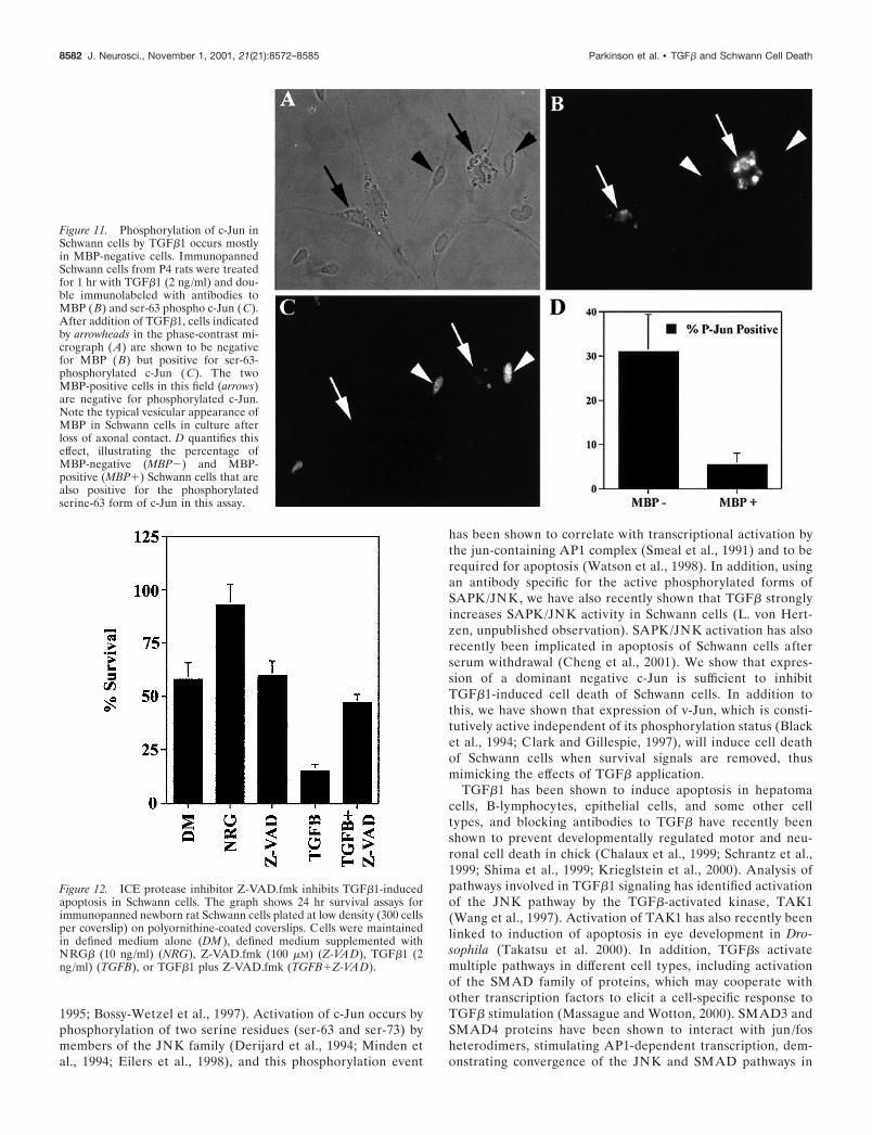
TGF� activation of c-Jun occurs predominantly inMBP-negative cellsThe previous experiment provided evidence that TGF� killsSchwann cells from newborn nerves by activating the c-Jun path-way. We now tested whether a failure to activate this pathwaycould provide an explanation for the failure of TGF� to kill moredifferentiated Schwann cells.
Immunopanned rat Schwann cells from P4 animals were platedat high density on laminin-coated coverslips in supplementeddefined medium. As discussed in a previous section, approxi-mately half of the Schwann cells in these cultures have startedmyelination as evidenced by expression of MBP, whereas theother half is less differentiated. One hour after TGF�1 (2 ng/ml)addition, the cells were immunolabeled with antibodies againstMBP and serine-63 phospho c-Jun. Figure 11 shows that TGF�1stimulated immunohistochemically detectable c-Jun phosphory-lation in 31 � 8.4% of MBP-negative cells but in only 5 � 2.6%of MBP-positive cells. Furthermore, the immunohistochemicallabeling was consistently stronger in the nuclei of the MBP-negative cells, indicating higher levels of phosphorylated c-Jun.This correlates well with our observation that MBP-positiveSchwann cells are relatively resistant to induction of cell death byTGF�1 and suggests that the inability of TGF� to kill differen-tiating Schwann cells can be explained in part by a failure toactivate c-Jun in these cells. Therefore, the molecular mechanismresponsible for the differentiation-related immunity to TGF�
killing may lie upstream of c-Jun activation.
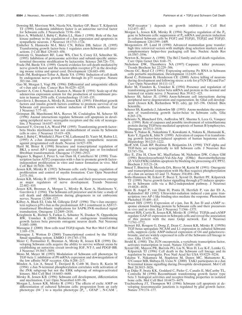
TGF�-activated death pathways involve theinterleukin-1�-converting enzyme-like protease pro-caspase 3The interleukin-1�-converting enzyme (ICE) protease inhibitorZ-VAD.fmk was used to investigate the role of ICE-like proteaseson Schwann cell death induced by TGF� and by the absence ofsurvival factors. This synthetic peptide inhibitor prevents othertypes of cell death by blocking irreversibly the activation of theICE-like protease, pro-caspase-3 (Chow et al., 1995; Zhu et al.,1995; Slee et al., 1996). It is not clear that Z-VAD.fmk is specificfor caspase-3 alone, and it might act similarly on the processing ofother ICE-like proteases. It was found that Z-VAD.fmk exerteddifferent effects on the two types of Schwann cell death: 100 �M
Z-VAD.fmk did not affect death of cells in medium alone,whereas its effects on the TGF�-mediated death were to increasesurvival by threefold, from 15 to 47% (Fig. 12). This effect wasdose dependent, with 50 �M Z-VAD.fmk increasing survivaltwofold (data not shown). We also tested the effects of oneproteasome inhibitor, lactacystin, a metabolite of streptomyces(Fenteany et al., 1995) on TGF�-induced cell death. LikeZ-VAD.fmk, lactacystin offered negligible protection against celldeath caused by the absence of survival factors, whereas it com-pletely blocked additional death induced by TGF� in a dose-dependent way (data not shown).
These results implicate ICE-like proteases, in particularcaspase-3, in the intracellular death pathways activated by TGF�in Schwann cells. They also suggest that these pathways are tosome extent different from those used when Schwann cells un-dergo cell death caused by the absence of survival-promotingfactors.
DISCUSSIONAlthough developmental and trauma-induced cell death in thenervous system has classically been considered to be regulated bypositive survival signals present in limiting amounts, it is apparentthat in some situations cell death is in fact controlled by aninterplay between survival factors and negative survival signalsthat actively induce cell death. The present work provides evi-dence that the survival of Schwann cells is in some circumstancesregulated by such a dual mechanism involving the negative sur-vival signal TGF�, a family of growth factors that is expressed bySchwann cells and secreted by purified Schwann cells in a signal-poor environment in vitro in an inactive form (Stewart et al.,1995b). We pinpoint phosphorylation of c-Jun as a key down-stream event in TGF�-induced Schwann cell death. We also showthat the ability of TGF� to kill Schwann cells, like normalSchwann cell death in vivo, is under strong developmental regu-lation and provide evidence that the decreasing ability of TGF�to kill older cells is caused by a decreasing ability of TGF� tophosphorylate c-Jun in more differentiated cells.
c-Jun is strongly expressed in cultured Schwann cells and inSchwann cells in the distal stump of transected nerves, whereasexpression levels in the cells of normal untransected newborn andadult nerves are low (De Felipe and Hunt, 1994; Stewart, 1995).We show here, using an antibody specific for the ser-63 phosphor-ylated and thus activated form, that TGF�1 activates c-Jun inSchwann cells. In confirmation of this, we find that TGF�1 willboth activate transcription of an AP1 reporter gene and upregu-late cyclin D1 mRNA, a known target of c-Jun [Wisdom et al.(1999) and references therein]. Activation of the c-Jun transcrip-tion factor is involved in cell death in a number of different celltypes, including sympathetic neurons and fibroblasts (Ham et al.,
Figure 10. Overexpression of v-Jun causes apoptosis of Schwann cells inserum-free medium. Schwann cells infected with either LexA control(Control ) or LexA-vJun (v-Jun) were plated at high density (5000 cells percoverslip). The medium of the cells was then changed to supplementeddefined medium, and the number of surviving cells was counted after 48 hr.
Parkinson et al. • TGF� and Schwann Cell Death J. Neurosci., November 1, 2001, 21(21):8572–8585 8581
1995; Bossy-Wetzel et al., 1997). Activation of c-Jun occurs byphosphorylation of two serine residues (ser-63 and ser-73) bymembers of the JNK family (Derijard et al., 1994; Minden etal., 1994; Eilers et al., 1998), and this phosphorylation event
has been shown to correlate with transcriptional activation bythe jun-containing AP1 complex (Smeal et al., 1991) and to berequired for apoptosis (Watson et al., 1998). In addition, usingan antibody specific for the active phosphorylated forms ofSAPK/JNK, we have also recently shown that TGF� stronglyincreases SAPK/JNK activity in Schwann cells (L. von Hert-zen, unpublished observation). SAPK/JNK activation has alsorecently been implicated in apoptosis of Schwann cells afterserum withdrawal (Cheng et al., 2001). We show that expres-sion of a dominant negative c-Jun is sufficient to inhibitTGF�1-induced cell death of Schwann cells. In addition tothis, we have shown that expression of v-Jun, which is consti-tutively active independent of its phosphorylation status (Blacket al., 1994; Clark and Gillespie, 1997), will induce cell deathof Schwann cells when survival signals are removed, thusmimicking the effects of TGF� application.
TGF�1 has been shown to induce apoptosis in hepatomacells, B-lymphocytes, epithelial cells, and some other celltypes, and blocking antibodies to TGF� have recently beenshown to prevent developmentally regulated motor and neu-ronal cell death in chick (Chalaux et al., 1999; Schrantz et al.,1999; Shima et al., 1999; Krieglstein et al., 2000). Analysis ofpathways involved in TGF�1 signaling has identified activationof the JNK pathway by the TGF�-activated kinase, TAK1(Wang et al., 1997). Activation of TAK1 has also recently beenlinked to induction of apoptosis in eye development in Dro-sophila (Takatsu et al. 2000). In addition, TGF�s activatemultiple pathways in different cell types, including activationof the SMAD family of proteins, which may cooperate withother transcription factors to elicit a cell-specific response toTGF� stimulation (Massague and Wotton, 2000). SMAD3 andSMAD4 proteins have been shown to interact with jun /fosheterodimers, stimulating AP1-dependent transcription, dem-onstrating convergence of the JNK and SMAD pathways in
Figure 11. Phosphorylation of c-Jun inSchwann cells by TGF�1 occurs mostlyin MBP-negative cells. ImmunopannedSchwann cells from P4 rats were treatedfor 1 hr with TGF�1 (2 ng/ml) and dou-ble immunolabeled with antibodies toMBP (B) and ser-63 phospho c-Jun (C).After addition of TGF�1, cells indicatedby arrowheads in the phase-contrast mi-crograph (A) are shown to be negativefor MBP (B) but positive for ser-63-phosphorylated c-Jun (C). The twoMBP-positive cells in this field (arrows)are negative for phosphorylated c-Jun.Note the typical vesicular appearance ofMBP in Schwann cells in culture afterloss of axonal contact. D quantifies thiseffect, illustrating the percentage ofMBP-negative (MBP�) and MBP-positive (MBP�) Schwann cells that arealso positive for the phosphorylatedserine-63 form of c-Jun in this assay.
Figure 12. ICE protease inhibitor Z-VAD.fmk inhibits TGF�1-inducedapoptosis in Schwann cells. The graph shows 24 hr survival assays forimmunopanned newborn rat Schwann cells plated at low density (300 cellsper coverslip) on polyornithine-coated coverslips. Cells were maintainedin defined medium alone (DM ), defined medium supplemented withNRG� (10 ng/ml) (NRG), Z-VAD.fmk (100 �M) (Z-VAD), TGF�1 (2ng/ml) (TGFB), or TGF�1 plus Z-VAD.fmk (TGFB�Z-VAD).
8582 J. Neurosci., November 1, 2001, 21(21):8572–8585 Parkinson et al. • TGF� and Schwann Cell Death

response to TGF� (Zhang et al., 1998). Indeed, treatment ofSchwann cells with TGF� causes nuclear localization ofSMAD4 (D. Parkinson, unpublished observation). The linkseen in the present experiments between TGF� stimulation,c-Jun phosphorylation, and cell death is of particular interest,because to our knowledge it has not been seen before in thesame cell type.
We find that TGF�-induced death is distinct from the celldeath observed after the withdrawal of survival signals such asNRG� or the autocrine survival mixture of IGF-2, NT3, andPDGF-BB (Grinspan et al., 1996; Syroid et al., 1996; Meier et al.,1999), because in Schwann cells the cell death induced by with-drawal of these growth factors is not inhibited by the generalcaspase inhibitor Z-VAD.fmk and apparently is not accompaniedby phosphorylation of c-Jun (C. Meier, unpublished observation).In contrast, the cell death induced by TGF�1 in Schwann cellsrequires the activity of caspases and is inhibited by the caspaseinhibitor Z-VAD.fmk, in keeping with previous findings ofcaspase activation in cells by TGF� (Schrantz et al., 1999; Shimaet al., 1999). TGF�-induced cell death is reduced but not pre-vented by autocrine survival signals or by NRG�, although incombination these signals allow survival of TGF�-treated cells.This requirement for a combination of survival signals may sug-gest a role for TGF�-mediated death during the embryonic andneonatal phase of Schwann cell development when autocrinesignals are less prominent than at later stages (Meier et al., 1999).TGF�1 has also been shown to reduce levels of NT3 mRNAexpression in Schwann cells (Cai et al., 1999). Because this is animportant component of the autocrine survival factors producedby Schwann cells, it may be an additional mechanism by whichTGF�1 induces cell death (Meier et al., 1999). The resistance ofSchwann cells to TGF�-induced killing as the nerve matures isparalleled by a failure to phosphorylate c-Jun in vitro. Neverthe-less, after some days in culture, presumably as they dedifferenti-ate, previously resistant cells become susceptible to TGF�-induced killing, suggesting that under some circumstances, evenin mature Schwann cells, TGF� could play a role in cell death,particularly in combination with other factors such as TNF-�(Skoff et al., 1998).
Induction of apoptosis is often related to regulation of theBcl-2 family of molecules, specifically an alteration in thebalance between pro- and anti-apoptotic members of thisgroup (Newton and Strasser, 1998). Upregulation of JNKactivity involved in apoptosis of Schwann cells after serumdeprivation is inhibited by Bcl-X(L) overexpression (Cheng etal., 2001). We have observed a transcriptional upregulation ofthe pro-apoptotic Bax mRNA by TGF�1 in Schwann cells, andfurthermore that Bax and p53 mRNAs are strongly downregu-lated during development in a manner that is inversely relatedto differentiation (data not shown). Regulation of such pro-apoptotic molecules may contribute to TGF�1-induced apo-ptosis in Schwann cells and the altering susceptibility of cells toapoptosis during development.
The present experiments argue that one of the functions ofTGF� in peripheral nerves is to take part in negative survivalregulation of developing Schwann cells. There is evidence thatNGF/p75 signaling acts in a comparable manner, whereas positivesurvival signals in developing nerves are likely to include NRG�,IGF-2, NT3, PDGF-BB, and LIF (for references, see introduc-tory remarks). In addition to taking part in this network of
survival-regulating signals, there is good evidence that TGF� iscapable of controlling Schwann cell proliferation and differenti-ation without necessarily inducing cell death (Mews and Meyer,1993; Morgan et al., 1994; Einheber et al., 1995; Guenard et al.,1995). This shows clearly that the effects of TGF� on Schwanncells are context dependent, a point illustrated in the presentwork in the interactions between TGF� and NRG� and auto-crine signals. It will be some time before we are in a position togenerate an integrated picture of the involvement of TGF� inSchwann cell biology.
REFERENCESAlbanese C, Johnson J, Watanabe G, Eklund N, Vu D, Arnold A, Pestell
RG (1995) Transforming p21ras mutants and c-Ets-2 activate the cy-clin D1 promoter through distinguishable regions. J Biol Chem270:23589–23597.
Bader AG, Hartl M, Bister K (2000) Conditional cell transformation bydoxycycline-controlled expression of the ASN17 v-Jun allele. Virology270:98–110.
Bakiri L, Lallemand D, Bossy-Wetzel E, Yaniv M (2000) Cell cycle-dependent variations in c-Jun and JunB phosphorylation: a role in thecontrol of cyclin D1 expression. EMBO J 19:2056–2068.
Berkner KL (1998) Development of adenovirus vectors for the expres-sion of heterologous genes. Biotechniques 6:616–629.
Black EJ, Catling AD, Woodgett JR, Kilbey A, Gillespie DA (1994)Transcriptional activation by the v-Jun oncoprotein is independent ofpositive regulatory phosphorylation. Oncogene 9:2363–2368.
Blanchard AD, Sinanan A, Parmantier E, Zwart R, Broos L, Meijer D,Meier C, Jessen KR, Mirsky R (1996) Oct-6 (SCIP/Tst-1) is expressedin Schwann cell precursors, embryonic Schwann cells, and postnatalmyelinating Schwann cells: comparison with Oct-1, Krox-20 and Pax-3.J Neurosci Res 46:630–640.
Bohmann D, Tjian R (1989) Biochemical analysis of transcriptional ac-tivation by Jun: differential activity of c- and v-Jun. Cell 59:709–717.
Bossy-Wetzel E, Bakiri L, Yaniv M (1997) Induction of apoptosis by thetranscription factor c-Jun. EMBO J 16:1695–1709.
Cai F, Campana WM, Tomlinson DR, Fernyhough P (1999) Transform-ing growth factor-beta1 and glial growth factor 2 reduce neurotrophin-3mRNA expression in cultured Schwann cells via a cAMP-dependentpathway. Brain Res Mol Brain Res 71:256–264.
Cassacia-Bonnefil P, Gu C, Chao MV (1999) Neurotrophins in cellsurvival /death decisions. Adv Exp Med Biol 468:275–282.
Chalaux E, Lopez-Rovira T, Rosa JL, Pons G, Boxer LM, Bartrons R,Ventura F (1999) A zinc-finger transcription factor induced by TGF-beta promotes apoptotic cell death in epithelial Mv1Lu cells. FEBSLett 457:478–482.
Cheng HL, Steinway ML, Xin X, Feldman EL (2001) Insulin-likegrowth factor-I and Bcl-X(L) inhibit c-jun N-terminal kinase activation,rescue Schwann cells from apoptosis. J Neurochem 76:935–943.
Cheng L, Mudge AW (1996) Cultured Schwann cells constitutively ex-press the myelin protein P0. Neuron 16:309–319.
Chow SC, Weis M, Kass GEN, Holmstroem TH, Eriksson JE, OrreniusS (1995) Involvement of multiple proteases during Fas-mediated apo-ptosis in T lymphocytes. FEBS Lett 364:134–138.
Clark W, Gillespie DA (1997) Transformation by v-Jun prevents cellcycle exit and promotes apoptosis in the absence of serum growthfactors. Cell Growth Differ 8:371–380.
Cryns V, Yuan J (1998) Proteases to die for. Genes Dev 12:1551–1570.De Felipe C, Hunt SP (1994) The differential control of c-Jun expression
in regenerating sensory neurons and their associated glial cells. J Neu-rosci 14:2911–2923.
Derijard B, Hibi M, Wu IH, Barrett T, Su B, Deng T, Karin M, Davis RJ(1994) JNK1: a protein kinase stimulated by UV light and Ha-Ras thatbinds and phosphorylates the c-Jun activation domain. Cell76:1025–1037.
Devary Y, Gottlieb RA, Lau LF, Karin M (1991) Rapid and preferentialactivation of the c-jun gene during the mammalian UV response. MolCell Biol 11:2804–2811.
Dong Z, Brennan A, Liu N, Yarden Y, Lefkowitz G, Mirsky R, JessenKR (1995) NDF is a neuron-glia signal and regulates survival, prolif-eration, and maturation of rat Schwann cell precursors. Neuron15:585–596.
Dong Z, Dean C, Walters JE, Mirsky R, Jessen KR (1997) Response ofSchwann cells to mitogens in vitro is determined by pre-exposure toserum, time in vitro and developmental age. Glia 20:219–230.
Dong Z, Sinanan A, Parkinson D, Parmantier E, Mirsky R, Jessen KR(1999) Schwann cell development in embryonic mouse nerves. J Neu-rosci Res 56:334–348.
Parkinson et al. • TGF� and Schwann Cell Death J. Neurosci., November 1, 2001, 21(21):8572–8585 8583
Dowsing BJ, Morrison WA, Nicola NA, Starkey GP, Bucci T, KilpatrickTJ (1999) Leukemia inhibitory factor is an autocrine survival factorfor Schwann cells. J Neurochem 73:96–104.
Eilers A, Whitfield J, Babij C, Rubin LL, Ham J (1998) Role of the Junkinase pathway in the regulation of c-Jun expression and apoptosis insympathetic neurons. J Neurosci 18:1713–1724.
Einheber S, Hannocks M-J, Metz CN, Rifkin DB, Salzer JL (1995)Transforming growth factor-beta 1 regulates axon-Schwann cell inter-actions. J Cell Biol 129:443–458.
Fenteany G, Standaert RF, Lane WS, Choi S, Corey EJ, Schreiber SL(1995) Inhibition of proteasome activities and subunit-specific amino-terminal threonine modification by lactacystin. Science 268:726–731.
Frade JM, Barde YA (1999) Genetic evidence for cell death mediated bynerve growth factor and the neurotrophin receptor p75 in the develop-ing mouse retina and spinal cord. Development 126:683–690.
Frade JM, Rodriguez-Tebar A, Barde YA (1996) Induction of cell deathby endogenous nerve growth factor through its p75 receptor. Nature383:166–168.
Gao M, Morgan I, Vogt PK (1996) Differential and antagonistic effectsof v-Jun and c-Jun. Cancer Res 56:4229–4235.
Garnier A, Cote J, Nadeau J, Kamen A, Massie B (1994) Scale-up of theadenovirus expression system for the production of recombinant pro-tein in human 293S cells. Cytotechnology 15:145–155.
Gavrilovic J, Brennan A, Mirsky R, Jessen KR (1995) Fibroblast growthfactors and insulin growth factors combine to promote survival of ratSchwann cell precursors without induction of DNA synthesis. EurJ Neurosci 7:77–85.
Grinspan JB, Marchionni MA, Reeves M, Coulaloglou M, Scherer SS(1996) Axonal interactions regulate Schwann cell apoptosis in devel-oping peripheral nerve: neuregulin receptors and the role of neuregu-lins. J Neurosci 16:6107–6118.
Guenard V, Gwynn LA, Wood PM (1995) Transforming growth factor-beta blocks myelination but not ensheathment of axons by Schwanncells in vitro. J Neurosci 15:419–428.
Ham J, Babij C, Whitfield J, Pfarr CM, Lallemand D, Yaniv M, Rubin LL(1995) A c-Jun dominant negative protects sympathetic neuronsagainst programmed cell death. Neuron 14:927–939.
Hartl M, Bister K (1998) Structure and transcriptional regulation ofBKJ, a novel AP-1 target gene activated during jun- or fos-inducedfibroblast transformation. Oncogene 17:2901–2913.
Huguier S, Baguet J, Perez S, van Dam H, Castellazzi M (1998) Tran-scription factor ATF2 cooperates with v-Jun to promote growth factor-independent proliferation in vitro and tumor formation in vivo. MolCell Biol 18:7020–7029.
Jessen KR, Mirsky R (1992) Schwann cells: early lineage, regulation ofproliferation and control of myelin formation. Curr Opin Neurobiol2:575–581.
Jessen KR, Mirsky R (1999) Schwann cells and their precursors emergeas major regulators of nerve development. Trends Neurosci22:402–410.
Jessen KR, Brennan A, Morgan L, Mirsky R, Kent A, Hashimoto Y,Gavrilovic J (1994) The Schwann cell precursor and its fate: a study ofcell death and differentiation during gliogenesis in rat embryonicnerves. Neuron 12:509–527.
Kilbey A, Black EJ, Unlu M, Gillespie DAF (1996) The v-Jun oncopro-tein replaces p39 c-Jun as the predominant AP-1 constituent in ASV17-transformed fibroblasts: implications for SAPK/JNK-mediated signaltransduction. Oncogene 12:2409–2418.
Krieglstein K, Richter S, Farkas L, Schuster N, Dunker N, OppenheimRW, Unsicker K (2000) Reduction of endogenous transforminggrowth factors beta prevents ontogenic neuron death. Nat Neurosci3:1085–1090.
Massague J (2000) How cells read TGF� signals. Nat Rev Mol Cell Biol1:169–178.
Massague J, Wotton D (2000) Transcriptional control by the TGF�/Smad signalling system. EMBO J 19:1745–1754.
Meier C, Parmantier E, Brennan A, Mirsky R, Jessen KR (1999) De-veloping Schwann cells acquire the ability to survive without axons byestablishing an autocrine circuit involving IGF, NT-3, and PDGF-BB.J Neurosci 19:3847–3859.
Mews M, Meyer M (1993) Modulation of Schwann cell phenotype byTGF-beta 1: inhibition of P0 mRNA expression and downregulation ofthe low affinity NGF receptor. Glia 8:208–217.
Minden A, Lin A, Smeal T, Derijard B, Cobb M, Davis R, Karin M(1994) c-Jun N-terminal phosphorylation correlates with activation ofthe JNK subgroup but not the ERK subgroup of mitogen-activatedkinases. Mol Cell Biol 14:6683–6688.
Mirsky R, Jessen KR (1996) Schwann cell development, differentiationand myelination. Curr Opin Neurobiol 6:89–96.
Morgan L, Jessen KR, Mirsky R (1991) The effects of cyclic AMP ondifferentiation of cultured Schwann cells: progression from an earlyphenotype (04 �) to a myelin phenotype (Po
�, GFAP �, N-CAM �,
NGF-receptor �) depends on growth inhibition. J Cell Biol112:457–467.
Morgan L, Jessen KR, Mirsky R (1994) Negative regulation of the P0gene in Schwann cells: suppression of P0 mRNA and protein inductionin cultured Schwann cells by FGF2 and TGF�1, TGF�2 and TGF�3.Development 120:1399–1409.
Morgenstern JP, Land H (1990) Advanced mammalian gene transfer:high titre retroviral vectors with multiple drug selection markers and acomplementary helper-free packaging cell line. Nucleic Acids Res18:3587–3596.
Newton K, Strasser A (1998) The Bcl-2 family and cell death regulation.Curr Opin Genet Dev 8:68–75.
Nicholson DW, Thornberry NA (1997) Caspases: killer proteases.Trends Biochem Sci 22:229–306.
Owens GC, Boyd CJ (1991) Expressing antisense Po RNA in Schwanncells perturbs myelination. Development 112:639–649.
Raoul C, Pettmann B, Henderson CE (2000) Active killing of neuronsduring development and following stress: a role for p75(NTR) and Fas?Curr Opin Neurobiol 10:111–117.
Rufer M, Flanders K, Unsicker K (1994) Presence and regulation oftransforming growth factor beta mRNA and protein in the normal andlesioned rat sciatic nerve. J Neurosci Res 39:412–423.
Scherer SS, Salzer JL (1996) Axon-Schwann cell interactions duringperipheral nerve degeneration and regeneration. In: Glial cell develop-ment (Jessen KR, Richardson WD, eds), pp 165–196. Oxford: BiosScientific.
Scherer SS, Kamholz J, Jakowlew SB (1993) Axons modulate the expres-sion of transforming growth factor-betas in Schwann cells. Glia8:265–276.
Schrantz N, Blanchard DA, Auffredou MT, Sharma S, Leca G, VasquezA (1999) Role of caspases and possible involvement of retinoblastomaprotein during TGFbeta-mediated apoptosis of human B lymphocytes.Oncogene 10:3511–3519.
Shima Y, Nakao K, Nakashima T, Kawakami A, Nakata K, Hamasaki K,Kato Y, Eguchi K, Ishii N (1999) Activation of caspase-8 in transform-ing growth factor-beta-induced apoptosis of human hepatoma cells.Hepatology 30:1215–1222.
Skoff AM, Lisak RP, Bealmar B, Benjamins JA (1998) TNF-alpha andTGF-beta act synergistically to kill Schwann cells. J Neurosci Res53:747–756.
Slee E, Zhu H, Chow SC, MacFarlane M, Nicholson DW, Cohen GM(1996) Benzyloxycarbonyl-Val-Ala-Asp (OMe) fluoromethylketone(Z-VAD.FMK) inhibits apoptosis by blocking the processing of CPP32.Biochem J 315:21–24.
Smeal T, Binetruy B, Mercola DA, Birrer M, Karin M (1991) Oncogenicand transcriptional cooperation with Ha-Ras requires phosphorylationof c-Jun on serines 63 and 73. Nature 354:494–496.
Soilu-Hanninen M, Eckert P, Bucci T, Syroid D, Bartlett PF, KilpatrickTJ (1999) Nerve growth factor signalling through p75 induces apopto-sis in Schwann cells via a Bcl-2-independent pathway. J Neurosci19:4828–4838.
Stein B, Angel P, van Dam H, Ponta H, Herrlich P, van der Eb A,Rahmsdorf HJ (1992) Ultraviolet-radiation induced c-jun gene tran-scription: two AP-1 like binding sites mediate the response. PhotochemPhotobiol 55:409–415.
Stewart HJS (1995) Expression of c-jun, Jun B, Jun D and cAMP re-sponse element binding protein by Schwann cells and their precursorsin vivo and in vitro. Eur J Neurosci 7:1366–1375.
Stewart HJS, Curtis R, Jessen KR, Mirsky R (1995a) TGF�s and cAMPregulate GAP-43 expression in Schwann cells and reveal the associationof this protein with the trans Golgi network. Eur J Neurosci7:1761–1772.
Stewart HJ, Rougon G, Dong Z, Dean C, Jessen KR, Mirsky R (1995b)TGF-betas upregulate NCAM and L1 expression in cultured Schwanncells, suppress cyclic AMP-induced expression of O4 and galactocere-broside, and are widely expressed in cells of the Schwann cell lineage invivo. Glia 15:419–436.
Struhl K (1988) The JUN oncoprotein, a vertebrate transcription factor,activates transcription in yeast. Nature 332:649–650.
Syroid DE, Maycox PR, Burrola PG, Liu N, Wen D, Lee K-F, Lemke G,Kilpatrick TJ (1996) Cell death in the Schwann cell lineage and itsregulation by neuregulin. Proc Natl Acad Sci USA 93:9229–9234.
Takatsu Y, Nakamura M, Stapleton M, Danos MC, Matsumoto K,O’Connor MB, Shibuya H, Ueno N (2000) TAK1 participates in c-JunN-terminal kinase signalling during Drosophila development. Mol CellBiol 20:3015–3026.
Ten Dijke P, Iwata KK, Goddard C, Pieler C, Canalis E, McCarthy TL,Centrella M (1990) Recombinant transforming growth factor typebeta 3: biological activities and receptor-binding properties in isolatedbone cells. Mol Cell Biol 10:4473–4479.
Trachtenberg JT, Thompson WJ (1996) Schwann cell apoptosis at de-veloping neuromuscular junctions is regulated by glial growth factor.Nature 379:174–177.
8584 J. Neurosci., November 1, 2001, 21(21):8572–8585 Parkinson et al. • TGF� and Schwann Cell Death

van Dam H, Wilhelm D, Herr I, Steffen A, Herrlich P, Angel P (1995)ATF-2 is preferentially activated by stress-activated protein kinases tomediate c-jun induction in response to genotoxic agents. EMBO J14:1798–1811.
Wang W, Zhou G, Hu MCT, Yao Z, Tan TH (1997) Activation of thehematopoietic progenitor kinase-1 (HPK1)-dependent, stress-activatedc-Jun N-terminal kinase (JNK) pathway by transforming growth factorbeta (TGF-beta)-activated kinase (TAK1), a kinase mediator of TGFbeta signal transduction. J Biol Chem 272:22771–22775.
Watson A, Eilers A, Lallemand D, Kyriakis J, Rubin LL, Ham J (1998)Phosphorylation of c-Jun is necessary for apoptosis induced by survivalsignal withdrawal in cerebellar granule neurons. J Neurosci 18:751–762.
Wisdom R, Johnson RS, Moore C (1999) c-Jun regulates cell cycleprogression and apoptosis by distinct mechanisms. EMBO J18:188–197.
Xia Z, Dickens M, Raingeaud J, Davis RJ, Greenberg ME (1995) Op-posing effects of ERK and JNK-p38 MAP kinases on apoptosis. Sci-ence 270:1326–1331.
Zhang Y, Feng XH, Derynk R (1998) Smad3 and Smad4 cooperate withc-Jun/Fos to mediate TGF-beta-induced transcription. Nature394:909–913.
Zhu H, Fearnhead HO, Cohen GM (1995) An ICE-like protease is acommon mediator of apoptosis induced by diverse stimuli in humanmonocytic THP.1 cells. FEBS Lett 374:303–308.
Parkinson et al. • TGF� and Schwann Cell Death J. Neurosci., November 1, 2001, 21(21):8572–8585 8585


![Spectrofluorometric Assays of Human Collagenase Activity ...€¦ · 02.03.2015 · V. Ejupi et al. 20 extracellular matrix [1]. Human collagenase is a MMP with the ability to cleave](https://img.pdfslide.us/doc/110x75/606320ee99e12b64ef3fa964/spectrofluorometric-assays-of-human-collagenase-activity-02032015-v-ejupi.jpg)