Embed Size (px)
Citation preview
1
Transforming growth factor-3 and recombinant human
bone morphogenetic protein-7 for the regeneration of
segmental mandibular defects in Papio ursinus.
NIKA VAFAEI
A research report submitted to the School of Oral Health Sciences, University of the
Witwatersrand, Johannesburg, in partial fulfilment of the requirements for the degree of Master
of Dentistry in the branch Maxillofacial and Oral Surgery.
Johannesburg 2014
2
DECLARATION
I, Nika Vafaei, declare that this report is my own work and has not been submitted before for any
degree or examination at this or any other institution.
Nika Vafaei
10 day of November 2014
3
ACKNOWLEDGEMENTS
I would like to thank:
Professor Carlo Ferretti for all his inspiration, motivation, and patience in the initiation and
creation of this research project. He has helped me understand the true importance and joy in
research and science.
Professor Ugo Ripamonti for his invaluable guidance, encouragement and mentorship
throughout the process of this research write up.
Mrs. Rookie Parak from the Bone Research Laboratory at the University of the Witwatersrand,
Johannesburg, for all her help in histological preparation and analysis.
The staff in the Central Animal Services of the University of the Witwatersrand, Johannesburg
for all their assistance and time in the animal work.
Marco Perrero for all his help with the data collection and analysis of the slides.
The Department of Maxillofacial and Oral Surgery, University of the Witwatersrand,
Johannesburg, where I am registered as a registrar during the production of this work.
The Bone Research Laboratory of the University of the Witwatersrand, Johannesburg. This work
was supported by the funds of the Bone Research Laboratory and ad hominem funds from the
Faculty of Health Sciences, University of the Witwatersrand, Johannesburg.
4
ABSTRACT
The reconstruction of osseous mandibular defects remains a significant challenge. The use of
autologous bone for mandibular reconstruction is associated with numerous limitations, and
alternatives to autologous bone would provide significant benefits for patients. The aim of this
study was to evaluate and compare binary application of recombinant human bone
morphogenetic protein 7 (rhBMP-7) and recombinant human transforming growth factor
(rhTGF-3) to solo application of recombinant human bone morphogenetic protein 7 (rhBMP-7)
in full-thickness mandibular defects in the non-human primate Papio ursinus. In four baboons, a
2.5cm segmental defect was created in the mandible and stabilized with a 2.7mm titanium
reconstruction plate. Two defects were implanted with rhBMP-7 solo, and the other two with
binary application rhBMP-7 and rhTGF-3 at a ratio of 20:1. All four baboons were euthanazed
at 180 days post implantation. All four specimens were radiographed prior to sectioning. Tissue
processing and histomorphometry were done on the undecalcified sections prepared from the
harvested mandible specimens. In all defects bone regeneration re-established bony continuity at
six months. The mean area of the regenerate was 336 ± 107.5 mm2 (range 229-444.7) in the solo
specimens, and 312 ± 63.5mm2 (range 249-376.6) in the binary specimens. Radiographic
examination confirmed complete bone healing in all defects but variable restitution of defect
volume. The regenerated bone had a trabecular pattern consistent with mature mandibular bone
and the defect interfaces were indiscernible. Due to the small sample size no performance
advantage could be identified between the two treatment groups. These results confirm that
successful bone regeneration by tissue induction in surgically created mandibular defects can be
achieved with osteogenic proteins of the transforming growth factor- superfamily.
5
CONTENTS
Declaration ii
Acknowledgements iii
Abstract iv
Contents v
List of figures vii
List of tables ix
1 Introduction
1.1 Mandibular defects 1
1.2 The induction of bone formation 2
1.3 The osteogenic proteins of the transforming growth factor- supergene family 4
1.4 Delivery systems 5
1.5 Synergy 6
1.6 Preclinical and clinical application of BMPs and TGF-
2 Aim 12
3 Materials and methods
3.1 Experimental animal selection 13
3.2 Morphogens 13
3.3 Preparation of implants 14
3.4 Surgery
3.4.1 General anaesthesia 15
6
3.4.2 Extraction 15
3.4.3 Preparation of mandible defect 16
3.4.4 Implantation protocol of osteogenic device into mandible defect 17
3.4.5 Euthanasia & tissue harvest 18
3.5 Analytical procedures
3.6.1 Histomorphometry 18
3.6.2 Radiographic assessment 21
3.6.3 Statistical analysis 21
4 Results
4.1 Macroscopic examination of resected mandibles 22
4.2 Qualitative Radiographic analysis 28
4.3 Histomorphometry 33
5 Limitations 38
6 Discussion 39
7 Conclusion 42
8 References 43
9 Appendix 1: Plagiarism declaration
2: Animal ethics clearance certificate
7
LIST OF FIGURES
Figure 1: ` Panoramic radiograph post implantation with BMP-2 in mandibular defect
Figure 2: ` Lateral view showing surgically created mandibular defect
Figure 3: ` Defect implanted with osteoinductive device
Figure 4: ` Photomicrograph of sagittal section through mandibular defect
Figure 5a: Specimen 005 – rhBMP-7 solo lateral view
Figure 5b: Specimen 005 – rhBMP-7 solo occlusal view
Figure 6a: ` Specimen 805 – rhBMP-7 solo lateral view
Figure 6b: ` Specimen 805 – rhBMP-7 solo inferior view
Figure 7a: Specimen 816 – rhBMP-7 + TGF-3 lateral view
Figure 7b: Specimen 816 – rhBMP-7 + TGF-3 occlusal view
Figure 8a: Specimen 811 – rhBMP-7 + TGF-3 lateral view
Figure 8b: Specimen 811 – rhBMP-7 + TGF-3 lingual view
Figure 9: Specimen 005 – rhBMP-7 solo sagittal section through regenerate
Figure 10a: Specimen 005 – rhBMP-7 solo lateral view of radiograph
Figure 10b: Specimen 005 – rhBMP-7 solo occlusal view of radiograph
Figure 11a: Specimen 805 – rhBMP-7 solo lateral view of radiograph
8
Figure 11b: Specimen 805 – rhBMP-7 solo occlusal view of radiograph
Figure 12a: Specimen 811 – rhBMP-7 + TGF-3 lateral view of radiograph
Figure 12b: Specimen 811 – rhBMP-7 + TGF-3 occlusal view of radiograph
Figure 13: Specimen 816 – rhBMP-7 + TGF-3 lateral view of radiograph
Figure 14: Specimen 005 – rhBMP-7 solo: Low power photomicrograph
Figure 15: Specimen 805 – rhBMP-7 solo: Low power photomicrograph
Figure 16: Specimen 811 – rhBMP-7 + TGF-3: Low power photomicrograph
Figure 17: Specimen 816 – rhBMP-7 + TGF-3: Low power photomicrograph
Figure 18a: Specimen 805 – rhBMP-7 solo: High power photomicrograph
Figure 18b: Specimen 805 – rhBMP-7 solo: High power photomicrograph

9
LIST OF TABLES
Table 1: Mineralised bone area measurements
10
1 INTRODUCTION
1.1 Mandibular defects
The reconstruction of osseous mandibular defects remains a significant challenge for
reconstructive surgeons. The complex anatomy of the mandible and the harsh environment in
which a reconstruction is needed magnify the complexities of the task.1 The goal of mandibular
reconstruction is to restore mandibular continuity and morphology to allow functional
rehabilitation with osseointegrated dental implants. Functional loading of the repaired mandible
demands that the regenerated bone is of sufficient height, width and strength to withstand the
forces of mastication to which it is exposed. Autogenous bone grafts (either free or pedicled) are
the most effective choice for mandibular reconstruction.2, 3
Free grafts can be either cortical,
cancellous or corticocancellous blocks, and can be obtained from local, regional or distant sites
depending on the size of the defect to be reconstructed. The most used donor site for bone
grafting is the anterior and posterior ilium, which provides large volumes of corticocancellous
bone. Costochondral grafts are required to provide for temporomandibular joint replacement.
Less commonly used alternatives include cranial and tibial grafts.3
Microvascular free flaps
(either from the fibula, iliac crest, scapula or radius) have the advantage of having their own
blood supply independent of the local tissue bed.3
Although autogenous bone graft provides an
effective reconstructive option, its use is associated with numerous draw backs including donor
site morbidity, costs of extended operative time, and difficulty of adapting the graft to
appropriate shape and contour. In addition, in young patients, insufficient bone at the donor sites
may preclude successful reconstruction.3
11
It has therefore been the hope of reconstructive surgeons to regenerate bone without harvesting
autogenous bone, by using homologous (allograft) and heterogenous grafts (xenograft). None of
these materials have equalled the outcomes of autogenous grafts.3 In light of these challenges
focus has shifted to investigate alternatives for effective bone tissue regeneration to rehabilitate
mandibular and other bony defects. Improved understanding of the biology of bone grafting, and
biotechnological advancements have provided reconstructive alternatives to autologous bone for
mandibular reconstruction.1
1.2 The induction of bone formation
Bone formation is initiated prenatally and continues throughout development. This process is
referred to as osteogenesis and involves the synthesis of new bone matrix by cells called
osteoblasts.4 Osteogenesis may occur either by intramembranous or endochondral ossification. In
the former, bone is formed in primitive mesenchyme. In endochondral ossification, bone
formation occurs over a precursor of cartilage. In post natal life, this process is recapitulated
during fracture healing.4 The cells involved in bone formation and remodelling are osteoblasts,
osteoclasts and osteocytes. Numerous growth factors play important roles during embryonic
formation of bone: bone morphogenetic proteins (BMPs), transforming growth factor-TGF-,
growth and differentiation factors (GDF) and cartilage derived morphogenetic proteins
(CDMP).4
12
Marshall Urist’s seminal work confirmed that demineralized bone matrix when implanted in
rabbits, rats, mice, guinea pigs and humans, induced formation of vital cellular bone.5 He
surmised that this induction was due to the activity of new morphogens which he termed bone
morphogenetic protein (BMP).5, 6
Sampath and Reddi demonstrated that the putative
osteoinductive proteins could be dissociatively extracted from the demineralized bone matrix
using chaotropic agents.7, 8
Demineralized bone matrix subjected to dissociative extraction,
yields two components: a protein extract, and an insoluble collagenous matrix or residue.7, 8
After chaotropic extraction, the osteoinductive capacity of the resultant insoluble collagenous
bone matrix (ICBM) is lost.7, 8
However, reconstitution of the extracted soluble signals with the
residual inactive collagenous matrix resulted in the restoration of osteoinductive activity.7, 8
This
bone-inductive potential is not species specific and osteogenic proteins from different species
induce bone formation in another species when reconstituted with their homologous insoluble
collagenous bone matrix.7, 8
The isolation of putative osteogenic tissues within the bone matrix proved to be a challenge
given the small quantity of proteins bound to the extracellular matrix of bone.9 The process of
purification requires solubilisation of bone inductive fractions from the bone matrix. Increasingly
refined purification schemes were instrumental to purify to homogeneity naturally-derived
BMPs.10
BMPs could be purified in sufficient quantity and purity to obtain amino acid sequence
information.10
From this information, full-length complementary DNA clones were isolated
encoding the human equivalent of several recombinant human BMPs (rhBMP).10
13
1.3 The osteogenic proteins of the transforming growth factor- supergene family
The transforming growth factor- β (TGF-β) supergene family is a large family of structurally
related cell regulatory proteins that are pleiotropic peptides that control cellular proliferation,
differentiation and a plethora of other functions in different cell types.11-16
BMPs are members of
the TGF-β supergene family that also include the three mammalian TGF- isoforms, the
amphibian TGF-β5 isoform, GDF and CDMPs.11-16
BMPs are critical morphogens in bone
induction and regeneration. There are at least 20 BMPs which have been identified, characterized
and cloned.3, 14-17
The induction of bone formation is conferred by these osteogenic molecular
signals collectively referred to as BMPs or osteogenic proteins (OPs). The BMPs are involved in
the formation of bone and cartilage during embryonic development, and are also known to be
involved in postnatal osteogenesis. Moreover, BMPs are involved in axial growth, hard tissue
development and repair, tooth morphogenesis, and neural development.18
Many members of the
TGF- superfamily possess the unique ability to induce bone formation not only orthotopically,
but also heterotopically, recapitulating embryonic development.2-4, 18
BMP-2 and 7 have been produced by recombinant DNA technology using mammalian cells.10, 16
Cloned rhBMPs have been shown to singly induce bone formation after implantation in
heterotopic sites in murine models. rhBMPs can induce in a dose dependant fashion
mesenchymal cell chemotaxis and infiltration, differentiation of the cells into chondrocytes,
chondrolysis, formation of bone with bone marrow elements, and ultimately normal remodeling
of the bone.10
At different concentrations of the rhBMPs, various amounts of cartilage and bone
14
can be formed, with the cartilage always being replaced by bone marrow after vascular invasion.
The larger the dose of rhBMPs, the earlier osteoinduction occurs, and the more significant the
amount of bone formed in preclinical studies in a range of animal models.19-21
Thus they can be referred to as differentiation and/or inducing factors. Ultimately, as
differentiation factors they cause the host to induce bone formation which functions and
remodels in a way appropriate for the environment into which it takes place. Despite the fact that
heterotopic implantation of single rhBMPs induce bone differentiation, there is a cascade of
events at molecular and cellular levels which involves the expression of different BMPs and
other growth factors in a particular spatial and temporal sequence culminating in the formation of
bone tissue and bone marrow organs.11, 18
1.4 Delivery system
Bone tissue engineering requires a scaffold to initiate and spatially regulate the process of
osteogenesis.11, 18
The use of biomimetic matrices controlling the expression of the soluble
molecular signals provides one of the three key components for bone regeneration. The use of
insoluble collagenous bone matrix (ICBM) and other collagen-based materials provide a
substratum for the functional expression of BMPs. This carrier is the inactive and insoluble
residue obtained after dissociative extraction of the bone matrix. It is the reconstitution of the
soluble signal (BMPs) with this insoluble signal or substratum of the bone matrix that
15
demonstrated the critical role of the carrier for the induction of bone formation.7
Inactive ICBM
has also been shown to play a role in cell recruitment, attachment and proliferation of
mesenchymal cells. The organic collagenous matrices however, have operative limitations which
include no structural support, immunogenic response, and potential transmission of viral
antigens. These drawbacks have motivated scientists to search for alternative substrata to deliver
the biological activity of rhBMPs.22, 23
1.5 Synergy
Synergy is the combination of two products to produce a result greater than the sum of their
individual effects. Serendipitously, Ripamonti et al. in 1997, discovered that TGF-1 and BMP-7
synergize to rapidly promote the induction of bone formation.24, 25
Binary application of TGF-1
and BMP-7 induce a several fold increase in bone volume as compared to solo use of either
protein heterotopically.24, 25
It was shown that significant angiogenesis characterizes the
synergistic induction of bone formation which thus enables a faster induction of bone formation.
It has been hypothesized that TGF-may regulate the expression of different BMPs, acting
upstream of the BMPs and may induce the induction of bone by expressing selected BMP gene
products eventually resulting in the induction of bone formation.24, 25
This phenomenon may be
exploited to improve clinical performance in human patients.
16
1.6 Preclinical and clinical application of BMPs and TGF-
Tissue engineering is defined as the construction of new tissues by regeneration for replacement
based on principles of developmental and molecular biology.26
Bone regeneration in preclinical
and clinical contexts requires a number of key components which includes an insoluble signal or
substratum, an inductive molecular signal, and responding host cells capable of differentiating
into osteoblasts.12-14
It is this fundamental understanding of bone induction that has become the
basis of hard tissue regeneration.
The theoretical potential of BMPs to be deployed therapeutically has been tested in several
animal models. Critical size bone defects were created in the mandible of animal models into
which rhBMP-2 and rhBMP-7 were implanted.19-21
The application of recombinant or natively
sourced BMPs have been found to successfully restore critical sized defects in rats, pigs, rabbits,
dogs, sheep, monkeys, and baboons.19-21
Combined use of rhBMP-2 and rhTGF-induce bone
formation in heterotopic sites in mice.27
Comparative histomorphometry of iliac crest biopsy
specimens from humans and Papio ursinus demonstrate a remarkable degree of similarity
between the two.28
This data indicates that the adult baboon is ideally suited for comparative
bone physiology and repair with relevance to humans.28
Preclinical studies in the non-human
primate Papio ursinus, have shown that a single application of the rhBMP-7 results in complete
regeneration of both craniofacial and periodontal defects. rhTGF- combined with allogeneic
ICBM induces regeneration of segmental mandibular defects as evaluated 30 days post
implantation in Papio ursinus, and induces periodontal tissue regeneration. 18, 23, 25, 29-31
17
Exploitation of bone induction was first attempted in humans with allogeneic demineralized bone
matrix to reconstruct congenital craniofacial defects and mandibular defects. These trials
determined success based on radiographic findings of bone formation with no histological
evidence confirming the induction of bone formation.32, 33
Reports of the use of BMPs in the craniofacial region are appearing on a regular basis. BMPs
have been used in sinus lifting, mandibular osteotomies and reconstruction.34-38
Bone induction
by rhBMP-2 in sinus augmentation was found to be an effective inductor of bone formation but
required massive doses and incurred significant costs.26, 31, 39, 40
The first reported attempt to reconstruct a defect of the maxillofacial skeleton with naturally
derived BMPs was done by Moghadam et al. in 2001. Their positive assessment of the outcome
is not supported by critical assessment of the radiographic evidence which did not reveal
ossification in the defect even nine months after implantation of BMPs.41
The first series of
patients to provide histological evidence of bone induction in mandibular segmental defects in
humans treated with naturally derived BMPs was done by Ferretti and Ripamonti.1 Thirteen
patients were enrolled in the trial, comparing autogenous iliac crest graft in seven patients, to six
patients receiving an osteogenic device human demineralized bone matrix as a carrier for highly
purified naturally-derived bovine BMPs. Histological analysis showed that the osteogenic
devices induced bone in only two of six patients treated, which provided valuable insights for the
use of BMPs in human mandibular defects, necessitating the need for further clinical research.1
18
Warnke et al. attempted the growth of a custom bone transplant in the latissimus dorsi of a
human patient using rhBMP-7, for transplantation into a mandibular defect. Seven months after
transplantation, the graft became exposed and secondarily infected leading to its failure.42, 43
Radiographic examination of the mandible did not support the conclusion of successful and
clinically significant bone induction. Despite this failure the authors felt it was a technique that
warranted further investigation. Heliotis et al. also prefabricated a hydroxyapatite/BMP-7
implant in a vascularized pedicled bone flap in the human chest for hemimandible
reconstruction. Despite histological evidence of osteogenesis within the hydroxyapatite carrier,
transplantation of the pedicled bone flap to the mandibular defect was not successful.44
Other studies conducted to investigate the use of BMPs in human subjects with mandibular
continuity defects have found sparse areas of bone production and poor radiographic correlation
to support the claim of osseous healing.41, 46-49
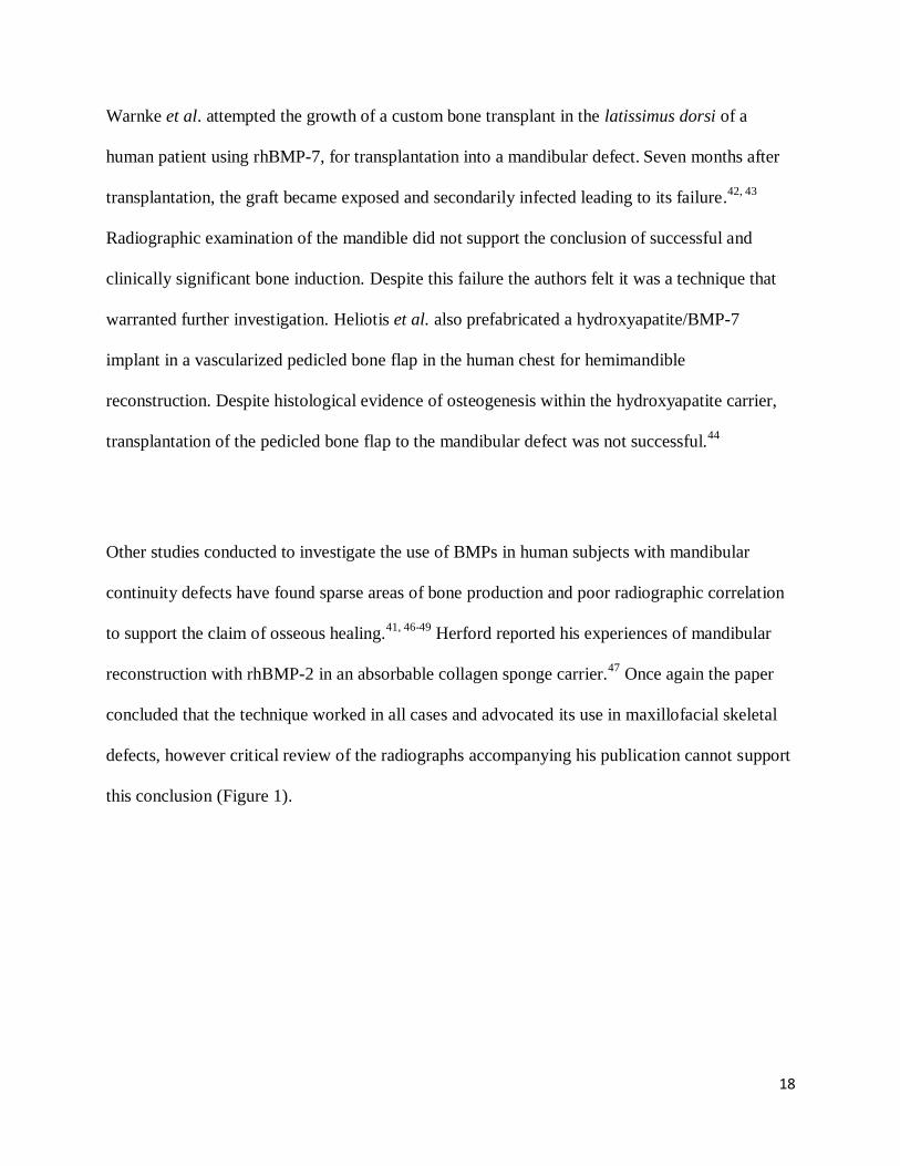
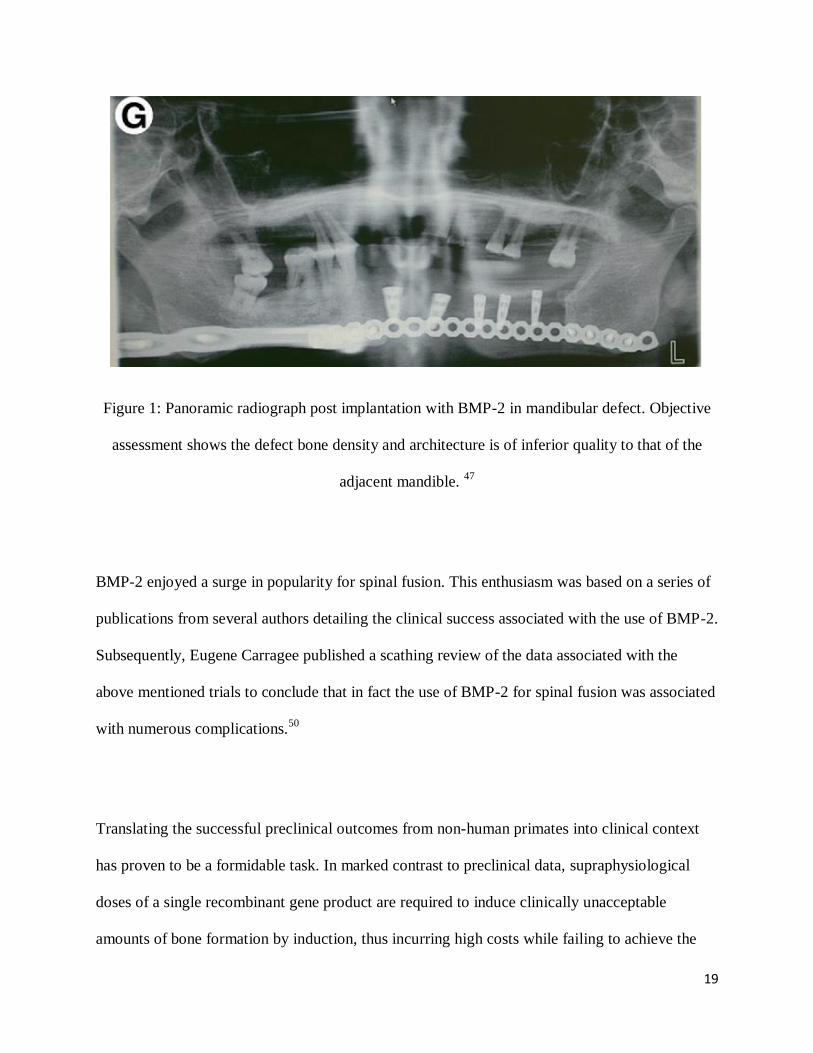
Herford reported his experiences of mandibular
reconstruction with rhBMP-2 in an absorbable collagen sponge carrier.47
Once again the paper
concluded that the technique worked in all cases and advocated its use in maxillofacial skeletal
defects, however critical review of the radiographs accompanying his publication cannot support
this conclusion (Figure 1).
19
Figure 1: Panoramic radiograph post implantation with BMP-2 in mandibular defect. Objective
assessment shows the defect bone density and architecture is of inferior quality to that of the
adjacent mandible. 47
BMP-2 enjoyed a surge in popularity for spinal fusion. This enthusiasm was based on a series of
publications from several authors detailing the clinical success associated with the use of BMP-2.
Subsequently, Eugene Carragee published a scathing review of the data associated with the
above mentioned trials to conclude that in fact the use of BMP-2 for spinal fusion was associated
with numerous complications.50
Translating the successful preclinical outcomes from non-human primates into clinical context
has proven to be a formidable task. In marked contrast to preclinical data, supraphysiological
doses of a single recombinant gene product are required to induce clinically unacceptable
amounts of bone formation by induction, thus incurring high costs while failing to achieve the

20
outcomes of autogenous bone grafts.26, 39, 40
It is clear that the promise of therapeutic bone tissue
engineering remains elusive.50
21
2 AIM
The aim of this study is to evaluate and compare binary application of rhBMP-7 and rhTGF-3,
to the single application of rhBMP-7 in full-thickness mandibular defects in the non-human
primate Papio ursinus. This investigation will assess whether clinically significant
osteoinduction by rhBMP-7 singly and in combination with rhTGF-3, can be obtained in
surgically created mandibular defects. Success in this binary application could lead to the clinical
application of rhBMPs and rhTGF-3 in human subjects for tissue regeneration and may provide
an alternative therapeutic option for human bone tissue engineering.
22
3 MATERIALS AND METHODS
3.1 Experimental animal selection
Ethics approval was obtained from the Animal Ethics Committee, University of the
Witwatersrand, Johannesburg, clearance code AESC 2006715. Four clinically healthy adult
Chacma baboons (Papio ursinus) with normal hematological and biochemical profiles 51
and
skeletal maturity confirmed by radiograph evidence of closure of the distal epiphyseal plates of
radius and ulna were selected from the primate colony of the University of Witwatersrand,
Johannesburg.13, 30
Non-human primates were housed at the Central Animal Services of the
University, Medical School in compliance with the National Code for Animal Use in Research
and Education in South Africa.52
3.2 Morphogens
3.2.1 rhBMP-7
Mature rhBMP-7 is a gylcosolated 36kDa homodimer of 139 amino acid residue chains. rhBMP-
7 prepared as previously described by Sampath et al., was kindly supplied by Stryker Biotech
(Hopkinton, Massachusetts, U.S.A.).53
It was supplied in 12 sterile vials each containing 1g of
bovine insoluble collagenous bone matrix preloaded with 2.5mg rhBMP-7.
23
3.2.2 Transforming Growth Factor-
Mature recombinant rhTFG-a gylcosolated 25kDa homodimer with a C-terminal domain of
112 amino acids with nine cysteine residues, was obtained from Novartis Pharma AG (Basel,
Switzerland) and supplied as a stock solution of rhTGF-3 9.37mg per mL of diluent. This was
further diluted with 5mmol acetic acid to obtain a concentration of 1µg rhTGF-3 per µl of
diluent.
3.3 Preparation of implants
Twelve vials each containing 2.5mg rhBMP-7 and 1g bovine insoluble collagenous bone matrix,
were provided for implantation. To six of the vials was added 125µl of the diluted rhTGF-3
solution for a final dose of 125µg of rhTGF-3 per vial to obtain a 20:1 ratio rhBMP-7: rhTGF-
3. This has been the ratio with the highest biological activity when implanted in heterotopic
sites of Papio ursinus.22
The other six vials were used for implantation of rhBMP-7 solo.
24
3.4 Surgery
3.4.1 General anaesthesia
Baboons were operated under general anaesthesia induced with a mixture of Ketamine
(100mg/ml, Bayer, India (Pty) Ltd) at a dose volume of 5mg/kg, and Midazolam (5mg/ml, Roche
Prod. (Pty) Ltd) at a dose of 0.25mg/kg given intramuscularly. Oro-tracheal intubation was done
to provide inhalation of Isoflurane (Forane 1.5% 250mL, Piramal Critical Care, Inc, Bethlehem
U.S.A.) and oxygen for maintenance. Buprenorphine Hydrochloride (Temgesic 0.3mg/ml, RB
Pharmaceutics Ltd, United Kingdom) at a dose of 0.01 - 0.03mg/kg and Meloxicam (Mobic,
10mg/ml, Ingelham Pharm. (Pty) Ltd) at a dose of 0.2-0.3mg/kg were given subcutaneously for
analgesia. All baboons were given an intramuscular dose of oxytetracycline (Phenix, Peni LA,
Phenix S.A. (Pty) Ltd 1ml/10kg) intraoperatively. Doxapram Hydrochloride (Dopram 20mg/ml,
Amdipharm Mercury Ltd. UK) was used as reversal agent at a dose of 0.5-1mg/kg intravenously
in all cases. Postoperatively Carpofen (Novox 100mg/tab, Impax Labs, U.S.A.) was given at a
dose of 4mg/kg per os, crushed in banana, as an anti-inflammatory.
3.4.2 Extractions
All baboons had extractions of teeth on the right side of the mandible and maxilla from the right
canines posteriorly. Eight weeks thereafter, the baboons were returned to theatre and the
operative sites inspected for complete healing of the oral wound in order to avoid contamination
when implanting the osteogenic devices.
25
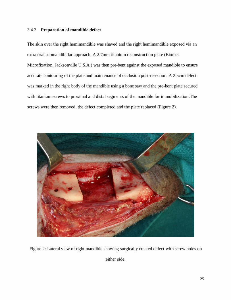
3.4.3 Preparation of mandible defect
The skin over the right hemimandible was shaved and the right hemimandible exposed via an
extra oral submandibular approach. A 2.7mm titanium reconstruction plate (Biomet
Microfixation, Jacksonville U.S.A.) was then pre-bent against the exposed mandible to ensure
accurate contouring of the plate and maintenance of occlusion post-resection. A 2.5cm defect
was marked in the right body of the mandible using a bone saw and the pre-bent plate secured
with titanium screws to proximal and distal segments of the mandible for immobilization.The
screws were then removed, the defect completed and the plate replaced (Figure 2).
Figure 2: Lateral view of right mandible showing surgically created defect with screw holes on
either side.
26
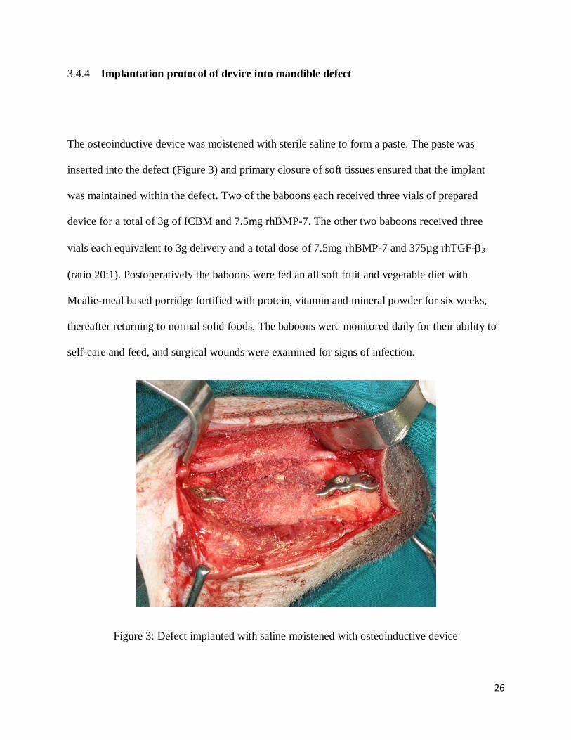
3.4.4 Implantation protocol of device into mandible defect
The osteoinductive device was moistened with sterile saline to form a paste. The paste was
inserted into the defect (Figure 3) and primary closure of soft tissues ensured that the implant
was maintained within the defect. Two of the baboons each received three vials of prepared
device for a total of 3g of ICBM and 7.5mg rhBMP-7. The other two baboons received three
vials each equivalent to 3g delivery and a total dose of 7.5mg rhBMP-7 and 375µg rhTGF-3
(ratio 20:1). Postoperatively the baboons were fed an all soft fruit and vegetable diet with
Mealie-meal based porridge fortified with protein, vitamin and mineral powder for six weeks,
thereafter returning to normal solid foods. The baboons were monitored daily for their ability to
self-care and feed, and surgical wounds were examined for signs of infection.
Figure 3: Defect implanted with saline moistened with osteoinductive device
27
3.4.5 Euthanasia and Tissue harvest
The baboons were anaesthetized 180 days post implantation, and the right and left carotid
arteries were exposed via a midline incision in the neck. The carotids were transected and
cannulated to allow perfusion of the mandible with 1 litre of saline in each carotid. The animals
were thus euthanazed with Nembutal sodium (Pentobarbital 50mg/mL, Lundbeck Inc. Deerfield,
IL U.S.A.) following which further perfusion of the carotids was done with 1 litre of formalin in
each carotid. The hemi-mandibles were resected and skeletonised for radiographic imaging and
histological processing. The defect and 1cm of normal bone was included in the final specimen.
3.5 Analytical procedures
3.5.1 Histomorphometry
Undecalcified tissues were processed according to the method of Donath and Breuner with minor
modifications.53
Briefly the tissues were processed in ascending grades of ethanol in an
automatic tissue processor (Tissue-Tek, V.I.P.; Miles Inc, Elkhart, USA), under pressure vacuum
cycles. Samples were infiltrated with ascending concentration of Technovit 7200 VLC (Heraeus
Kulzer GmbH, Wehrheim, Germany) and embedded in a fresh solution of the same resin.
Undecalcified sections were then ground and polished to m and stained with a modified
Goldner’s trichrome. All sample preparation was performed using the EXAKT precision cutting
and grinding system (EXAKT Apparatebau, Nordestedt, Hamburg, Germany).
28
Digital images were acquired using the Olympus BX16 microscope with built-in camera.
Quantification of regenerated tissue was carried out using Stream Essentials software. Stream
Essentials consists of four separate units including Olympus UPS, Microscope AX70, free
standing control unit, a camera and associated software (Shinjuku Monolith, 3-1 Nishi-Shinjuku
2-chrome, Tokyo, Japan). Stream Essentials can perform various functions which include
acquiring an image through the microscope camera, performing measurements on that image,
and exporting data to create a spreadsheet for analysis. Stream essentials software identifies bone
based on the colour taken up by the program, distinction cannot be made between osteoid and
mineralised tissue both of which take up the program similarly, therefore the amount of new
bone formation includes both osteoid and mineralised tissue.
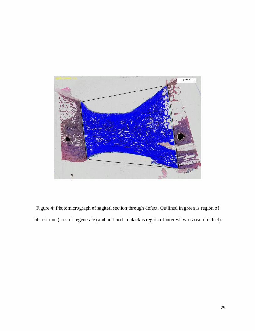
Two sagittal sections were selected from each specimen for analysis. Each section included the
entire defect and the interface with 1cm of intact mandible. A magnification factor of 10x was
used for all specimens. The outline of the regenerated bone was selected to calculate the amount
of regenerated bone formed within this outline (referred to as region of interest one (ROI 1))
(Figure 4). The original defect area was created by a quadrilateral whose sides are formed by the
two interfaces and the superior and inferior borders by constructed lines joining the superior and
the inferior ends of the interface lines. This was denoted as region of interest two (ROI 2). The
total area of new bone was measured in mm2 and the percentage within each ROI was calculated.
The regenerate as a percentage of ROI 2 reflects the percentage of the original defect area filled
with mineralized bone, whereas the ROI 1 reflects the density of mineralized bone within the
regenerate. All data captured was recorded in Microsoft Office Excel 2007 spreadsheet.
29
Figure 4: Photomicrograph of sagittal section through defect. Outlined in green is region of
interest one (area of regenerate) and outlined in black is region of interest two (area of defect).
30
3.6.2 Radiographic assessment
Although subjective, with critical examination, radiographs serve as an important tool to
determine clinically significant osteoinduction, remodelling, and integration. It is more reliable
than the histological examination. Clinically significant osteoinduction refers to the concept that
the quality of regenerated bone by osteoinductive agents is adequate to be identified
radiographically as normal bone (in radiodensity and trabecular pattern) and that the morphology
of the reconstructed segment resembles that of the former skeletal segment.54
Although objective
quantification of radiodensity and trabecular pattern on routine radiography is impossible it does
provide an unequivocal overview of osseous regeneration in a defect.
All four resected specimens were radiographed prior to sectioning. Specimens were radiographed
from a lateral and occlusal view at kV 53 and mA 0.09. Radiographs were taken to assess the
radiodensity of the regenerated bone and the trabecular pattern. Moreover, the size and
morphology of the regenerate will be assessed as will the visibility of the osseous interfaces (as a
guide to remodelling). With this evaluation the density of bone regenerate in the defect can be
compared to the adjacent normal mandible.
3.6.3 Statistical analysis
Due to the limited number of animals as well as of each treatment modality, statistical analysis
was not performed.
31
4 RESULTS
4.1 Macroscopic examination of resected mandibles
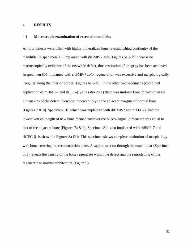
All four defects were filled with highly mineralized bone re-establishing continuity of the
mandible. In specimen 005 implanted with rhBMP-7 solo (Figures 5a & b), there is no
macroscopically evidence of the erstwhile defect, thus restitution of integrity has been achieved.
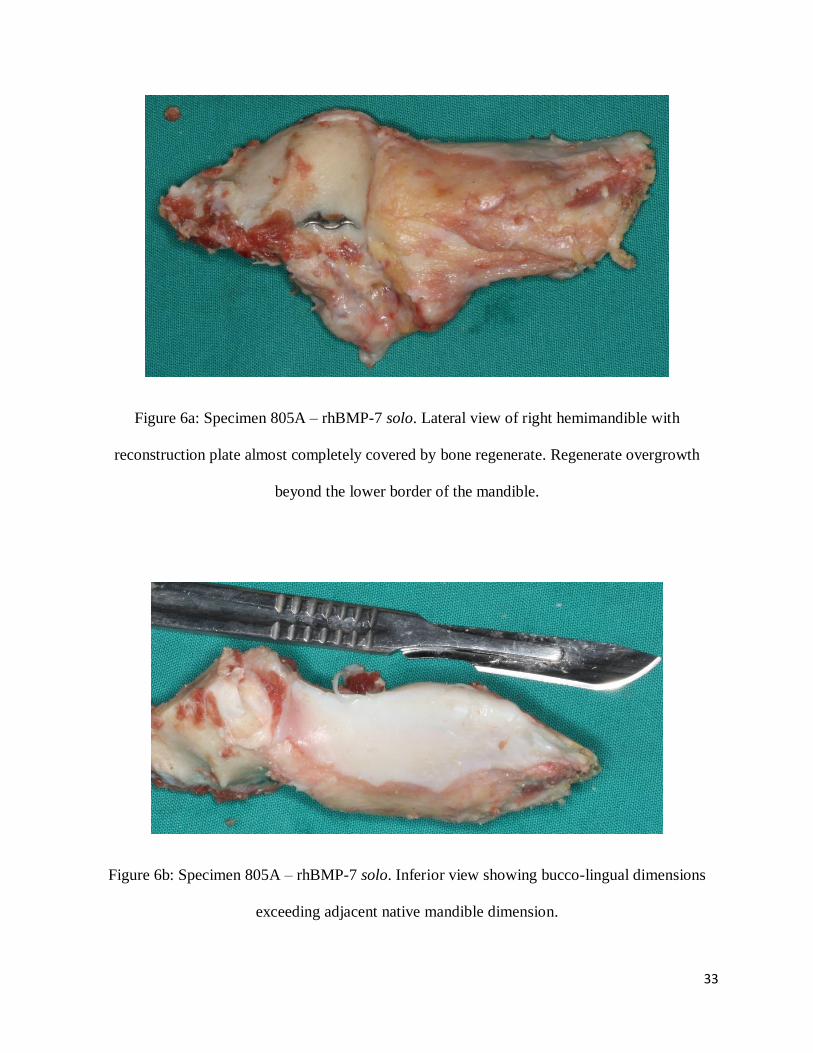
In specimen 805 implanted with rhBMP-7 solo, regeneration was excessive and morphologically
irregular along the inferior border (Figures 6a & b). In the other two specimens (combined
application of rhBMP-7 and rhTFG-3 at a ratio 20:1) there was uniform bone formation in all
dimensions of the defect, blending imperceptibly to the adjacent margins of normal bone
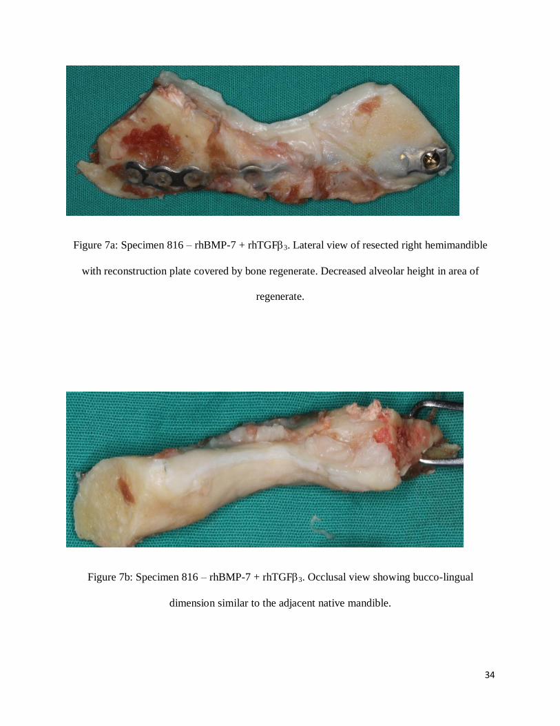
(Figures 7 & 8). Specimen 816 which was implanted with rhBMP-7 and rhTFG-3 had the
lowest vertical height of new bone formed however the bucco-lingual dimension was equal to
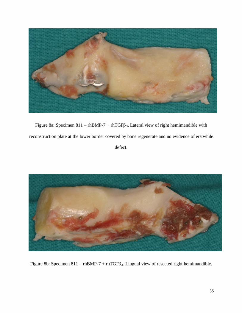
that of the adjacent bone (Figures 7a & b). Specimen 811 also implanted with rhBMP-7 and
rhTFG-3 is shown in Figures 8a & b. This specimen shows complete restitution of morphology
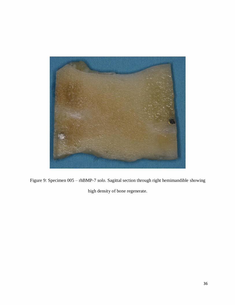
with bone covering the reconstruction plate. A sagittal section through the mandibular (Specimen
005) reveals the density of the bone regenerate within the defect and the remodelling of the
regenerate to normal architecture (Figure 9).
32
Figure 5a: Specimen 005 – rhBMP-7 solo. Lateral view of resected right hemimandible with
reconstruction plate covered by bone regenerate with defect margins not apparent. Restitutio ad
integrum of the mandibular defect.
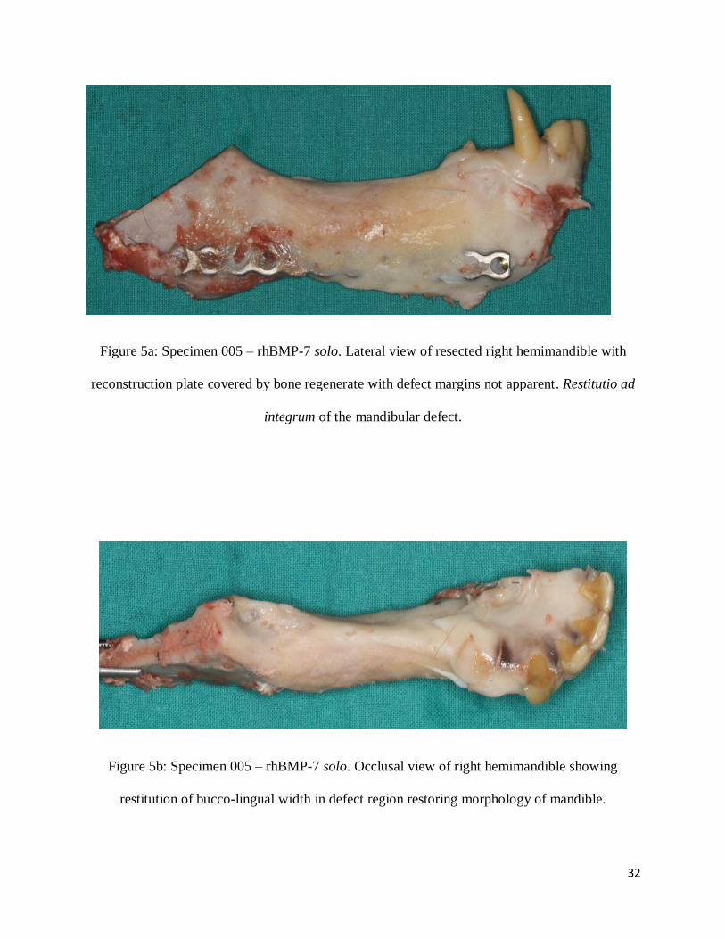
Figure 5b: Specimen 005 – rhBMP-7 solo. Occlusal view of right hemimandible showing
restitution of bucco-lingual width in defect region restoring morphology of mandible.
33
Figure 6a: Specimen 805A – rhBMP-7 solo. Lateral view of right hemimandible with
reconstruction plate almost completely covered by bone regenerate. Regenerate overgrowth
beyond the lower border of the mandible.
Figure 6b: Specimen 805A – rhBMP-7 solo. Inferior view showing bucco-lingual dimensions
exceeding adjacent native mandible dimension.
34
Figure 7a: Specimen 816 – rhBMP-7 + rhTGF3. Lateral view of resected right hemimandible
with reconstruction plate covered by bone regenerate. Decreased alveolar height in area of
regenerate.
Figure 7b: Specimen 816 – rhBMP-7 + rhTGF3. Occlusal view showing bucco-lingual
dimension similar to the adjacent native mandible.
35
Figure 8a: Specimen 811 – rhBMP-7 + rhTGF3. Lateral view of right hemimandible with
reconstruction plate at the lower border covered by bone regenerate and no evidence of erstwhile
defect.
Figure 8b: Specimen 811 – rhBMP-7 + rhTGF3. Lingual view of resected right hemimandible.
36
Figure 9: Specimen 005 – rhBMP-7 solo. Sagittal section through right hemimandible showing
high density of bone regenerate.
37
4.2 Qualitative Radiographic analysis
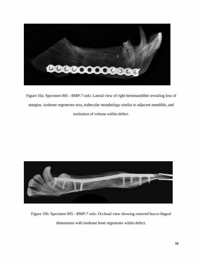
Assessment of radiographic images was qualitative and considered bone volume, visibility of
interface, and trabecular architecture. In all four specimens the defect margins were
imperceptible, confirming complete remodelling and integration of the regenerate with the
recipient bone. The newly formed bone was isodense (in some cases hyperdense) to the normal
mandible (Figures 10-13). The radiographic appearance of all specimens was uniform throughout
the defect area, with no areas of radiolucency noted to indicate poor bone formation or density.
Cortical continuity was evident both on the buccal and lingual borders of the mandible, with no
steps, breaks or radiolucency noted on either aspect of all four specimens. The trabecular
architecture was indistinguishable from normal mandible confirming restitutio ad integrum of
regenerated bone. Results thus confirm the achievement of “clinically significant
osteoinduction” as the quality of the regenerated bone is identified radiographically as normal
bone both in radiodensity and trabecular architecture.54
38
Figure 10a: Specimen 005 - BMP-7 solo. Lateral view of right hemimandible revealing loss of
margins, isodense regenerate area, trabecular morphology similar to adjacent mandible, and
restitution of volume within defect.
Figure 10b: Specimen 005 - BMP-7 solo. Occlusal view showing restored bucco-lingual
dimensions with isodense bone regenerate within defect.
39
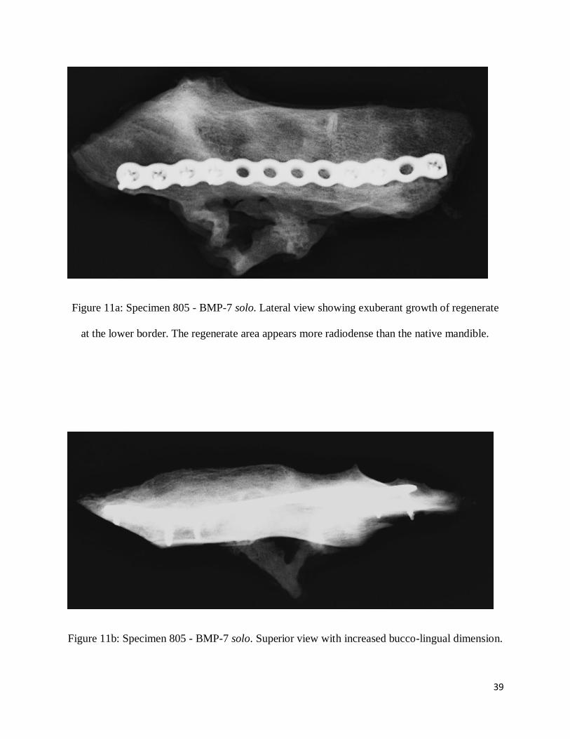
Figure 11a: Specimen 805 - BMP-7 solo. Lateral view showing exuberant growth of regenerate
at the lower border. The regenerate area appears more radiodense than the native mandible.
Figure 11b: Specimen 805 - BMP-7 solo. Superior view with increased bucco-lingual dimension.
40
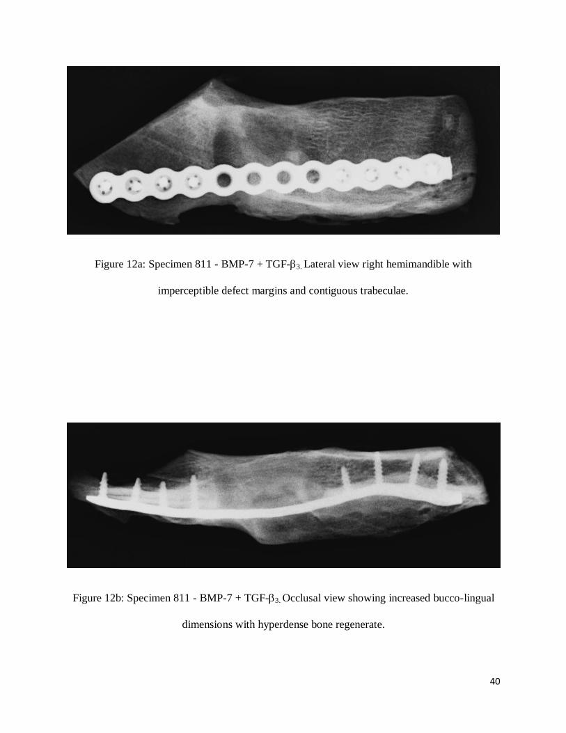
Figure 12a: Specimen 811 - BMP-7 + TGF-3. Lateral view right hemimandible with
imperceptible defect margins and contiguous trabeculae.
Figure 12b: Specimen 811 - BMP-7 + TGF-3. Occlusal view showing increased bucco-lingual
dimensions with hyperdense bone regenerate.
41
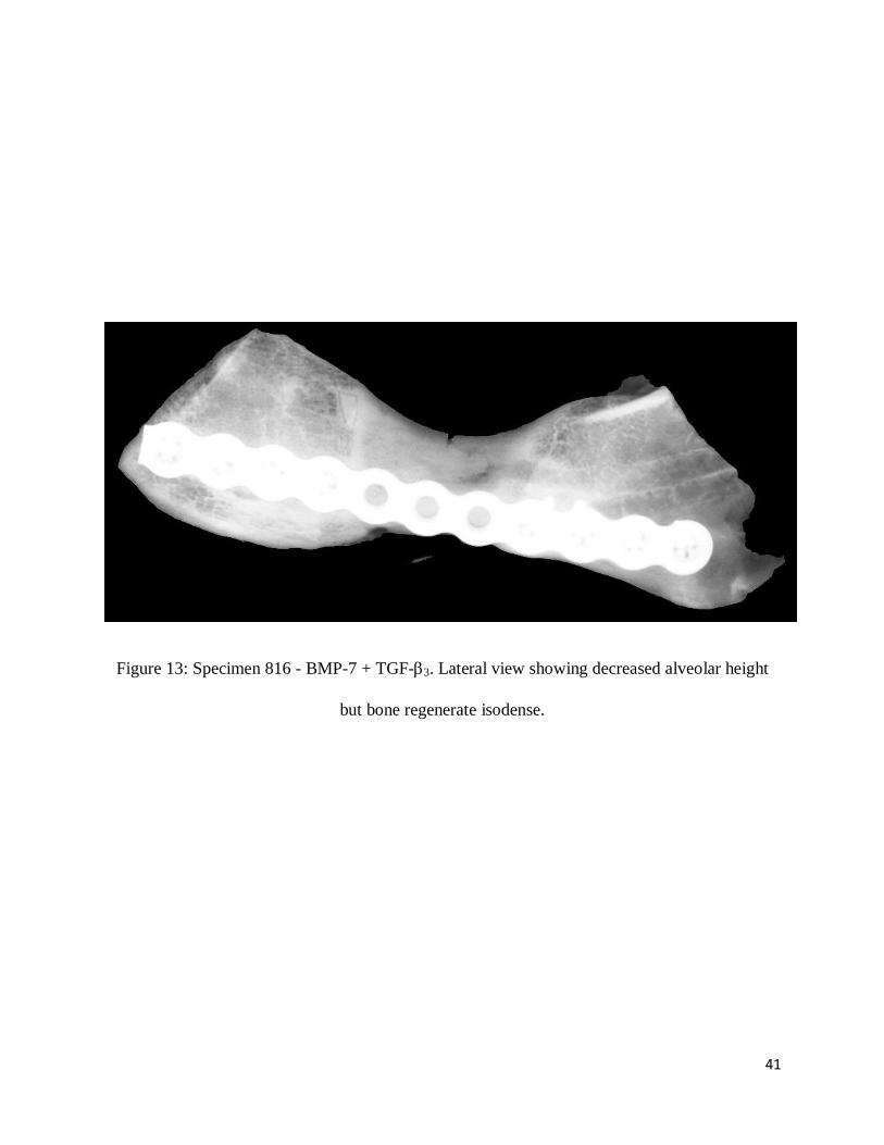
Figure 13: Specimen 816 - BMP-7 + TGF-3. Lateral view showing decreased alveolar height
but bone regenerate isodense.
42
4.3 Histomorphometry
Representative photomicrographs of sagittal histological sections at 5x magnification are shown
in figures 14-17a through each specimen. Low power histology confirms regeneration with
highly mineralized bone trabeculae. Trabecular density is greater than the adjacent normal
mandible. The superior and inferior cortical plates are restored. Bone formation can be seen
along the recipient margins with more delicate mineralizing trabeculae of woven bone towards
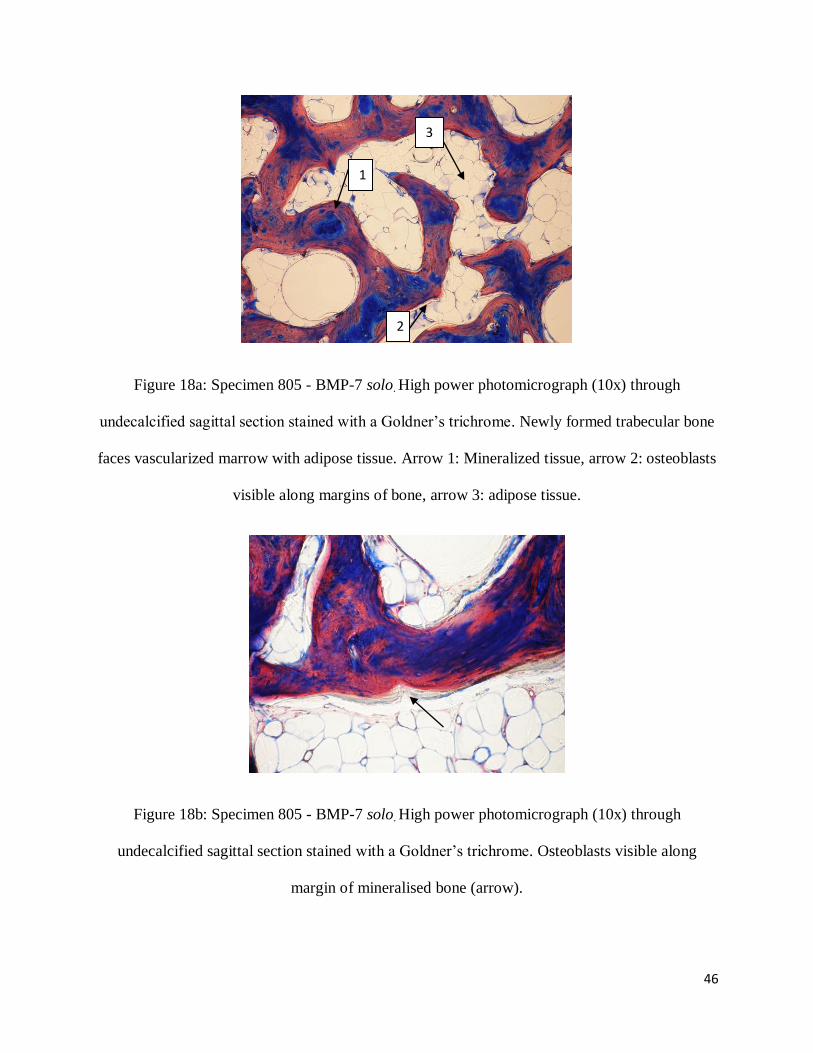
the centre of the previous defect area. High power magnification (10x) shows newly formed
trabecular bone facing vascularised marrow with adipose tissue. Osteoblasts can be seen along
the margin of woven bone (Figures 18a & b).
In specimen 005 implanted with rhBMP-7 solo, the mean amount of new bone formation within
the defect was 229.1 ± 12.7 mm2 (range 220-238mm
2), which is 43 ± 4.9% (range 40-47%) of
the regenerate area, and 40.5 ± 6.4% (range 36-45%) of the total defect area. Thus the
mineralised tissue occupied 43% of the total area of regenerate and 40.5% of the original defect
area. In specimen 805 implanted with rhBMP-7 solo, the mean amount of new bone formation
within the regenerate is 444.7 ± 68.7 mm2 (range 396-493mm
2) which is 50.7 ± 7.8% (range 45-
56%) of regenerate, and 48.5 ± 3.5% (range 41-56%) of the original defect area. In specimen 811
implanted with rhBMP-7 and rhTGF-, the mean amount of new bone formation is 376.6 ±
68.6mm2 (range 328-425mm
2) which is 53.3 ± 4.9% (range 50-57%) of regenerate area, and 48.9
± 4.9% (range 45-52%) of the total defect area. In specimen 816 also implanted with rhBMP-7
and rhTGF-, the average amount of new bone formation is 249.0 ± 22.6mm2 (range 233-
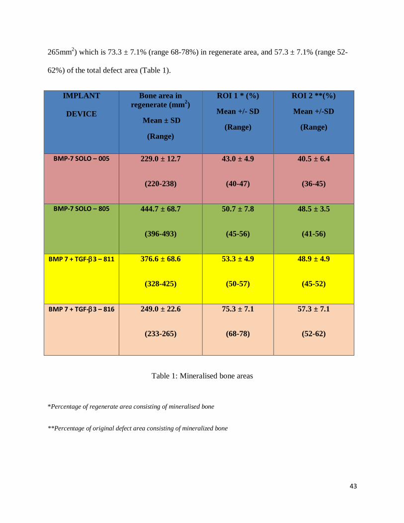
43
265mm2) which is 73.3 ± 7.1% (range 68-78%) in regenerate area, and 57.3 ± 7.1% (range 52-
62%) of the total defect area (Table 1).
IMPLANT
DEVICE
Bone area in
regenerate (mm2)
Mean ± SD
(Range)
ROI 1 * (%)
Mean +/- SD
(Range)
ROI 2 **(%)
Mean +/-SD
(Range)
BMP-7 SOLO – 005 229.0 ± 12.7
(220-238)
43.0 ± 4.9
(40-47)
40.5 ± 6.4
(36-45)
BMP-7 SOLO – 805 444.7 ± 68.7
(396-493)
50.7 ± 7.8
(45-56)
48.5 ± 3.5
(41-56)
BMP 7 + TGF-3 – 811 376.6 ± 68.6
(328-425)
53.3 ± 4.9
(50-57)
48.9 ± 4.9
(45-52)
BMP 7 + TGF-3 – 816 249.0 ± 22.6
(233-265)
75.3 ± 7.1
(68-78)
57.3 ± 7.1
(52-62)
Table 1: Mineralised bone areas
*Percentage of regenerate area consisting of mineralised bone
**Percentage of original defect area consisting of mineralized bone
44
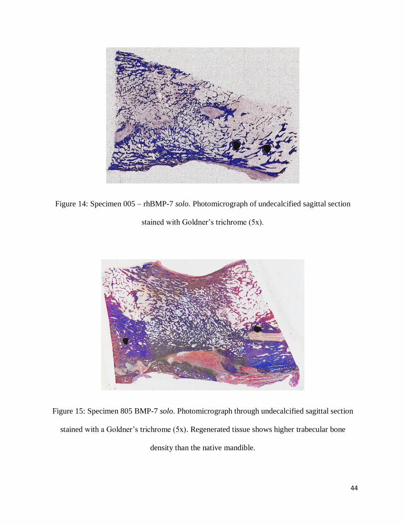
Figure 14: Specimen 005 – rhBMP-7 solo. Photomicrograph of undecalcified sagittal section
stained with Goldner’s trichrome (5x).
Figure 15: Specimen 805 BMP-7 solo. Photomicrograph through undecalcified sagittal section
stained with a Goldner’s trichrome (5x). Regenerated tissue shows higher trabecular bone
density than the native mandible.
45
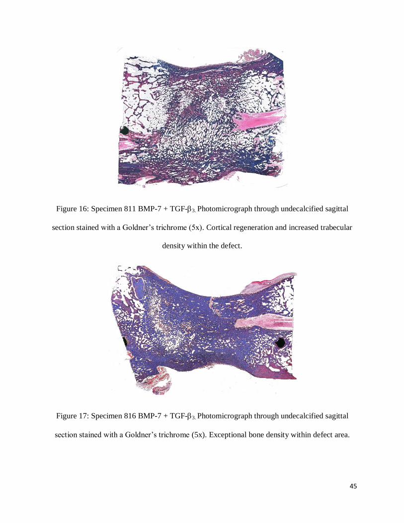
Figure 16: Specimen 811 BMP-7 + TGF-3. Photomicrograph through undecalcified sagittal
section stained with a Goldner’s trichrome (5x). Cortical regeneration and increased trabecular
density within the defect.
Figure 17: Specimen 816 BMP-7 + TGF-3. Photomicrograph through undecalcified sagittal
section stained with a Goldner’s trichrome (5x). Exceptional bone density within defect area.
46
Figure 18a: Specimen 805 - BMP-7 solo. High power photomicrograph (10x) through
undecalcified sagittal section stained with a Goldner’s trichrome. Newly formed trabecular bone
faces vascularized marrow with adipose tissue. Arrow 1: Mineralized tissue, arrow 2: osteoblasts
visible along margins of bone, arrow 3: adipose tissue.
Figure 18b: Specimen 805 - BMP-7 solo. High power photomicrograph (10x) through
undecalcified sagittal section stained with a Goldner’s trichrome. Osteoblasts visible along
margin of mineralised bone (arrow).
1
1
1
2
2
1
2
1
1
2
2
1
3
2
1
47
5 Limitations
The small number of animals available for the study was a significant limitation. With only two
animals in each study group, it was not possible to identify differences between the treatment
groups. The lack of a control group is another limitation, however given the scarcity of animals
available for scientific research, and that previous work in calvaria confirmed that defects of
2.5cm in diameter do not heal spontaneously, it was decided that a control group was
unnecessary, since a 2.5cm segmental defect in the mandible is a non-healing defect. Within the
limit of the study however, binary application of BMP-7 and TGF-3 (ratio of 20:1) showed
significant restitution of integrity as evaluated histologically and radiographically (Figures 10-
18), but above all, superb morphological healing with substantial bone regeneration and
cortication of the newly formed segment of the mandibular defect with superior bone density
across the treated defect area (Figures 5-9).
48
6 DISCUSSION
This study found that all the implanted devices regenerated bone and restituted the osseous micro
and macro anatomy of the treated mandibular defect. Comparing the area of bone regenerate in
all four specimens, specimen 805 implanted with rhBMP-7 solo produced the largest area of
bone regenerate within the defect. Following this was specimen 811 which was implanted with
rhBMP-7 and rhTGF-3. Specimen 816 (rhBMP-7 and rhTGF-3) had bone areas slightly lower
than 811, and the least amount of bone was produced in specimen 005 which was implanted with
rhBMP-7 solo. Specimen 816 was volumetrically deficient but the density of the regenerate was
significantly higher in this specimen than all others. This small study failed to identify a
difference in bone regeneration between the two treatment groups.
Many studies have used histology as the arbitrate of success or failure. The contention of this
study is that radiography is a far more important tool to assess whether a regenerate has achieved
preclinical success. High power histology demonstrating new bone formation is often used to
erroneously conclude success. Although histology is a useful adjunct, it often hides the bone
architecture, density and mineralization where high power images are zoomed in on only small
areas of bone formation. Radiographic examination of the entire defect provides unequivocal
evidence of mineralized tissue regeneration within a defect, the volume of tissue regenerated, the
amount of remodelling that has occurred (thus resulting in the loss of an obvious defect
interface) and finally the restitution of the micro anatomical structure of osseous trabeculation.
49
The assessment of all specimens was subjective and qualitative to replicate the clinical scenario
of bone graft assessment. This confirmed that new bone regeneration had occurred within the
mandible defects. The bone was isodense to the adjacent normal bone indicating good
regenerated bone quality. Remodelling was so extensive that the defect margins had become
imperceptible. The main difference between the various specimens was that of the bone height
within the defect, where specimen 805 showed greatest bone height, followed by 811 and 005,
with 816 having the lowest levels of bone height. Although the bone height of 816 was lowest,
this specimen defect appeared to be most radiopaque. These results were supported by the
histomorphometry analysis. Finally trabecular morphology is a final confirmation of remodelling
and once again the regenerate was indistinguishable from the adjacent mandible.
The results of the study have reconfirmed that the morphogens of the TGF- superfamily
regenerate tissue within surgically created defects that is histologically identifiable as
mineralized bone. More telling is the radiographic assessment which shows outstanding restitutio
ad integrum of the surgically created defect. This allows us to conclude that clinically significant
osseoinduction has been achieved.
Binary application of osteoinductive growth factors has previously shown to significantly
increase the volume of bone regenerate in orthotopic and heterotopic sites (calvarium) of non-
human primates.24, 26
In this small study no performance advantage was found between the
single morphogens and binary treated defects, beside the radiographic and histomorphometric
examinations as outlined under the limitations of the study.
50
A final fundamental question still remains: Are these results translatable into successful therapy
for human patients? It is becoming clear that this may not be the case as previous attempts at
therapeutic osseoinduction in humans have shown. Although described as clinically successful
by its author, critical examination of published radiographic evidence (Figure 1) cannot support
this contention. Moreover, increasing concern regarding efficacy and safety of Infuse (rhBMP-2)
raised in several articles by Spine Journal50
, led to a 16 month investigation into Medtronic
(Medtronic Inc., Minneapolis, U.S.A.) the makers of Infuse. This investigation found
questionable ties between the company and the physician consultants commissioned with testing,
reviewing and reporting on Medtronic products. From this investigation by the Senate Finance
Committee, it was found that the studies published by Medtronic were inaccurately representing
the risks of InFuse in addition to placing added weight on alternative treatment side effects.
Medtronic was found in violation of the trust of patients in their medical care and as a result
sullied the reputation of bone inductive agents.55
Resolving these challenges will require renewed vigour and courage on the part of scientists,
physicians, patients, and regulatory authorities.
51
7 CONCLUSION
The osteogenic proteins of the TGF- superfamily, solo or in combination, regenerate osseous
defects of the mandible in Papio ursinus. Qualitative assessment of the regenerate reveals that
clinically significant osteoinduction has been achieved.
52
8 REFERENCES
1. Ferretti C, Ripamonti U. (2002) Human segmental mandibular defects treated with
naturally derived bone morphogenetic proteins. J Craniofac Surg. vol. 13:434-444.
2. Habal MB, Reddi H. Bone grafts & bone substitutes. (1992) W. B. Saunders Comp.
Harcourt Brace Jovanovich, Inc. Philadelphia, Pennsylvania 19106. pg: 18-134.
3. Peterson LJ, Indresano AT, Marciani RD, Roser SM. (2009) Principles of Oral and
Maxillofacial Surgery. 2nd
ed. Philadelphia: JB Lippincott Co. pg: 783-798.
4. Reddi AH. (1981) Cell biology and biochemistry of endochondral bone development.
Coll Rel Res. vol. 1:209-226.
5. Urist MR. (1965) Bone: Formation by Autoinduction. Science. vol. 150:893-899.
6. Urist MR, Strates BS. (1971) Bone Morphogenetic Protein. J Dent Res. vol. 50:1392-
1406.
7. Sampath TK, Reddi AH. (1981) Dissociative extraction and reconstitution of
extracellular matrix components involved in local bone differentiation. Proc Natl Acad
Sci. U.S.A. vol. 78:7599-7602.
8. Sampath TK, Reddi AH. (1983) Homology of bone-inductive proteins from human,
monkey, bovine and rat extracellular matrix. Proc Natl Acad Sci. U.S.A. vol. 80:6591-
6595.
53
9. Wang EA, Rosen V, Cordes P, Hewick RM, Kriz MJ, Luxenberg DP, Sibley BS,
Wozney JM. (1988) Purification and characterization of other distinct bone inducing
factors. Proc Natl Acad Sci. U.S.A. vol. 85:9484-9488.
10. Wozney JM, Rosen V, Celeste AJ, Mitstock LM, Whitters MJ, Kriz RW, Hewick RM,
Wang EA. (1988) Novel regulators of bone formation: Molecular Clones and activities.
Science. Vol. 242: 1528-1534.
11. Reddi AH. (2001) Morphogenesis and tissue engineering of bone and cartilage: inductive
signals, stem cells, and biomimetic biomaterials. Tissue Eng. vol. 6:351-359.
12. Ripamonti U, Van Den Heerver B, Crooks J, Rueger DC, Reddi AH. (2001) Long-term
evaluation of bone formation by osteogenic protein-1 in the baboon and relative efficacy
of bone derived morphogenetic proteins delivered by irradiated xenogeneic collagenous
matrices. J Bone Mineral Res. vol. 15(9):1798-1809.
13. Ripamonti U, Ramoshebi LN, Matsaba T, Tasker J, Crooks J, Teare J. (2001) Bone
induction by BMPs/OP and related family members in primates: The critical role of
delivery systems. J Bone Joint Surg Am. vol. 83:116-127.
14. Ripamonti, U. (2006) Soluble osteogenic molecular signals and the induction of bone
formation. Biomaterial. vol. 27:807-822.
15. Celeste AJ, Iannazzi JA, Taylor RC. (1990) Identification of transforming growth factor-
family members present in bone-inductive protein purified from bovine bone. Proc Natl
Acad Sci USA. vol. 87:9843-9847.
16. Ozkaynak E, Rueger DC, Drier EA, Corbett C, Ridge RJ, Sampath T, Oppermann H.
(1990) OP-1 cDNA encodes an osteogenic protein in the TGF- family. EMBO J. vol.
9:2085-93.
54
17. De Biase P, Capanna R. (2005) Clinical applications of BMPs. Injury 36S, S43-S46.
18. Ripamonti, U. (2006) Soluble osteogenic molecular signals and the induction of bone
formation. Biomaterials. vol. 27:802-822.
19. Murakami N, Saito N, Horiuchi H, Okada T, Nozaki K, Takaoka K. (2002) Repair of
segmental defects in rabbit humeri with titanium fiber mesh cylinders containing
recombinant human bone morphogenetic protein-2 and a synthetic polymer. J Biomed
Mater Res. vol. 62: 169-174.
20. Chen D, Zhao M, Mundy R. (2004) Bone Morphogenetic Proteins. Growth Factors. vol.
22:233-241.
21. Terheyden H, Warnke P, Dunsche A, Jepson S, Brenner W, Palmie S. (2001) Mandibular
reconstruction with prefabricated vascularised bone grafts using recombinant human
osteogenic protein-1: An experimental study in miniature pigs. Part II: Transplantation.
Int J Oral Maxillofac Surg. vol. 30:469-478.
22. Ripamonti U, and Reddi AH. (1995) Bone morphogenetic proteins: Applications in
plastic and reconstructive surgery. Adv Plast Reconstr Surg. vol. 11:47-65.
23. Ripamonti U, Crooks J, Khoali L, Roden L. (2009) The induction of bone formation by
coral-derived calcium carbonate/hydroxyapatite constructs. Biomaterials. vol. 30:1428-
1439.
24. Ripamonti U, Duneas N, Van Der Heever, B, Bosch C, Crooks J. (1997) Recombinant
transforming growth factor- β1 induces endochondral bone in the baboon and synergizes
with recombinant osteogenic protein-1 (bone morphogenetic protein-7) to initiate rapid
bone formation. J Bone Mineral Res. vol. 12:1584-1595
55
25. Ripamonti U, Klar RM, Renton LF, Ferretti C. (2010) Synergistic induction of bone
formation by hOP-1, hTGF-β3 and inhibition by zoledronate in macroporous coral-
derived hydroxyapatites. Biomaterials. vol. 31:6400-6410
26. Ripamonti U, Ferretti C, Teare J, Blann L. (2009) Transforming growth factor-beta
isoforms and the induction of bone formation: implications for reconstructive craniofacial
surgery. J of Craniofac Surg. vol. 20:1544-1555.
27. Si X, Jin Y, Yang L. (1998) Induction of new bone by ceramic bovine bone with
recombinant human bone morphogenetic protein 2 and transforming growth factor beta.
Int J Maxillofac Surg. vol. 27: 310-314.
28 Schnitzler CM, Ripamonti U, Mesquita JM. (1993) Histomorphometry of iliac crest
trabecular bone in adult male baboons in captivity. Calcif Tissue Int. vol.53:447-454.
29. Ripamonti U, Parak R, Petit J-C. (2009) Induction of cementogenesis and periodontal
ligament regeneration by recombinant human transforming growth factor beta-3 in
Matrigel with rectus abdominis responding cells. J Perio Res. vol. 44:81-87.
30. Ripamonti U. (1991) Bone induction in nonhuman primates: An experimental study on
the baboon (Papio ursinus). J Clin Orthop Rel Res. vol. 269:293-303.
31. Ripamonti U. (2005) Bone induction by recombinant human osteogenic protein-1 (hOP-
1, BMP-7) in the primate Papio ursinus with expression of mRNA of gene products of the
TGF-superfamily. J Cell Mol Med. vol. 9:911-928.
32. Glowaki G, Kaban LB, Murray JE, Folkman J, Mulliken JB. (1981) Application of the
biological principle of induced osteogenesis for craniofacial defects. Lancet. vol. 1: 959-
983.
56
33. Kaban LB, Mulliken JB, Glowacki J. (1982) Treatment of jaw defects with demineralized
bone implants. J Oral Maxillofac Surg. vol. 40:623-626.
34. Sailer HF, Kolb E. (1994a) Application of purified bone morphogenetic protein (BMP) in
cranio-maxillo-facial surgery. BMP in compromised surgical reconstructions using
titanium implants. J Craniomaxillofac Surg. 22:2-11.
35. Gilbert Triplett R, Marx RE, Oates TW, Boyne PJ. (2009) Pivotal, randomized, parallel
evaluation of recombinant human bone morphogenetic protein-2/absorbable collagen
sponge and autogenous bone graft for maxillary sinus floor augmentation. J Oral
Maxillofac Surg. vol. 67:1947-1960.
36. Boyne PJ, Lilly AC, Marx RE. (2005) De novo bone induction by recombinant human
bone morphogenetic protein-2 (rhBMP-2) in maxillary sinus floor. J Oral Maxillofac
Surg. vol. 63:1693-1707.
37. Smith DM, Cooper GM, Mooney MP, Marra KG, Losee JE. (2008) Bone morphogenetic
protein-2 therapy for craniofacial surgery. J Craniofac Surg. vol. 19:1244-1259.
38. Carstens MH, Chin M, Ng T. (2005) Reconstruction of L7 facial cleft with distraction-
assisted in situ osteogenesis (DISO): role of recombinant human bone morphogenetic
protein-2 with Helistat-activated collagen implant. J Craniofac Surg. vol. 16:1023-1032.
39. Garrison KR, Donell S, Ryder J, Shemilt I, Mugford M, Harvey I, Song F. (2007)
Clinical effectiveness and cost-effectiveness of bone morphogenetic proteins in the non-
healing of fractures and spinal fusion: a systematic review. Health Technol. Assess. vol.
11:1–150.
40. Mussano F, Ciccone G, Ceccarelli M, Baldi I, Bassi F. (2007) Bone morphogenetic
proteins and bone defects: a systematic review. Spine. vol. 32: 824–830.
57
41. Moghadam HG, Urist MR, Sandor GK, Clokie CM. (2001) Successful mandibular
reconstruction using a BMP bioimplant. J Craniofac Surg. vol. 12:119–127.
42. Warnke PH, Wiltfang J, Springer I, Acil Y, Eufinger H, Wehmoller M. (2004) Growth
and transplantation of a custom vascularised bone graft in a man. Lancet. vol. 364:766-
770.
43. Warnke PH, Wiltfang J, Springer I, Acil Y, Bolte H, Kosmahl M, Russo PAJ, Sherry E,
Lutzen U, Wolfart S, Terheyden H. (2006) Man as living bioreactor: Fate of an
exogenously prepared customized tissue-engineered mandible. Biomaterials. vol.
27:3163–3167.
44. Heliotis M, Lavery KM, Ripamonti U, Tsiridis E, Di Silvio L. (2006) Transformation of
a prefabricated hydroxyapatite/osteogenic protein-1 implant into a vascularised pedicled
bone flap in the human chest. Int. J. Oral Maxillofac Surg. vol. 35:265–269.
45. Chao M, Donovan T, Sotelo C, Carstens MH. (2006) In situ osteogenesis of
hemimandible with rhBMP-2 in a 9-year-old boy: Osteoinduction via stem cell
concentration. J Craniofac Surg. vol. 17:405-412.
46. Hernandez-Alfaro F, Ruiz-Magaz V, Chatakun P, Guijarro-Martínez R. (2012)
Mandibular reconstruction with tissue engineering in multiple recurrent ameloblastoma.
J of Periodon & Resto Dent. vol. 32:82-86.
47. Herford A, Boyne PJ. (2008) Reconstruction of mandibular continuity defects with bone
morphogenetic protein-2. J Oral Maxillofac Surg. vol. 66:616-624.
48. Carter TG, Brar PS, Tolas A, Beirne OR. (2008) Off-label use of recombinant human
bone morphogenetic protein-2 reconstruction of mandibular bone defects in humans. J
Oral Maxillofac Surg. vol. 66: 1417-1425.
58
49. Clokie CML, Sandor GKB. (2008) Reconstruction of 10 mandibular defects using
bioimplants containing BMP 7. J Canadian Dent Association. vol. 74:67-72.
50. Carragee EJ, Hurwitz EL, Weiner BK. (2011) A critical review of recombinant human
bone morphogenetic protein-2 trials in spinal surgery: emerging safety concerns and
lessons learned. Spine. vol. 11:471–491.
51. Melton DA, Melton CL. (1982) Blood parameters of the wild Chacma baboon, Papio
ursinus South Afr J Zool. vol. 17:85-90.
52. Public Service Department 1990 National Code for Animal Use in Research, Education,
Diagnosis and Testing of Drugs and Related Substances South Africa. Public Service
Dept, Pretoria, South Africa.
53. Donath K, Breuner G. (1982) A method for the study of undecalcified bones and teeth
with attached soft tissues. The Sage-Schliff (sawing and grinding) technique. J Oral
Pathol. vol 11:318-326.
54. Ferretti C, Ripamonti U, Tsiridis E, Kerawala CJ, Mantalaris A, Heliotis M. (2010)
Osteoinduction: translating preclinical promise into clinical reality. Brit. J Oral &
Maxillofac Surg. vol. 48:536-539.
55. Baucus-Grassley. (2012) Investigation into Medtronic. Committee on Finance. U.S.A.
Senate.