Embed Size (px)
Citation preview

J. Embryol. exp. Morph. 86, 205-218 (1985) 2 0 5Printed in Great Britain © Company of Biologists Limited 1985
Transferrin in foetal and adult mouse tissues:synthesis, storage and secretion
JENNIFER MEEK AND EILEEN D. ADAMSON
La Jolla Cancer Research Foundation, 10901 North Torrey Pines Road, LaJolla, California 92037, USA
SUMMARY
Transferrin is an important growth-promoting serum glycoprotein synthesized chiefly in theliver in adults. The transferrin found in the mouse foetus is thought to be wholly a product ofthe foetus itself and its synthesis starts at least as early as the 7th day of gestation. The majorsites of synthesis in mouse foetuses are the visceral yolk sac (VYS) and liver (Adamson, 1982).We now report that other murine foetal tissues synthesize readily detectable amounts, namelylung, spleen, spinal cord and rib cage. Very low levels are also synthesized by the brain, muscleand pancreas. We can detect no synthesis of transferrin in late foetal thymus, heart or skinalthough mid-gestation foetal skin may make a very small amount. No synthesis of transferrincan be detected in adult brain, lung and spleen, but approximately equal rates of synthesis aredetected in adult liver and adult ear pinna.
Transferrin is accumulated by foetal and adult tissues in widely varying amounts and thesehave been measured by enzyme-linked immunosorbent assays of extracts. In addition to VYSand liver, high levels of transferrin are found in foetal skin, lung and rib cage with loweramounts in spinal cord, spleen and muscle tissues. Tissues of the 15th day foetus accumulatethe highest concentrations of transferrin. A role for the mediation of transferrin in the stimula-tion of growth and differentiation by interacting tissues is discussed.
INTRODUCTION
Transferrin is a j3i-globulin glycoprotein (Mr = 80 000) present in serum at2 -4mgmr 1 (Putnam, 1975), and capable of tightly binding two ferric and othermetal ions. Its principal function is regarded as the transport of iron, and its targetsare those tissues which are actively growing such as embryonal and tumour cellsand those cells with special iron requirements such as haematopoietic cells. Iron(and zinc) is required for all dividing cells to maintain enzyme and co-factor levelsin new cells. Transferrin is known to be adsorbed to cells through a specific receptorglycoprotein (Mr = 180000) and is probably endocytosed into vesicles calledreceptosomes. From here an acidic compartment removes iron and this is foundlater in the cytosol, bound to ferritin. Transferrin, unlike most ligands which entervia receptors, appears to be recycled out of the cell and reutilized in the extra-cellular fluid (reviewed by Octave, Schneider, Trouet & Crichton, 1983).
Embryonal cells have special demands for transferrin for the synthesis of iron-containing haem and enzymes. Since there appears to be a barrier to the passage of
Key words: Mouse, transferrin, glycoprotein, liver, yolk sac.

206 JENNIFER MEEK AND EILEEN D. ADAMSON
transferrin from the maternal circulation (Faulk & Galbraith, 1979) at least up tothe 18th day of gestation (Renfree & McLaren, 1974), it follows that transferrinshould be an early product of the murine embryo. The earliest time that transferrinsynthesis has been detected (Adamson, 1982) is on the 7th day of gestation. Theendoderm layer of the two-layered egg cylinder appears to be the source and laterthis layer develops into the endoderm of the visceral yolk sac (VYS). The VYS andthe foetal liver are major sources of transferrin during gestation. Both of theseendodermally derived tissues also synthesize alphafoetoprotein (Dziadek & Adam-son, 1978), but only transferrin continues to be synthesized by the liver in adults.
It is not yet known if transferrin is synthesized by any other foetal mouse organs,an important question in view of the ability of transferrin to stimulate the prolifera-tion and differentiation of mouse foetal kidney (Ekblom et al. 1983), chick muscle(Markelonis, Oh, Eldefrawi & Guth, 1982a; Ii, Kimura & Ozawa, 1982; Beach,Popiela & Festoff, 1983), and lymphocyte growth in vitro (Brock, 1981; Anderson,Chase & Tomasi, 1982). Transferrin is an essential component of serum-free media(Sato et al. 1979) for all kinds of cell types. We have therefore made a survey ofmurine foetal tissues to identify those that synthesize transferrin and which maystimulate their own development or the development of associated tissues.
We have analysed the content of transferrin in several adult and foetal organs inorder to identify transferrin which may be sequestered or accumulated to sig-nificantly high levels. The hypothesis is that transferrin could be released locally orpreferentially to influence the development of associated tissues. These datatogether with transferrin synthesis rates are assessed to determine the possibility ofa role for transferrin in tissue interactions which are known to stimulate organo-genesis or cell proliferation.
MATERIALS AND METHODS
Metabolic labelling and extraction of foetal tissuesRandom bred mice (CDI Flow Laboratories) were mated and the day of finding the copulation
plug was designated the first day of gestation. Pregnant females were killed by cervical dislocation,the uteri removed and foetuses dissected to collect the tissues. Care was taken to remove skin andbrain samples before opening the body cavity and dissections were made so that the liver fragmentsdid not contaminate any other tissue. Tissues (0-05-0-1 g wet weight) were washed in Trypsin-EDTA for 10-30 min and twice in medium (DME with 10 % foetal bovine serum) before pre-incubating them in 0-5 ml DME containing 1/20 normal concentration of methionine and 10 %dialysed foetal bovine serum. After 30min, [35S]methionine (lOOjuCiml"1, l-2Ciiumor1, NewEngland Nuclear), in fresh medium was added and incubation continued at 37 °C in 7 % CO2 in air.After 3 h, tissues were separated from the incubation medium, and the latter was centrifuged at15000 g for 10 min at 4 °C. Tissues were washed once in phosphate-buffered saline and homogenizedin 0-5 ml hypotonic buffer. Lysing buffer contained 10mM-KCl, lmM-MgCl2, pH7-5, 0-1 mM-PMSF, and after 15 min at 0 °C, Triton X-100 was added to 0-5 %. After centrifugation at 15000gfor10 min the supernatant was removed for analysis and the pellet was dissolved in 0-5 ml 0-5 N-NaOHto assay for protein by the Coomassie blue-dye-binding procedure (Sedmak & Grossberg, 1977).
The medium and extracts were analysed as shown in the flow diagram (Fig. 1) for protein, totaland acid-insoluble radioactivity, total transferrin content by enzyme-linked immunosorbentassay (ELISA) (see below) and for immunoprecipitated radioactive transferrin (see below).

Transferrin synthesis and secretion 207
Dissected tissue 3h inmedium + [3SS|methionine
Aliquotsfor TCA ppt.
Medium
Aliquotsfor ELISA
Tissue
Most forimmunoppt& PAGE
Aliquots forprotein assay
Aliquots forTCA ppt.
Aliquotsfor ELISA
Most forimmunoppt& PAGE
Hypotonic buffer, 0-5 7c Triton,inhibitors, ccntrifugation
15000/?, 20min.4°C
Dissolve in 0-5 N NaOH
Aliquots forprotein assay
Fig. 1 Flow diagram to show foetal tissue extraction and analysis procedures.
Analysis of radioactive proteinFrom the medium and extracts prepared as above, 2jul aliquots were examined for total
radioactive protein by precipitation on filter paper squares with ice-cold 10 % (w/v) trichloraceticacid (TCA) (30min at 0°C). The squares were washed with cold 10 % TCA containing 1 mM-methionine, followed by ethanol. They were dried and counted in 3 ml Beta-Max (West ChemProducts, San Diego, CA) in a scintillation counter.
Appropriate aliquots of medium and extract were taken to contain 2 X 10⁵ and 4 x 10⁵ c.p.m.TCA-insoluble radioactivity, respectively. Aliquots of medium were made up to 1 to 1-2 ml withNET buffer (0-4M-NaCl, 5mM-EDTA, 005M-Tris-HCl pH8, 1% NP-40, 0-lmM-PMSF and0-02 % sodium azide) and one portion was treated with 5 [A (approx. 5 ]Ug) affinity-purified anti-transferrin (see below) while to another, 5 [il non-immune rabbit immunoglobulin was added.After incubation at room temperature for lV^h or overnight at 4°C, 30 jul of a 50 % suspensionof protein A-agarose (Sigma) was added and the mixture mixed end-over-end for lVzh at 4°C.The beads containing adsorbed antibody/antigen complexes were washed twice in NET bufferand once in 0-0625 M-Tris-HCl pH6-8 before being boiled in 30//1 two-fold-concentrated gelsample buffer (Laemmli, 1970). Tissue extracts were analysed similarly except that the dilutingand washing medium was RIPA buffer (0-01 M-Tris-HCl, pH7-5, 0-15 M-NaCl, 1 % NP-40,1 %sodium deoxycholate, 0-1 % sodium dodecyl sulphate). Radioactive proteins were analysed byelectrophoresis on 7-5% polyacrylamide gels (Laemmli, 1970), stained with 0-2% (w/v)Coomassie brilliant blue, destained and soaked in fluor (Autofluor, National Diagnostics, Somer-ville, NJ), dried and exposed to preflashed Kodak XAR-5 film for 5-21 days at - 70 C (Bonner& Laskey, 1974). The bands on films were analysed by scanning densitometry and quantitatedby integration of the peaks.
Analysis of transferrin by ELISAPooled mouse plasma was processed by the method described by Sawatzki, Anselstetter &
Kubanek (1981) to isolate pure transferrin. Briefly, a 45% saturated-ammonium-sulphate-supernatant fraction from mouse serum was salted out on a column of Sepharose CL-6B, and thetransferrin peak was rechromatographed on DEAE-Sepharose. Peak fractions from this columnwere homogeneous by several criteria (Adamson, 1982). Transferrin (200 fig) dissolved in PBSwas mixed with an equal volume of Freund's complete adjuvant and injected into several sub-cutaneous sites in one rabbit. Immunizations were repeated at 4-week intervals using Freund'sincomplete adjuvant. Serum from the third bleed onwards contained anti-transferrin activity in

208 JENNIFER MEEK AND EILEEN D. ADAMSON
double-diffusion tests. Antibodies were affinity purified by passing a fraction which wasprecipitated by 40 % saturated ammonium sulphate over a conjugate of 50 mg purified transfer-rin linked to cyanogen-bromide-activated Sepharose (10 ml). After washing away unadsorbedprotein, antibodies were eluted with O-lM-glycine-HCl, pH2-6. Fractions with the highestprecipitating activity were pooled, dialysed against 0 05 M-NH4HCO3 solution and lyophilized.The antibody was shown to react specifically with mouse transferrin by double-diffusionanalyses (Adamson, 1982) and by lack of crossreaction with whole mouse, calf and rabbit serain ELISA.
A portion (1 mg) of affinity-purified anti-transferrin antibodies was crosslinked to 5 mg alkalinephosphatase (bovine intestine, Sigma type VII) according to the method described by Engvall(1980) and this was used as the third layer in a sandwich ELISA. The wells of 96-well polystyreneplates (Linbro/Titertek, cat. no. 76-38104, Flow Laboratories, Inc., McLean, VA) were coatedwith affinity-purified rabbit anti-mouse transferrin (100 fA of 5 fig. ml"1 antibodies dissolved inOlM-sodium bicarbonate buffer at pH9-5). This was allowed to adsorb for 16 h at room tem-perature. The plate was washed three times with 0-9 % (w/v) NaCl, 0-05 % (v/v) Tween 20 aftereach layer. The second layer was a solution (100 jul) containing an unknown or standard transfer-rin concentration from 2 to 200ng.ml"1 in PBS, 5% calf serum, 0*05% Tween-20 (Bovinetransferrin does not react with the anti-mouse transferrin antibodies, Adamson, 1982, andtherefore transferrin in calf serum does not interfere in the assay). Incubation and washingfollowed as described above. Rabbit antimouse transferrin coupled to alkaline phosphatase(200 jul of 0-5/ig. ml"1) was added to each well for 5h at room temperature and finally, 250 (j\substrate for alkaline phosphatase (Sigma 104 at 1 mg. ml"1 in 0-1 M-diethanolamine-HCl, 1 mM-MgCl2, pH9-8) allowed the enzyme activity in each well to be revealed as a yellow colour. Theabsorption at 405 nm was measured in a Flow titertek 96-well reader after 20 min and 30 min ofcolour development. Figure 2 shows a typical standard curve used to measure the concentrationof transferrin in tissue extracts. The lower limit of sensitivity is 10 ng. ml"1 transferrin.
Measurement of transferrin in adult tissuesAdult mice were perfused by passing 0-9 % NaCl solution into the heart of anaesthetized
heparin-injected animals using a 20 cm pressure head. Tissues were dissected and approximately1 gm was homogenized and processed as described for foetal tissues.
5 10 20 40 100 200Transferrin concentration (ng. ml"1)
Fig. 2 Standard curve for transferrin ELISA. The concentrations of transferrin inindividual wells (averages of duplicates) of a 96-well plate are shown on the abscissa (logscale). The optical densities of the products of alkaline phosphatase-linked (third layer)anti-transferrin antibodies is shown on the ordinate.
Transferrin synthesis and secretion 209
RESULTS
Transferrin synthesis by foetal tissuesFigure 3 A shows that large amounts of transferrin are synthesized by the liver and
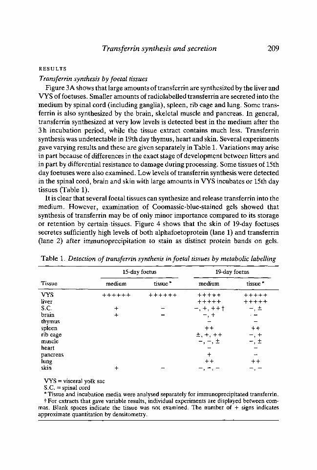
VYS of foetuses. Smaller amounts of radiolabelled transferrin are secreted into themedium by spinal cord (including ganglia), spleen, rib cage and lung. Some trans-ferrin is also synthesized by the brain, skeletal muscle and pancreas. In general,transferrin synthesized at very low levels is detected best in the medium after the3h incubation period, while the tissue extract contains much less. Transferrinsynthesis was undetectable in 19th day thymus, heart and skin. Several experimentsgave varying results and these are given separately in Table 1. Variations may arisein part because of differences in the exact stage of development between litters andin part by differential resistance to damage during processing. Some tissues of 15thday foetuses were also examined. Low levels of transferrin synthesis were detectedin the spinal cord, brain and skin with large amounts in VYS incubates or 15th daytissues (Table 1).
It is clear that several foetal tissues can synthesize and release transferrin into themedium. However, examination of Coomassie-blue-stained gels showed thatsynthesis of transferrin may be of only minor importance compared to its storageor retention by certain tissues. Figure 4 shows that the skin of 19-day foetusessecretes sufficiently high levels of both alphafoetoprotein (lane 1) and transferrin(lane 2) after immunoprecipitation to stain as distinct protein bands on gels.
Table 1. Detection of transferrin synthesis in foetal tissues by metabolic labelling
15-day foetus 19-day foetus
Tissue medium tissue* medium tissue'
VYSliverS.C.brainthymusspleenrib cagemuscleheart - -pancreas + —lung ++ + +skin + - - , - , - - , -
VYS = visceral yolk sacS.C. = spinal cord* Tissue and incubation media were analysed separately for immunoprecipitated transferrin.f For extracts that gave variable results, individual experiments are displayed between com-
mas. Blank spaces indicate the tissue was not examined. The number of + signs indicatesapproximate quantitation by densi tome try.

210 JENNIFER MEEK AND EILEEN D. ADAMSON
Similarly, spinal cord (lane 4), brain (lane 6), ribs (lane 11), muscle (lane 13) andheart (lane 15), in addition to VYS (lane 8) and liver (lane 10) release readilydetectable transferrin into the medium during the 3 h incubation period. We there-fore measured the transferrin content of various foetal tissues at several stages.
Transferrin content of foetal and adult tissuesTo establish the size of the compartment of retained transferrin, total transferrin
1 2 3 4 5 6 7 8 9 1 0 1 2 3 4 5MrX _
m200-
_ -TF92.5-
68-
43-
(A) _ _ (0
1 2 3 4 5 6 MrX10-3 1 2 3 4
1 0-3 200 -
200-
92.5-
6 8 - ' '
43-
(B)

Transferrin synthesis and secretion 211in tissue extracts and incubation media was measured by ELISA. These values areexpressed as /ig transferrin. ing"1 protein in Figure 5 and as jug. g"1 wet weight oftissue in Table 2. Among tisues of the 13th day foetuses, the placenta holds the
M x M 1 2 3 4 5 6 7 8 9 10111213 14 15
10-3 ^
200 -
-TF
«**
Fig. 4 Coomassie-blue-stained polyacrylamide gel showing the products analysed afterimmunoprecipitation of 19th day foetal tissues. Lanes 1-10 are the same as Figure 3A;lane 11, ribs, anti-transferrin; lane 12, skeletal muscle, control; lane 13, skeletal muscle,anti-transferrin; lane 14, heart, control; lane 15, heart, anti-transferrin. MT markers areshown on the left and the positions of transferrin (TF) and alphafoetoprotein (AFP) onthe right. Specifically immunoprecipitated protein bands are indicated by spots on theleft hand side.
Fig. 3 Autofluorographs of immunoprecipitated metabolically labelled extracts.[35S]methionine was incorporated into tissues in organ culture for 3 h and media or tissueswere immunoprecipitated with anti-alphafoetoprotein, anti-transferrin or preimmunerabbit control Ig as described in Materials and Methods. (A) Immunoprecipitatedproducts from the medium of 19th day foetal tissues: lane 1, skin, anti-alphafoetoprotein;lane 2, skin, anti-transferrin; lane 3, spinal cord, control; lane 4, spinal cord, anti-transferrin; lane 5, brain, control (this precipitate appears to have missed the washingsteps but serves to indicate the range of total biosynthesized proteins); lane 6, brain, anti-transferrin; lane 7, VYS, anti-alphafoetoprotein; lane 8, VYS, anti-transferrin; lane 9,liver, anti-alphafoetoprotein; lane 10, liver, anti-transferrin. (B) From 19th day foetaltissues: lane 1, lung, anti-transferrin; Lane 2, lung, control; lane 3, spleen, anti-transferrin; lane 4, spleen, control; lane 5, pancreas, anti-transferrin; lane 6, pancreas,control. (C) Immunoprecipitated products from the medium of 18th day foetal tissues:lane 1, spinal cord, control; lane 2, spinal cord, anti-transferrin; lane 3, VYS, anti-transferrin; lane 4, liver, anti-transferrin; lane 5, ribs, anti-transferrin. (D) adult tissues:lane 1, ears, control; lane 2, ears, anti-transferrin; lane 3, liver, control; lane 4, liver, anti-transferrin. The positions of relative molecular mass (Mr) markers are shown on the leftand transferrin (TF) and alphafoetoprotein (AFP) on the right. Lower Mr bands in theheavy immunoprecipitates obtained from VYS and liver are likely to be breakdownproducts in these tissues; the higher Mr band seen in (B), lanes 5 and 6 and in (C), lane 5, isfibronectin which is specifically adsorbed by protein A. Exposures were for 14-38 days.
212
20 -
JENNIFER MEEK AND EILEEN D. ADAMSON
15
10
I
I
I i
2 > _J CQ
13th 15th 19thTissues (day of development)
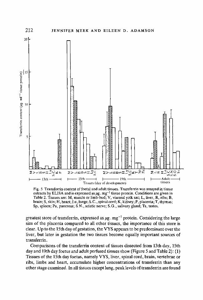
Fig. 5 Transferrin content of foetal and adult tissues. Transferrin was assayed in tissueextracts by ELISA and is expressed as /ig. mg"1 tissue protein. Conditions are given inTable 2. Tissues are: M, muscle or limb-bud; V, visceral yolk sac; L, liver, R, ribs; B,brain; S, skin; H, heart; Lu, lungs; S.C., spinal cord; K, kidney; P, placenta; T, thymus;Sp, spleen; Pa, pancreas; S.N., sciatic nerve; S.G., salivary gland; Ts, testes.
greatest store of transferrin, expressed as jug. mg"1 protein. Considering the largesize of the placenta compared to all other tissues, the importance of this store isclear. Up to the 15th day of gestation, the VYS appears to be predominant over theliver, but later in gestation the two tissues become equally important sources oftransferrin.
Comparisons of the transferrin content of tissues dissected from 13th day, 15thday and 19th day foetus and adult perfused tissues show (Figure 5 and Table 2): (1)Tissues of the 15th day foetus, namely VYS, liver, spinal cord, brain, vertebrae orribs, limbs and heart, accumulate higher concentrations of transferrin than anyother stage examined. In all tissues except lung, peak levels of transferrin are found

Transferrin synthesis and secretion 213
Table 2. Transferrin content of washed foetal and perfused adult mouse tissues
Tissue
VYSliver
S.C.brainS.N.
thymusspleenrib cage
muscleheart
pancreaskidneylungsal. gl.testes
skin
15th day foetusi"g • g"1
wet wt.
100 ± 22197
14 ±827 ±2
43-3
92-39-2
11-5
15-3
19th day foetusJ"g • g"1
wet wt.
775 ± 625599 ± 506
49 (5-173)29 ± 9
2063
125 (28-278)
110 ± 8271 ±16
28191128 (41-215)
406 ± 232
adultjwg • g"1
wet wt.
352 ± 224
28143 ± 101170 (35-285)
114 ± 19145 ± 36
359 ± 71132139 ± 47
The transferrin content of extracts was estimated by ELISA (see Methods section for details).Washed or perfused tissues were incubated for 3 h in culture media before separate analyses oftotal transferrin that was present in the medium and in the tissue. These figures were also usedto derive those in Figure 5. VYS, visceral yolk sac; S .C, spinal cord; S.N., sciatic nerve; sal. gl.,submandibulary salivary gland.
Means of 2-4 experiments ± standard deviations with at least triplicate samples. In some cases,parentheses showing the range are given. Single value indicates the result of one experiment withat least duplicate samples. Blank spaces indicate no determinations.
in the 15th day foetus; (2) In the lung, transferrin continues to accumulate throughoutgestation and into the adult stage where levels become similar to the stores in liver; (3)Nervous tissues (spinal cord and brain) of the foetus accumulate transferrin tomoderate levels which decline after the 15th day. The adult sciatic nerve contains con-siderable concentrations of transferrin; in some individuals this level is higher than inthe liver when expressed as a proportion of the total protein concentration in thattissue; (4) The total store of transferrin in skeletal muscle is very high in adults and latefoetuses considering that a high proportion of body mass is muscle. In addition, 15thday limb (predominantly developing muscle and cartilage) contains the highestconcentrations of transferrin of any tissues examined. The rib cage and vertebraeof mid- to late-gestation mouse foetuses also contain high levels.
Release of transferrin by foetal tissuesWe made a further comparison to determine the rates at which tissues could
release transferrin into the medium during organ culture. Figure 6 shows that most

214 JENNIFER MEEK AND EILEEN D. ADAMSON
15
10
I 1C/3
Tissues (day of development)
Fig. 6 Transferrin released by dissected tissues incubated in organ culture for 3 h at37 °C. Transferrin was determined by ELISA as described in the Materials and Methodssection. Tissues are those listed in Figure 5; data given in Figure 5 were obtained in thesame experiments.
15th day foetal tissues (muscle/limbs, ribs/vertebrae, brain, skin and heart) arecapable of releasing transferrin into the incubation medium at rates higher than thatof liver. Similar observations hold for 19th day foetal muscle, rib cage, brain, skin,heart, lung and kidney. Very slow rates of release are detected from spinal cord,thymus, spleen, and pancreas when expressed as fig released. mg"1 tissue protein.
Transferrin synthesis by adult tissuesIn addition to foetal liver and VYS, mouse spinal cord, brain, skin, lung, spleen,
rib cage and pancreas in foetuses from the 15th to the 19th day of gestationsynthesize transferrin as shown by metabolic labelling and electrophoretic analysisof immunoprecipitates (Fig 3A,B,C and Table 1). If we are to conclude that thesesources of newly synthesized transferrin are significant for the development ormaturation of tissues, we might expect that they are switched off in adult tissueswhen they would no longer be needed. Adult mouse lung, spleen, brain, liver andpinna of the ear (skin and cartilage, etc.) were labelled with p5S]methionine asdescribed in Materials and Methods. Autofluorographs show that synthesis of

Transferrin synthesis and secretion 215
transferrin is undetectable in lung, spleen and brain, but is occurring atapproximately equal rates (expressed as c.p.m. g"1 wet weight) both in liver and inear pinna (Fig. 3D). The latter is a surprising and rich source that could derive fromthe sebaceous glands, epidermis, connective tissue, cartilage or blood elementscontained within the pinna. Further studies will be needed to answer this question.
DISCUSSION
Synthesis of transferrinWe report here that transferrin may be synthesized by a variety of cell types in
the foetal mouse, since several tissues secrete a metabolically labelled polypeptidethat is antigenically related to adult mouse transferrin. It has previously beenreported that several adult mammalian tissues (liver, leucocytes, macrophages,Sertoli cells, salivary gland, testis, ovary and lactating mammary gland; Thorbeckeet al 1973; Imrie & Mueller, 1968; Tormey, Imrie & Mueller, 1972; Haurani,Meyer & O'Brien, 1973; Skinner & Griswold, 1980 and 1983) synthesize transferrinbut no foetal tissues other than liver and visceral endoderm have been identified tohave this ability in human (Gitlin & Perricelli, 1970), rat (Yeoh & Morgan, 1974)and mouse foetuses (Adamson, 1982). Recently, however, it has been reported thatchick embryo spinal cord synthesizes transferrin (Stamatos, Squicciarini & Fine,1983). We find that midgestation (15th day) foetal spinal cord, brain and skinsynthesize low but detectable levels of transferrin while very large amounts areproduced by the VYS and liver (Table 1). Late foetal spinal cord continues tosynthesize transferrin as do several other tissues (Fig. 3). The list of foetal tissuesthat synthesize transferrin may be much larger since it is possible that our detectionsystem is insufficiently sensitive or that tissues are damaged during trypsin-EDTAor other treatments. Of those adult tissues examined, only the liver and the pinnaof the ear synthesize readily detectable amounts (Fig. 3D). These results suggestthat multiple synthetic sources of transferrin are required for the special demandsof certain foetal tissues during gestation.
Importance of the visceral yolk sacThe VYS has long been known to play an important role in acting as a protective
barrier, as a transporter of selected molecules as well as producing nutritivematerials to the developing foetus. Transferrin is one of several nutritivemacromolecules synthesized very early in mouse development (7th day) by thevisceral endoderm and later by the VYS (Adamson, 1982). Other VYS productswith carrier and nutritive roles are AFP (Yeoh & Morgan, 1974; Dziadek & Adam-son, 1978) and apolipoprotein (Shi & Heath, 1984). The major source of transferrinfor the rapidly proliferating cells of the developing embryo is clearly the VYS, sincethe foetal liver may only commence significant synthesis by the 13th day or later(15th day in rat, Yeoh & Morgan, 1974). The immense importance of the VYS insupporting embryonic development cannot be overstated. It therefore may seem

216 JENNIFER MEEK AND EILEEN D. ADAMSON
that other tissues that synthesize transferrin must be relatively minor sources.However, the proximity of a transferrin source or delivery by a special mechanismmay be important for some developing tissues especially since arteries and bloodcapillaries would not yet be organized in the primordial organ buds of the embryo.
Retention of transferrin by tissuesIn perfused tissues, most of the serum transferrin has been removed and what
remains is present in these compartments: (1) Intracellular transferrin newlysynthesized in that tissue; (2) Transferrin that has been endocytosed or that isbound to cell surface receptors; (3) Transferrin in the interstitial fluid and remainingserum. All of the compartments may release transferrin readily to the surroundingmedium. The time taken for intracellular transferrin to be released after synthesisis less than H h (our unpublished observations) and it has been documented thatendocytosed transferrin may be released intact within minutes of entry into cells(Octave, Schneider, Trouet & Crichton, 1983). In these studies we have measuredthe release of transferrin from these combined compartments in order to estimatethe influence that each tissue may have on nearby target tissues in terms of provid-ing an essential nutrient or stimulator, namely, transferrin.
Transferrin is found in foetuses at levels far in excess of those in adults, therefore weconclude that transferrin is as necessary to embryonic development in general as it isto cells cultured in defined media. Transferrin is present in 15-day foetal tissues, at atime when rapid organ growth is occurring, at two- to fifty-fold higher levels than in13th or 19th day foetuses. These findings indicate (a) possible specialized roles incertain tissues such as muscle, bone and skin, and (b) the importance of a store oftransferrin at stages when the circulatory system is not well developed.
A developmental role for transferrinTransferrin is a required factor for the proper development of muscle (Cohen &
Fischbach, 1977; Podlewski et al. 1978; Kuromi, Gonoi & Hasegawa, 1981; Mar-kelonis etal. 19S2a,b; Stamatos etal. 1983; Matsuda, Spector & Strohman, 1984a;Matsuda, Spector, Micou-Eastwood & Strohman, 19846), kidney (Ekblom, Thes-leff, Miettinen & Saxen, 1981; Ekblom et al 1983), teeth (Partanen, Thesleff &Ekblom, 1984) and blood (Broxmeyer et al. 1980; Pelus, Broxmeyer, de Sousa &Moore, 1981; Gentile & Broxmeyer, 1983). We show here that transferrin levelsare highest when the foetal tissues' proliferative demands are very great and con-clude that developmental requirements could be met by one or all of the following:(a) synthesis in the individual tissue; (b) retention and reuse; (c) delivery fromadjacent tissues; (d) delivery by nerves.
There is some evidence that transferrin exerts a mitogenic effect independentlyof its ability to supply iron to proliferating tissues (Imrie & Mueller, 1968; Tormeyet al. 1972). The recent findings that there is some homology between the oncogeneBlym and transferrin (Goubin et al. 1983) also support the notion that transferrinmay be a general mitogen.
Transferrin synthesis and secretion 217We are indebted to Drs. A. Grover, S. Edwards, and D. Mercola for constructive criti-
cisms of the work. We thank G. Sandford for excellent photography and D. Lowe for typingthe manuscript. This study was supported by grants from the NIH (HD18782, P30 CA 30199 andCA 28427).
REFERENCESADAMSON, E. D. (1982). The location and synthesis of transferrin in mouse embryos and
teratocarcinoma cells. Devi Biol. 91, 227-236.ANDERSON, W. L., CHASE, C. G. & TOMASI, T. B. (1982). Transferrin support of stimulated
lymphocytes. In Vitro 18, 766-774.BEACH, R. L., POPIELA, H. & FESTOFF, B. W. (1983). The identification of neurotrophic factor
as transferrin. FEBS Lett. 156,151-156.BONNER, W. M. & LASKEY, R. A. (1974). A film detection method for tritium-labelled proteins
and nucleic acids in polyacrylamide gels. Eur. J. Biochem. 46, 83-88.BROCK, J. H. (1981). The effect of iron and transferrin on the response of serum-free cultures of
mouse lymphocytes to Concanavalin A and lipopolysaccharide. Immunology 43, 387-398.BROXMEYER, H. E., DE SOUSA, M., SMITHYMAN, A., RALPH, P., HAMILTON, J., KURLAND, J. I. &
BOGNACKI, J. (1980). Specificity and modulation of the action of lactoferrin, a negative feed-back regulator of myelopoiesis. Blood 55, 324-333.
COHEN, S. A. & FISCHBACH, G. D. (1977). Clusters of acetylcholine receptors located at identifiednerve-muscle synapses in vitro. Devi Biol. 59, 24-38.
DZIADEK, M. & ADAMSON, E. D. (1978). Localization and synthesis of alphafetoprotein in post-implantation mouse embryos. /. Embryol. exp. Morph. 43, 289-313.
EKBLOM, P., THESLEFF, I., MIETTINEN, A. & SAXEN, L. (1981). Organogenesis in a definedmedium supplemented with transferrin. Cell Differ. 10, 281-288.
EKBLOM, P., THESLEFF, I., SAXEN, L., MIETTINEN, A. & TIMPL, R. (1983). Transferrin as a fetalgrowth factor: Acquisition of responsiveness related to embryonic induction. Proc. natn. Acad.Sci., U.S.A. 80, 2651-2655.
ENGVALL, E. (1980). Enzyme immunoassay ELISA and EMIT. Meth. Enzymol. 70, 419-439.FAULK, W. P. & GALBRAITH, G. M. P. (1979). Trophoblast transferrin and transferrin receptors
in the host-parasite relationship of human pregnancy. Proc. Roy. Soc. London B. 204, 83-97.GENTILE, P. & BROXMEYER, H. E. (1983). Suppression of mouse myelopoiesis by administration
of human lactoferrin in vivo and the comparative action of human transferrin. Blood 61,982-993.
GITLIN, D. & PERRICELLI, A. (1970). Synthesis of serum albumin, prealbumin, a-foetoprotein,a-antitrypsin and transferrin by the human yolk sac. Nature 228, 995-997.
GOUBIN, G., GOLDMAN, D. S., LUCE, J., NEIMAN, P. E. & COOPER, G. M. (1983). Molecularcloning and nucleotide sequence of a transforming gene detected by transfection of chicken B-cell lymphoma DNA. Nature 302, 114-119.
HAURANI, F. E., MEYER, A. & O'BRIEN, R. (1973). Production of transferrin by the macrophage./. Retic. Soc. 14, 309-316.
Ii, I., KIMURA, J. & OZAWA, E. (1982). A myotrophic protein from chick embryo extract: itspurification, identity to transferrin and indispensability for avian myogenesis. Devi Biol. 94,366-377.
IMRIE, R. C. & MUELLER, G. C. (1968). Release of a lymphocyte growth promoter in leukocytecultures. Nature 219, 1277-1279.
KUROMI, H., GONOI, T. & HASEGAWA, S. (1981). Neurotrophic substance develops tetrodotoxin-sensitive action potential and increases curare-sensitivity of acetylcholine responses in culturedrat myotubes. Devi Brain Res. 1, 369-379.
LAEMMLI, U. (1970). Cleavage of structural proteins during the assembly of the head of bac-teriophage T4. Nature 227, 680-685.
MARKELONIS, G. J., OH, T. H., ELDEFRAWI, M. E. & GUTH, L. (1982a). Sciatin: a myotrophicprotein increases the number of acetylcholine receptors and receptor clusters in culturedskeletal muscle. Devi Biol. 89, 353-361.

218 JENNIFER MEEK AND EILEEN D. ADAMSON
MARKELONIS, G. J., BRADSHAW, R. A., OH, T. H., JOHNSON, J. L. & BATES, O. J. (19826). Sciatinis a transferrin-like polypeptide. /. Neurochem. 39, 315-320.
MATSUDA, R., SPECTOR, D. & STROHMAN, R. C. (1984a). There is selective accumulation of agrowth factor in chicken skeletal muscle. I. Transferrin accumulation in adult anteriorlatissimus dorsi. Devi Biol. 103, 267-275.
MATSUDA, R., SPECTOR, D., MICOU-EASTWOOD, J. & STROHMAN, R. C. (19846). II. Transferrinaccumulation in dystrophic fast muscle. Devi Biol. 103, 276-284.
OCTAVE, J.-N., SCHNEIDER, Y.-J., TROUET, A. & CRICHTON, R. R. (1983). Iron uptake andutilization by mammalian cells. I. Cellular uptake of transferrin and iron. Trends in Biochemi-cal Science 8, 217-220.
PARTANEN, A. M., THESLEFF, I. & EKBLOM, P. (1984). Transferrin is required for early toothmorphogenesis. Differentiation 27, 59-66.
PELUS, L. M., BROXMEYER, H. E., DE SOUSA, M. & MOORE, M. A. S. (1981). Heterogeneityamong resident murine peritoneal macrophages: Separation and functional characterization ofmonocytoid cells producing granulocyte-macrophage colony stimulating factor (GM-CSF) andresponding to regulation by lactoferrin. /. Immunol. 126, 1016-1021.
PODLEWSKI, T. R., AXELROD, D., RAVDIN, P., GREENBERG, L., JOHNSON, M. M. &SALPETER, M.M. (1978). Nerve extract induces increase and redistribution of acetylcholine receptors oncloned muscle cells. Proc. natn. Acad. ScL, U.S.A. 75, 2035-2039.
PUTNAM, F. W. (1975). In The Serum Proteins (ed. F. W. Putnam), Vol. 1, pp. 265-316. NewYork: Academic Press.
RENFREE, M. B. & MCLAREN, A. (1974). Foetal origin of transferrin in mouse amniotic fluid.Nature 252, 150-161.
SATO, G. H. et al. (1979). In Methods in Enzymology (ed. W. B. Jakoby & I. H. Pastan), Vol.58, p. 89. New York: Academic Press.
SAWATZKI, G., ANSELSTETTER, V. & KUBANEK, B. (1981). Isolation of mouse transferrin usingsalting-out chromatography on Sepharose CL-6B. Biochim. biophys. Acta 667, 132-138.
SEDMAK, J. J. & GROSSBERG, S. E. (1977). A rapid, sensitive and versatile assay for protein usingCoomassie brilliant blue G250. Analyt. Biochem. 79, 544-552.
SHI, W.-K. & HEATH, J. K. (1984). Apolipoprotein expression by murine visceral yolk sac endo-derm. /. Embryol. exp. Morph. 81, 143-152.
SKINNER, M. K. & GRISWOLD, M. D. (1980). Sertoli cells synthesize and secrete a transferrin-likeprotein. /. biol. Chem. 255, 9523-9525.
SKINNER, M. K. & GRISWOLD, M. D. (1983). Multiplication stimulating activity (MSA) cansubstitute for insulin to stimulate the secretion of testicular transferrin by cultured Sertoli cells.Cell Biol. int. Rep. 7, 441-446.
STAMATOS, C , SQUICCIARINI, J. & FINE, R. E. (1983). Chick embryo spinal cord neuronssynthesize a transferrin-like myotrophic protein. FEBS Lett. 153, 387-390.
THORBECKE, G. J., LIEM, H. H., KNIGHT, S., COX, K. & MULLER-EBERHARD, U. (1973). Sites offormation of the serum proteins transferrin and hemopexin. J. din. Invest. 52, 725-731.
TORMEY, D. C , IMRIE, R. C. & MUELLER, G. C. (1972). Identification of transferrin as a lympho-cyte growth promoter in human serum. Expl Cell Res. 74,163-169 and 220-226.
YEOH, G. C. T. & MORGAN, E. H. (1974). Albumin and transferrin synthesis during developmentin the rat. Biochem. J. 144, 215-224.
{Accepted 15 November 1984)