Embed Size (px)
Citation preview
Transdifferentiation of glioblastoma cells intovascular endothelial cellsYasushi Sodaa, Tomotoshi Marumotoa,b, Dinorah Friedmann-Morvinskia, Mie Sodaa, Fei Liua, Hiroyuki Michiuec,Sandra Pastorinod, Meng Yange, Robert M. Hoffmane,f, Santosh Kesarid, and Inder M. Vermaa,1
aLaboratory of Genetics, Salk Institute for Biological Studies, La Jolla, CA 92037; bDivision of Molecular and Clinical Genetics, Department of MolecularGenetics, Medical Institute of Bioregulation, Kyushu University, Higashi-ku, Fukuoka 812-8582, Japan; cDepartment of Physiology, Okayama UniversityGraduate School of Medicine, Dentistry, and Pharmaceutical Sciences, Okayama 700-8558, Japan; dDepartment of Neurosciences, Moore’s Cancer Center,University of California at San Diego, La Jolla, CA 92093; eAntiCancer, Inc., San Diego, CA 92111; and fDepartment of Surgery, University of California at SanDiego, San Diego, CA 92103
This Feature Article is part of a series identified by the Editorial Board as reporting findings of exceptional significance.
Edited by Douglas Hanahan, Swiss Institute for Experimental Cancer Research and Swiss Federal Institute of Technology Lausanne, Lausanne, Switzerland, andapproved December 27, 2010 (received for review October 26, 2010)
Glioblastoma (GBM) is the most malignant brain tumor and ishighly resistant to intensive combination therapies and anti-VEGFtherapies. To assess the resistance mechanism to anti-VEGF ther-apy,we examined the vessels of GBMs in tumors thatwere inducedby the transduction of p53+/− heterozygous mice with lentiviralvectors containing oncogenes and themarker GFP in the hippocam-pus of GFAP-Cre recombinase (Cre) mice. We were surprised toobserve GFP+ vascular endothelial cells (ECs). Transplantation ofmouse GBM cells revealed that the tumor-derived endothelial cells(TDECs) originated from tumor-initiating cells and did not resultfrom cell fusion of ECs and tumor cells. An in vitro differentiationassay suggested that hypoxia is an important factor in the differ-entiation of tumor cells to ECs and is independent of VEGF. TDECformationwas not only resistant to an anti-VEGF receptor inhibitorin mouse GBMs but it led to an increase in their frequency. A xe-nograft model of human GBM spheres from clinical specimens anddirect clinical samples from patients with GBM also showed thepresence of TDECs. We suggest that the TDEC is an importantplayer in the resistance to anti-VEGF therapy, and hence a potentialtarget for GBM therapy.
mouse model | glioma | angiogenesis | hypoxia-inducible factor 1
Glioblastoma multiforme (GBM) is the most common andlethal form of brain cancer. Despite optimal treatment and
evolving standard of care, the median survival of patients di-agnosed with GBM is only 12–15 mo (1). Because GBM is one ofthe most vascular-rich tumors and VEGF is produced by tumorcells, the anti-VEGF antibody bevacizumab (Avastin) is beingused in clinical trials (2). In a phase II clinical trial, more thanhalf of the patients with GBM responded to the combinationtreatment of bevacizumab and irinotecan, but this effect wastransient in most patients (3). Mechanisms proposed to explainresistance to anti-VEGF therapy include activation of otherproangiogenic signaling pathways, recruitment of bone marrow(BM)-derived myeloid cells that protect and nurture vascularcells, protection of blood vessels by increased pericyte coverage,and increased tumor invasion (4, 5). In GBMs, the antitumoreffect of the antiangiogenic therapies is likely attributable to nor-malization of vasculature, which also decreases edema (3, 6).Recent studies have shown that tumor cells become more ag-gressive after antiangiogenic therapy (7).The tumor vessels in GBMs are different from normal blood
vessels morphologically and functionally. As with other tumors,the tumor vessels in GBMs are tortuous; disorganized; highly per-meable; and abnormal in the endothelial walls, pericyte coverage,and basement membrane, resulting in loss of the blood–brain bar-rier (2). In addition, angiogenesis in GBMs has a unique feature,so-called “glomeruloid tufts,” exhibiting aggressive proliferation ofendothelial cells (ECs) compared with those of anaplastic glioma(grade III) (2). Recently, the possibility of EC differentiation oftumor cells has been suggested in lymphoma, myeloma, chronic
myeloid leukemia (CML), breast cancer, and neuroblastoma (8–12). Therefore, we surmised that the mechanism of vascular for-mation in GBM may also be different from that of regular tumorvascular formation.To investigate angiogenesis in GBMs, we examined our recent
mouse GBM model in which tumors developed 2–6 mo afterinjection with viral vectors. The tumors showed all the featuresof GBM, including hypervascularity (13). Furthermore, tumorswere GFP+, because the vectors contained GFP in addition toactivated oncogenes and loss of p53. Interestingly, in thesetumors, we found many GFP+ cells with EC characteristics,particularly in the deep area of the lesions, by confocal micros-copy and flow cytometry, indicating the presence of tumor-derivedendothelial cells (TDECs). Here, we demonstrate that the TDECsoriginated from tumor-initiating cells but not from con-taminated EC progenitors or cell-to-cell fusion between tumorcells and ECs. The TDECs are functional because blood flowsthrough them. Additionally, it has been suggested that hypoxia-inducible factor 1 (HIF-1) is an important enhancer of EC dif-ferentiation of tumor cells and that the formation of TDECsis independent of VEGF. Finally, direct clinical samples frompatients show EC cells with tumor markers. Our report showsdirect evidence of TDEC formation in GBMs, which may playa role in the resistance to anti-VEGF therapy.
ResultsECs Express Tumor-Specific Marker in Mouse GBM Models. To in-vestigate angiogenesis in GBMs, we used our recent mouseGBM model (13). As previously reported, Cre recombinase(Cre)-loxP–controlled lentiviral vectors encoding the activatedform of oncogenes H-Ras and Akt [pTomo vectors (13)] wereinjected stereotaxically into the hippocampus of GFAP-Cre-p53+/− mice. In this model, the oncogenes were expressed spe-cifically in GFAP+ cells and tumors expressed GFP, H-Ras, andAkt and showed loss of p53. Additionally, they were positivefor the neural progenitor cell marker nestin, which is oftenexpressed in human GBMs (14). To investigate the tumor vas-culature, we carried out immunofluorescence by confocal mi-croscopy using the endothelial antigens von Willebrand factor(vWF), CD31, CD34, and vascular endothelium (VE)-cadherin(CD144) as markers. Fig. 1A shows a normal EC, where the GFP
Author contributions: Y.S. and I.M.V. designed research; Y.S., T.M., D.F.-M., M.S., F.L., andH.M. performed research; S.P., M.Y., R.M.H., and S.K. contributed new reagents/analytictools; Y.S., D.F.-M., and I.M.V. analyzed data; and Y.S., D.F.-M., S.K., and I.M.V. wrotepaper.
The authors declare no conflict of interest.
This article is a PNAS Direct Submission.
See Commentary on page 4271.1To whom correspondence should be addressed. E-mail: [email protected].
This article contains supporting information online at www.pnas.org/lookup/suppl/doi:10.1073/pnas.1016030108/-/DCSupplemental.
4274–4280 | PNAS | March 15, 2011 | vol. 108 | no. 11 www.pnas.org/cgi/doi/10.1073/pnas.1016030108

in tumor cells is completely distinct from vWF, the endothelialantigen (Fig. 1A, i–iii). Surprisingly, we found that some ECsexpressed not only endothelial antigens but also GFP, whichmost likely originated from tumor cells (Fig. 1B, i–iii, comparewith the merge in Fig. 1 A, iii, and B, iii, and Fig. S1A). Addi-tionally some GFP+ ECs formed vessels with GFP− regular ECs,exhibiting a mosaic pattern [Fig. 1B, iv–vi; a z-series assay furtherconfirmed the mosaic pattern (Fig. S1A)]. Immunofluorescencewith other EC-specific antigens like CD34 and CD144 alsoshowed EC cells containing GFP, further supporting the for-mation of TDECs (Fig. 1B, vii–xii). The GFP+ ECs expressedthe transduced oncogene Flag-tagged H-RasV12 (Fig. 1C, i–v).Similar results were obtained with HA-tagged Akt. Nestin ex-pressed in tumor cells can be detected in both GFP+ EC cells(marked T) and GFP− EC cells (marked R), further strength-
ening the notion that GFP+ ECs (TDECs) most likely originatedfrom the tumor cells (Fig. 1C vi–x). The TDECs were also ob-served when GBM was generated using lentivectors (LVs) con-taining only activated H-Ras and small interfering p53 (sip53).To confirm the presence of GFP+ ECs, we also examined dis-sociated tumors by flow cytometry. Similar to the results ofconfocal microscopy, 10–25% of ECs (CD45−CD31+CD34+)were positive for GFP (Fig. 1D). The TDECs were mostly foundin the deep part of the tumor rather than on its surface, and thefrequency of vessels containing the GFP+ ECs was 6.4–37.8%(average of 24.6 ± 12.7%) in the deep area, depending onthe size of the tumor. In the surface area, 2.0–12.7% (average of8.33 ± 4.15%) of vessels contained the GFP+ ECs (Table S1). Ingeneral, the frequency of the TDECs was higher in large tumorsthan in smaller tumors. By injecting Hypoxyprobe-1 (NaturalPharmacia International) into tail veins of tumor-harboringmice, we showed that the deep area of the tumor was morehypoxic than the surface area (Fig. 2A), suggesting that hypoxiamay be an important factor for TDEC formation. Hypoxia leadsto angiogenesis by induction of VEGF through increasing levelsof HIF-1. Furthermore, tumors produced VEGF (13, 15). Weare now pursuing HIF-1 expression by immunofluorescencestudies. Interestingly, the majority of TDECs did not expressVEGF receptor 2 (R2) (Fig. 2B, vi–x), whereas most of theregular ECs (GFP−) expressed VEGF-R2 (Fig. 2B, i–v). TheTDECs also did not show expression of VEGF-R1 and VEGF-R3. Because FGF-2 is another important growth factorexpressed by the ECs and the GBM cells, we also examined theexpression of FGF receptor 1 (FGFR-1). In contrast to theresults of VEGF receptors, the FGFR-1 was expressed in bothTDECs (marked T) and regular ECs (marked R) as well as insurrounding tumor cells (Fig. 2C).
TDEC-Forming Vessels Are Functional. To assess if TDECs arefunctional, we determined blood flow in TDEC-containing ves-sels. We injected biotinylated lectin i.v., which can bind to ECsin mice harboring brain tumors, 15 min before euthanasia. To
GFP vWF
A
Merge
GFP CD31 Merge
GFP CD34 Merge
B
GFP vWF Merge
GFP CD144 Merge
i ii iii
i ii iii
iv v vi
vii viii ix
x xi xii
C
GFP vWF Merge Nestin DAPI vi vii viii ix x
R R R R T T T T
GFP CD31 Flag(Ras) DAPI Merge i ii iii iv v
D
100 101 102 103 104GFPGFP
24.4%
Count
100 101 102 103 104PE
100 101 102 103 104PE
CD34
CD
31
1.1* 7.1
62.6 29.2
100 101 102 103 104PC5.5
100 101 102 103 104PC5.5CD45
SS
C-H
CD31+CD34+
CD45-
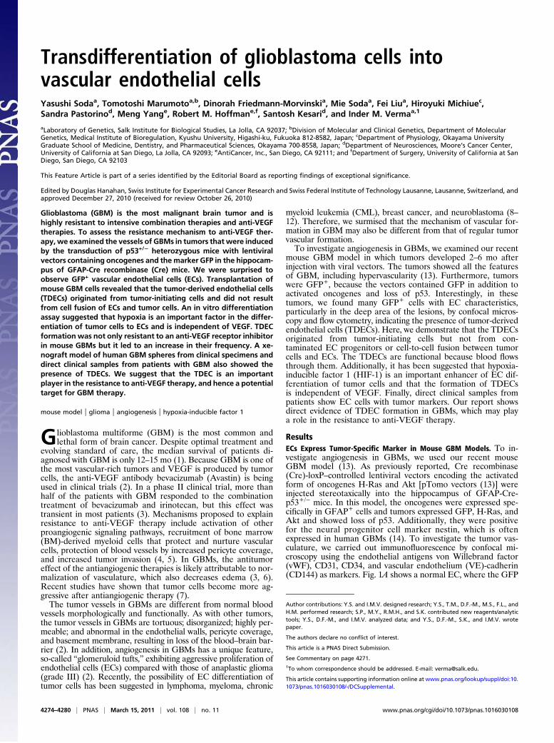
Fig 1. TDECs. GFAP-Cre/p53+/− mouse brain was transduced by Tomo H-RasV12 LVs and Tomo Akt LVs as described (13). A representative image of ECsobserved by confocal microscopy is shown. (A) Regular ECs lined the vessellumen and expressed EC marker vWF (ii) but not the tumor marker GFP (i).DAPI was used as the nuclear marker, and the image was incorporated in themerge panel (iii). (B) In contrast, TDECs expressed both the GFP marker (i, iv,vii, and x) and EC markers vWF (ii), CD31 (v), CD34 (viii), and CD144 (xi). SomeGFP+ ECs formed vessels with GFP− regular ECs (vi, arrowheads). DAPI wasused as the nuclear marker, and the image was incorporated in the mergepanels (iii, vi, ix, and xii). (C) TDECs expressed Flag-tagged H-RasV12 in addi-tion to GFP and CD31 (i–v, arrows). They also expressed nestin in addition toGFP and vWF (vi–x, R and T indicate regular ECs and TDECs, respectively). (D)Representative result of flow cytometry for dissociated brain tumors. In theCD45− population (Top), ECs were CD31+CD34+ and constituted 7.1% of thewhole tumor (Middle) and GFP+ ECs (TDECs) represented 24.4% of total ECs(Bottom). The asterisk represents the percentage of cells in each quadrant. Allconfocal pictures are single-slice images at an Airy factor of 1.0. [Magnifica-tion: all confocal images were taken at 63× with 3× (A and B) or 2× (C)electrical zoom (total magnification: 189× or 126×).]
GFP vWF Merge VEGF-R2 DAPI
GFP vWF Merge VEGF-R2 DAPI
Regular EC
TDEC
i ii iii iv v
vi vii viii ix x
GFP CD31 Merge FGF-R1 DAPI
T
R
DAPI i ii iii v iv
Transparency GFP Hypoxyprobe Merge
A
B
C
Fig. 2. Hypoxia and expression of HIF-1α and receptors of angiogenicgrowth factors. (A) Hypoxyprobe assay of the tumor. The hypoxic area (p02 <10 mmHg) was detected by the anti-Hypoxyprobe antibody. The dotted lineshows the approximate border of the hypoxic area. (Scale bar: 1 mm.) (B)Regular tumor ECs expressed VEGF-R2 (i–v), but GFP+ TDECs did not expressVEGF-R2 (vi–x). (C) GFP+ TDECs (T) expressed FGF-R1, as did regular ECs (R)and surrounding tumor cells. All confocal pictures are single-slice images atan Airy factor of 1.0. [Magnification: 63× with 3× electrical zoom (totalmagnification: 189×) except A, which was 1.25×.]
Soda et al. PNAS | March 15, 2011 | vol. 108 | no. 11 | 4275
CELL
BIOLO
GY
FEATU
REART
ICLE
SEECO
MMEN
TARY

visualize the blood flow, tumor sections were stained with fluo-rescence-labeled streptavidin, which binds to biotinylated lectin-labeled ECs. Just like regular ECs, lectin-bound TDECs wereobserved in many tumors, indicating that the TDEC-formingvessels are functional (Fig. 3 A and B). There were also non-functional TDEC-forming vessels (Fig. 3C).
TDECs in Transplanted Tumors. We next investigated the ability oftumor cells to differentiate into ECs by transplanting mouseGBM cell line 005, which is a tumor-initiating cell line estab-lished from our lentiviral vector-induced tumor model, into thebrain of a nonobese diabetic (NOD)-SCID mouse. On exami-nation of the tumor vessels in the transplanted mice, GFP+ ECswere observed (Fig. 4A, i–iv). We further established 14 sub-clones of the 005 cells and transplanted them into NOD-SCIDmouse brains. Most clones formed tumors in the same way as theparental 005 cells and contained GFP+ ECs in these tumors.Data from one such subclone are shown using CD34 as a markerof ECs (Fig. 4B, i–iv). Because it has been reported that someGFAP+ neural stem cells (NSCs) can transdifferentiate into ECs(16), it is possible that 005 cells may contain transduced GFAP+
NSCs in addition to tumor-initiating cells and that these GFAP+
NSCs differentiate into the GFP+ ECs. We therefore generateda cell line from another tumor (006) induced by pTomo vector.Results from 006 cells also show GFP+ ECs (Fig. 4C, i–iv), thusminimizing the possibility of the presence of GFAP+ NSCs intumor-initiating cells that differentiated into the ECs.
Fusion-Independent Mechanism in TDEC Formation. To exclude thepossibility that GFP+ ECs (TDECs) result from cell fusion oftumor cells and ECs but are not derived from tumor cells, wetransplanted 005 tumor cells into the brains of DsRed-transgenicnude mice and examined the expression of GFP, DsRed, and ECmarkers in the transplanted tumors. In these transgenic mice, theDsRed is driven by the CAG promoter and all cell types excepthair and red blood cells express DsRed (17). In tumors de-veloped in these mice, many DsRed+ host cells, including ECs,were infiltrating into the tumors (Fig. S1B). Confocal microscopyrevealed that GFP− ECs were expressing DsRed (Fig. 4D, i–iv),whereas GFP+ ECs were not expressing DsRed (Fig. 4D, v–viii),thus confirming that the GFP+ TDECs were derived from afusion-independent mechanism. To confirm these results, we alsoexamined dissociated tumors by flow cytometry. Similar to theresults of confocal microscopy, most of the GFP+ ECs wereDsRed−, whereas DsRed+ ECs were GFP− (Fig. 4E). We furtherexamined the cell fusion in the NOD-SCIDmouse transplantationmodel by flow cytometry. In this model, MHC class I H-2Kd
is expressed in the host cells but not in 005-derived tumor cells.
The majority of H-2Kd+ host-derived ECs were GFP−, whereasGFP+ ECs were H-2Kd− (Fig. 4F), again suggesting a fusion-independent mechanism of TDEC formation.
In Vitro Differentiation of GBM Cells to ECs: Role of Hypoxia and HIF-1.We next attempted to induce GBM initiating cells (005 cells) todifferentiate into ECs in vitro. Because HIF-1 is likely to be animportant factor for TDEC formation, we added an iron chelator,deferoxamine (DFO), into the culture media to mimic hypoxicconditions by blocking proline hydroxylase (18, 19), which sta-bilizes HIF-1α. Culturing 005 cells in DMEM/F-12 mediumsupplemented with FBS (DFS) and endothelial cell growth me-dium (EGM; Lonza), which contains FBS, human VEGF, humanEGF, human FGF-2, insulin-like growth factor, cortisol, andheparin, induced significant morphological changes. DFO en-
DAPI GFP CD31 MergeSAV(Lectin)
DAPI GFP CD31 MergeSAV(Lectin)
i ii iii iv v
DAPI GFP CD31 MergeSAV(Lectin)
A
B
C
i ii iii iv v
i ii iii iv v
Fig. 3. Blood flow in TDEC-forming vessels. Biotinylated lectin injection assayshowedthatboth regular vessels (A) andTDEC-formingvessels (B) are functionaland allowed blood flow. (C) Nonfunctional TDEC-forming vessels were alsoobserved. All confocal pictures are single-slice images at an Airy factor of 1.0.[Magnification: 63×with 3× electrical zoom (total magnification: 189×).]
DAPI GFP vWF Merge i ii iii iv
DAPI GFP CD34 Merge i ii iii iv
DAPI GFP vWF Merge i ii iii iv
GFP vWF Merge DsRed i ii iii iv
GFP vWF Merge DsRed v vi vii viii
100 101 102 103 104100 101 102 103 104
CD
31
CD45
100 101 102 103 104100 101 102 103 104
CD45-CD31+
DsR
ed
78.1* 5.6
GFP
2.1 14.2 100 101 102 103 104
GFP100 101 102 103 104
GFP
CD45-CD31+
78.0 5.7
0.8 15.5
H-2
Kd
GFP
A
B
C
D
E F
Regular EC
TDEC
Fig. 4. TDECs in tumor-initiating cells transplanted into immunocompro-mised mice. Representative images of TDECs in brain tumors developed inNOD-SCID mice transplanted with tumor-initiating cell line. Cell line 005 (A),005 subclone cells (B), and another tumor-initiating cell line 006 (C). TDECsexpressed GFP and EC markers vWF and CD34. (D) Representative image ofregular ECs and TDECs in brain tumors developed in DsRed nude micetransplanted with 005 cells by confocal microscopy. Regular tumor vessel ECsexpressed vWF and host marker DsRed but not tumor marker GFP (i–iv),whereas GFP+ vWF+ TDECs did not express DsRed (v–viii). TDECs are indicatedby arrows, and regular ECs are indicated by arrowheads (v–viii). All confocalpictures are single-slice images at an Airy factor of 1.0. [Magnification: 63×with 3× electrical zoom (total magnification: 189×).] (E) Representativeresults of flow cytometry of dissociated tumors developed in DsRed nudemice transplanted with 005 subclone cells. In the CD45−CD31+ EC fraction(Left), DsRed-positive cells did not express GFP (Right). (F) Representativeresults of flow cytometry for dissociated tumors developed in NOD-SCIDmice. In the CD45−CD31+ EC fraction, cells expressing NOD-SCID mouse-specific H-2Kd did not express GFP. The asterisk represents the percentage ofcells in each quadrant.
4276 | www.pnas.org/cgi/doi/10.1073/pnas.1016030108 Soda et al.
hanced the change to endothelial-likemorphology inDFSmediumor EGM but not in N2 medium, which is used to maintain theNSCs (Fig. 5A). In the DFSmedium and EGM,DFO significantlyenhanced the HIF-1α expression in the 005 cells (Fig. 5B) andinduced expression of endothelial antigens vWF and CD31 (Fig. 5C and D). However, VEGF-R2 expression was induced only ina small population of cells (Fig. 5D). Additionally, these differ-entiated cells formed a tube structure on Matrigel (BectonDickinson) (Fig. 5E). To confirm that this endothelial differen-tiation results from HIF-1α accumulation but not from a non-specific effect of DFO, we cultured the 005 cells in 2% O2(hypoxia). Tubular structures could be observed in the absence ofDFO when these cells were cultured in DFS medium or EGM butnot in N2 medium (Fig. 5E), suggesting that hypoxia, presumablythrough the activation of HIF-1 α (Fig. 5B), is playing an im-portant role in the endothelial differentiation.
VEGF-Independent Transdifferentiation of Tumor Cells. BecauseVEGF is a critical factor in tumor angiogenesis and is induced byhypoxia through accumulation of HIF-1α (15), we investigated therole of VEGF in the formation of TDECs. VEGF was releasedconstitutively from 005 cells in N2 medium at a low level (32.2 ±8.8 pg/mL per 106 cells), and the amount of VEGF release in-creased about threefold in DFS medium and EGM. In the pres-ence of DFO, however, secretion of VEGF in both DFS medium(243± 22.2 pg/mL) and EGM (368± 32.6 pg/mL) was significantly
increased. No effect was observed in N2 medium (Fig. 6A). Theseresults suggest that VEGF may play a role in endothelial differ-entiation of 005 cells. We therefore blocked autocrine VEGFfunction with anti-mouse VEGF neutralization antibody (NAb) inaddition to using EGM devoid of human VEGF. Tube formationof 005 cells cultured in DFS with DFO medium or in EGM withor without DFO was not inhibited at all despite the addition of1 μg/mL NAb (Fig. 6B), which completely inhibited activity of100 ng/mL VEGF on growth of human umbilical vein endothelialcells (HUVECs), whereas 10 ng/mL VEGF can enhance tubeformation of HUVECs (Fig. S2). We also added the anti-VEGFreceptor–specific small molecule inhibitor AG28262 (Pfizer),which inhibits autophosphorylation of VEGF-R1, VEGF-R2, andVEGF-R3 selectively at a subnanomolar concentration (20). Therewas no inhibition of tube formation even at a 20-nM concentrationof the inhibitor (Fig. 6C). Because the TDECs were expressingFGFR-1 in our mouse GBMs (Fig. 2D), we used a high concen-tration of the AG28262 (200 and 1,000 nM), which inhibits not onlyVEGF receptors but FGFR-1. However, there was no significantinhibition of tube formation (Fig. 6C). These results reaffirm thatGBM-initiating cells are able to differentiate into ECs by a VEGF-or FGF-independent mechanism.
Resistance of TDECs to Anti-VEGF Receptor Inhibitor. To confirm theresistance of TDECs to anti-VEGF therapies and that this re-sistance is playing an important role in the resistance of patients
N2 EGM
(+)
DFO DFS
(-)
DFO
HIF-1
Lamin
B1
N2 EGMDFS - + - + - +
N2
DFO(-)
DAPI vWF GFP Merge
EGM
DFO(+)
EGM
0.7* 0.3
98.2 0.7
1.7 1.0
95.1 2.2
2.0 5.8
76.1 16.1
0.7 0.3
97.3 1.7
0.4 2.0
50.4 47.2
0.5 6.6
24.7 68.3
N2 EGMDFS
CD31
(+)
(-)
VEGF -R2
DFO
A
B
C
D
E N2 DFS
DFO (-)
DFO(+)
Low O2
Normal O2
Normal O2
DFO(-)
Fig. 5. Endothelial differentiation of tumor-initiatingcells in vitro. (A) Morphological changes of 005 cells cul-tured in N2 medium, DFS medium, and EGM with orwithout DFO. (B) Expression of HIF-1α in 005 cells culturedin the indicated conditions. Nuclear protein was extractedfrom the cells and analyzed by Western blotting usinganti-HIF-1α antibody (Upper) or anti-lamin B1 antibody(Lower). (C) Representative confocal microscopy images of005 cells cultured in N2 medium without DFO (Upper) or inEGM with DFO (Lower). The confocal microscopy imagesare maximum projection images of consecutive single-sliceimages at an Airy factor of 1.0. (Magnification: 40×.) (D)Flow cytometry of CD31 and VEGF-R2 expression in 005cells cultured in various conditions. (E) Tube formationassay of 005 cells cultured in the indicated conditions andseeded on Matrigel. The 005 cells were cultured in DFSmedium with DFO and in EGM with or without DFO undernormoxia-formed tube structure, and all these cells wereGFP+. The hypoxic condition (low O2) in DFS medium andEGM without DFO also induced tube formation.
Soda et al. PNAS | March 15, 2011 | vol. 108 | no. 11 | 4277
CELL
BIOLO
GY
FEATU
REART
ICLE
SEECO
MMEN
TARY

with GBM to anti-VEGF therapies, we have examined the effectof VEGF receptor inhibitor on tumor development and TDECformation in vivo using our mouse GBM model. We have ad-ministrated the VEGF receptor inhibitor AG28262 from week6–12 following LV transduction. As shown in Fig. 6D, there wasno significant difference in survival between the control groupand AG28262 group (P = 0.3688), indicating that the VEGFinhibitor had almost no effect on tumor growth as observedin clinical studies. Examination of tumor vessels revealed thatTDECs increased in the treated mice compared with controlmice, however. Although the regular ECs decreased in thetreated mice, TDECs significantly increased in ratio comparedwith control mice (Fig. 6E). Furthermore, the increase of TDECsin the AG28262-treated mice was particularly significant in the
border area of the tumor, which contains fewer TDECs than thedeep area in control mice (Fig. 6E). These results indicate thatTDEC formation is resistant to the anti-VEGF therapy andstrongly suggest the contribution of TDECs in the clinical re-sistance of GBM to anti-VEGF therapies.
TDECs in Xenograft Tumors of Human GBM Spheres.We next asked ifTDECs were also found in human GBMs. We obtained threelenti-GFP–transduced human GBM spheres (21) and trans-planted them in the brains of NOD-SCID mice. The resultingtumors examined by immunofluorescence show that regularvascular ECs express vWF but not human nestin or GFP (Fig.7A). In contrast, some ECs express not only vWF but humannestin and GFP (Fig. 7B). Fig. 7C further shows that regularGFP− ECs were human CD31 (hCD31)-negative but mouseCD31 (mCD31)-positive, whereas GFP+ EC cells expressedhCD31 but not mCD31 (Fig. 7D). It thus appears that like themouse GBMs, human GBMs are also capable of formingTDECs. The average ratio of TDECs in total ECs in threetransplanted GBMs was 15–44% in the deep area and 4–22% inthe border area (Fig. S3). Therefore, as in the mouse model,hypoxia may also play an important role in TDEC formation inhuman GBMs.
Presence of EGF Receptor-Positive ECs in Clinical Samples of Patientswith GBM. Finally, we wanted to determine if direct clinicalsamples from patients with GBM also contained TDECs. Wetook advantage of the genetic abnormalities in the form of EGFreceptor (EGFR) amplification in these tumors and asked ifsome ECs contained both human vWF and EGFR. Fig. 7E showsthe ECs in the normal human brain by immunofluorescence withvWF antigen (Fig. 7E, i, iii, and iv) but no reactivity to anti-EGFR antigen (Fig. 7E, ii, iii, and iv). In contrast, Fig. 7F showsthat some ECs in the clinical tumor sample express both vWFand EGFR (Fig. 7F i–iv), offering strong evidence for the pres-ence of TDECs in human GBMs.
DiscussionIn tumor angiogenesis, BM-derived circulating endothelial pre-cursors (CEPs) are known to be the main source of the vascularECs (22). A recent study suggested that the BM-derived CEPsdid not contribute to the vascular endothelium, however (23). Todate, the presence of TDECs has been suggested in severalneoplasias, such as CML, lymphoma, and myeloma, by analyzingclinical samples (8–10). In these tumors, tumor-specific fusiongenes resulting from chromosomal translocation were used forthe tumor-specific markers. Here, we have demonstrated thepresence of blood vessel ECs expressing the tumor marker GFPin our recently developed mouse GBM model, in human GBMxenografts, and clinical samples from patients. In contrast to theconventional theory of tumor angiogenesis in which the ECs arederived from mesodermal BM progenitor cells (22), the presenceof TDECs in GBM suggests that the ECs transdifferentiatedfrom the neuroectoderm and that tumor cells can also be in-volved in tumor angiogenesis. The endothelial transdifferentia-tion of the tumor cells may result from the aberrant stem cellcharacter of the tumor progenitor cells. The other possiblemechanism is that the endothelial differentiation of GBM cells isnot the result of transdifferentiation but reflects the normaldifferentiation pathway of the NSC, which has previously beendescribed to differentiate into ECs (16). If this is also observed inthe normal differentiation of human NSCs, perhaps the termi-nology of transdifferentiation needs reconsideration.Vasculogenic mimicry (VM) has been reported in melanoma
as fluid-conducting channels formed by the tumor cell itself. Incontrast to the regular blood vessels, VM lacks ECs; therefore,VM was easily distinguishable from regular blood vessels. VMwas also reported in nonmelanoma tumors, including GBM (24–26). The TDECs in this study are likely to be different from theVM because the TDECs are indistinguishable from regular ECs,except for the tumor-specific markers (e.g., GFP) or chromo-
N2 EGM DFS
VEG
F [p
g/m
L/10
6 cel
ls]
- + - + - +0
100
200
300
400
500
(-)
(+)
EGM/VEGF(-) DFS/DFO(+) DFO (-) DFO (+)
-VEGF NAb
0
20
Bright GFP AG28262 [nM]
200
1000
0
0.2
0.4
0.6
0.8
1.0
0 10 20 30 40 50 60 70 80 90Days after vector injection
Cum
ulat
ed s
urvi
val r
ate
AG28262
(100 mg/kg/d)
0.5%CMC
(vehicle)
Treatment
10
20
30
40
50
60
70
TDEC
+ ve
ssel
s [%
]
0
Vehicle control AG28262
**
**
NS
A
B
C
D E
Deep Area Border
Fig. 6. Effect of inhibition of VEGF on TDEC formation. (A) Concentrationof mVEGF in culture supernatant of 005 cells in various conditions. VEGF wasreleased from 005 cells constitutively, and DFO treatment enhanced theproduction of VEGF significantly in DFS medium and EGM. Data representmean ± SD of triplicate assays. (B) Effect of anti-VEGF NAb on tube forma-tion. The 005 cells cultured in the indicated conditions were seeded onMatrigel, and tube formation was observed after 20 h. We omitted humanVEGF from the EGM in this assay and used 1 μg/mL anti-VEGF NAb. (C) Effectof anti-VEGF receptor small molecule inhibitor AG28262 on tube formationof 005 cells. We cultured cells and observed tube formation under the samecondition indicated in B, except for the addition of NAb. (D) Survival curve ofthe GBM mice treated with AG28262. GFAP-Cre transgenic mice receivedstereotaxic injection of LVs in the hippocampus of the brain. Mice wereadministrated 100 mg·kg−1·d−1 AG28262 orally for 6 wk from the sixth weekafter lentiviral injection. Control mice were administrated vehicle (0.5%carboxyl methyl cellulose). The survival curve was obtained by the Kaplan–Meier method, and the statistical difference was examined by the log-ranktest. (E) Frequency of TDEC-forming vessels in the mouse GBM. Tumors wereobtained from the mice that developed tumors and examined by immuno-fluorescence assay using a confocal microscope. Data represent mean ± SDfrom six (control) or five (AG28262) mice. *<0.5% by the Mann–Whitney Utest; **<0.5% by the Wilcoxon signed-rank test; NS, not significant by theWilcoxon signed-rank test.
4278 | www.pnas.org/cgi/doi/10.1073/pnas.1016030108 Soda et al.

somal rearrangements. There is another aberrant tumor vessel,the “mosaic tumor vessel,” which was reported in colon cancer(27). The mosaic blood vessels are lumens formed with both ECsand tumor cells lacking EC markers. Because the TDECs areexpressing EC markers and behave as regular ECs, the TDECsare likely to be different from the mosaic vessels.The transdifferentiation of tumor cells into vessel formation in
GBM was not previously recognized, probably because of the lackof a good tumor marker. We also suggest that hypoxia is an im-portant factor of endothelial differentiation in addition to regulartumor vessel formation. In the hypoxic condition, induction ofVEGF expression through the stabilization of HIF-1α is an im-portant factor for tumor angiogenesis (15). In contrast to regularendothelial differentiation, however, in vitro assays have suggestedthat the formation of TDECs is independent of VEGF and FGF(Fig. 6 B and C). In addition, administration of the anti-VEGFreceptor inhibitor AG28262 did not improve survival of the GBMmice (Fig. 6D), and TDEC formation increased in contrast to
regular ECs (Fig. 6E). Therefore, the involvement of TDECs intumor angiogenesis might be one of the resistance mechanismsagainst anti-VEGF therapies and may require novel combina-tion therapies.While this paper was under review, two articles (28, 29) were
published that further support the notion that a proportion ofECs contributing to the formation of blood vessels in humanGBMs originate from tumor cells. The findings of these twogroups show that ECs (ranging from 20–90%) in the tumorscarry genetic abnormalities found in the tumor cells themselves.Thus, together with the findings reported here, it is clear thatpart of the vasculature in GBMs originates from tumor cells,bypassing the normal mechanisms of angiogenesis, thus offeringan additional therapeutic opportunity to treat the disease.
Materials and MethodsEstablishment of Mouse GBM Model by Lentiviral Vector Injection. The mouseGBM model was established as described (13). Briefly, we injected the Cre-inducible LVs Tomo H-RasV12 LV and Tomo AKT LV stereotaxically into thehippocampus of GFAP-Cre/p53+/− transgenic mice. More recently, mouseGBM models have also been generated in GFAP-Cre mice using a singlelentiviral vector containing activated H-Ras and sip53. We have killed miceto take tumor samples when the mice show tumor-related signs, such asa domed head, a hunched position, lethargy, and weight loss. In most cases,it takes 3–4 mo after vector injection before tumor-related signs appear.
Cell Culture. Mouse GBM-initiating cell lines 005 and 006 were established asdescribed (13). The 005 and 006 cells were maintained in N2 medium, whichcontains DMEM/F-12 (Omega Scientific), 1%N2 supplement (Invitrogen), 20 ng/mL human FGF-2 (Peprotech), 20 ng/mL human EGF (Promega), and 40 μg/mLheparin (Sigma). In the differentiation-induction assay, cells were cultured inDFS medium [10% (vol/vol) FBS] or EGM-2 (Lonza). To reproduce the hypoxiccondition, we added 100 μg/mL DFO mesylate (Sigma) into the above media.The 005 cellswere also cultured in the2%O2 conditionusinganN2O2 incubator.Mouse GBM-initiating 005 cells were transplanted into the hippocampus ofNOD-SCIDmice or DsRed transgenic mice. HUVECs were cultured in the EGM-2.
Transplantation of Mouse GBM-Initiating Cells. Mouse GBM-initiating 005 and006 cells were transplanted into brains of NOD-SCIDmice or DsRed transgenicmice. A total of 3 × 10⁵ cells were suspended in 1–1.5 μL of PBS and injectedstereotaxically in the right hippocampus. These mice developed GBM about1–2 mo after transplantation. In some cases, as few as 5,000 cells wereinjected, except the tumors took longer to develop.
Immunofluorescence Assay. Mouse brain tumors were processed as described(13). The primary antibodies used in this study are as follows: rabbit anti-vWF(Abcam), rat anti-mCD31 (MEC13.3; Becton Dickinson), rat anti-mCD34(RAM34; Becton Dickinson), rat anti-mCD144 (11D4.1; Becton Dickinson),chicken anti-nestin (Abcam), rabbit anti-DYKDDDK (Cell Signaling), goatanti-VEGF-R2 (Abcam), and rabbit anti-FGF-R1 (Abcam). The secondaryantibodies used were as follows (all from Invitrogen): Alexa Fluor 568 anti-rabbit IgG, Alexa Fluor 568 anti-rat IgG, Alexa Fluor 647 anti-rabbit IgG,Alexa Fluor 647 anti-rat IgG, Alexa Fluor 647 anti-chicken IgG, and AlexaFluor 647 anti-goat IgG. The nucleus was stained by DAPI. The images wereobtained by confocal laser scanning microscopy (TCS SP2 ABS; Leica or LSM 5PASCAL; Carl Zeiss), and the obtained images were processed by Photoshopsoftware (Adobe).
Hypoxyprobe Assay. To detect hypoxic regions of the brain tumors, a Hypoxyp-robe-1Omnikit (NaturalPharmacia International)wasused.Weinjected45mgofHypoxyprobe-1 into the tail veins of tumor-harboring mice 30 min beforeeuthanasia. Brain sections were stained with rabbit anti-Hypoxyprobe anti-body, followed by staining with Alexa Fluor 647-labeled anti-rabbit IgG an-tibody (Invitrogen). The images were obtained by confocal laser scanningmicroscopy.
Blood Flow Detection Assay. We injected 50 μg of biotinylated lectin (VectorLaboratories) into the tail veins of tumor-harboring mice 15 min before eu-thanasia. Brain sectionswere stainedwith Alexa Fluor 647-labeled streptavidin(Invitrogen). The images were obtained by confocal laser scanningmicroscopy.
Flow Cytometry. The brain tumors were dissociated using a Neural TissueDissociation Kit (Miltenyi Biotec), and 005 cells were collected after differ-
DAPI GFP vWF Merge hNestin i ii iii v iv
DAPI GFP vWF Merge hNestin i ii iii v iv
DAPI GFP mCD31 Merge hCD31 i ii iii v iv
DAPI GFP mCD31 Merge hCD31 i ii iii v iv
vWF EGFR
vWF+EGFR vWF+EGFR +Hoechst
i ii
iii iv
vWF EGFR
vWF+EGFR vWF+EGFR +Hoechst
i ii
iii iv
A
B
C
D
E F
Fig. 7. TDEC formation in a xenograft model using human GBM spheres andpatient samples. Representative images of regular ECs (A and C) and TDECs (Band D) in brain tumors developed in NOD-SCIDmice transplanted with humanGBM spheres. (A–D) (i) DAPI nuclear staining; (ii) GFP; (iii) and (iv) expressionof indicated antigens; (v) merging image. (A and B) Tumors were stained withanti-vWF antibody, which reacts with both mouse and human vWF, and withan antibody specific for human nestin. (A) Regular ECs expressed vWF (iii) butnot GFP (ii) or human nestin (iv). (B) TDECs expressed vWF (iii), GFP (ii), andhuman Nestin (iv; v, showing the merge with an arrow). A regular EC is in-dicated by the arrowhead (v). (C and D) Tumors were also stained withantibodies specific for mouse CD31 or human CD31. (C) Regular ECs expressedmouse CD31 (iii) but not human CD31 (iv). (D) TDECs expressed human CD31(iv) but not mouse CD31 (iii). (E and F) Representative images of blood vesselsof clinical samples of patients with GBM. (i) vWF; (ii) EGFR; (iii) merging image;(iv) merging image with Hoechst 33258 nuclear staining. (E) Vessels of normalbrain expressed vWF (i) but not EGFR (ii). (F) vWF+ ECs (i) strongly expressedEGFR (ii), and surrounding tumor cells expressed EGFR (ii).
Soda et al. PNAS | March 15, 2011 | vol. 108 | no. 11 | 4279
CELL
BIOLO
GY
FEATU
REART
ICLE
SEECO
MMEN
TARY
entiation induction. These cells were stained with the following fluorescence-labeled antibodies: peridinin chlorophyll protein-Cy5.5 anti-mCD45 (30-F11;Becton Dickinson), phycoerythrin (PE) anti-mCD34 (RAM34; Becton Dick-inson), Alexa Fluor 647 anti-mCD31 (MEC13.3; BioLegend), and PE anti-mH-2Kd (SF1-1.1; Becton Dickinson). They were then analyzed on a BD LSR I flowcytometer (Becton Dickinson).
Western Blotting. Nuclear proteins from 005 cells cultured in various con-ditions were extracted and subjected to SDS/PAGE. Proteins were transferredto a PVDF membrane and probed with mouse anti-HIF-1α antibody (Novus),followed by probing with HRP-labeled anti-mouse IgG (Santa Cruz). The blotwas reprobed with rabbit anti-lamin B1 antibody (Abcam) and HRP-labeledanti-rabbit IgG (GE Healthcare) after treatment with a ReBlot Plus kit (Mil-lipore). A fluorescence signal was generated using an ECL kit (GE Healthcare).
ELISA. The VEGF concentration of culture supernatant of 005 cells was mea-sured by ELISA using a Duo Set Mouse VEGF kit (R&D Systems). Optical densityat 450 nm was measured by an HTS 7000+ microplate reader (Perkin–Elmer).
Tube Formation Assay. The 005 cells cultured in various conditions wereseeded on Matrigel (Becton Dickinson). HUVECs were suspended in RPMI1640 medium supplemented with 1% FBS and seeded on Matrigel in thepresence or absence of 10 ng/mL mouse VEGF (mVEGF) and 1 μg/mL anti-mVEGF NAb. After 20 h, images of the cells were taken using an invertedfluorescence microscope (Axiovert 100; Zeiss).
Human GBM Sphere Cultures. The human GBM sphere lines (BT37, BT70, andBT74) were derived from GBM biopsies, implanted into NOD-SCID mice, andpassaged serially in mice to maintain authentic biology (30). Dissectedxenografts were washed in artificial cerebrospinal fluid and manually dis-sociated into single cells. Red blood cells were removed using Lympholyte-M(Cedarlane). The cells were cultured in DMEM/F12 (with L-glutamine; Invi-trogen) medium containing glucose (0.3%), penicillin/streptomycin (50 μg/mL), Apo-transferrin (0.1 mg/mL), progesterone (20 nM), sodium selenite (30nM), putrescine (60 μM), insulin (25 μM/mL), sodium bicarbonate (3 mM),Hepes (10 mM), 20 ng/mL EGF, 10 ng/mL leukemia inhibitory factor (LIF), and20 ng/mL FGF. Live cells were counted using a hemocytometer and trypanblue exclusion.
Lentiviral Transduction. Lentiviral vector stocks of pLKO-GFP lentiviral vectorswere produced as previously described (31). For neurosphere transduction,110 mL of virus was concentrated by ultracentrifugation using an SW-28rotor (Beckman Coulter) and rotated at 19,500 rpm at 4 °C for 3 h. The pelletwas resuspended in 360 μL of serum-free DMEM overnight. Fifty microlitersof virus was used to infect 100,000 viable cells.
In Vivo Human Xenograft Model. Animal husbandry was performed accordingto University of California at San Diego guidelines under Institutional AnimalCare and Use Committee-approved protocols. For orthotopic transplants, 2 ×10⁵ cells in 2 μL of HBSS were injected stereotaxically. Mice were killed whenmorbid, and brain tumors were perfused with PBS and 4% paraformal-dehyde (wt/vol), excised, and processed for histological studies.
Human GBM Clinical Samples. We retrospectively reviewed the cases ofpatients with GBM who were treated at Okayama University Hospital. Alltumor samples were fixed with formalin and embedded in paraffin. Thesesamples were approved by the patients for research use. The primary anti-bodies used in this assaywere anti-EGFR antibody (MS-378-P; NeoMarker) andanti-human vWF (A0082; Dako). The secondary antibodies were anti-mouseIgG Cy3 (c-2181; Sigma) and anti-rabbit IgG FITC (F-4890; Sigma). The nucleuswas stained by Hoechst 33258. The images were obtained by confocal laserscanning microscopy (LSM510; Zeiss).
ACKNOWLEDGMENTS. We thank Drs. D. Cheresh, F. H. Gage, K. Shimozaki,and H. Kato for useful discussions and Dr. K. Suzuki, G. Estepa, M. Schmitt,B. Coyne, and the other members of the Verma and Gage laboratories ofthe Salk Institute for help. We also thank Pfizer for AG28262. T.M. wassupported by the American Brain Tumor Association. I.M.V. is an AmericanCancer Society Professor of Molecular Biology and holds the Irwin MarkJacobs Chair in Exemplary Life Sciences. I.M.V. was supported by theNational Institutes of Health (Grant HL053670), National Cancer Insti-tute (Grant P30CA014195), Merieux Foundation, Ellison Medical Founda-tion, Ipsen/Biomeasure, Sanofi Aventis, and H. N. and Frances C. BergerFoundation. S.K. was supported by the National Institutes of Health(Grants K08CA124804 and 3P30CA023100-25S8) and James S. McDonnellFoundation. M.Y. was supported by the National Cancer Institute (GrantR01CA132971-01A1).
1. Wen PY, Kesari S (2008) Malignant gliomas in adults. N Engl J Med 359:492–507.2. Jain RK, et al. (2007) Angiogenesis in brain tumours. Nat Rev Neurosci 8:610–622.3. Vredenburgh JJ, et al. (2007) Phase II trial of bevacizumab and irinotecan in recurrent
malignant glioma. Clin Cancer Res 13:1253–1259.4. Bergers G, Hanahan D (2008) Modes of resistance to anti-angiogenic therapy. Nat Rev
Cancer 8:592–603.5. Shojaei F, Ferrara N (2008) Refractoriness to antivascular endothelial growth factor
treatment: Role of myeloid cells. Cancer Res 68:5501–5504.6. Pope WB, Lai A, Nghiemphu P, Mischel P, Cloughesy TF (2006) MRI in patients with high-
grade gliomas treated with bevacizumab and chemotherapy. Neurology 66:1258–1260.7. Pàez-Ribes M, et al. (2009) Antiangiogenic therapy elicits malignant progression of
tumors to increased local invasion and distant metastasis. Cancer Cell 15:220–231.8. Gunsilius E, et al. (2000) Evidence from a leukaemiamodel for maintenance of vascular
endothelium by bone-marrow-derived endothelial cells. Lancet 355:1688–1691.9. Streubel B, et al. (2004) Lymphoma-specific genetic aberrations in microvascular
endothelial cells in B-cell lymphomas. N Engl J Med 351:250–259.10. Rigolin GM, et al. (2006) Neoplastic circulating endothelial cells in multiple myeloma
with 13q14 deletion. Blood 107:2531–2535.11. Pezzolo A, et al. (2007) Tumor origin of endothelial cells in human neuroblastoma. J
Clin Oncol 25:376–383.12. Bussolati B, Grange C, Sapino A, Camussi G (2009) Endothelial cell differentiation of
human breast tumour stem/progenitor cells. J Cell Mol Med 13:309–319.13. Marumoto T, et al. (2009) Development of a novel mouse glioma model using
lentiviral vectors. Nat Med 15:110–116.14. Dahlstrand J, Collins VP, Lendahl U (1992) Expression of the class VI intermediate
filament nestin in human central nervous system tumors. Cancer Res 52:5334–5341.15. Bergers G, Benjamin LE (2003) Tumorigenesis and the angiogenic switch. Nat Rev
Cancer 3:401–410.16. Wurmser AE, et al. (2004) Cell fusion-independent differentiation of neural stem cells
to the endothelial lineage. Nature 430:350–356.17. Yang M, Reynoso J, Bouvet M, Hoffman RM (2009) A transgenic red fluorescent
protein-expressing nude mouse for color-coded imaging of the tumor microenvi-ronment. J Cell Biochem 106:279–284.
18. Ivan M, et al. (2001) HIFalpha targeted for VHL-mediated destruction by prolinehydroxylation: Implications for O2 sensing. Science 292:464–468.
19. Jaakkola P, et al. (2001) Targeting of HIF-alpha to the von Hippel-Lindauubiquitylation complex by O2-regulated prolyl hydroxylation. Science 292:468–472.
20. Zou HY, et al. (2004) AG-028262, a novel selective VEGFR tyrosine kinase antagonistthat potently inhibits KDR signaling and angiogenesis in vitro and in vivo. Proc AmAssoc Cancer Res 45:A2578.
21. Chen J, et al. (2010) Inhibition of Notch Signaling Blocks Growth of Glioblastoma CellLines and Tumor Neurospheres. Genes Cancer 1:822–835.
22. Bertolini F, Shaked Y, Mancuso P, Kerbel RS (2006) The multifaceted circulatingendothelial cell in cancer: Towards marker and target identification. Nat Rev Cancer6:835–845.
23. Purhonen S, et al. (2008) Bone marrow-derived circulating endothelial precursors donot contribute to vascular endothelium and are not needed for tumor growth. ProcNatl Acad Sci USA 105:6620–6625.
24. Hendrix MJ, Seftor EA, Hess AR, Seftor RE (2003) Vasculogenic mimicry and tumour-cell plasticity: Lessons from melanoma. Nat Rev Cancer 3:411–421.
25. Yue WY, Chen ZP (2005) Does vasculogenic mimicry exist in astrocytoma? J HistochemCytochem 53:997–1002.
26. El Hallani S, et al. (2010) A new alternative mechanism in glioblastoma vasculari-zation: Tubular vasculogenic mimicry. Brain 133:973–982.
27. Chang YS, et al. (2000) Mosaic blood vessels in tumors: Frequency of cancer cells incontact with flowing blood. Proc Natl Acad Sci USA 97:14608–14613.
28. Ricci-Vitiani L, et al. (2010) Tumour vascularization via endothelial differentiation ofglioblastoma stem-like cells. Nature 468:824–828.
29. Wang R, et al. (2010) Glioblastoma stem-like cells give rise to tumour endothelium.Nature 468:829–833.
30. Lee J, et al. (2006) Tumor stem cells derived from glioblastomas cultured in bFGF andEGF more closely mirror the phenotype and genotype of primary tumors than doserum-cultured cell lines. Cancer Cell 9:391–403.
31. Moffat J, et al. (2006) A lentiviral RNAi library for human and mouse genes applied toan arrayed viral high-content screen. Cell 124:1283–1298.
4280 | www.pnas.org/cgi/doi/10.1073/pnas.1016030108 Soda et al.