Embed Size (px)
Citation preview
1
Transcriptional responses of Candida albicans to epithelial and
endothelial cells
Hyunsook Park1, Yaoping Liu
1, Norma Solis
1, Joshua Spotkov
1, Jessica Hamaker
2, Jill R.
Blankenship2†
, Michael R. Yeaman,1,4
Aaron P. Mitchell3†
, Haoping Liu4, and Scott G.
Filler1,3
*
1Los Angeles Biomedical Research Institute at Harbor-UCLA Medical Center, Torrance, CA;
2Columbia University, New York, NY;
3David Geffen School of Medicine at UCLA, Los
Angeles, CA; 4Department of Biological Chemistry, College of Medicine, University of
California, Irvine, Irvine, CA;
Running Title: C. albicans response to host cells
*Corresponding author: Mailing address: Division of Infectious Diseases, Los Angeles
Biomedical Research Institute at Harbor-UCLA Medical Center, 1124 W. Carson St.,
Torrance, CA 90502 USA. Phone: (310) 222-3813. FAX: (310) 782-2016. E-mail:
†Current address: Department of Microbiology, Carnegie Mellon University, Pittsburg, PA
Copyright © 2009, American Society for Microbiology and/or the Listed Authors/Institutions. All Rights Reserved.Eukaryotic Cell doi:10.1128/EC.00165-09 EC Accepts, published online ahead of print on 21 August 2009
on Decem
ber 24, 2019 by guesthttp://ec.asm
.org/D
ownloaded from

2
ABSTRACT
Candida albicans interacts with oral epithelial cells during oropharyngeal candidiasis,
and with vascular endothelial cells when it disseminates hematogenously. We set out to
identify C. albicans genes that govern interactions with these host cells in vitro. The
transcriptional response of C. albicans to the FaDu oral epithelial cell line and primary
endothelial cells was determined by microarray analysis. Contact with epithelial cells caused a
decrease in transcript levels of genes related to protein synthesis and adhesion, whereas contact
with endothelial cells did not did significantly influence any specific functional category of
genes. Many genes whose transcripts were increased in response to either host cell had not
been previously characterized. We constructed mutants with homozygous insertions in 22 of
these uncharacterized genes to investigate their function during host-pathogen interaction. By
this approach, we found that YCK2, VPS51, and UEC1 are required for C. albicans to cause
normal damage to epithelial cells and resist the antimicrobial peptides. YCK2 is also necessary
for maintenance of cell polarity. VPS51 is necessary for normal vacuole formation, resistance
to multiple stressors, and induction of maximal endothelial cell damage. UEC1 encodes a
unique protein that is required for resistance to cell membrane stress. Therefore, some C.
albicans genes whose transcripts are increased upon contact with epithelial or endothelial cells
are required for the organism to damage these cells and withstand the stresses that it likely
encounters within during growth in the oropharynx and bloodstream.
on Decem
ber 24, 2019 by guesthttp://ec.asm
.org/D
ownloaded from

3
INTRODUCTION
Candida albicans is the most common cause of oropharyngeal candidiasis in patients
with HIV/AIDS, Sjogren’s syndrome, diabetes mellitus, and head and neck cancers (52, 54, 55,
61, 74). This organism is also the most common cause of hematogenously disseminated
candidiasis in neonates, and in adult patients with central venous catheters, cancer, or recent
surgery (3, 23, 51). The predominance of C. albicans in these infections suggests that this
organism expresses unique virulence factors that enable it to cause disease in susceptible hosts.
Some aspects of the pathogenesis of oropharyngeal candidiasis and disseminated
candidiasis are comparable. For example, during the development of oropharyngeal
candidiasis, the organism adheres to and invades the epithelial cells of the oral mucosa (8, 14,
30, 31, 40, 69, 78). Similarly, during the initiation of hematogenously disseminated
candidiasis, C. albicans adheres to and invades the endothelial cell lining of the blood vessels
(19, 21, 49, 57). In addition, this organism invades both oral epithelial cells and endothelial
cells in vitro by inducing its own endocytosis (14, 15, 19, 49, 57, 78). This endocytosis
requires functional host cell microfilaments and is induced in part by Als3 on the fungal
surface binding to E-cadherin on epithelial cells and N-cadherin on endothelial cells (19, 47,
49, 50, 57). Invasion of both cell types by live C. albicans results in damage and eventual
death of these host cells (47, 48, 50, 59, 78). Moreover, the capacity of C. albicans to form
hyphae is required for maximal virulence during both oropharyngeal and disseminated
candidiasis (34, 47, 62).
There are also distinct differences between the pathogenesis of oropharyngeal and
disseminated candidiasis. The microenvironment of the oropharynx, which contains epithelial
cells, antimicrobial salivary proteins, and numerous species of bacteria is distinct from that of
on Decem
ber 24, 2019 by guesthttp://ec.asm
.org/D
ownloaded from

4
the blood vessels, which contain endothelial cells, blood cells, plasma proteins, and is normally
sterile. Furthermore, some C. albicans genes, such as TPK2 and CKA2 are required for normal
virulence in a murine model oropharyngeal candidiasis, but not disseminated candidiasis (9,
47). In contrast, C. albicans IRS4 and NRG1 are necessary for maximal virulence during
disseminated candidiasis, but not oropharyngeal candidiasis (4, 41, 43). These findings have
led us to hypothesize that different virulence determinants and regulatory pathways enable C.
albicans to initiate and maintain an infection in different host micro-environments.
Here we set out to test this hypothesis through transcriptional profiling and subsequent
functional analysis. We have used the interactions of C. albicans with epithelial and
endothelial cells as surrogates for animal models of disease in order to compare these distinct
pathogen-cell interactions under controlled in vitro conditions. Our results distinguish gene
expression patterns and functional requirements for each of these pathogenic interactions, and
lend overall support to the evolving view that niche-specific responses are critical for infection
(6).
MATERIALS AND METHODS
C. albicans strains and growth conditions. The C. albicans strains used in this study
are listed in Table 1. All strains were maintained on YPD agar (1% yeast extract [Difco], 2%
peptone [Difco], and 2% glucose, 2% Bacto agar). C. albicans transformants were selected on
synthetic complete medium (2% dextrose, 0.67% yeast nitrogen base with ammonium sulfate,
and auxotrophic supplements). For use in the experiments, the strains were grown in YPD
broth at 30oC in a shaking incubator overnight. The resulting blastospores were harvested by
centrifugation and enumerated with a hemacytometer as previously described (19). The strains
on Decem
ber 24, 2019 by guesthttp://ec.asm
.org/D
ownloaded from

5
were adjusted to the desired concentration in RPMI 1640 medium and warmed to 37oC prior to
adding them to the host cells.
Epithelial and endothelial cells. The FaDu oral epithelial cells, originally isolated
from a pharyngeal carcinoma, were purchased from the American Type Culture Collection.
This cell line was maintained in Eagle’s minimum essential medium with Earle's balanced salt
solution (Irvine Scientific) containing 10% fetal bovine serum, 1 mM pyruvic acid, 2 mM L-
glutamine, 0.1 mM non-essential amino acids, 100 IU/ml penicillin, and 100 IU/ml
streptomycin. Endothelial cells were harvested from human umbilical veins by the method of
Jaffe et al. (28). The cells were grown in M-199 medium (Gibco) supplemented with 10%
fetal bovine serum, 10% defined bovine calf serum, and 2mM L-glutamine with penicillin and
streptomycin (16). All cell cultures were grown at 37°C in a humidified environment
containing 5% CO2.
Microscopic observation of C. albicans. C. albicans blastospores were added to
tissue-culture treated plastic, or 95% confluent FaDu oral epithelial cells and human umbilical
vein endothelial cells at 105 blastospores per cm
2. After incubation for various times at 37
oC,
the cells were rinsed once with Hanks balanced salt solution (HBSS; Irvine Scientific) to
remove the unbound organisms and then fixed with 3% parformaldehyde. The cells were
washed three times with phosphate buffered saline (PBS) and imaged using differential
interference contrast with a Leica Microsystems confocal microscope.
The vacuolar morphology of the C. albicans strains was visualized by pulse-chase
staining with FM4-64 by a minor modification of the method of Subramanian et al., (68).
Briefly, each strain was grown to log phase in YPD broth at 30°C. The cells were harvested by
centrifugation and resuspended in YPD broth, after which FM4-64 (Invitrogen) was added to
on Decem
ber 24, 2019 by guesthttp://ec.asm
.org/D
ownloaded from
6
achieve a final concentration of 25 µM. The cells were incubated at 30°C for 30 min and then
harvested by centrifugation. They were resuspended in fresh YPD broth and incubated for an
additional 90 min. During the last 60 min of this incubation, a polyclonal anti-C. albicans
antibody (Biodesign International) conjugated with Alexa Fluor 488 (Molecular Probes) was
added to the medium to label the cell surface of the organisms. Next the cells were rinsed once
in PBS, resuspended in YNB broth (0.17 % yeast nitrogen base, 2% glucose) and imaged by
confocal microscopy.
Adherence and endocytosis assay. The time course of C. albicans adherence to and
endocytosis by oral epithelial and endothelial cells was determined by our standard differential
fluorescence assay, as described previously (47, 48). Briefly, the host cells were grown on 12
mm diameter glass coverslips and inoculated with 105 blastospores of C. albicans CAI4-URA
in RPMI 1640 medium. After, 45, 90 and 180 min, the non-adherent organisms were removed
by rinsing with HBSS, after which the cells were fixed with 3% paraformaldehyde. The
adherent, extracellular organisms were stained with the anti-C. albicans rabbit antiserum
conjugated with Alexa Fluor 568 (red fluorescence; Molecular Probes). Next, the host cells
were permeablized in 0.5% Triton X-100, after which the cell-associated organisms (the
adherent plus endocytosed organisms) were stained with anti-C. albicans antiserum conjugated
with Alexa Fluor 488 (green fluorescence). The coverslips were mounted inverted on a
microscope slide, and the number of endocytosed and cell-associated organisms was
determined by viewing the cells with an epifluorescent microscope. At least 100 organisms
were counted on each coverslip, and organisms that were partially internalized were scored as
being endocytosed. Each experiment was performed in triplicate on at least three separate
occasions.
on Decem
ber 24, 2019 by guesthttp://ec.asm
.org/D
ownloaded from
7
Damage assay. The time course of oral epithelial and endothelial cell damage caused
by C. albicans was determined using a 51
Cr release assay using a minor modification of our
previously described method (19, 47, 70). Briefly, the oral epithelial or endothelial cells were
grown to 95% confluency in a 24-well tissue culture plate and loaded with 51
Cr. They were
infected with 106 blastospores of C. albicans CAI4-URA to yield the same ratio of organisms
to host cells as was used in the transcriptional profiling experiments (see below). After 45, 90
and 180 min, the medium was aspirated from each well and the 51
Cr content was determined
by gamma counting. Next, the cells were lysed by the sequential application of 6 N NaOH and
RadiacWash (Atomic Products), after which the 51
Cr content of the lysate was measured. The
amount of 51
Cr released by epithelial or endothelial cells infected with the various C. albicans
strains was compared with the amount of 51
Cr released by uninfected host cells to calculate the
specific release of 51
Cr using the following formula: (experimental release – spontaneous
release)/(total incorporation – spontaneous release). Experimental release was the amount of
51Cr released into the medium by cells infected with C. albicans. Spontaneous release was the
amount of 51
Cr released into the medium by uninfected host cells. Total incorporation was the
sum of the amount of 51
Cr released into the medium and remaining in the host cells. Each
experiment was performed in triplicate on at least three separate occasions.
Isolation of C. albicans RNA. C. albicans cells suspended in pre-warmed RPMI 1640
medium were added to 15 cm diameter polystyrene tissue culture plates containing either oral
epithelial or endothelial cells. As a control, organisms were added to empty tissue culture
plates that did not contain host cells (hereafter called polystyrene). In all experiments, the final
concentration of organisms was 5 x 10⁵ cells/cm
2 and the same RNA extraction procedure was
used for both the experimental and control conditions. The organisms were incubated with the
on Decem
ber 24, 2019 by guesthttp://ec.asm
.org/D
ownloaded from

8
host cells or plastic for 45, 90 and 180 min. At the end of each incubation period, the non-
adherent organisms were removed by rinsing with ice-cold distilled water. To reduce the
amount of host cell RNA, 10 ml of mammalian cell RNA extraction buffer (4 M guanidine
thiocyanate, 25 mM sodium citrate, 0.5% sarkosyl [N-lauroyl-sarcosine], and 0.1 M β-
mercaptoethanol) (10) was added to each dish. This buffer lyses the host cells, but leaves the
fungal cells intact (18). The fungal cells were collected by centrifugation at 4oC, washed once
with ice-cold DEPC-treated water, and snap frozen in liquid nitrogen. Next, the cells were
thawed on ice and the fungal RNA was extracted by the hot phenol method (32). The quality
of RNA was determined using a Bioanalyzer (Agilent Inc.).
To verify that the RNA extraction procedure did not significantly alter the gene
expression profile of C. albicans, an alternative approach was also used. C. albicans cells
suspended in pre-warmed RPMI 1640 medium were added to either empty polystyrene tissue
culture plates, or tissue culture plates containing endothelial cells. After 90 min, the non-
adherent organisms were removed by rinsing once with ice-cold distilled water. Next, 8 ml of
ice-cold DEPC-treated water was added to each dish, and the cells were removed with a cell
scraper. They were vortexed vigorously for 30 sec to lyse the host cells, after which the fungal
cells were collected by a 2 min centrifugation at 4oC. They were quickly resuspended in 400
µL TES buffer (10mM Tris, 10mM EDTA, 0.5% SDS), and then snap frozen in liquid
nitrogen. The total time from rinsing the cells to freezing them in liquid nitrogen was less than
5 min. The total RNA was extracted from these cells as described above. This RNA was used
for the confirmatory real-time PCR experiments described below.
C. albicans microarray. The microarrays were constructed using the C. albicans
Genome Oligo Set (Qiagen), which consists of 70-mer oligonucleotide probes designed from
on Decem
ber 24, 2019 by guesthttp://ec.asm
.org/D
ownloaded from

9
6,266 predicted open reading frames (ORFs) in Assembly 6 of the C. albicans genome
sequencing project. Because multiple ALS genes were misassembled in this assembly, we
added custom designed oligonucleotides to detect ALS1, ALS2, ALS3, ALS4, ALS5/6, ALS7,
and ALS9. In addition, we added probes to detect the following genes that were not
represented in the original set of oligonucleotides: CDC24, CLN2, EFG1, HDA1, HOS2,
HSP12, MAD2, PCL2, PLC2.3, PHO11, RBT2, RBT4, RBT7, RHO3, SNF1, SRA1, STT4,
TEM1, TPK1, and TPK2. The oligonucleotides were spotted onto glass slides.
cDNA labeling and microarray hybridization. Preparation and labeling of the cDNA
and hybridization with the microarrays were performed following standard protocols
(http://microarrays.org). Briefly, 10µg of total RNA was reverse transcribed with Superscript
II reverse transcriptase (Invitrogen) in the presence of 5-(3-aminoallyl)-2′-deoxyuridine 5′-
triphosphate (aa-dUTP), using both oligo dT and random primers (Stratagene). The cDNAs
from the control and experimental conditions were coupled with Cy3 or Cy5 monoreactive
dyes (Amersham Biosciences), mixed at 1:1 ratio, and concentrated in a Microcon-30 spin
column (Millipore). The concentrated probes were hybridized with the microarray slides in
Hybridization Buffer #3 (Ambion) at 50°C for 16 h. The arrays were visualized with 428TM
array scanner (Affymetrix). At least three hybridizations were performed for each time point,
host cell type, and strain of C. albicans.
Data analysis. The scanned images of both channels were quantified using ImaGene
5.0 (Biodiscovery). Spots were quantified as the median value of all pixel intensities in the
spot region. The local background values were calculated from the area surrounding each
feature and subtracted from respective spot signal values. The data from each array were
on Decem
ber 24, 2019 by guesthttp://ec.asm
.org/D
ownloaded from
10
normalized by locally weighted linear regression (lowess) analysis using the MIDAS program
(http://www.tigr.org/software/).
Strain CAI4-URA, which was originally derived from the clinical isolate SC5314 (59),
was used as the reference strain and its response to both oral epithelial cells and endothelial
cells was determined. To increase the power of our analysis, we also determined the
transcriptional response to oral epithelial cells of another oral isolate, strain 7392 (50). We
combined the data from both CAI4-URA and 7392 and considered a gene to a significant
change in transcript levels in response to oral epithelial cells only when these two criteria were
met: 1) there had to be at least a 2-fold difference in transcript levels between the two
conditions, and 2) this difference had to be statistically significant by the Cyber-T test (p ≤
0.05) (71). We also determined the transcriptional response to endothelial cells of both CAI4-
URA and the blood isolate, strain 36082 (17), and analyzed the data in a similar manner.
Strain 7392 adheres to, invades and damages oral epithelial cells in vitro, and is virulent in a
mouse model of oropharyngeal candidiasis (50) (Park and Filler, unpublished data). Similarly,
strain 36082 adheres to, invades, and damages endothelial cells in vitro and is virulent in a
mouse model of disseminated candidiasis (17, 19, 25, 27, 38). The transcriptional profiling
data have been deposited in Gene Expression Omnibus (GEO;
http://www.ncbi.nlm.nih.gov/geo/) under accession number GSE5340 (transcriptional response
of Candida albicans to oral epithelial cells) and GSE5344 (transcriptional response of Candida
albicans to endothelial cells).
Real-Time PCR. C. albicans CAI4-URA was incubated with oral epithelial cells,
endothelial cells, or polystyrene as in the microarray experiments. Next C. albicans RNA was
extracted, treated with DNase I (Ambion), and then used to synthesize cDNA with MMLV
on Decem
ber 24, 2019 by guesthttp://ec.asm
.org/D
ownloaded from

11
reverse transcriptase (Ambion). Quantitative real-time PCR was carried out using the SYBR
green PCR kit (Applied Biosystems) and an ABI 7000 Real-Time PCR System (Applied
Biosystems) following the manufacturer’s protocol. The primers used in these experiments are
listed on Table S1. The results were analyzed by the 2
∆∆CT method (33) using the transcript
level of CaACT1 as the endogenous control. The mRNA levels for each gene were determined
in at least two biological replicates and the results were combined.
Construction of insertion mutants. C. albicans mutants with homozygous insertions
in genes that were verified to be up-regulated in response to oral epithelial cells or endothelial
cells were constructed using the Tn7-UAU1 transposon (13, 44). Insertion cassettes were
obtained from a library of Tn7-UAU1 insertions in C. albicans CAI4 DNA that were
sequenced from one end. The insertion sites for each clone are listed at
http://www.tigr.org/tigr-scripts/e2k1/qzhao/page.cgi?num=1, and the specific sequence from
the end of each insertion may be found through the Seq_ID link. Because insertion cassettes
for orf19.403 and orf19.1148 were not present in this library, they were constructed using the
Tn7-UAU1 transposon using the GPS-M Mutagenesis System (NEB), following the
manufacturer’s protocol. Each cloned DNA insert, including the Tn7-UAU1 insertion, was
excised from the plasmid backbone by digestion with NotI, and then transformed into strain
BWP17 (75). Arg+ Ura
+ clones with homozygous insertions were selected as previously
described (13, 44). The presence of homozygous insertions was verified by whole cell PCR
using the primer sets listed in Table S1. The homozygous insertion mutant strains were
transformed with pGEM-HIS1 (75) linearized with NruI to generate His+ strains.
Complementation of insertion mutants. The same approach was used to complement
the yck2/yck2, vps51/vps51, and uec1/uec1 insertion mutants. DNA fragments encompassing
on Decem
ber 24, 2019 by guesthttp://ec.asm
.org/D
ownloaded from

12
the protein coding regions of YCK2 (orf19.7001), VPS51 (orf19.5568) or UEC1 (orf19.4646)
plus approximately 1000 bp of upstream sequence and 500 bp downstream sequence was
amplified from genomic DNA of strain CAI4-URA with high fidelity polymerase. The
primers were YCK2 comp-5 and YCK2 comp-3 for YCK2, VPS51 comp-f and VPS51 comp-r
for VPS51, and UEC1 comp-f and UEC1 comp-r for UEC1 (Table S1). Each fragment was
cloned into pGEM-T easy. The fragment containing YCK2 was then subcloned into the SphI
site of pGEM-HIS1 to yield pHIS1-YCK2. The fragments containing VPS51 and UEC1 were
subcloned into the NruI site of pGEM-HIS1 to produce pHIS1-VPS51 and pHIS1-UEC1,
respectively. pHIS1-YCK2 was linearized with PacI and transformed into strain JJH34.
pHIS1-VPS51 and pHIS1-UEC1 were linearized with salI and transformed into strains BL35
and BL41, respectively. Correct integration of the complementation plasmids was verified by
PCR.
Susceptibility to antimicrobial peptides and environmental stressors. Agar dilution
assays were used to test the susceptibility of the various C. albicans strains to protamine and
environmental stressors. Serial 10-fold dilutions of the strains (range 104 to 10
1 CFU) in 10 µl
were plated onto YPD agar containing 2.8 mg/ml protamine sulfate (Sigma-Aldrich), 200
µg/ml Congo red, 1 M NaCl, 0.05 % SDS, or 5 mM H2O2 and incubated at 30°C. The plates
were photographed after 5 days in the protamine experiments, and after 2 days when the other
stressors were tested.
The susceptibility of the C. albicans strains to human β defensin 2 was determined by a
radial diffusion assay as previously described (77). Briefly, late logarithmic phase organisms
were suspended in 1% agarose, pH 7.5 at a final concentration of 106 organisms/ml. The agar
was solidified in Petri dishes and 4 mm diameter sample wells were bored. Ten micrograms of
on Decem
ber 24, 2019 by guesthttp://ec.asm
.org/D
ownloaded from
13
human β defensin 2 was added to each well, after which the dishes were overlaid with yeast-
nitrogen base agar. After incubation at 30oC for 2 d, the diameters of the zone of growth
inhibition around each of the wells were measured.
Statistical analysis. Differences in C. albicans adherence to, endocytosis by, and
damage to the two different types of host cells were compared by analysis of variance.
P values ≤ 0.05 were considered to be significant.
RESULTS
Interactions of C. albicans with oral epithelial cells and endothelial cells. The
interaction of C. albicans with host cells includes three features: germination of C. albicans to
produce hyphae, binding to and endocytosis by host cells, and damage to the host cells. We
first compared germination of C. albicans after contact with FaDu oral epithelial cells,
endothelial cells, or polystyrene. We added blastospores of C. albicans to the different host
cells or polystyrene and observed the morphology of the organisms. After 45 min, the
blastospores had just begun to germinate (Fig. 1A). At subsequent time points, the resulting
hyphae progressively elongated. We tested three different clinical isolates of C. albicans and
all had similar morphologies at these time points (data not shown). At each time point, the
hyphae that formed in the presence of either host cell type were of similar length to those
grown on polystyrene. Thus, the host cells did not visibly influence candidal germination or
hyphal elongation at the time points studied. These findings also indicate that differences
discussed below in the transcriptional profile in C. albicans exposed to these conditions were
the result of factors other than differences in morphology.
on Decem
ber 24, 2019 by guesthttp://ec.asm
.org/D
ownloaded from

14
Next, we compared the time course of C. albicans adherence to, invasion of, and
damage to oral epithelial cells and endothelial cells. At the 45 min time point, slightly more
organisms adhered to the epithelial cells compared to the endothelial cells, and very few
organisms had been endocytosed (Fig. 1B). There was no detectable damage to either type of
host cell at this time (Fig. 1C). After 90 min, 24% of the cell-associated organisms had been
endocytosed by oral epithelial cells, whereas 50% of the cell-associated organisms had been
endocytosed by endothelial cells. The epithelial cells experienced significantly less C.
albicans-induced damage than did endothelial cells at this time. By 180 min, endocytosis was
largely complete as the majority of cell-associated organisms had been endocytosed by both
types of host cell. It is noteworthy that the oral epithelial cells endocytosed significantly more
organisms than the endothelial cells, yet C. albicans caused approximately 50% less damage to
the epithelial cells compared to the endothelial cells. The delayed endocytosis of C. albicans
by oral epithelial cells and the relative resistance of these cells to C. albicans-induced damage
is consistent with the model that C. albicans interacts differently with epithelial cells than
endothelial cells.
C. albicans responded differently to oral epithelial cells compared to endothelial
cells. We used microarray analysis to determine the transcriptional response of C. albicans to
contact with oral epithelial cells and endothelial cells. Organisms grown on polystyrene were
used as the reference condition. The response of strain CAI4-URA, which was derived from
strain sequence strain SC5314, to both cell types was determined. To increase the power of our
analysis, we also determined the oral epithelial cell response of strain 7392, which was isolated
from a patient with oropharyngeal candidiasis. As our goal was to determine the core response
of C. albicans to oral epithelial cells, we focused our analysis on genes whose mRNA levels
on Decem
ber 24, 2019 by guesthttp://ec.asm
.org/D
ownloaded from
15
changed in the same direction in both 7392 and CAI4-URA (73). Similarly, we determined the
endothelial cell response of strain 36082, which was isolated from a patient with candidemia,
and focused on genes whose transcript levels changed in both this strain and in CAI4-URA.
The transcript levels of only 29 C. albicans genes changed significantly in response to
both epithelial and endothelial cells at 45 min, and the transcript levels of all of these genes
were decreased (Table 3). A complete list of C. albicans genes whose transcript levels
changed significantly in response to either host cell type is provided in Table S2. The mRNA
levels of 116 genes changed in response to epithelial cells at 90 and/or 180 min (Table 3).
Interestingly, contact with endothelial cells did not alter the transcript levels of any of these
genes. These results indicate that the response of C. albicans to oral epithelial cells is
substantially different from its response to endothelial cells.
Different strains of C. albicans were used in the epithelial cell experiments compared to
the endothelial cell experiments. Therefore, it was possible that some of the differences in the
response of C. albicans to the two types of host cells were due to the difference in C. albicans
strains. To evaluate this possibility, we analyzed the microarray data of strain C. albicans
CAI4-URA alone to oral epithelial cells and endothelial cells. As expected, when the response
of a single strain was used, many more genes had changes in transcript levels. A total of 1424
genes had significant changes in transcript levels after 45, 90, and 180 min of contact with
epithelial or endothelial cells compared to polystyrene. However, only 160 (11%) of these
genes had transcript levels that changed in the same direction in response to both cell types.
These results support our conclusion that the response of C. albicans to oral epithelial cells is
substantially different from its response to endothelial cells.
on Decem
ber 24, 2019 by guesthttp://ec.asm
.org/D
ownloaded from

16
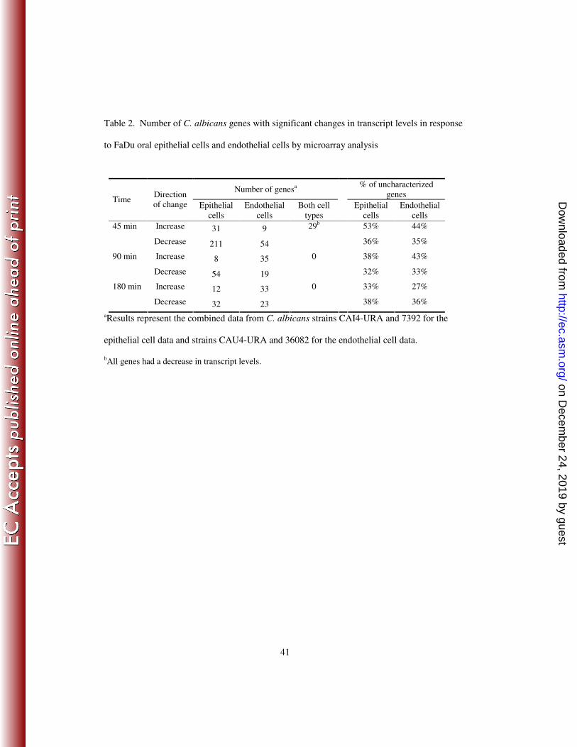
Next, we used Gene Ontology (GO) term analysis to determine the functional
categories of the C. albicans genes whose transcript level changed in response to epithelial and
endothelial cells. A large percentage of these genes did not have GO terms associated with
them (Table 2). As a result, the genes whose transcripts were increased in response to either
type of host cell were not significantly enriched for any GO term. Also, no GO term was
significantly overrepresented among the genes whose transcripts were decreased in response to
endothelial cells. Interestingly, the genes with reduced transcript levels in response to
epithelial cells were significantly enriched in three functional categories. Genes related to
adhesion were overrepresented at all 3 time points (p < 0.04 at each time point; Table 3). Also,
genes involved in ribonucleoprotein complex biogenesis were significantly enriched at 45 min
(p = 0.004). Finally, genes related to protein catabolic process were overrepresented at 90 min.
Collectively, these data suggest that C. albicans responds to contact with epithelial cells by
reducing expression of adhesins. There is also decreased protein synthesis that may be
counteracted by a reduction in protein catabolism.
Functional analysis of differentially induced genes. We used real-time PCR using
RNA from strain CAI4-URA to verify the increased transcript levels of selected genes
identified in the microarray experiments. The verification experiments focused on genes for
which Tn7-UAU1 transposon insertion cassettes (7, 13, 42) were available and whose function
had not been studied previously. Real-time PCR assays verified that the transcripts of 13 C.
albicans genes were significantly increased in response to oral epithelial cells compared to
polystyrene (Table 2). Similarly, we confirmed that the transcripts of 15 C. albicans genes
were significantly increased in response to endothelial cells. These measurements confirm our
on Decem
ber 24, 2019 by guesthttp://ec.asm
.org/D
ownloaded from
17
microarray results, thus strengthening the argument that C. albicans interacts with and
responds differently with these distinct host cell types.
To verify that the observed changes in gene transcript levels were induced by contact
with the host cells and not by the RNA isolation procedure, we infected endothelial cells and
polystyrene with C. albicans CAI4 for 90 min. Next, the host cells were lysed in ice-cold
distilled water, after which the C. albicans cells were collected and then snap frozen. The total
time between adding the distilled water and snap freezing the fungal cells was less than 5 min.
We then used real-time PCR the measure the transcript levels of YCK2, orf19.5568, and
orf19.4646. When CaACT1 was used as the endogenous control gene, the transcripts of YCK2,
orf19.5568, and orf19.464 increased by 1.8-, 2.2-, and 1.8-fold, respectively in response to
endothelial cells compared to polystyrene. A similar increase was also observed when either
CaTDH3 or CaTEF1 was used as the endogenous control gene (data not shown), indicating
that CaACT1 was an appropriate endogenous control gene for the real-time PCR experiments.
For genes whose transcripts were found by real-time PCR to be increased by at least
1.5-fold in response to epithelial cells or endothelial cells, we constructed homozygous
insertion mutants using the Tn7-UAU1 transposon insertion cassettes. We generated mutant
strains with homozygous insertions in 8 of the 13 genes whose transcripts were increased in
response to epithelial cells and 14 of the 15 genes whose transcripts were increased in response
to endothelial cells (Tables 1 and 4). Among the six genes for which we were unable to
construct homozygous insertion mutants, orf19.202 and orf19.3260 have essential orthologs in
S. cerevisiae (MCM7 and CAB3, respectively). The others are predicted to participate in vital
cellular processes including cell wall biogenesis, transcription, or mitochondrial function, so
that homozygous mutations may cause lethality or very slow growth.
on Decem
ber 24, 2019 by guesthttp://ec.asm
.org/D
ownloaded from

18
The 22 insertion mutants were screened for defects in virulence related phenotypes,
including hyphal formation and capacity to damage epithelial and endothelial cells (34, 47, 59,
62). In addition, because C. albicans is likely exposed to host antimicrobial peptides during
oropharyngeal and disseminated candidiasis, the mutants were tested for their susceptibility to
protamine. Protamine is a helical cationic polypeptide that is often used to screen for
antimicrobial peptide susceptibility (22, 76).
We found that mutants with insertions in YCK2 (orf19.7001), orf19.5568, orf19.4646,
and orf19.1939 had defects in at least one of the following characteristics: hyphal formation,
host cell damage, and protamine resistance (Table 4). These phenotypes were shared with at
least one additional independent insertion mutant for each gene. To verify that each insertion
mutation caused the phenotypic defect, the mutants were complemented with the respective
wild-type allele. This approach linked the mutant phenotypes to insertions in YCK2,
orf19.5568, and orf19.4646. YCK2 encodes the highly conserved serine/threonine kinase,
casein kinase 1 (CK1). Orf19.5568 has no S. cerevisiae ortholog, but the predicted protein
product has limited homology to S. cerevisiae Vps51, a component of the Vps53 complex.
This complex is required for fusion of endosome-derived vesicles with the late Golgi (11, 53,
64). Based on our phenotypic analysis below, we suggest the provisional name VPS51 for
orf19.5568. Orf19.4646 specifies a unique protein that does share significant homology with
any other protein, including the predicted proteins of the closely related species C. dubliniensis.
Because expression of orf19.4646 was increased in response to endothelial cells we named it
UEC1 (up-regulated by endothelial cells).
YCK2 governs cell polarity and is necessary for maximal epithelial cell damage.
YCK2 mRNA was increased in response to contact with either oral epithelial or endothelial
on Decem
ber 24, 2019 by guesthttp://ec.asm
.org/D
ownloaded from

19
cells, as indicated by real-time PCR measurements (Table 4). The yck2/yck2 insertion mutant
produced multiple short hyphae on epithelial cells, endothelial cells, and plastic, suggesting
that it had a defect in polarized growth (Fig. 2 and data not shown). It had significantly
reduced capacity to damage oral epithelial cells, but not endothelial cells, and this defect was
reversed by complementation (Fig. 3). Prior studies indicate that mutant germination defects
are invariably associated with endothelial cell damage defects (48, 59), so the yck2/yck2
mutant is novel in separating these two phenotypes.
The yck2/yck2 mutant also had impaired response to stress. It had markedly increased
susceptibility to protamine, Congo red, and SDS (Fig. 4). As predicted by the protamine results,
this mutant also had increased susceptibility to human β defensin 2 (Fig. 5), which is made by
oral epithelial cells (1, 29, 39), All defects of the yck2/yck2 were due to the insertions in YCK2
because complementing this mutant with a wild-type copy of YCK2 restored the wild-type
phenotype in all assays. Collectively, these results suggest that in C. albicans, YCK2 governs
polarized growth, capacity to damage epithelial cells, and resistance to environmental stress.
VPS51 is required for hyphal elongation, maximal damage to host cells, and stress
resistance. VPS51 mRNA levels are also increased in response to contact with either oral
epithelial or endothelial cells, as indicated by real-time PCR measurements (Table 4). The
vps51/vps51 insertion mutant produced unbranched hyphae that were shorter than those of the
wild-type strain when it was grown on epithelial cells, endothelial cells, or plastic (Fig. 2 and
data not shown). This mutant had a severe defect in its capacity to damage both epithelial and
endothelial cells (Fig. 3). Furthermore, the vps51/vps51 insertion mutant had significantly
increased susceptibility to all stressors tested (Figs. 4 and 5). To verify that VPS51 did indeed
specify a protein involved in vacuolar function, we stained the vps51/vps51 insertion mutant
on Decem
ber 24, 2019 by guesthttp://ec.asm
.org/D
ownloaded from

20
with FM4-64, which stains the vacuolar membrane (72). The vacuoles of this mutant were
highly fragmented (Fig. 6), similar to the vacuolar morphology of the S. cerevisiae vps51∆
mutant (53). In contrast, the vacuoles of the wild-type cells were much larger and either single
or multilobed. Complementing the vps51/vps51 insertion mutant with a wild-type copy of
VPS51 rescued all of these defects. Therefore, VPS51 is necessary in C. albicans for normal
vacuolar morphology as well as many virulence related phenotypes.
UEC1 is required for maximal epithelial cell damage and protamine resistance.
UEC1 mRNA levels were increased by contact with endothelial cells, but not epithelial cells
(Table 4). Under all conditions tested, the uec1/uec1 insertion mutant produced hyphae that
were as long as those of the wild-type strain but slightly thicker (Fig. 2 and data not shown).
Although UEC1 transcript levels were not increased by contact with epithelial cells, the
uec1/uec1 insertion mutant had significantly reduced capacity to damage these cells (Fig. 3).
However, it did not have an endothelial cell damage defect. The uec1/uec1 insertion mutant
also had significantly increased susceptibility to protamine, SDS and human β defensin 2, as
well as a slight increase susceptibility to NaCl (Figs. 4 and 5). These defects were rescued by
complementing the insertion mutant with a wild-type copy of UEC1. Taken together, these
results indicate that UEC1 is necessary for C. albicans to cause normal damage epithelial cells
in vitro, perhaps by governing cell membrane integrity.
DISCUSSION
The results presented here demonstrate that C. albicans interacts differently with oral
epithelial cells compared to endothelial cells. C. albicans hyphae were endocytosed more
slowly by epithelial cells and caused less damage to these cells than endothelial cells. Also, the
on Decem
ber 24, 2019 by guesthttp://ec.asm
.org/D
ownloaded from

21
transcriptional response of C. albicans to epithelial cells was different from its response to
endothelial cells. Interestingly, the C. albicans genes whose transcripts were increased in
response to either of these cell types were also different from those that have been reported to
be induced by exposure to monocytes or neutrophils (35, 58). These divergent responses to
different types of host cells likely enable C. albicans to survive and proliferate in diverse
anatomic sites within the host.
To our knowledge, the transcriptional response of C. albicans to endothelial cells has
not been determined previously. However, two other groups of investigators have studied the
response of this organisms to contact with vaginal, cervical, and intestinal epithelial cell lines
as compared to polystyrene (60, 65). Both groups found that contact with epithelial cells
caused only a 2- to 4-fold change in C. albicans gene transcript levels. This magnitude of
differential transcript levels is comparable to the results reported here. Sohn et al. (65) found
that, when C. albicans was added to either vaginal or intestinal cell lines, the transcript levels
of most genes changed within 30 to 60 min of contact; the mRNA of very few genes changed
after 120 min of contact with these epithelial cells. Our results reported here lead to a similar
conclusion.
The prior epithelial cell-interaction studies (60, 65) reported that contact with epithelial
cells increased mRNA levels of C. albicans cell surface protein genes, including ALS2, ALS5,
HWP1, PRA1, and PGA7. In contrast, we found that the transcript levels of some adhesin
genes actually fell in response to epithelial cells. Sohn et al. (65) noted that HWP1 transcripts
were induced simply by contact with a solid support, so it is possible that that HWP1 transcript
levels were stimulated more strongly by contact with polystyrene than by contact with
epithelial cells. Also, PRA1 orthologs in S. cerevisiae and A. fumigatus are members of a zinc-
on Decem
ber 24, 2019 by guesthttp://ec.asm
.org/D
ownloaded from

22
responsive regulon (36, 63), and recent results verify the induction of C. albicans PRA1 by
zinc limitation (Nobile et al., in preparation). Thus it seems likely that competition for zinc by
host cells and C. albicans cells may have caused the PRA1 induction reported by Sandovsky-
Losica et al., (60) and our failure to detect its up-regulation may simply reflect a higher zinc
level in our medium. Other key differences between these other microarray studies and ours
include differences in the epithelial cell type, growth media and strains of C. albicans used.
The microarray studies also suggested that contact with oral epithelial cells resulted in
an early reduction of protein synthesis followed by a decrease in protein catabolism. Inhibition
of genes involved in protein synthesis also occurs when the organism is ingested by
macrophages and neutrophils, and in response to nitric oxide (20, 24, 35). The down-
regulation of genes involved in protein synthesis may be a response to stress (24, 35). It is
known that oral epithelial cells can inhibit the growth of C. albicans (66, 67). Even though we
did not observe any reduction in growth or hyphal elongation at the time points studied, it is
possible that inhibition of protein synthesis may have preceded growth inhibition, which might
have been detectable after longer incubation times.
Zakikhany et al., (78) used microarrays to analyze the transcript levels of C. albicans
during infection of reconstituted human epithelium in vitro. They also analyzed C. albicans
gene expression in samples from patients with pseudomembranous OPC. In these analyses, the
reference condition was C. albicans blastospores grown to mid-log phase in YPD broth.
Because contact with epithelial cells induces hyphal formation, the microarray data of
Zakikhany et al. were enriched in hyphal-associated genes, such as ALS3, HYR1, RBT1, and
ECE1 (78). In the current experiments, the reference condition (contact with polystyrene)
caused the organisms to form hyphae, and thus increased transcript levels that were induced by
on Decem
ber 24, 2019 by guesthttp://ec.asm
.org/D
ownloaded from

23
hyphal formation alone were not detected. However, Zakikhany et al. found that a large
proportion of genes with increased transcript levels during the early phase of infection had not
been previously characterized (78), similar to the results presented here.
The main value of our expression profiling results comes from the implication of new
genes in the process of C. albicans-host cell interaction. Our analysis of mutants defective in
up-regulated genes indicates that a relatively modest change in gene expression can
nonetheless be biologically significant. Of the 22 unique insertion mutants reported here, 3
(14%) had defective capacity to damage epithelial or endothelial cells. In contrast, our
previous screen of unselected C. albicans insertion mutants yielded only 3 (1.6%) of 183
mutants with consistent defects (9). Therefore, the use of microarray data to guide candidate
gene selection significantly improves the efficiency of virulence-associated gene identification.
Mutants with insertion in YCK2, VPS51, and UEC1 all had defects in damaging oral
epithelial cells, while only the vps51/vps51 insertion mutant had a defect in damaging
endothelial cells. We also found that the wild-type strain of C. albicans caused less damage to
epithelial cells than to endothelial cells. We hypothesize that oral epithelial cells may be
relatively resistant to C. albicans-induced damage because oral epithelial cells frequently
encounter C. albicans even in normal hosts, whereas endothelial cells do not. Our microarray
data also suggest the possibility that oral epithelial cells may actually induce down-regulation
of some C. albicans genes (such as those involved in protein synthesis) that are likely required
for the organism to cause maximal epithelial cell damage.
Damage to epithelial and endothelial cells requires that C. albicans adheres to and
invades these cells, and that it secretes lytic enzymes (9, 26, 43, 47, 50). Therefore, it is
probable that the yck2/yck2, vps51/vps51, and uec1/uec1 insertion mutants have defects in one
on Decem
ber 24, 2019 by guesthttp://ec.asm
.org/D
ownloaded from
24
or more of these processes. Furthermore, mutants with reduced capacity to damage epithelial
cells or endothelial cells in vitro have a high probability of having attenuated virulence in
mouse models of oropharyngeal or hematogenously disseminated candidiasis (9, 47, 59).
Collectively, our results suggest that YCK2, VPS51, and UEC1 may influence the virulence of
C. albicans.
It was notable that all three insertion mutants also had increased susceptibility to the
antimicrobial peptides, protamine and human β defensin 2. C. albicans is exposed to
antimicrobial peptides, such as histatins and defensins when it is in the oral cavity. The
organism also encounters defensins and antimicrobial chemokines as it disseminates
hematogenously and interacts with leukocytes and endothelial cells. Our finding that three of
the genes that were up-regulated in response to either epithelial or endothelial cells were
required for C. albicans to resist antimicrobial peptides suggests that contact with these host
cells induces a protective reaction that enables C. albicans to withstand these peptides. The
capacity to tolerate antimicrobial peptides is important for C. albicans virulence because the
extent of antimicrobial peptide resistance is directly related to virulence in animal models of
disseminated candidiasis (22, 76)
C. albicans Yck2 shares extensive homology with S. cerevisiae Yck2 and slightly less
homology with S. cerevisiae Yck1, which are the two plasma membrane-associated isoforms
of CK1 in this yeast. BLAST searches of the C. albicans genome did not identify a YCK1
ortholog, suggesting that C. albicans has only a single plasma membrane-associated isoform of
CK1. In S. cerevisiae, Yck2 and Yck1 are required for normal bud morphogenesis, cytokinesis,
and endocytosis (2, 56). Strains of S. cerevisiae with reduced plasma membrane CK1 activity
have defects in cell polarity (56). The early branching phenotype of the C. albicans yck2/yck2
on Decem
ber 24, 2019 by guesthttp://ec.asm
.org/D
ownloaded from
25
insertion mutant suggests that Yck2 is also required for maintenance of cell polarity in C.
albicans.
Vps51 was first identified in S. cerevisiae, where it functions in a complex with Vps52,
Vps53, and Vps54 (53, 64). This complex is required for retrograde protein traffic from the
early endosome to the late Golgi. Mutants that lack any of the subunits of this complex have
missorting of vacuolar proteins, abnormal Golgi membrane proteins, and fragmented vacuoles
(12, 53). While vacuoles are not required for growth under nutrient rich conditions, they are
important for S. cerevisiae to resist stress due to starvation, changes in environmental pH, and
hyperosmolarity (5). The Vps51-54 complex has not been studied previously in C. albicans.
However, a vps11∆/vps11∆ mutant of C. albicans, which does not contain a vacuole, has
delayed germination and increased susceptibility to osmotic stress due to glycerol or high NaCl
(45). It also has markedly reduced secretion of secreted aspartyl proteinases and lipases,
enzymes that have been implicated in damaging host cells (26, 46). The abnormal vacuole,
shortened hyphae, and increased susceptibility to environmental stress of the C. albicans
vps51/vps51 insertion mutant are consistent with the probable role of Vps51 in vacuolar
function. The impaired capacity of the vps51/vps51 insertion mutant to damage epithelial and
endothelial cells is probably due to multiple factors, including abnormal hyphal formation and
possibly reduced secretion of hydrolytic enzymes.
Our recommendation that orf19.5568 be named VPS51 is based upon three
considerations. First, as discussed above, the C. albicans mutant has several phenotypes that
would be expected to result from a Vps51 defect. Second, although the homology between
CaVps51 and ScVps51 is limited (64% similarity over 56 residues, P = 4.6e-06
), it spans the
most highly conserved region of ScVps51 among fungi (residues 90-145; see
on Decem
ber 24, 2019 by guesthttp://ec.asm
.org/D
ownloaded from
26
http://www.yeastgenome.org/cache/fungi/YKR020W.html). Third, we note that ScVps51 does
not have a closer homolog that CaVps51 among C. albicans predicted proteins. Thus this gene
name recommendation is based upon independent lines of evidence.
UEC1 encodes a unique 145 amino acid protein. The function of Uec1 cannot be
inferred from its primary amino acid sequence as it has no close orthologs and Pfam analysis
does not reveal any conserved domains. Although UEC1 is listed as a dubious open reading
frame in the Candida Genome Database, our results suggest that it is a functional gene for the
following reasons. First, the microarray and real-time PCR data indicate that UEC1 is
transcribed. Second, homozygous insertions in the UEC1 locus induced a mutant phenotype.
In principle these properties might be expected for a 5’ regulatory transcript, as has been
described for the S. cerevisiae SER3 gene (37). However, we were able to complement the
uec1/uec1 insertion mutant with a wild-type copy of UEC1, integrated ectopically at the HIS1
locus, to restore the wild-type phenotype. This observation argues that the uec1::Tn7-UAU1
insertion phenotype does not result from a cis-acting effect on a neighboring gene. While the
exact function of Uec1 remains to be determined, our finding that the uec1/uec1 insertion
mutant had increased susceptibility to the cell membranes stressors, antimicrobial peptides and
SDS, suggest that Uec1 may be required for maintenance of cell membrane integrity.
In summary, our microarray analysis demonstrates that the transcriptional response of
C. albicans to oral epithelial cells is significantly different from its response to vascular
endothelial cells. Furthermore, a significant fraction of the C. albicans genes whose transcript
levels are increased upon contact with either of these host cells are uncharacterized. Some of
these genes, such as YCK2, VPS51, and UEC1 are required for the organism to damage host
on Decem
ber 24, 2019 by guesthttp://ec.asm
.org/D
ownloaded from

27
cells and resist the types of environmental stress that it likely encounters during growth in the
oropharynx and bloodstream.
on Decem
ber 24, 2019 by guesthttp://ec.asm
.org/D
ownloaded from

28
ACKNOWLEDGEMENTS
We thank Q. Trang Phan for expert support with tissue culture, Deborah Kupferwasser
for technical assistance, and the perinatal nurses of the Harbor-UCLA General Clinical
Research Center for collection of umbilical cords. We are also grateful for Shelley Lane’s
assistance with the microarray experiments and Dr. Matthew Schibler of the
Microscopy/Spectroscopy Core Facility at the California NanoSystems Institute of the
University of California, Los Angeles for help with confocal microscopy. We appreciate the
Candida Genome Database (http://www.candidagenome.org/) for providing genomic sequence
data and protein information for Candida albicans. We thank Qi Zhao and William C.
Nierman (J. Craig Venter Institute) and Frank J. Smith (Columbia University) for providing
Candida gene disruption cassettes. The insertion library project was accomplished with the
support of NIH 1R01AI057804. This study was supported by Public Health Service grants
5R01DE13974, 1R01DE017088, R01AI054928, RO1AI19990 and MO1RR00425 from the
National Institutes of Health.
on Decem
ber 24, 2019 by guesthttp://ec.asm
.org/D
ownloaded from

29
REFERENCES
1. Abiko, Y., Y. Jinbu, T. Noguchi, M. Nishimura, K. Kusano, P. Amaratunga, T.
Shibata, and T. Kaku. 2002. Upregulation of human beta-defensin 2 peptide
expression in oral lichen planus, leukoplakia and candidiasis. an immunohistochemical
study. Pathol Res Pract 198:537-42.
2. Babu, P., J. D. Bryan, H. R. Panek, S. L. Jordan, B. M. Forbrich, S. C. Kelley, R.
T. Colvin, and L. C. Robinson. 2002. Plasma membrane localization of the Yck2p
yeast casein kinase 1 isoform requires the C-terminal extension and secretory pathway
function. J Cell Sci 115:4957-68.
3. Bader, M. S., S. M. Lai, V. Kumar, and D. Hinthorn. 2004. Candidemia in patients
with diabetes mellitus: epidemiology and predictors of mortality. Scand J Infect Dis
36:860-4.
4. Badrane, H., S. Cheng, M. H. Nguyen, H. Y. Jia, Z. Zhang, N. Weisner, and C. J.
Clancy. 2005. Candida albicans IRS4 contributes to hyphal formation and virulence
after the initial stages of disseminated candidiasis. Microbiology 151:2923-31.
5. Banta, L. M., J. S. Robinson, D. J. Klionsky, and S. D. Emr. 1988. Organelle
assembly in yeast: characterization of yeast mutants defective in vacuolar biogenesis
and protein sorting. J Cell Biol 107:1369-83.
6. Brown, A. J., F. C. Odds, and N. A. Gow. 2007. Infection-related gene expression in
Candida albicans. Curr Opin Microbiol 10:307-13.
7. Bruno, V. M., and A. P. Mitchell. 2005. Regulation of azole drug susceptibility by
Candida albicans protein kinase CK2. Mol Microbiol 56:559-73.
on Decem
ber 24, 2019 by guesthttp://ec.asm
.org/D
ownloaded from

30
8. Cawson, R. A., and K. C. Rajasingham. 1972. Ultrastructural features of the invasive
phase of Candida albicans. Br J Dermatol 87:435-43.
9. Chiang, L. Y., D. C. Sheppard, V. M. Bruno, A. P. Mitchell, J. E. Edwards, Jr.,
and S. G. Filler. 2007. Candida albicans protein kinase CK2 governs virulence during
oropharyngeal candidiasis. Cell Microbiol 9:233-45.
10. Chomczynski, P., and N. Sacchi. 1987. Single-step method of RNA isolation by acid
guanidinium thiocyanate-phenol-chloroform extraction. Anal Biochem 162:156-9.
11. Conibear, E., J. N. Cleck, and T. H. Stevens. 2003. Vps51p mediates the association
of the GARP (Vps52/53/54) complex with the late Golgi t-SNARE Tlg1p. Mol Biol
Cell 14:1610-23.
12. Conibear, E., and T. H. Stevens. 2000. Vps52p, Vps53p, and Vps54p form a novel
multisubunit complex required for protein sorting at the yeast late Golgi. Mol Biol Cell
11:305-23.
13. Davis, D. A., V. Bruno, L. Loza, S. G. Filler, and A. P. Mitchell. 2002. C. albicans
Mds3p, a conserved regulator of pH responses and virulence identified through
insertional mutagenesis. Genetics 162:1573-81.
14. Eversole, L. R., P. A. Reichart, G. Ficarra, A. Schmidt-Westhausen, P. Romagnoli,
and N. Pimpinelli. 1997. Oral keratinocyte immune responses in HIV-associated
candidiasis. Oral Surg Oral Med Oral Pathol Oral Radiol Endod 84:372-80.
15. Farah, C. S., R. B. Ashman, and S. J. Challacombe. 2000. Oral candidosis. Clin
Dermatol 18:553-62.
on Decem
ber 24, 2019 by guesthttp://ec.asm
.org/D
ownloaded from

31
16. Filler, S. G., B. O. Ibe, A. S. Ibrahim, M. A. Ghannoum, J. U. Raj, and J. E.
Edwards, Jr. 1994. Mechanisms by which Candida albicans induces endothelial cell
prostaglandin synthesis. Infect Immun 62:1064-9.
17. Filler, S. G., B. O. Ibe, P. M. Luckett, J. U. Raj, and J. E. Edwards, Jr. 1991.
Candida albicans stimulates endothelial cell eicosanoid production. J Infect Dis
164:928-35.
18. Filler, S. G., A. S. Pfunder, B. J. Spellberg, J. P. Spellberg, and J. E. Edwards, Jr.
1996. Candida albicans stimulates cytokine production and leukocyte adhesion
molecule expression by endothelial cells. Infect Immun 64:2609-17.
19. Filler, S. G., J. N. Swerdloff, C. Hobbs, and P. M. Luckett. 1995. Penetration and
damage of endothelial cells by Candida albicans. Infect Immun 63:976-83.
20. Fradin, C., P. De Groot, D. MacCallum, M. Schaller, F. Klis, F. C. Odds, and B.
Hube. 2005. Granulocytes govern the transcriptional response, morphology and
proliferation of Candida albicans in human blood. Mol Microbiol 56:397-415.
21. Fu, Y., A. S. Ibrahim, D. C. Sheppard, Y. C. Chen, S. W. French, J. E. Cutler, S. G.
Filler, and J. E. Edwards. 2002. Candida albicans Als1p: an adhesin that is a
downstream effector of the EFG1 filamentation pathway. Mol Microbiol 44:61-72.
22. Gank, K. D., M. R. Yeaman, S. Kojima, N. Y. Yount, H. Park, J. E. Edwards, Jr.,
S. G. Filler, and Y. Fu. 2008. SSD1 is integral to host defense peptide resistance in
Candida albicans. Eukaryot Cell 7:1318-27.
23. Hajjeh, R. A., A. N. Sofair, L. H. Harrison, G. M. Lyon, B. A. Arthington-Skaggs,
S. A. Mirza, M. Phelan, J. Morgan, W. Lee-Yang, M. A. Ciblak, L. E. Benjamin, L.
T. Sanza, S. Huie, S. F. Yeo, M. E. Brandt, and D. W. Warnock. 2004. Incidence of
on Decem
ber 24, 2019 by guesthttp://ec.asm
.org/D
ownloaded from
32
bloodstream infections due to Candida species and in vitro susceptibilities of isolates
collected from 1998 to 2000 in a population-based active surveillance program. J Clin
Microbiol 42:1519-27.
24. Hromatka, B. S., S. M. Noble, and A. D. Johnson. 2005. Transcriptional response of
Candida albicans to nitric oxide and the role of the YHB1 gene in nitrosative stress and
virulence. Mol Biol Cell 16:4814-26.
25. Ibrahim, A. S., S. G. Filler, M. A. Ghannoum, and J. E. Edwards, Jr. 1993.
Interferon-gamma protects endothelial cells from damage by Candida albicans. J Infect
Dis 167:1467-70.
26. Ibrahim, A. S., S. G. Filler, D. Sanglard, J. E. Edwards, Jr., and B. Hube. 1998.
Secreted aspartyl proteinases and interactions of Candida albicans with human
endothelial cells. Infect Immun 66:3003-5.
27. Ibrahim, A. S., F. Mirbod, S. G. Filler, Y. Banno, G. T. Cole, Y. Kitajima, J. E.
Edwards, Jr., Y. Nozawa, and M. A. Ghannoum. 1995. Evidence implicating
phospholipase as a virulence factor of Candida albicans. Infect Immun 63:1993-8.
28. Jaffe, E. A., R. L. Nachman, C. G. Becker, and C. R. Minick. 1973. Culture of
human endothelial cells derived from umbilical veins. Identification by morphologic
and immunologic criteria. J Clin Invest 52:2745-56.
29. Joly, S., C. Maze, P. B. McCray, Jr., and J. M. Guthmiller. 2004. Human beta-
defensins 2 and 3 demonstrate strain-selective activity against oral microorganisms. J
Clin Microbiol 42:1024-9.
on Decem
ber 24, 2019 by guesthttp://ec.asm
.org/D
ownloaded from

33
30. Kamai, Y., M. Kubota, T. Hosokawa, T. Fukuoka, and S. G. Filler. 2002.
Contribution of Candida albicans ALS1 to the pathogenesis of experimental
oropharyngeal candidiasis. Infect Immun 70:5256-8.
31. Kamai, Y., M. Kubota, T. Hosokawa, T. Fukuoka, and S. G. Filler. 2001. New
model of oropharyngeal candidiasis in mice. Antimicrob Agents Chemother 45:3195-7.
32. Kohrer, K., and H. Domdey. 1991. Preparation of high molecular weight RNA.
Methods Enzymol 194:398-405.
33. Livak, K. J., and T. D. Schmittgen. 2001. Analysis of relative gene expression data
using real-time quantitative PCR and the 2(-Delta Delta C(T)) Method. Methods
25:402-8.
34. Lo, H. J., J. R. Kohler, B. DiDomenico, D. Loebenberg, A. Cacciapuoti, and G. R.
Fink. 1997. Nonfilamentous C. albicans mutants are avirulent. Cell 90:939-49.
35. Lorenz, M. C., J. A. Bender, and G. R. Fink. 2004. Transcriptional response of
Candida albicans upon internalization by macrophages. Eukaryot Cell 3:1076-87.
36. Lyons, T. J., A. P. Gasch, L. A. Gaither, D. Botstein, P. O. Brown, and D. J. Eide.
2000. Genome-wide characterization of the Zap1p zinc-responsive regulon in yeast.
Proc Natl Acad Sci U S A 97:7957-62.
37. Martens, J. A., P. Y. Wu, and F. Winston. 2005. Regulation of an intergenic
transcript controls adjacent gene transcription in Saccharomyces cerevisiae. Genes Dev
19:2695-704.
38. Mayer, C. L., S. G. Filler, and J. E. Edwards, Jr. 1992. Candida albicans adherence
to endothelial cells. Microvasc Res 43:218-26.
on Decem
ber 24, 2019 by guesthttp://ec.asm
.org/D
ownloaded from
34
39. Meyer, J. E., J. Harder, T. Gorogh, J. B. Weise, S. Schubert, D. Janssen, and S.
Maune. 2004. Human beta-defensin-2 in oral cancer with opportunistic Candida
infection. Anticancer Res 24:1025-30.
40. Montes, L. F., and W. H. Wilborn. 1968. Ultrastructural features of host-parasite
relationship in oral candidiasis. J Bacteriol 96:1349-56.
41. Murad, A. M., P. Leng, M. Straffon, J. Wishart, S. Macaskill, D. MacCallum, N.
Schnell, D. Talibi, D. Marechal, F. Tekaia, C. d'Enfert, C. Gaillardin, F. C. Odds,
and A. J. Brown. 2001. NRG1 represses yeast-hypha morphogenesis and hypha-
specific gene expression in Candida albicans. Embo J 20:4742-52.
42. Nobile, C. J., V. M. Bruno, M. L. Richard, D. A. Davis, and A. P. Mitchell. 2003.
Genetic control of chlamydospore formation in Candida albicans. Microbiology
149:3629-37.
43. Nobile, C. J., N. Solis, C. L. Myers, A. J. Fay, J. S. Deneault, A. Nantel, A. P.
Mitchell, and S. G. Filler. 2008. Candida albicans transcription factor Rim101
mediates pathogenic interactions through cell wall functions. Cell Microbiol 10:2180-
96.
44. Norice, C. T., F. J. Smith, Jr., N. Solis, S. G. Filler, and A. P. Mitchell. 2007.
Requirement for Candida albicans Sun41 in biofilm formation and virulence. Eukaryot
Cell 6:2046-55.
45. Palmer, G. E., A. Cashmore, and J. Sturtevant. 2003. Candida albicans VPS11 is
required for vacuole biogenesis and germ tube formation. Eukaryot Cell 2:411-21.
on Decem
ber 24, 2019 by guesthttp://ec.asm
.org/D
ownloaded from
35
46. Paraje, M. G., S. G. Correa, M. S. Renna, M. Theumer, and C. E. Sotomayor.
2008. Candida albicans-secreted lipase induces injury and steatosis in immune and
parenchymal cells. Can J Microbiol 54:647-59.
47. Park, H., C. L. Myers, D. C. Sheppard, Q. T. Phan, A. A. Sanchez, J. E. Edwards,
Jr., and S. G. Filler. 2005. Role of the fungal Ras-protein kinase A pathway in
governing epithelial cell interactions during oropharyngeal candidiasis. Cell Microbiol
7:499-510.
48. Phan, Q. T., P. H. Belanger, and S. G. Filler. 2000. Role of hyphal formation in
interactions of Candida albicans with endothelial cells. Infect Immun 68:3485-3490.
49. Phan, Q. T., R. A. Fratti, N. V. Prasadarao, J. E. Edwards, Jr., and S. G. Filler.
2005. N-cadherin mediates endocytosis of Candida albicans by endothelial cells. J Biol
Chem 280:10455-61.
50. Phan, Q. T., C. L. Myers, Y. Fu, D. C. Sheppard, M. R. Yeaman, W. H. Welch, A.
S. Ibrahim, J. E. Edwards, and S. G. Filler. 2007. Als3 is a Candida albicans invasin
that binds to cadherins and induces endocytosis by host cells. PLoS Biol 5:e64.
51. Rangel-Frausto, M. S., T. Wiblin, H. M. Blumberg, L. Saiman, J. Patterson, M.
Rinaldi, M. Pfaller, J. E. Edwards, Jr., W. Jarvis, J. Dawson, and R. P. Wenzel.
1999. National epidemiology of mycoses survey (NEMIS): variations in rates of
bloodstream infections due to Candida species in seven surgical intensive care units
and six neonatal intensive care units. Clin Infect Dis 29:253-8.
52. Redding, S. W., R. C. Zellars, W. R. Kirkpatrick, R. K. McAtee, M. A. Caceres, A.
W. Fothergill, J. L. Lopez-Ribot, C. W. Bailey, M. G. Rinaldi, and T. F. Patterson.
on Decem
ber 24, 2019 by guesthttp://ec.asm
.org/D
ownloaded from
36
1999. Epidemiology of oropharyngeal Candida colonization and infection in patients
receiving radiation for head and neck cancer. J Clin Microbiol 37:3896-900.
53. Reggiori, F., C. W. Wang, P. E. Stromhaug, T. Shintani, and D. J. Klionsky. 2003.
Vps51 is part of the yeast Vps fifty-three tethering complex essential for retrograde
traffic from the early endosome and Cvt vesicle completion. J Biol Chem 278:5009-20.
54. Revankar, S. G., O. P. Dib, W. R. Kirkpatrick, R. K. McAtee, A. W. Fothergill, M.
G. Rinaldi, S. W. Redding, and T. F. Patterson. 1998. Clinical evaluation and
microbiology of oropharyngeal infection due to fluconazole-resistant Candida in
human immunodeficiency virus-infected patients. Clin Infect Dis 26:960-3.
55. Rhodus, N. L., C. Bloomquist, W. Liljemark, and J. Bereuter. 1997. Prevalence,
density, and manifestations of oral Candida albicans in patients with Sjogren's
syndrome. J Otolaryngol 26:300-5.
56. Robinson, L. C., C. Bradley, J. D. Bryan, A. Jerome, Y. Kweon, and H. R. Panek.
1999. The Yck2 yeast casein kinase 1 isoform shows cell cycle-specific localization to
sites of polarized growth and is required for proper septin organization. Mol Biol Cell
10:1077-92.
57. Rotrosen, D., J. E. Edwards, Jr., T. R. Gibson, J. C. Moore, A. H. Cohen, and I.
Green. 1985. Adherence of Candida to cultured vascular endothelial cells: mechanisms
of attachment and endothelial cell penetration. J Infect Dis 152:1264-74.
58. Rubin-Bejerano, I., I. Fraser, P. Grisafi, and G. R. Fink. 2003. Phagocytosis by
neutrophils induces an amino acid deprivation response in Saccharomyces cerevisiae
and Candida albicans. Proc Natl Acad Sci U S A 100:11007-12.
on Decem
ber 24, 2019 by guesthttp://ec.asm
.org/D
ownloaded from

37
59. Sanchez, A. A., D. A. Johnston, C. Myers, J. E. Edwards, Jr., A. P. Mitchell, and S.
G. Filler. 2004. Relationship between Candida albicans virulence during experimental
hematogenously disseminated Infection and endothelial cell damage In vitro. Infect
Immun 72:598-601.
60. Sandovsky-Losica, H., N. Chauhan, R. Calderone, and E. Segal. 2006. Gene
transcription studies of Candida albicans following infection of HEp2 epithelial cells.
Med Mycol 44:329-34.
61. Sangeorzan, J. A., S. F. Bradley, X. He, L. T. Zarins, G. L. Ridenour, R. N. Tiballi,
and C. A. Kauffman. 1994. Epidemiology of oral candidiasis in HIV-infected patients:
colonization, infection, treatment, and emergence of fluconazole resistance. Am J Med
97:339-46.
62. Saville, S. P., A. L. Lazzell, C. Monteagudo, and J. L. Lopez-Ribot. 2003.
Engineered control of cell morphology in vivo reveals distinct roles for yeast and
filamentous forms of Candida albicans during infection. Eukaryot Cell 2:1053-60.
63. Segurado, M., R. Lopez-Aragon, J. A. Calera, J. M. Fernandez-Abalos, and F.
Leal. 1999. Zinc-regulated biosynthesis of immunodominant antigens from Aspergillus
spp. Infect Immun 67:2377-82.
64. Siniossoglou, S., and H. R. Pelham. 2002. Vps51p links the VFT complex to the
SNARE Tlg1p. J Biol Chem 277:48318-24.
65. Sohn, K., I. Senyurek, J. Fertey, A. Konigsdorfer, C. Joffroy, N. Hauser, G. Zelt,
H. Brunner, and S. Rupp. 2006. An in vitro assay to study the transcriptional
response during adherence of Candida albicans to different human epithelia. FEMS
Yeast Res 6:1085-93.
on Decem
ber 24, 2019 by guesthttp://ec.asm
.org/D
ownloaded from

38
66. Steele, C., J. Leigh, R. Swoboda, and P. L. Fidel, Jr. 2000. Growth inhibition of
Candida by human oral epithelial cells. J Infect Dis 182:1479-85.
67. Steele, C., J. Leigh, R. Swoboda, H. Ozenci, and P. L. Fidel, Jr. 2001. Potential role
for a carbohydrate moiety in anti-Candida activity of human oral epithelial cells. Infect
Immun 69:7091-9.
68. Subramanian, S., C. A. Woolford, and E. W. Jones. 2004. The Sec1/Munc18 protein,
Vps33p, functions at the endosome and the vacuole of Saccharomyces cerevisiae. Mol
Biol Cell 15:2593-605.
69. Sundstrom, P., E. Balish, and C. M. Allen. 2002. Essential role of the Candida
albicans transglutaminase substrate, hyphal wall protein 1, in lethal oroesophageal
candidiasis in immunodeficient mice. J Infect Dis 185:521-30.
70. Tsuchimori, N., L. L. Sharkey, W. A. Fonzi, S. W. French, J. E. Edwards, Jr., and
S. G. Filler. 2000. Reduced virulence of HWP1-deficient mutants of Candida albicans
and their interactions with host cells. Infect Immun 68:1997-2002.
71. Tusher, V. G., R. Tibshirani, and G. Chu. 2001. Significance analysis of microarrays
applied to the ionizing radiation response. Proc Natl Acad Sci U S A 98:5116-21.
72. Vida, T. A., and S. D. Emr. 1995. A new vital stain for visualizing vacuolar
membrane dynamics and endocytosis in yeast. J Cell Biol 128:779-92.
73. Williams, R. M., M. Primig, B. K. Washburn, E. A. Winzeler, M. Bellis, C.
Sarrauste de Menthiere, R. W. Davis, and R. E. Esposito. 2002. The Ume6 regulon
coordinates metabolic and meiotic gene expression in yeast. Proc Natl Acad Sci U S A
99:13431-6.
on Decem
ber 24, 2019 by guesthttp://ec.asm
.org/D
ownloaded from

39
74. Willis, A. M., W. A. Coulter, C. R. Fulton, J. R. Hayes, P. M. Bell, and P. J. Lamey.
1999. Oral candidal carriage and infection in insulin-treated diabetic patients. Diabet
Med 16:675-9.
75. Wilson, R. B., D. Davis, and A. P. Mitchell. 1999. Rapid hypothesis testing with
Candida albicans through gene disruption with short homology regions. J Bacteriol
181:1868-74.
76. Yeaman, M. R., S. S. Soldan, M. A. Ghannoum, J. E. Edwards, Jr., S. G. Filler,
and A. S. Bayer. 1996. Resistance to platelet microbicidal protein results in increased
severity of experimental Candida albicans endocarditis. Infect Immun 64:1379-84.
77. Yount, N. Y., and M. R. Yeaman. 2004. Multidimensional signatures in antimicrobial
peptides. Proc Natl Acad Sci U S A 101:7363-8.
78. Zakikhany, K., J. R. Naglik, A. Schmidt-Westhausen, G. Holland, M. Schaller,
and B. Hube. 2007. In vivo transcript profiling of Candida albicans identifies a gene
essential for interepithelial dissemination. Cell Microbiol 9:2938-54.
on Decem
ber 24, 2019 by guesthttp://ec.asm
.org/D
ownloaded from

40
Table 1. Strains used in this study
Strain Genotype Source (reference)
CAI4-
URAa
ura3∆::imm434/ura3∆::imm434::URA3 Y. Fu (59)
36082 Wild type (human blood isolate) American Type Culture Collection
7392 Wild type (human oral isolate) T. Patterson (50)
BWP17 ura3∆::λimm434/ura3∆::λimm434 his1::hisG/his1::hisG
arg4::hisG/arg4::hisG
A. Mitchell (75)
DAY286 ura3∆::λimm434/ura3∆::λimm434
pARG4::URA3::arg4::hisG/arg4::hisG
A. Mitchell (13)
DAY185 pARG4::URA3::arg4::hisG/arg4::hisG pHIS1::his1::his1/his1::hisG A. Mitchell (13)
EpKO1 atm1-Tn::UAU1/atm1-Tn::URA3 This study
EpKO2 orf19.4656-Tn::UAU1/orf19.4656-Tn::URA3 This study
EpKO3 orf19.4793-Tn::UAU1/orf19.4793-Tn::URA3
EpKO4 orf19.2398-Tn::UAU1/orf19.2398-Tn::URA3 This study
EpKO5 clp1-Tn::UAU1/clp1-Tn::URA3 This study
EpKO6 opt26-Tn::UAU1/opt26-Tn::URA3 This study
EpKO7 cdc47-Tn::UAU1/cdc47-Tn::URA3 This study
EpKO8 orf19.1504-Tn::UAU1/orf19.1504-Tn::URA3 This study
JJH34 yck2-Tn::UAU1/yck2-Tn::URA3 This study
JJH34H yck2-Tn::UAU1/yck2-Tn::URA3::pHIS1 This study
JJH34C yck2-Tn::UAU1/yck2-Tn::URA3::pHIS1-YCK2 This study
BL35-1 vps51-Tn::UAU1/vps51-Tn::URA3 This study
BL35-2 vps51-Tn::UAU1/vps51-Tn::URA3 This study
BL35-1H vps51-Tn::UAU1/vps51-Tn::URA3::pHIS1 This study
BL35-1C vps51-Tn::UAU1/vps51-Tn::URA3::pHIS1-VPS51 This study
BL36 orf19.6168-Tn::UAU1/orf19.6168-Tn::URA3 This study
BL37 orf19.1766-Tn::UAU1/orf19.1766-Tn::URA3 This study
BL38 orf19.3740-Tn::UAU1/orf19.3740-Tn::URA3 This study
BL39 orf19.4142-Tn::UAU1/orf19.4142-Tn::URA3 This study
BL40 orf19.4791-Tn::UAU1/orf19.4791-Tn::URA3 This study
BL41-1 uec1-Tn::UAU1/uec1-Tn::URA3 This study
BL41-2 uec1-Tn::UAU1/uec1-Tn::URA3 This study
BL41-1H uec1-Tn::UAU1/uec1-Tn::URA3::pHIS1 This study
BL41-1C uec1-Tn::UAU1/uec1-Tn::URA3::pHIS1-UEC1 This study
BL42 orf19.3664-Tn::UAU1/orf19.3664-Tn::URA3 This study
BL43 orf19.4894-Tn::UAU1/orf19.4894-Tn::URA3 This study
BL44 orf19.1980-Tn::UAU1/orf19.1980-Tn::URA3 This study
BL45 orf19.1939-Tn::UAU1/orf19.1939-Tn::URA3 This study
BL46 orf19.6392-Tn::UAU1/orf19.6392-Tn::URA3 This study
BL48 orf19.403-Tn::UAU1/orf19.403-Tn::URA3 This study
BL88 orf19.1148-Tn::UAU1/orf19.1148-Tn::URA3 This study
on Decem
ber 24, 2019 by guesthttp://ec.asm
.org/D
ownloaded from
41
Table 2. Number of C. albicans genes with significant changes in transcript levels in response
to FaDu oral epithelial cells and endothelial cells by microarray analysis
aResults represent the combined data from C. albicans strains CAI4-URA and 7392 for the
epithelial cell data and strains CAU4-URA and 36082 for the endothelial cell data.
bAll genes had a decrease in transcript levels.
Number of genesa
% of uncharacterized
genes Time
Direction
of change Epithelial
cells
Endothelial
cells
Both cell
types
Epithelial
cells
Endothelial
cells
Increase 31 9 53% 44% 45 min
Decrease 211 54
29b
36% 35%
Increase 8 35 38% 43% 90 min
Decrease 54 19
0
32% 33%
Increase 12 33 33% 27% 180 min
Decrease 32 23
0
38% 36%
on Decem
ber 24, 2019 by guesthttp://ec.asm
.org/D
ownloaded from
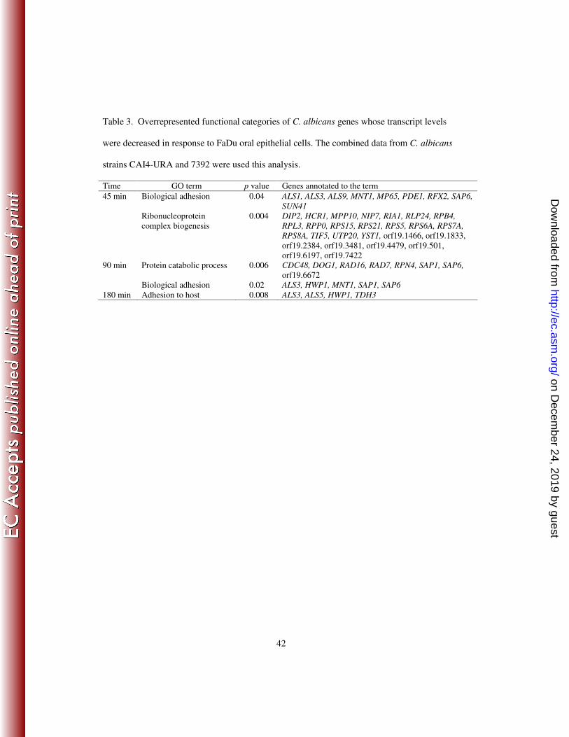
42
Table 3. Overrepresented functional categories of C. albicans genes whose transcript levels
were decreased in response to FaDu oral epithelial cells. The combined data from C. albicans
strains CAI4-URA and 7392 were used this analysis.
Time GO term p value Genes annotated to the term
45 min Biological adhesion 0.04 ALS1, ALS3, ALS9, MNT1, MP65, PDE1, RFX2, SAP6,
SUN41
0.004
Ribonucleoprotein
complex biogenesis
DIP2, HCR1, MPP10, NIP7, RIA1, RLP24, RPB4,
RPL3, RPP0, RPS15, RPS21, RPS5, RPS6A, RPS7A,
RPS8A, TIF5, UTP20, YST1, orf19.1466, orf19.1833,
orf19.2384, orf19.3481, orf19.4479, orf19.501,
orf19.6197, orf19.7422
90 min Protein catabolic process 0.006 CDC48, DOG1, RAD16, RAD7, RPN4, SAP1, SAP6,
orf19.6672
Biological adhesion 0.02 ALS3, HWP1, MNT1, SAP1, SAP6
180 min Adhesion to host 0.008 ALS3, ALS5, HWP1, TDH3
on Decem
ber 24, 2019 by guesthttp://ec.asm
.org/D
ownloaded from

43
Table 4. List of genes for generation of insertion mutant strains
Fold change by
microarraya
Fold change by
real time PCRa
Host cell damage
(% WT ± SD)b ORF 19 no.
Gene
Name Possible Function
Epi Endo Epi Endo
TIGR
insertion ID
Insertion
mutation Hyphae
Epi Endo
Protamine
susceptibility
orf19.348 Uncharacterized 2.2 1.0 2.5 NDc CAGOO23 No
orf19.1077 ATM1 Member of MDR subfamily of ABC family 2.2 1.1 3.5 ND CAGQD15 Yes normal 91±5 ND normal
orf19.4656 Uncharacterized 2.2 -1.1 3.8 ND CAGPD46 Yes normal 112±4 ND normal
orf19.4793 uncharacterized 2.0 -1.1 1.6 ND CAGAQ76 Yes normal 111±15 ND normal
orf19.6266 Uncharacterized, CCR4-NOT core
complex
2.0 -1.1 2.5 ND CAGB385 No
orf19.2398 Uncharacterized 2.1 1.2 2.5 ND CAGN836 Yes normal 114±8 ND normal
orf19.6931 CLP1 Uncharacterized , probable cleaveage/
polyadenylation factor
2.0d -1.5 1.9 ND CAGI909 Yes normal 109±24 ND normal
orf19.200 THO1 Uncharacterized 2.1e -1.1
e 2.5
e ND CAGDN85 No
orf19.4655 OPT6 Putative oligopeptide transporter 2.4e -1.7
e 3.1
e ND CAGFI33 Yes normal 94±9 ND normal
orf19.7001 YCK2 Casein kinase I (by homology) 2.2e -1.4
e 3.5
e 2.0
e CAGDI41 Yes short/
multiple
48±8 79±8 increased
orf19.202 CDC47 Cell division control protein 2.5e -1.5
e 1.6
e ND CAGCJ23 No
orf19.1504 Uncharacterized, putative patatin-like
phospholipase
2.6 1.1 4.3 ND CAGBR73 Yes normal 107±13 ND normal
orf19.3455 LPE10 Uncharacterized, putative magnesium
transporter activity
2.7 2.5 2.6 ND CAGDF12 No
orf19.4922 Uncharacterized 2.3 1.0 1.6 ND CAGF844 Yes normal 122±10 ND normal
orf19.3260 SIS2 Uncharacterized -1.2 2.1d ND 3.0
d CAGB279 No
orf19.5568 VPS51 Protein targeting to vacuole (by homology) 1.2d 2.1
d 2.2
d 2.9
d CAGDP76 Yes short 2±13 3±4 increased
orf19.6168 Uncharacterized, possible structural
constituent of cytoskeleton
1.2 2.3 ND 2.4 CAGEP09 Yes normal ND 97±9 normal
orf19.1766 IFO3 Uncharacterized, similar to Streptomyces
coelicolor putative hydrolase
1.3 2.1 ND 1.8 CAGL436 Yes normal ND 80±3 normal
orf19.3740 PGA23 Putative GPI-anchored protein with
unknown function
1.0d 2.7
d ND 2.8
d CAGN829 Yes normal ND 99±2 normal
orf19.4142 Uncharacterized 1.0d 2.4
d ND 1.6
d CAGC641 Yes normal ND 71±16 normal
orf19.4791 Uncharacterized -1.8d 2.0
d ND 2.2
d CAGE249 Yes normal ND 107±9 normal
orf19.4646 UEC1 Uncharacterized 1.2 2.0 1.1 3.4 CAGLQ62 Yes thickened 36±18 91±5 increased
orf19.3664 HSP31 Heat shock protein (by homology) -1.8 2.2 ND 4.9 CAGJM81 Yes normal ND 84±13 normal
orf19.4894 Uncharacterized -1.5d 3.4
d ND 2.3
d CAGKC82 Yes normal ND 73±11 normal
on Decem
ber 24, 2019 by guesthttp://ec.asm
.org/D
ownloaded from

44
orf19.1980 GIT4 Putative glycerophosphoinositol permease 1.1d 2.5
d ND 1.7
d CAGP819 Yes normal ND 77±12 normal
orf19.1939 Uncharacterized -1.1d 2.1
d ND 1.5
d CAGM083 Yes normal ND 67±6 normal
orf19.6392 Uncharacterized -1.1d 2.3
d ND 1.5
d CAGJP50 Yes normal ND 96±16 normal
orf19.403 PCL2 Cyclin-dependent protein kinase -1.3d 2.3
d ND 2.0
d --
f Yes normal ND 94±6 normal
orf19.1148 CIS312 Uncharacterized 1.0 2.4 ND 2.5 --f Yes normal ND 96±6 normal
Epi; epithelial cells, Endo; endothelial cells aAfter 45 min incubation unless otherwise specified
bThe wild-type control strain was DAY185
cNot determined
dAfter 90 min incubation eAfter 180 min incubation
fInsertion cassettes for orf19.403 and orf19.1148 were made in the current study
on Decem
ber 24, 2019 by guesthttp://ec.asm
.org/D
ownloaded from
45
FIGURE LEGENDS
Fig. 1. Time course of C. albicans interaction with host cells and polystyrene.
Blastospores of C. albicans strain CAI4-URA were incubated with the FaDu oral epithelial
cells, human umbilical vein endothelial cells, or polystyrene. (A) At the indicated times, the
cells were fixed and the organisms were imaged by differential interference contrast. Scale bar
indicates 5 µm. All images were taken at the same magnification. (B) The number of
organisms that were endocytosed by and cell-associated with the host cells the indicated times
was determined by a differential fluorescence assay. Results are the mean ± SD of three
independent experiments. *P = < 0.015 compared to oral epithelial cells at the same time
point . (C) The extent of C. albicans induced host cell damage was measured using a 51
Cr
release assay. Results are the mean ± SD of three independent experiments. *P < 0.001
compared to oral epithelial cells at the same time point. Epi, oral epithelial cells; Endo,
endothelial cells; HPF, high-powered field.
Fig. 2. Morphology of the yck2/yck2, vps51/vps51, and uec1/uec1 insertion mutants.
Blastospores of indicated strains were incubated on plastic for 180 min and then were fixed
and imaged by differential interference contrast. Scale bar indicates 5 µm. All images were
taken at the same magnification. WT, wild-type strain DAY185.
Fig. 3. Damage to oral epithelial cells and endothelial cells caused by the yck2/yck2,
vps51/vps51, and uec1/uec1 insertion mutants. The indicated strains were incubated with
FaDu oral epithelial cells or endothelial cells for 180 min, after which the extent of host cell
damage was assessed using a 51
Cr release assay. *P < 0.01 compared to both the wild-type
(DAY185) and complemented strains.
on Decem
ber 24, 2019 by guesthttp://ec.asm
.org/D
ownloaded from
46
Fig. 4. Susceptibility of the yck2/yck2, vps51/vps51, and uec1/uec1 insertion mutants to
stressors. Serial 10-fold dilutions of the indicated strains were plated onto YPD agar
containing 2.8 mg/ml protamine sulfate, 200 µg/ml Congo red, 1 M NaCl, 0.05 % SDS, or 5
mM H2O2 and incubated at 30°C. The plates containing protamine were photographed after 5
days, and the other plates were photographed after 2 days. WT, wild-type strain DAY185.
Fig. 5. Susceptibility of the yck2/yck2, vps51/vps51, and uec1/uec1 insertion mutants to
human β defensin 2. The susceptibility of the indicated strains of C. albicans to human β
defensin 2 was measured using a radial diffusion assay. The zones of growth inhibition were
imaged after incubation at 30oC for 2 days. WT, wild-type strain DAY185.
Fig. 6. Abnormal vacuolar morphology of the vps51/vps51 insertion mutant. Blastospores
of the indicated strains were grown to mid-log phase and then pulse-chased with FM4-64 to
label their vacuolar membranes. The cell walls were stained with an Alexa Fluor 488 labeled
anti-C. albicans antibody, after which the cells were imaged by confocal microscopy. The cell
walls are shown in green and the vacuolar membranes are show in red. Scale bar indicates 5
µm. WT, wild-type strain DAY185.
Supplementary Data
Table S1. C. albicans genes that were differentially expressed during exposure to FaDu oral
epithelial cells or primary endothelial cells compared to polystyrene. Results represent the
combined data from C. albicans strains CAI4-URA and 7392 for the epithelial cell data and
strains CAU4-URA and 36082 for the endothelial cell data.
Table S2. Primers used in this study
on Decem
ber 24, 2019 by guesthttp://ec.asm
.org/D
ownloaded from