Embed Size (px)
Citation preview
Research Article TRANSPARENTPROCESS
OPENACCESSNew therapeutic target in Pompe disease
Transcription factor EB (TFEB) is a newtherapeutic target for Pompe disease
Carmine Spampanato1,2,3y, Erin Feeney4y, Lishu Li4z, Monica Cardone1z, Jeong-A Lim4z,Fabio Annunziata1z, Hossein Zare4, Roman Polishchuk1, Rosa Puertollano5, Giancarlo Parenti1,6,Andrea Ballabio1,2,3,6*, Nina Raben4**
Keywords: acid alpha-glucosidase;
autophagy; lysosomal storage; Pompe
disease; TFEB
DOI 10.1002/emmm.201202176
Received October 18, 2012
Revised February 25, 2013
Accepted February 27, 2013
(1) Telethon Institute of Genetics and Medicine (TIGEM
(2) Department of Molecular and Human Genetics
Medicine, Houston, TX, USA
(3) Jan and Dan Duncan Neurological Research Insti
Hospital, Houston, TX, USA
(4) Laboratory of Muscle Stem Cells and Gene Regulatio
of Arthritis and Musculoskeletal and Skin Diseases,
Health, Bethesda, MD, USA
(5) Laboratory of Cell Biology, National Heart, Lung,
National Institutes of Health, Bethesda, MD, USA
(6) Department of Pediatrics, Federico II University, Na
*Corresponding author: Tel: þ39 081 613 2207; Fax:
E-mail: [email protected]
**Corresponding author: Tel: þ1 301 496 1474; Fax:
E-mail: [email protected]
ySpampanato and Feeney contributed equally as the twzLi, Cardone, Lim, and Annunziata contributed equally
Published 2013. This is a US Government work and is inLtd on behalf of EMBO. This is an open access article undwhich permits use, distribution and reproduction in any m
A recently proposed therapeutic approach for lysosomal storage disorders (LSDs)
relies upon the ability of transcription factor EB (TFEB) to stimulate autophagy
and induce lysosomal exocytosis leading to cellular clearance. This approach is
particularly attractive in glycogen storage disease type II [a severe metabolic
myopathy, Pompe disease (PD)] as the currently available therapy, replacement of
the missing enzyme acid alpha-glucosidase, fails to reverse skeletal muscle
pathology. PD, a paradigm for LSDs, is characterized by both lysosomal abnorm-
ality and dysfunctional autophagy. Here, we show that TFEB is a viable thera-
peutic target in PD: overexpression of TFEB in a new muscle cell culture system
and in mouse models of the disease reduced glycogen load and lysosomal size,
improved autophagosome processing, and alleviated excessive accumulation of
autophagic vacuoles. Unexpectedly, the exocytosed vesicles were labelled with
lysosomal and autophagosomal membrane markers, suggesting that TFEB
induces exocytosis of autophagolysosomes. Furthermore, the effects of TFEB
were almost abrogated in the setting of genetically suppressed autophagy,
supporting the role of autophagy in TFEB-mediated cellular clearance.
INTRODUCTION
Pompe disease (PD; OMIM 232300) is a severe metabolic
myopathy caused by the deficiency of acid alpha-glucosidase
(GAA; acid maltase, E.C.3.2.1.20), an enzyme responsible for
), Naples, Italy
, Baylor College of
tute, Texas Children
n, National Institute
National Institutes of
and Blood Institute,
ples, Italy
þ39 081 579 0919;
þ1 301 480 63 67;
o co-first authors.
among themselves.
the public domain in the Uer the terms of the Creativeedium, provided the origin
breaking down glycogen to glucose within the acidic environment
of lysosomes. The functional deficiency or complete absence of
the enzyme results in accumulation of glycogen within this
cellular compartment (Kroos et al, 2012; Van der Ploeg & Reuser,
2008). PD pathology is also characterized by secondary
accumulation of autophagic debris (autophagic build-up),
typically found in skeletal muscle fibres (Raben et al, 2007a,b,
2012). Although PD is a rare disorder (Martiniuk et al, 1998), it
shares common pathological features with many other lysosomal
storage disorders (LSDs), which encompass more than 60
nosological entities (Cox & Cachon-Gonzalez, 2012).
GAA deficiency is a systemic disorder – distended glycogen-
filled lysosomes can be found in multiple tissues – but skeletal
and cardiac muscles are particularly vulnerable to the
accumulation of storage material. In the most serious infantile
form, the disease manifests as profound weakness, hyper-
trophic cardiomyopathy, heart failure, feeding difficulties,
respiratory infections and, if left untreated, causes death
within the first year of life. In the attenuated phenotypes,
characterized by later (childhood, juvenile or adult) onset,
cardiac muscle is usually spared, but the illness remains
a serious condition with progressive motor impairment,
SA. Published by John Wiley and Sons,Commons Attribution License (CC BY 3.0),al work is properly cited. EMBO Mol Med (2013) 5, 691–706 691
Research Article www.embomolmed.orgNew therapeutic target in Pompe disease
692
respiratory failure and premature death (Van der Ploeg &
Reuser, 2008).
Enzyme replacement therapy (ERT) is now available for
several LSDs including PD. Unfortunately, in many of these
diseases the target tissues are not easily accessible to the
replacement enzymes (Lachmann, 2011; Urbanelli et al, 2011).
In PD, skeletal muscle – the major target for ERT – is particularly
challenging to treat. Even with extremely high dosages of
the drug, recombinant human GAA (rhGAA; alglucosidase
alpha; Myozyme1 and Lumizyme1, Genzyme Corporation,
Framingham, MA), many patients experience limited clinical
benefit or show signs of disease progression (Angelini &
Semplicini, 2012; Kishnani et al, 2007; Schoser et al, 2008;
Strothotte et al, 2010; Van den Hout et al, 2004; Van der Ploeg
et al, 2010). Furthermore, the greatest success of ERT in PD –
significantly improved survival of infants due to the restoration
of cardiac function – comes at a price: many long-term survivors
suffer from debilitating myopathy, often more severe than in
late-onset cases.
Several factors contribute to the resistance of skeletal muscle
to ERT. These include the sheer mass of muscle tissue, a
relatively low density of the mannose-6-phosphate receptor
that is responsible for the uptake of the enzyme, inefficient
trafficking of the internalized enzyme to lysosomes, immune
response to the recombinant enzyme in cross-reactive immu-
nologic material (CRIM)-negative patients and the presence of
large areas of autophagic build-up alongside expanded lyso-
somes (Banugaria et al, 2011; Funk et al, 1992; Kishnani et al,
2010; Koeberl et al, 2012; Raben et al, 2002, 2010, 2012).
Significant efforts are aimed at improving the current therapy,
such as the development of ‘‘second generation’’ recombinant
enzymes with enhanced targeting properties (Maga et al, 2012;
Tiels et al, 2012; Zhu et al, 2009), pharmacological chaperones
that assist the folding of the mutant enzyme (Okumiya et al,
2007; Parenti et al, 2007) or increase the stability of the
recombinant enzyme (Porto et al, 2009, 2012), and gene therapy
(Byrne et al, 2011).
A recently proposed novel approach to therapy for LSDs –
modulation of transcription factor EB (TFEB) – circumvents
the problem of inefficient enzyme delivery by exploiting the
ability of lysosomes to expel their content into the extracellular
space, thus providing clearance of the stored material (Medina
et al, 2011). It has been shown that overexpression of TFEB, a
master regulator of lysosomal biogenesis and autophagy, leads
to the generation of new lysosomes and increased numbers of
autophagosomes in a variety of cell types (Sardiello et al, 2009;
Settembre & Ballabio, 2011; Settembre et al, 2011). Further-
more, since TFEB has been shown to promote lysosomal–
autophagosomal fusion (Settembre & Ballabio, 2011; Settembre
et al, 2011), this approach, unlike most current efforts, has the
potential to prevent or resolve autophagic build-up.
Here, we have characterized the effect of TFEB overexpres-
sion on lysosomal and autophagic accumulation in previously
described and newly developed PD models. Expression of TFEB
in myotubes and muscle fibres resulted in lysosomal docking/
fusion with the plasma membrane, lysosomal exocytosis and a
dramatic reduction of intra-lysosomal glycogen accumulation.
Published 2013. This is a US Government work and is in the public domain in the UPublished by John Wiley and Sons, Ltd on behalf of EMBO.
In addition, TFEB overexpression in PD muscle alleviated
autophagic pathology by promoting the formation and removal
of autophagolysosomes. Thus, modulation of TFEB activity
holds promise for the development of a better therapy for this
devastating disorder.
RESULTS
Establishment and characterization of new in vitro and
in vivo PD models
To test new therapeutic approaches for PD, we established
conditionally immortalized myogenic cell lines (Supporting
Information Fig S1). PD myotubes but not myoblasts or
fibroblasts (Supporting Information Fig S2) replicated lysosomal
pathology, namely the enlargement of lysosomes and abnormal
glycogen storage (Fig 1A and D). Disappointingly, another
abnormality in PD muscle fibres – autophagic accumulation
(Raben et al, 2012) – was not reproduced in PD myotubes, as
demonstrated by immunostaining and western analysis with LC3
[a highly specific autophagosomal marker (Kabeya et al, 2000)]
(shown for western blot in Supporting Information Fig S1C).
In contrast, autophagic pathology was clearly visible in
muscle fibres from a newly developed PD mouse model, in
which autophagosomes are labelled with GFP-LC3 (GFP-
LC3:GAA�/�). In this new strain (but not in the myoblast
cell line derived from these mice; Supporting Information Fig
S3), large areas of autophagic accumulation can be seen in live
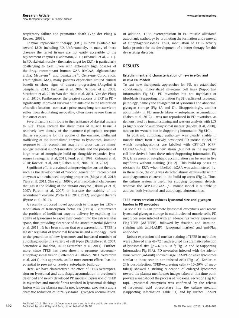
myofibres without staining (Fig 2). This build-up poses an
obstacle for ERT: when labelled rhGAA was administered i.v.
in these mice, the drug was detected almost exclusively within
autophagosomes clustered in the build-up areas (Fig 2). Thus,
the culture system is useful for studying lysosomal defects,
whereas the GFP-LC3:GAA�/� mouse model is suitable to
address both lysosomal and autophagic abnormalities.
TFEB overexpression reduces lysosomal size and glycogen
burden in PD myotubes
To see if TFEB can promote lysosomal exocytosis and rescue
lysosomal glycogen storage in multinucleated muscle cells, PD
myotubes were infected with an adenovirus vector expressing
Flag-TFEB (Ad-TFEB), followed by fixation and immuno-
staining with anti-LAMP1 (lysosomal marker) and anti-Flag
antibodies.
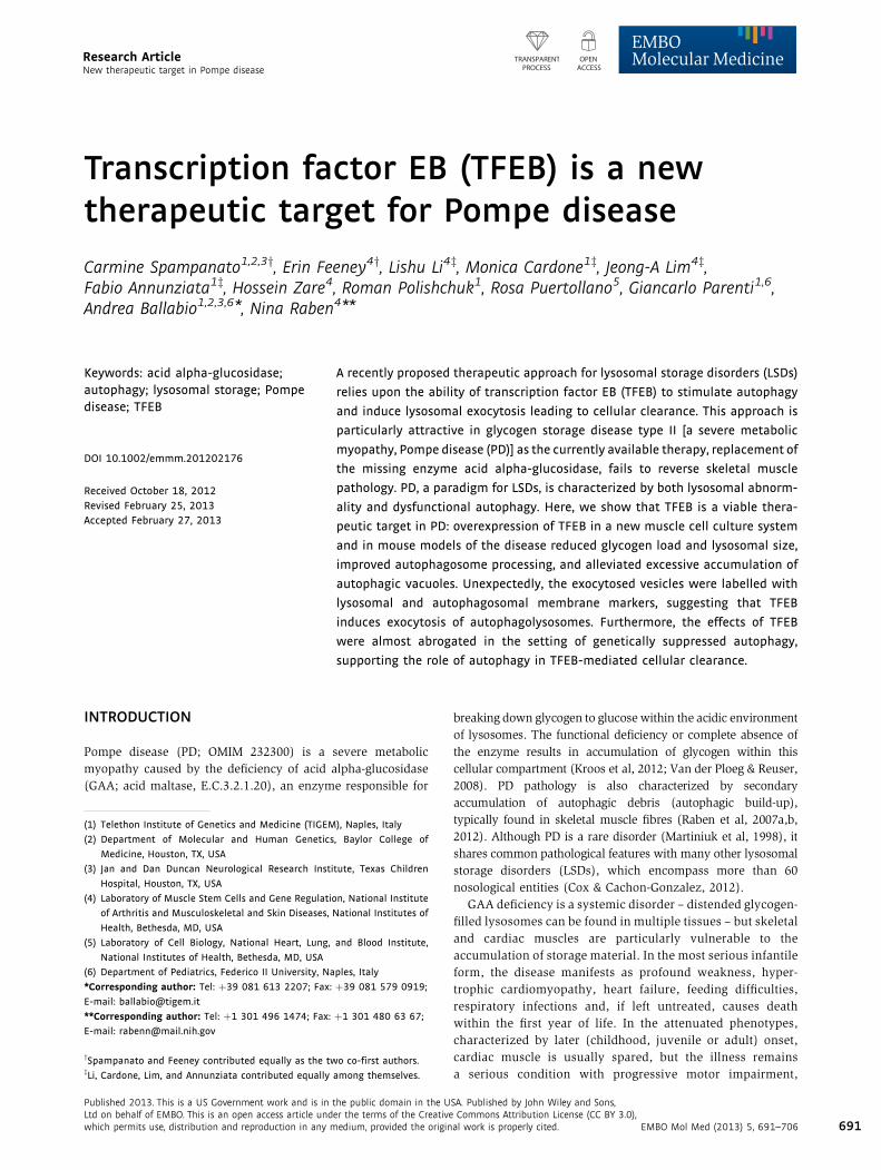
Robust expression and nuclear staining of TFEB in myotubes
were achieved after 48–72 h and resulted in a dramatic reduction
of lysosomal size (p¼ 6.32� 10�8; Fig 1A and B; Supporting
Information Fig S4A). PD myotubes infected with the adeno-
virus vector (Ad-null) showed large LAMP1-positive lysosomes
similar to those seen in non-infected cells (Fig 1A). Earlier, at
24 h post-infection, TFEB-expressing cells (�10–20% of myo-
tubes) showed a striking relocation of enlarged lysosomes
toward the plasma membrane; images taken at this time point
provide a snapshot of the process of lysosomal secretion (Fig 1C,
top). Lysosomal exocytosis was confirmed by the release
of lysosomal acid phosphatase into the culture medium
(Supporting Information Table S1) and by surface LAMP1
SA.EMBO Mol Med (2013) 5, 691–706
www.embomolmed.org Research ArticleCarmine Spampanato et al.
Figure 1. TFEB stimulates clearance of enlarged lysosomes and reduces glycogen burden in PD myotubes.
A. Confocal microscopy image of PD myotubes (cl. 3LE8) infected for 72 h with adenovirus containing TFEB (PDþ Ad-TFEB 72 h) shows a dramatic reduction in the
number of large LAMP1-positive lysosomes (red) compared to that in untreated (PD) or adenovirus (PDþAd-null)-treated PD myotubes. WT myotubes are
shown on the left panel. Nuclei are stained with Hoechst (blue). TFEB was detected with anti-Flag antibody (green).
B. Distribution of lysosomal size differs significantly in Ad-null and Ad-TFEB PD myotubes ( p¼6.32� 10�8; Kolmogorov–Smirnov test). Lysosomal size
is expressed as number of pixels representing lysosomal area (LAMP1-positive structures). The median lysosomal size of Ad-TFEB infected myotubes
(m¼367.13 pixels, n¼703, range 208–2659) was significantly lower than that of Ad-null infected myotubes (m¼ 491.16 pixels, n¼ 1395, range 200–2857;
p¼ 6.5�10�12; Wilcoxon rank sum test).
C. Confocal microscopy image of PD myotubes infected for 24 h with Ad-TFEB shows relocation of lysosomes to the plasma membrane (top). Images showing
LAMP1 staining (red) on plasma membrane in a PD myotube infected with Ad-TFEB (bottom; arrows) but not in a non-infected cell (middle).
Non-permeabilized cells were incubated with anti-LAMP1 antibody at 48C for 40 min, followed by fixation and staining with secondary antibody.
D. Confocal microscopy images of live non-infected PD myotubes (left) or PD myotubes infected for 72 h with Ad-TFEB (right) show a dramatic reduction in the
amount of accumulated glycogen in TFEB-treated cells. The cells were incubated with the fluorescent glucose (2-NBDG; green), extensively washed, and
analysed by confocal microscopy. Bar: 10 mm for all panels.
assay, which showed the presence of lysosomal membrane
marker on the plasma membrane in TFEB-treated myotubes
(Fig 1C, lower) but not in untreated cells (Fig 1C, middle).
TFEB also stimulated autophagy in PD myotubes, as evidenced
by an increase in LC3 levels (Supporting Information Fig S4B
and C).
In addition, we tested the effect of constitutively active
mutant TFEB (S211A; TFEBmt) (Martina et al, 2012; Roczniak-
Ferguson et al, 2012; Settembre et al, 2012) in PD myotubes.
Massive accumulation of TFEB in the nuclei resulted in a
striking clearance of large lysosomes without any decrease in
the total amount of LAMP1 protein (Fig 3A and B). In fact, levels
of LAMP1 appear to increase in TFEBmt-treated cells, consistent
with the role of TFEB in stimulating lysosomal biogenesis
(Sardiello et al, 2009; Settembre & Ballabio, 2011).
As expected, the removal of enlarged lysosomes from PD
myotubes was associated with a significant decrease in the
amount of accumulated storage material, as shown by the
EMBO Mol Med (2013) 5, 691–706PublishePublishe
incorporation of the fluorescent glucose derivative 2-NBDG into
glycogen (Fig 1D). Thus, muscle cells in PD can be cleared of
lysosomal glycogen accumulation by TFEB induction. Of note,
many TFEB-overexpressing muscle cells, particularly those
treated with TFEBmt, change their morphology: normally
elongated myotubes become spindle-like and contain centrally
located nuclei (Fig 3A). However, no cytotoxicity was observed
in TFEB-treated cells as evidenced by lactate dehydrogenase
(LDH)-measurements in culture medium (not shown). Although
TUNEL assay revealed occassional apoptotic cells, no activation
of caspase-3 was detected by western (Fig 3C and D).
Next, we attempted to activate endogenous TFEB by
pharmacologically targeting mTORC1, which has been shown
to negatively regulate TFEB (Martina et al, 2012; Roczniak-
Ferguson et al, 2012; Settembre et al, 2012). Addition of Torin 1
and Torin 2 inhibited mTORC1 (Liu et al, 2010) and stimulated
autophagy, but failed to reduce lysosomal size (Supporting
Information Fig S5A–C; shown for Torin 2). These data suggest
d 2013. This is a US Government work and is in the public domain in the USA.d by John Wiley and Sons, Ltd on behalf of EMBO. 693
Research Article www.embomolmed.orgNew therapeutic target in Pompe disease
Figure 2. Therapeutic enzyme is trapped in the area of autophagic
build-up in PD fibres. Confocal microscopy of live unstained fibres from a
GFP-LC3:WT mouse (WT) and from untreated (GAA�/�) or ERT-treated
(GAA�/�; þERT) GFP-LC3:GAA�/� mice. Images show typical centrally located
autophagic build-up with multiple clusters of LC3-positive autophagosomes.
Structures like these are never seen in WT muscle. Autophagic build-up is
present virtually in all fibres derived from EDL or gastrocnemius muscles (not
shown), but only in 60–70% of FDB fibres. Labelled recombinant human GAA
(red) was administered i.v. into 3–4 month-old GFP-LC3:GAA�/�mice (n¼ 5) at
a dose of 100 mg/kg twice with a 24 h interval. Mice were sacrificed the
next day, and live fibres (shown for EDL muscle) were analysed by confocal
microscopy. The labelled recombinant GAA was detected almost exclusively in
the autophagic vesicles. At least 500 fibres were analysed for each condition.
Bar: 10 mm.
694
that the amount of endogenous TFEB may not be sufficient to
support lysosomal clearance; alternatively, another kinase –
MAPK (ERK1/2) kinase – may be involved in the process of
TFEB activation (Settembre et al, 2011). A striking increase in
the levels of p-ERK1/2 in TFEB-treated PD myotubes points
to such a possibility in muscle cells (Supporting Information
Fig S5D).
TFEB overexpression in PD muscle induces cellular clearance
by promoting lysosomal/autophagosomal exocytosis
To evaluate the effect of TFEB on lysosomal and autophagic
pathologies in vivo, three knockout mouse strains were used:
the previously described GAA�/� and autophagy-deficient
GAA�/� (Atg7:GAA DKO) models (Raben et al, 1998, 2010),
and a newly developed GFP-LC3:GAA�/� strain. Flexor
digitorum brevis (FDB) muscles from each line were transfected
by electroporation with plasmids containing TFEB and/or
LAMP1 (Table 1). Four to six days after transfection, live single
muscle fibres were analysed by time-lapse confocal microscopy;
Published 2013. This is a US Government work and is in the public domain in the UPublished by John Wiley and Sons, Ltd on behalf of EMBO.
alternatively, live or fixed fibres were stained and imaged. The
efficiency of the transfection in all conditions ranged from 10 to
30%.
Both Flag-TFEB/mCherry-LAMP1- and GFP-TFEB/mCherry-
LAMP1-transfected PD fibres (Table 1, conditions 2 and 3)
showed a striking decrease in the number of large lysosomes,
translocation of enlarged lysosomes to the periphery of the fibre,
docking and fusion of lysosomes with the plasma membrane,
the emergence of multiple normal dot-like size lysosomes,
and a markedly increased motility and fusion of lysosomes
(Fig 4A–C; Movie 1) compared to untreated PD controls
(Table 1, condition 1; Movie 2). The mean maximum velocity
of lysosomes in TFEB-treated fibres was 0.247� 0.01 mm/min
[pooled data from conditions 2 (n¼ 24) and 3 (n¼ 19)], whereas
this value was 0.130� 0.006 mm/min (n¼ 26) in untreated
controls (p¼ 2.07� 10�17). Importantly, the number of large
(>3.5 mm in length) lysosomes was significantly reduced in
TFEB-treated fibres [4.41� 0.7 and 1.37� 0.2 lysosomes/mm2
for untreated (n¼ 10) and treated (n¼ 24) fibres, respectively;
p¼ 1.0� 10�3] (Fig 4C; Supporting Information Fig S6A). This
dramatic decrease in the number of large lysosomes was
confirmed by lysotracker and LAMP1 staining of live or fixed
fibres transfected with GFP-TFEB alone (condition 7; Supporting
Information Fig S6B and C).
The vast majority of TFEB-transfected fibres (freshly isolated
or maintained for days in culture) had normal shape and
appearance, suggesting that overexpression of TFEB does not
cause any appreciable muscle damage. Furthermore, no major
mitochondrial abnormalities were observed in TFEB-treated
fibres as evidenced by MitoTracker labelling and immunostain-
ing for cytochrome c (Supporting Information Fig S7A and B).
However, stress of fibres exposed to hours of time-lapse
microscopy resulted in a massive translocation of TFEB into the
nuclei and – in occasional fibres – the detachment of plasma
membrane (Fig 4B, arrow and inset; Supporting Information
Fig S8A; Movie 3). This phenomenon was not seen in control
experiments, in which the muscle of GFP-LC3:GAA�/�mice was transfected with mCherry-LAMP1 only (Table 1,
condition 1).
The choice of the GFP-LC3:GAA�/� strain for control
experiments enabled us not only to evaluate the motility of
lysosomes, but also to examine the possibility of impaired
lysosomal/autophagosomal fusion as a mechanism underlying
autophagic build-up. Unexpectedly, the expression of mCherry-
LAMP1 was seen almost exclusively in fibres or in regions of
fibres free from autophagic build-up (�80–90% of all trans-
fected fibres exhibited these two expression patterns in roughly
equal proportions; Supporting Information Fig S8B), suggesting
a compromised lysosomal biogenesis in areas of autophagic
accumulation. However, in those fibres in which mCherry-
LAMP1 was expressed in the build-up area (�10–20%),
lysosomal-autophagosomal fusion events appear rare or non-
existent (Movie 4).
It was reasonable to assume that TFEB overexpression may
increase lysosomal–autophagosomal fusion, thus preventing or
even resolving autophagic build-up in PD muscle. Indeed,
none of the Flag-TFEB/mCherry-LAMP1 transfected fibres from
SA.EMBO Mol Med (2013) 5, 691–706
www.embomolmed.org Research ArticleCarmine Spampanato et al.
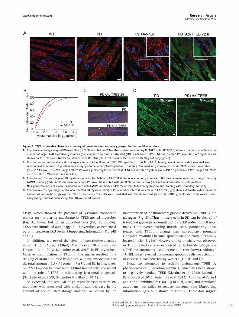
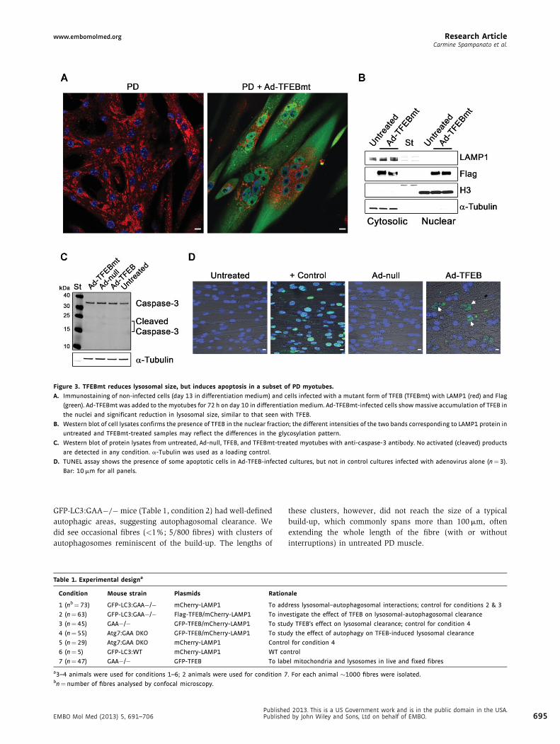
Figure 3. TFEBmt reduces lysosomal size, but induces apoptosis in a subset of PD myotubes.
A. Immunostaining of non-infected cells (day 13 in differentiation medium) and cells infected with a mutant form of TFEB (TFEBmt) with LAMP1 (red) and Flag
(green). Ad-TFEBmt was added to the myotubes for 72 h on day 10 in differentiation medium. Ad-TFEBmt-infected cells show massive accumulation of TFEB in
the nuclei and significant reduction in lysosomal size, similar to that seen with TFEB.
B. Western blot of cell lysates confirms the presence of TFEB in the nuclear fraction; the different intensities of the two bands corresponding to LAMP1 protein in
untreated and TFEBmt-treated samples may reflect the differences in the glycosylation pattern.
C. Western blot of protein lysates from untreated, Ad-null, TFEB, and TFEBmt-treated myotubes with anti-caspase-3 antibody. No activated (cleaved) products
are detected in any condition. a-Tubulin was used as a loading control.
D. TUNEL assay shows the presence of some apoptotic cells in Ad-TFEB-infected cultures, but not in control cultures infected with adenovirus alone (n¼3).
Bar: 10 mm for all panels.
GFP-LC3:GAA�/�mice (Table 1, condition 2) had well-defined
autophagic areas, suggesting autophagosomal clearance. We
did see occasional fibres (<1%; 5/800 fibres) with clusters of
autophagosomes reminiscent of the build-up. The lengths of
Table 1. Experimental designa
Condition Mouse strain Plasmids Ration
1 (nb¼73) GFP-LC3:GAA�/� mCherry-LAMP1 To add
2 (n¼ 63) GFP-LC3:GAA�/� Flag-TFEB/mCherry-LAMP1 To inv
3 (n¼ 45) GAA�/� GFP-TFEB/mCherry-LAMP1 To stu
4 (n¼ 55) Atg7:GAA DKO GFP-TFEB/mCherry-LAMP1 To stu
5 (n¼ 29) Atg7:GAA DKO mCherry-LAMP1 Contro
6 (n¼ 5) GFP-LC3:WT mCherry-LAMP1 WT co
7 (n¼ 47) GAA�/� GFP-TFEB To lab
a3–4 animals were used for conditions 1–6; 2 animals were used for condition 7bn¼ number of fibres analysed by confocal microscopy.
EMBO Mol Med (2013) 5, 691–706PublishePublishe
these clusters, however, did not reach the size of a typical
build-up, which commonly spans more than 100 mm, often
extending the whole length of the fibre (with or without
interruptions) in untreated PD muscle.
ale
ress lysosomal–autophagosomal interactions; control for conditions 2 & 3
estigate the effect of TFEB on lysosomal-autophagosomal clearance
dy TFEB’s effect on lysosomal clearance; control for condition 4
dy the effect of autophagy on TFEB-induced lysosomal clearance
l for condition 4
ntrol
el mitochondria and lysosomes in live and fixed fibres
. For each animal �1000 fibres were isolated.
d 2013. This is a US Government work and is in the public domain in the USA.d by John Wiley and Sons, Ltd on behalf of EMBO. 695
Research Article www.embomolmed.orgNew therapeutic target in Pompe disease
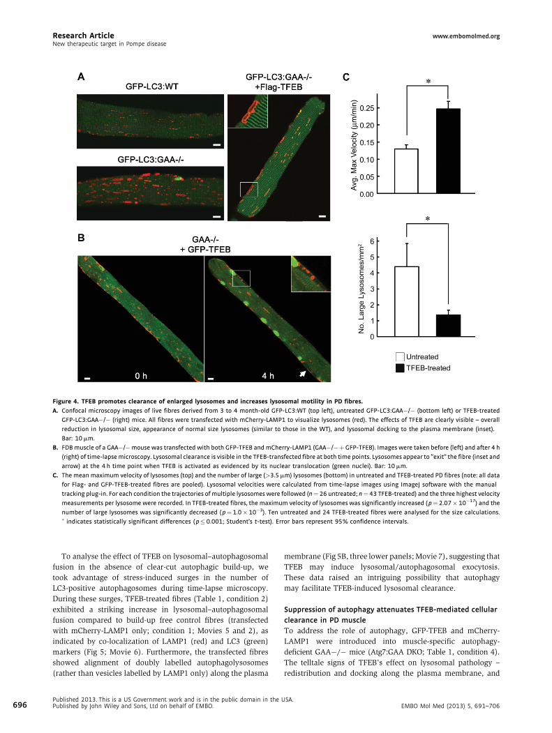
Figure 4. TFEB promotes clearance of enlarged lysosomes and increases lysosomal motility in PD fibres.
A. Confocal microscopy images of live fibres derived from 3 to 4 month-old GFP-LC3:WT (top left), untreated GFP-LC3:GAA�/� (bottom left) or TFEB-treated
GFP-LC3:GAA�/� (right) mice. All fibres were transfected with mCherry-LAMP1 to visualize lysosomes (red). The effects of TFEB are clearly visible – overall
reduction in lysosomal size, appearance of normal size lysosomes (similar to those in the WT), and lysosomal docking to the plasma membrane (inset).
Bar: 10 mm.
B. FDB muscle of a GAA�/�mouse was transfected with both GFP-TFEB and mCherry-LAMP1 (GAA�/�þGFP-TFEB). Images were taken before (left) and after 4 h
(right) of time-lapse microscopy. Lysosomal clearance is visible in the TFEB-transfected fibre at both time points. Lysosomes appear to ‘‘exit’’ the fibre (inset and
arrow) at the 4 h time point when TFEB is activated as evidenced by its nuclear translocation (green nuclei). Bar: 10 mm.
C. The mean maximum velocity of lysosomes (top) and the number of large (>3.5 mm) lysosomes (bottom) in untreated and TFEB-treated PD fibres (note: all data
for Flag- and GFP-TFEB-treated fibres are pooled). Lysosomal velocities were calculated from time-lapse images using ImageJ software with the manual
tracking plug-in. For each condition the trajectories of multiple lysosomes were followed (n¼ 26 untreated; n¼43 TFEB-treated) and the three highest velocity
measurements per lysosome were recorded. In TFEB-treated fibres, the maximum velocity of lysosomes was significantly increased ( p¼2.07� 10�17) and the
number of large lysosomes was significantly decreased ( p¼1.0� 10�3). Ten untreated and 24 TFEB-treated fibres were analysed for the size calculations.� indicates statistically significant differences ( p�0.001; Student’s t-test). Error bars represent 95% confidence intervals.
696
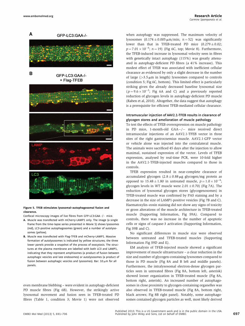
To analyse the effect of TFEB on lysosomal–autophagosomal
fusion in the absence of clear-cut autophagic build-up, we
took advantage of stress-induced surges in the number of
LC3-positive autophagosomes during time-lapse microscopy.
During these surges, TFEB-treated fibres (Table 1, condition 2)
exhibited a striking increase in lysosomal–autophagosomal
fusion compared to build-up free control fibres (transfected
with mCherry-LAMP1 only; condition 1; Movies 5 and 2), as
indicated by co-localization of LAMP1 (red) and LC3 (green)
markers (Fig 5; Movie 6). Furthermore, the transfected fibres
showed alignment of doubly labelled autophagolysosomes
(rather than vesicles labelled by LAMP1 only) along the plasma
Published 2013. This is a US Government work and is in the public domain in the UPublished by John Wiley and Sons, Ltd on behalf of EMBO.
membrane (Fig 5B, three lower panels; Movie 7), suggesting that
TFEB may induce lysosomal/autophagosomal exocytosis.
These data raised an intriguing possibility that autophagy
may facilitate TFEB-induced lysosomal clearance.
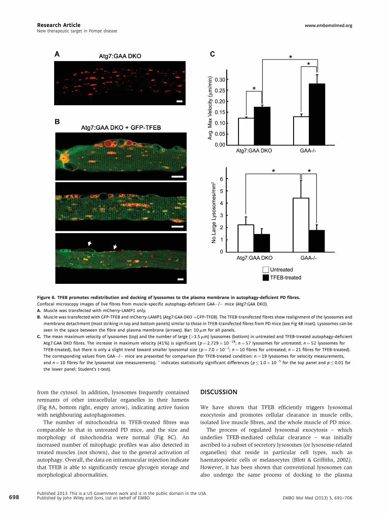
Suppression of autophagy attenuates TFEB-mediated cellular
clearance in PD muscle
To address the role of autophagy, GFP-TFEB and mCherry-
LAMP1 were introduced into muscle-specific autophagy-
deficient GAA�/� mice (Atg7:GAA DKO; Table 1, condition 4).
The telltale signs of TFEB’s effect on lysosomal pathology –
redistribution and docking along the plasma membrane, and
SA.EMBO Mol Med (2013) 5, 691–706
www.embomolmed.org Research ArticleCarmine Spampanato et al.
Figure 5. TFEB stimulates lysosomal–autophagosomal fusion and
clearance.
Confocal microscopy images of live fibres from GFP-LC3:GAA�/� mice.
A. Muscle was transfected with mCherry-LAMP1 only. The image (a single
frame from the time-lapse series presented in Movie 5) shows lysosomes
(red), LC3-positive autophagosomes (green) and a number of autolyso-
somes (yellow).
B. Muscle was transfected with Flag-TFEB and mCherry-LAMP1. Massive
formation of autolysosomes is indicated by yellow structures; the three
lower panels provide a snapshot of the process of exocytosis. The struc-
tures at the plasma membrane are labelled with both LC3 and LAMP1,
indicating that they represent amphisomes (a product of fusion between
autophagic vesicles and late endosomes) or autolysosomes (a product of
fusion between autophagic vesicles and lysosomes). Bar: 10 mm for all
panels.
even membrane blebbing – were evident in autophagy-deficient
PD muscle fibres (Fig 6B). However, the strikingly active
lysosomal movement and fusion seen in TFEB-treated PD
fibres (Table 1, condition 3; Movie 1) were not observed
EMBO Mol Med (2013) 5, 691–706PublishePublishe
when autophagy was suppressed. The maximum velocity of
lysosomes (0.174� 0.005 mm/min; n¼ 52) was significantly
lower than that in TFEB-treated PD mice (0.279� 0.02;
p¼ 7.01� 10�6; n¼ 19) (Fig 6C, top; Movie 8). Furthermore,
the TFEB-induced increase in lysosomal velocity seen in fibres
with genetically intact autophagy (115%) was greatly attenu-
ated in autophagy-deficient PD fibres (a 41% increase). This
modest effect of TFEB was associated with inefficient cellular
clearance as evidenced by only a slight decrease in the number
of large (>3.5 mm in length) lysosomes compared to controls
(condition 5; Fig 6C, bottom). This limited effect is particularly
striking given the already decreased baseline lysosomal size
(p¼ 9.6� 10�3; Fig 6A and C) and a previously reported
reduction of glycogen levels in autophagy-deficient PD muscle
(Raben et al, 2010). Altogether, the data suggest that autophagy
is a prerequisite for efficient TFEB-mediated cellular clearance.
Intramuscular injection of AAV2.1-TFEB results in clearance of
glycogen stores and amelioration of muscle pathology
To test the effects of TFEB overexpression on muscle pathology
in PD mice, 1-month-old GAA�/� mice received direct
intramuscular injections of an AAV2.1-TFEB vector in three
sites of the right gastrocnemius muscle. AAV2.1-GFP vector
or vehicle alone was injected into the contralateral muscle.
The animals were sacrificed 45 days after the injection to allow
maximal, sustained expression of the vector. Levels of TFEB
expression, analysed by real-time PCR, were 10-fold higher
in the AAV2.1-TFEB-injected muscles compared to those in
controls.
TFEB expression resulted in near-complete clearance of
accumulated glycogen (2.8� 0.88 mg glycogen/mg protein as
opposed to 15.48� 1.80 in untreated muscle, p¼ 1.0� 10�4;
glycogen levels in WT muscle were 2.01� 0.70) (Fig 7A). The
reduction of lysosomal glycogen stores (glycogenosomes) in
TFEB-treated muscle was confirmed by PAS staining and by a
decrease in the size of LAMP1-positive vesicles (Fig 7B and C).
Haematoxylin–eosin staining did not show any signs of toxicity
or gross alterations of the muscle architecture in TFEB-treated
muscle (Supporting Information, Fig S9A). Compared to
controls, there was no increase in the number of apoptotic
cells or signs of caspase-3 activation (Supporting Information,
Fig S9B and C).
No significant differences in muscle size were observed
between untreated and TFEB-treated muscles (Supporting
Information Fig S9D and E).
EM analysis of TFEB-injected muscle showed a significant
improvement of muscle ultrastructure – a clear reduction in the
size and number of glycogen-containing lysosomes compared to
those in PD muscle (Fig 8A and B left and middle panels).
Furthermore, the intralysosomal electron-dense glycogen par-
ticles seen in untreated fibres (Fig 8A, bottom left, asterisk)
showed looser organization in TFEB-treated muscle (Fig 8A,
bottom right, asterisk). An increased number of autophago-
somes in close proximity to glycogen-containing organelles was
also observed in TFEB-treated muscle (Fig 8A, bottom right,
black arrows; Fig 8B right panel). Notably, some autophago-
somes contained glycogen particles as well, most likely derived
d 2013. This is a US Government work and is in the public domain in the USA.d by John Wiley and Sons, Ltd on behalf of EMBO. 697
Research Article www.embomolmed.orgNew therapeutic target in Pompe disease
Figure 6. TFEB promotes redistribution and docking of lysosomes to the plasma membrane in autophagy-deficient PD fibres.
Confocal microscopy images of live fibres from muscle-specific autophagy-deficient GAA�/� mice (Atg7:GAA DKO).
A. Muscle was transfected with mCherry-LAMP1 only.
B. Muscle was transfected with GFP-TFEB and mCherry-LAMP1 (Atg7:GAA DKOþGFP-TFEB). The TFEB-transfected fibres show realignment of the lysosomes and
membrane detachment (most striking in top and bottom panels) similar to those in TFEB-transfected fibres from PD mice (see Fig 4B inset). Lysosomes can be
seen in the space between the fibre and plasma membrane (arrows). Bar: 10 mm for all panels.
C. The mean maximum velocity of lysosomes (top) and the number of large (>3.5 mm) lysosomes (bottom) in untreated and TFEB-treated autophagy-deficient
Atg7:GAA DKO fibres. The increase in maximum velocity (41%) is significant ( p¼ 2.729� 10�18; n¼57 lysosomes for untreated; n¼ 52 lysosomes for
TFEB-treated), but there is only a slight trend toward smaller lysosomal size ( p¼ 7.0�10�2; n¼ 10 fibres for untreated; n¼21 fibres for TFEB-treated).
The corresponding values from GAA�/� mice are presented for comparison (for TFEB-treated condition: n¼ 19 lysosomes for velocity measurements,
and n¼10 fibres for the lysosomal size measurements). � indicates statistically significant differences ( p�1.0� 10�5 for the top panel and p� 0.01 for
the lower panel; Student’s t-test).
698
from the cytosol. In addition, lysosomes frequently contained
remnants of other intracellular organelles in their lumens
(Fig 8A, bottom right, empty arrow), indicating active fusion
with neighbouring autophagosomes.
The number of mitochondria in TFEB-treated fibres was
comparable to that in untreated PD mice, and the size and
morphology of mitochondria were normal (Fig 8C). An
increased number of mitophagic profiles was also detected in
treated muscles (not shown), due to the general activation of
autophagy. Overall, the data on intramuscular injection indicate
that TFEB is able to significantly rescue glycogen storage and
morphological abnormalities.
Published 2013. This is a US Government work and is in the public domain in the UPublished by John Wiley and Sons, Ltd on behalf of EMBO.
DISCUSSION
We have shown that TFEB efficiently triggers lysosomal
exocytosis and promotes cellular clearance in muscle cells,
isolated live muscle fibres, and the whole muscle of PD mice.
The process of regulated lysosomal exocytosis – which
underlies TFEB-mediated cellular clearance – was initially
ascribed to a subset of secretory lysosomes (or lysosome-related
organelles) that reside in particular cell types, such as
haematopoietic cells or melanocytes (Blott & Griffiths, 2002).
However, it has been shown that conventional lysosomes can
also undergo the same process of docking to the plasma
SA.EMBO Mol Med (2013) 5, 691–706
www.embomolmed.org Research ArticleCarmine Spampanato et al.
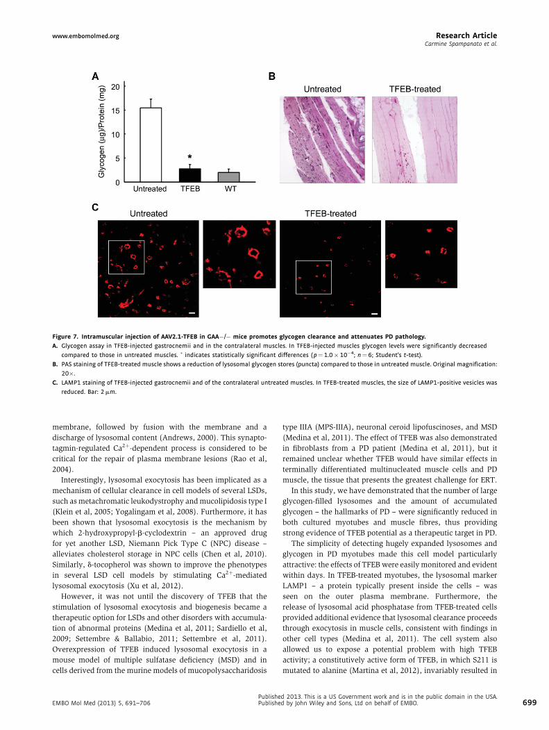
Figure 7. Intramuscular injection of AAV2.1-TFEB in GAA�/� mice promotes glycogen clearance and attenuates PD pathology.
A. Glycogen assay in TFEB-injected gastrocnemii and in the contralateral muscles. In TFEB-injected muscles glycogen levels were significantly decreased
compared to those in untreated muscles. � indicates statistically significant differences ( p¼1.0�10�4; n¼ 6; Student’s t-test).
B. PAS staining of TFEB-treated muscle shows a reduction of lysosomal glycogen stores (puncta) compared to those in untreated muscle. Original magnification:
20�.
C. LAMP1 staining of TFEB-injected gastrocnemii and of the contralateral untreated muscles. In TFEB-treated muscles, the size of LAMP1-positive vesicles was
reduced. Bar: 2 mm.
membrane, followed by fusion with the membrane and a
discharge of lysosomal content (Andrews, 2000). This synapto-
tagmin-regulated Ca2þ-dependent process is considered to be
critical for the repair of plasma membrane lesions (Rao et al,
2004).
Interestingly, lysosomal exocytosis has been implicated as a
mechanism of cellular clearance in cell models of several LSDs,
such as metachromatic leukodystrophy and mucolipidosis type I
(Klein et al, 2005; Yogalingam et al, 2008). Furthermore, it has
been shown that lysosomal exocytosis is the mechanism by
which 2-hydroxypropyl-b-cyclodextrin – an approved drug
for yet another LSD, Niemann Pick Type C (NPC) disease –
alleviates cholesterol storage in NPC cells (Chen et al, 2010).
Similarly, d-tocopherol was shown to improve the phenotypes
in several LSD cell models by stimulating Ca2þ-mediated
lysosomal exocytosis (Xu et al, 2012).
However, it was not until the discovery of TFEB that the
stimulation of lysosomal exocytosis and biogenesis became a
therapeutic option for LSDs and other disorders with accumula-
tion of abnormal proteins (Medina et al, 2011; Sardiello et al,
2009; Settembre & Ballabio, 2011; Settembre et al, 2011).
Overexpression of TFEB induced lysosomal exocytosis in a
mouse model of multiple sulfatase deficiency (MSD) and in
cells derived from the murine models of mucopolysaccharidosis
EMBO Mol Med (2013) 5, 691–706PublishePublishe
type IIIA (MPS-IIIA), neuronal ceroid lipofuscinoses, and MSD
(Medina et al, 2011). The effect of TFEB was also demonstrated
in fibroblasts from a PD patient (Medina et al, 2011), but it
remained unclear whether TFEB would have similar effects in
terminally differentiated multinucleated muscle cells and PD
muscle, the tissue that presents the greatest challenge for ERT.
In this study, we have demonstrated that the number of large
glycogen-filled lysosomes and the amount of accumulated
glycogen – the hallmarks of PD – were significantly reduced in
both cultured myotubes and muscle fibres, thus providing
strong evidence of TFEB potential as a therapeutic target in PD.
The simplicity of detecting hugely expanded lysosomes and
glycogen in PD myotubes made this cell model particularly
attractive: the effects of TFEB were easily monitored and evident
within days. In TFEB-treated myotubes, the lysosomal marker
LAMP1 – a protein typically present inside the cells – was
seen on the outer plasma membrane. Furthermore, the
release of lysosomal acid phosphatase from TFEB-treated cells
provided additional evidence that lysosomal clearance proceeds
through exocytosis in muscle cells, consistent with findings in
other cell types (Medina et al, 2011). The cell system also
allowed us to expose a potential problem with high TFEB
activity; a constitutively active form of TFEB, in which S211 is
mutated to alanine (Martina et al, 2012), invariably resulted in
d 2013. This is a US Government work and is in the public domain in the USA.d by John Wiley and Sons, Ltd on behalf of EMBO. 699
Research Article www.embomolmed.orgNew therapeutic target in Pompe disease
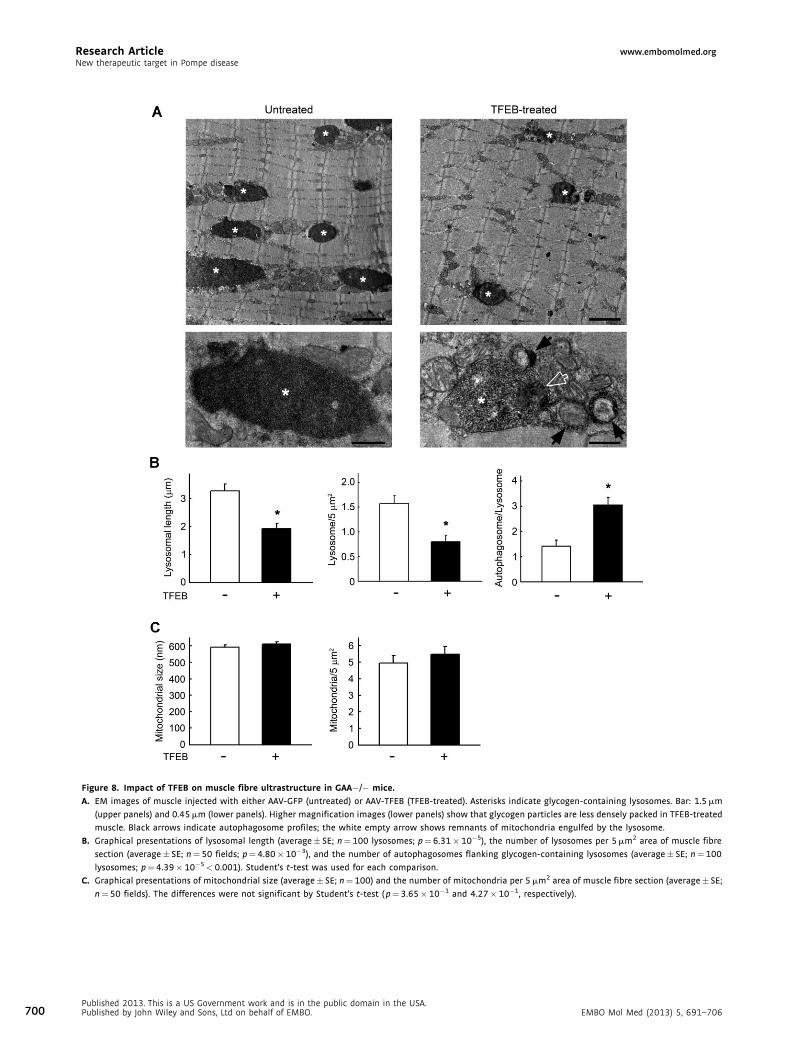
Figure 8. Impact of TFEB on muscle fibre ultrastructure in GAA�/� mice.
A. EM images of muscle injected with either AAV-GFP (untreated) or AAV-TFEB (TFEB-treated). Asterisks indicate glycogen-containing lysosomes. Bar: 1.5 mm
(upper panels) and 0.45 mm (lower panels). Higher magnification images (lower panels) show that glycogen particles are less densely packed in TFEB-treated
muscle. Black arrows indicate autophagosome profiles; the white empty arrow shows remnants of mitochondria engulfed by the lysosome.
B. Graphical presentations of lysosomal length (average� SE; n¼ 100 lysosomes; p¼6.31� 10�5), the number of lysosomes per 5 mm2 area of muscle fibre
section (average� SE; n¼50 fields; p¼ 4.80�10�3), and the number of autophagosomes flanking glycogen-containing lysosomes (average� SE; n¼100
lysosomes; p¼4.39� 10�5< 0.001). Student’s t-test was used for each comparison.
C. Graphical presentations of mitochondrial size (average� SE; n¼ 100) and the number of mitochondria per 5 mm2 area of muscle fibre section (average� SE;
n¼50 fields). The differences were not significant by Student’s t-test ( p¼ 3.65�10�1 and 4.27� 10�1, respectively).
700Published 2013. This is a US Government work and is in the public domain in the USA.Published by John Wiley and Sons, Ltd on behalf of EMBO. EMBO Mol Med (2013) 5, 691–706
www.embomolmed.org Research ArticleCarmine Spampanato et al.
morphological changes of myotubes, perhaps because of
massive expulsion of the lysosomes.
Live fibre experiments employing time-lapse confocal
microscopy provided a unique opportunity to track lysosomal
movements and fusion, to estimate the motility of lysosomes,
and to examine the interactions between lysosomes and
autophagosomes following TFEB activation and nuclear trans-
location. Similar to what was observed in the cell system, in vivo
TFEB delivery efficiently triggered lysosomal exocytosis and
promoted cellular clearance, as evidenced by a significant
reduction in glycogen levels and the number of large lysosomes
in PD muscle. Thus, lysosomal pathology in PD muscle can be
corrected by TFEB overexpression.
The development of a new PD mouse model, the GFP-
LC3:GAA�/� strain, allowed us to address the effect of TFEB on
autophagic accumulation – the major secondary abnormality
in PD skeletal muscle (Raben et al, 2007a,b, 2012). Defects in
autophagy, a major lysosome-dependent degradative system
(Yang & Klionsky, 2010)], significantly contribute to the
pathophysiology of several lysosomal storage diseases (Cao
et al, 2006; Cox & Cachon-Gonzalez, 2012; Fukuda et al,
2006a,b; Liao et al, 2007; Lieberman et al, 2012; Settembre et al,
2008). In PD skeletal muscle, the autophagic defect is
particularly striking, manifesting as a massive build-up which
poses an additional problem for ERT.
Remarkably, none of the TFEB-transfected fibres had large
areas of autophagic build-up, which are so prominent in most
non-transfected PD fibres. The lack of typical build-up suggests
that TFEB may have rescued autophagic pathology in PD
muscle. The mechanism of this rescue is not clear, but the
unexpected surge in autophagy – a dramatic increase in the
number of LC3-positive autophagosomes during hours of
time-lapse microscopy – provided a clue. TFEB-treated fibres
exhibited a significant increase in fusion between LC3- and
LAMP1-positive vesicles, an appearance of multiple LC3/LAMP1-
positive autophagolysosomes, and docking of these double-
positive structures to the plasma membrane. These data suggest
that TFEB may relieve autophagic build-up by stimulating the
formation and secretion of autophagolysosomes – a product of
lysosomal-autophagosomal fusion.
We hypothesize that lysosomal exocytosis may in fact not
be a purely lysosomal event, but rather a process involving
autophagy and secretion of autophagolysosomes. Our data
on the much attenuated effects of TFEB on lysosomal velocity
and clearance in autophagy-deficient PD mice support this
idea. Lysosomal function depends on the ability of lysosomal
membranes to fuse with other vesicles’ membranes and
with the plasma membrane (Luzio et al, 2007). Abnormal
accumulation of cholesterol in the endolysosomal membranes
in cell models of two neurodegenerative LSDs (MSD and
MPS-IIIA) was shown to sequester SNARE receptors – essential
components of the membrane fusion machinery – in
cholesterol-enriched membrane regions, resulting in a sig-
nificantly reduced ability of lysosomes to fuse with vesicles
of endocytic and autophagic pathways (Fraldi et al, 2010).
Perhaps the merging of lysosomal and autophagosomal
membranes is required for successful fusion with the plasma
EMBO Mol Med (2013) 5, 691–706PublishePublishe
membrane and for fully efficient TFEB-mediated cellular
clearance.
Two kinases, mTORC1 and ERK, have been implicated
in TFEB regulation in different cells (Martina et al, 2012;
Roczniak-Ferguson et al, 2012; Settembre et al, 2011, 2012). The
regulation of TFEB in skeletal muscle remains an open question,
which is beyond the scope of this paper. An unexpected
activation of ERK in TFEB-overexpressing cells could be an
attempt to down-regulate TFEB and restore homeostasis; it is
also possible that activation of ERK could be a downstream
event. Whatever the case, the search for compounds which can
activate TFEB in muscle would benefit the development of a
TFEB-mediated therapeutic approach. In thinking about TFEB
as a target for potential therapeutic intervention, one can
speculate that a single event of TFEB-mediated clearance of the
stored substrate and autophagic material may provide pro-
longed relief since the rate of muscle glycogen accumulation
in late-onset PD is relatively slow. Furthermore, ERT may
function more efficiently in TFEB-treated muscle, particularly
because of the reduction in autophagic build-up. A combination
of TFEB-targeted therapy with ERT, perhaps at much lower
dosages than those currently employed (and therefore at
reduced cost), could then maintain muscle tissue in normal
or nearly normal condition. In the meantime, pre-clinical
studies are needed to fully evaluate the consequences of TFEB
overexpression on muscle function and to exclude long-term
toxicity.
Finally, the potential applications of our newly developed
models, PD myotubes and GFP-LC3:GAA�/� mice, extend
beyond testing the effects of TFEB. For example, an in vitro PD
model is much needed for large-scale drug testing. Recently,
several attempts have been made to develop such a model: these
include the generation of cardiomyocyte-like cells from human
induced pluripotent stem (iPS) cells (Huang et al, 2011), muscle
cells from reprogrammed murine iPS cells (Kawagoe et al,
2011), and murine muscle cells transduced by viral constructs
containing immortalizing genes (Douillard-Guilloux et al, 2009;
Takikita et al, 2009). Our new PD muscle cell lines have several
advantages: they are highly myogenic, derived from individual
clones and, most importantly, they maintain the capacity to
differentiate when the immortalizing gene, shown to perturb
myogenic program (Douillard-Guilloux et al, 2009; Mouly et al,
1996), is inactivated. These cell lines replicate lysosomal
pathology but fail to recapitulate autophagic build-up. Possible
explanations for this failure – common to all in vitro PD models –
are the relatively short survival time of myotubes in culture and
the need for nutrient-rich conditions (Fig S3C). Additional
studies are needed to find a way to ‘‘trick’’ these cells into
displaying the autophagic accumulation that is so striking in
Pompe muscle fibres.
In the interim, the newly generated GFP-LC3:GAA�/�mouse
model provides an excellent tool to monitor autophagic
accumulation in live fibres, in muscle bundles, and even in
muscle of a living mouse [i.e. by intravital imaging (our
unpublished data)]. In addition to using these mice to address
TFEB’s effects, we have utilized the model to examine
intracellular trafficking of rhGAA and to explore possible
d 2013. This is a US Government work and is in the public domain in the USA.d by John Wiley and Sons, Ltd on behalf of EMBO. 701
Research Article www.embomolmed.orgNew therapeutic target in Pompe disease
702
mechanisms of autophagic build-up. The near absence of
lysosomal–autophagosomal fusion and strikingly low expres-
sion of LAMP1 in the build-up area suggest inefficient local
lysosomal biogenesis. LAMPs are considered to be of key
importance for lysosomal movement, maturation of autophago-
somes and lysosomal exocytosis (Saftig & Klumperman, 2009).
Therefore, these low local LAMP1 concentrations in the build-
up area may have far reaching consequences. The build-up
process appears to begin with the failure of a subset of
lysosomes to fuse with autophagosomes, perhaps because of
lysosomal rupture during muscle contractions. It is possible that
this fusion defect and the paucity of the newly formed lysosomes
create conditions that favour expansion of the autophagic build-
up. With the capacity to resolve both of these deficits by
promoting lysosomal–autophagosomal fusion and biogenesis,
TFEB is uniquely suited to remedy autophagic and lysosomal
pathologies in PD.
MATERIALS AND METHODS
Antibodies: rat anti-LAMP1 (1DB4), mouse anti-cytochrome c, and
mouse anti-MyoD (MoAb 5.8A) (BD Pharmingen, San Diego, CA);
rabbit anti-LC3B and mouse anti-Flag (M2) (Sigma–Aldrich, St. Louis,
MO); mouse anti-myogenin (F5D) (Dako, Glostrup, Denmark);
mouse anti-myosin heavy chain (Clontech, Mountain View, CA);
rabbit anti-caspase-3, rabbit anti-p44/42 MAPK (Erk1/2), rabbit
anti-phospho-p44/42 MAPK (Erk1/2; Thr202/Thr204), rabbit anti-
phospho-4EBP1 (Thr37/46), and rabbit anti-Histone H3 (D1H2) (Cell
Signaling, Beverly, MA). Rabbit anti-alpha tubulin (Abcam, Cambridge,
MA) or mouse anti-vinculin antibody (Sigma–Aldrich) was used as a
loading control for western blot. Alexa Fluor-conjugated secondary
antibodies (Life Technologies, Grand Island, NY) were used for
immunostaining and western blot analysis. Alexa Fluor 546 labelled
recombinant human GAA (6.2mg/ml) was provided by Genzyme Corp.
(Framingham, MA).
Plasmids: plasmid containing the full-length 3xFlag-tagged human
TFEB (referred to as Flag-TFEB) was described previously (Sardiello
et al, 2009); plasmid containing the full-length rat LAMP1 (mCherry-
LAMP1) was obtained from Dr. Kristien Zaal (Light Imaging Section,
Office of Science & Technology, NIAMS, NIH, Bethesda, MD, USA);
plasmid-containing GFP-TFEB was described previously (Martina et al,
2012).
Generation of mice and isolation of single live myofibres
The generation of GFP-LC3:GAA�/�, immorto:GAA�/�, and immor-
to:GFP-LC3:GAA�/� mice is described in the Supporting Information.
Single live myofibres from gastrocnemius and extensor digitorum
longus (EDL) muscles from 3 to 4 month-old mice were isolated as
described (Rosenblatt et al, 1995). Isolation of single live fibres from
FDB muscle was done with some modifications of the previously
published protocol (Raben et al, 2010) (Supporting Information). Live
fibres were either monitored by time-lapse microscopy or used for
labelling acidic organelles [LysoTracker1 Red DND-99 (Life Technol-
ogies), 100 nM] or mitochondria [MitoTracker1 Red CMXRos (Life
Technologies), 100 nM]. Alternatively, fibres were fixed in 2%
paraformaldehyde for 30min and immunostained with anti-cyto-
Published 2013. This is a US Government work and is in the public domain in the UPublished by John Wiley and Sons, Ltd on behalf of EMBO.
chrome c and anti-LAMP1 antibodies. Both live and fixed fibres were
analysed on a LSM 510 confocal microscope (Carl Zeiss, Gottingen,
Germany; see Supporting Information) equipped with Plan-Neofluar
40X oil immersion objective.
In vivo electroporation of skeletal muscle and enzyme
replacement therapy
Electric pulse-mediated gene transfer into FDB muscle was performed
as described (DiFranco et al, 2009). Three to four month-old GAA�/�,GFP-LC3:GAA�/�, or muscle-specific autophagy-deficient GAA�/�(MLCcre:Atg7F/F:GAA�/�; referred to as Atg7:GAA DKO) mice were
used for the experiments (Table 1). The animals were sacrificed 4–
6 days after the procedure. Live FDB fibres were isolated and analysed
by time-lapse confocal microscopy. Alexa-Fluor 546 labelled recombi-
nant human GAA (rhGAA) was administered into 3–4 month-old GFP-
LC3:GAA�/�mice at a dosage of 100mg/kg twice with a 24h interval.
Mice were sacrificed 24h after the last injections. Single fibres,
isolated from EDL and gastrocnemius muscle, were analysed by
confocal microscopy.
Fibroblast and myoblast culture systems
Primary skin fibroblasts, derived from two unrelated patients with
severe infantile PD, were obtained from Coriell Cell Repositories
(Camden, New Jersey, USA). Clinical diagnosis was confirmed by
genetic analysis. Fibroblasts were maintained in DMEM (high glucose)
supplemented with 10% (v/v) FBS, 1� P/S/L-Glutamine. Adult mouse
fibroblasts were isolated from the tail tip according to a standard
procedure.
Immortalized PD cell lines were derived from immortoGAA�/� and
immortoGFP-LC3:GAA�/� mice. Single fibres from EDL muscle of 3–
4 month-old mice were plated in each well (�10 fibres per well) of a
Matrigel-coated six-well plate in plating medium (10% horse serum in
DMEM, 0.5% chick embryo extract, and 1� P/S/L-Glutamine) at 378C,
5% CO2. After 3 days, when the satellite cells began to migrate away
from fibres, most of the medium (3/4) was replaced with proliferation
medium (20% foetal bovine serum, 10% horse serum, 1% chick
embryo extract, 1� P/S/L-Glutamine in high glucose DMEM). Myogenic
colonies were isolated with cloning cylinders (Corning, Corning, NY) on
day 5 or 6. Several individual myogenic clones were isolated and
analysed for their proliferation capacity and the ability to differentiate
into myotubes. To induce the expression of H-2Kb-tsA58 SV40 large
T-antigen for immortalization of the satellite cells and myoblasts,
recombinant IFN-g (Life Technologies) was added to the proliferation
medium (100units/ml). The cells were then incubated at 338C
in an atmosphere of 5% CO2 (Jat et al, 1991). When myoblasts
became nearly confluent, the medium was changed to differentiation
medium (DMEM containing 2% horse serum, 0.5% chick embryo
extract, 1� P/S/L-Glutamine) and the cells were moved to 378C in
an atmosphere of 5% CO2. Myotubes began to form within 2 days.
Glucose concentration was either 1 g/L (low) or 4.5 g/L (high).
Myotubes survived in culture for 2–3 weeks. A wild type immortalized
muscle cell line served as the control.
Infection of myotubes with adenovirus expressing TFEB and
immunofluorescence microscopy
Adenoviruses expressing either WT or mutant (S211A) TFEB were
described previously (Martina et al, 2012). Myotubes were infected
SA.EMBO Mol Med (2013) 5, 691–706
www.embomolmed.org Research ArticleCarmine Spampanato et al.
The paper explained
PROBLEM:
The major problem with the current ERT for several LSDs is
inefficient drug delivery to lysosomes in particular target tissues.
Furthermore, the therapy does not address autophagic
abnormalities, which are commonly found in LSDs. These
limitations are apparent in PD, a severe metabolic myopathy due
to a deficiency of glycogen-degrading lysosomal enzyme, GAA.
Experimental data and clinical experience over the past decade
provide strong evidence of skeletal muscle resistance to therapy
with recombinant human GAA. Here, we have tested a recently
developed approach for therapy of LSDs in PD mouse models. This
new approach circumvents the problem of ineffective drug
delivery and involves manipulation of TFEB.
RESULTS:
For testing the novel therapeutic approach, we developed new
PD models. Experiments in PD cell culture and animal models
demonstrate that TFEB has the capacity to rid muscle cells of
excessive glycogen burden and accumulation of autophagic
debris. Overexpression of TFEB in muscle cells or in Pompe mice
stimulated fusion between lysosomes and autophagosomes,
resulting in an increased formation and exocytosis of auto-
lysosomes. Unexpectedly, the effects of TFEB were almost
abolished when autophagy was genetically suppressed.
IMPACT:
Our study provides strong evidence that TFEB is a valid
therapeutic target for PD as well as other LSDs. The appeal for PD
is that unlike the current therapy, modulation of TFEB holds
promise to address both lysosomal and autophagic pathologies
in skeletal muscle. The study also reveals a previously
unrecognized role of autophagy in TFEB-mediated cellular
clearance.
with either adenovirus (Ad-null) or adenovirus expressing WT TFEB
(Ad-TFEB) or S211A TFEB (Ad-TFEBmt) for 24, 48 or 72 h. Myotubes
were fixed in 2% paraformaldehyde (Electron Microscopy Sciences,
Hatfield, PA) for 15min at room temperature, washed twice in PBS,
and permeabilized in 0.2% Triton X-100 (Sigma–Aldrich, St. Louis, MO).
Immunostaining with LAMP1, LC3 or Flag antibodies was done using
M.O.M. kit (Vector Laboratories, Burlingame, CA) as previously
described (Raben et al, 2009). The cell nuclei were stained with
2mg/ml Hoechst 33342 (Life Technologies) in PBS for 10min. After
staining, the cells were imaged on a Carl Zeiss LSM 510 confocal
microscope with a 40� or 63� oil immersion objective.
Fluorescent glycogen detection, LDH measurements, in situ
detection of apoptotic cells, surface LAMP1 analysis, and acid
phosphatase assay
Lysosomal glycogen in live cells was detected by the incorporation of
2-NBDG, a D-glucose fluorescent derivative (2-deoxyglucose), into
glycogen as described (Louzao et al, 2008). The amount of LDH
released into the medium was assayed using the LDH-Cytotoxicity
Assay Kit II (Abcam) according to the manufacturer’s instructions.
Apoptotic cells were detected with TUNEL using the ApopTag1
Fluorescein Direct In Situ Apoptosis Detection Kit (Millipore, Billerica,
MA). Lysosomal exocytosis in TFEB-treated cell cultures was evaluated
by the surface LAMP1 assay as previously described (Medina et al,
2011) and by the measurement of acid phosphatase released into the
medium. The acid phosphatase assay was performed in cultures
treated with Ad-TFEB for 2 days according to the standard procedure
(Supporting Information, Table S1).
Western blot
Cells were homogenized in RIPA buffer [PBS containing 1% NP-40,
0.5% sodium deoxycholate, 0.1% SDS and Protease/Phosphatase
Inhibitor Cocktail (Cell Signaling Technology, Danvers, MA)] and
EMBO Mol Med (2013) 5, 691–706PublishePublishe
centrifuged for 30min at 13,000 rpm at 48C. Alternatively, for
nuclear/cytosolic fractionation, cells were lysed in 0.5% Triton X-100
buffer [50mM Tris–HCl, 0.5% Triton X-100, 137.5mM NaCl, 10%
glycerol, 5mM EDTA and protease and phosphatase inhibitors] and
centrifuged at 3000 rpm at 48C for 5min. The supernatant constituted
the cytosolic fraction. The pellet was then washed with 0.5% Triton
X-100 buffer, re-suspended in Triton X-100 buffer with 0.5% SDS,
sonicated, and centrifuged at 13,000 rpm for 15min at 48C. The
supernatant constituted the nuclear fraction. Protein concentrations
were measured using the Bio-Rad Protein Assay (Bio-Rad Laboratories,
Inc., Hercules, CA). Western blots were performed according to
standard procedures. Blots were scanned on an Odyssey Infrared
Imager (LI-COR Biosciences, Lincoln, NE).
Intramuscular injection of AAV-TFEB, muscle staining and
glycogen assay, and electron microscopy (EM)
Six 1-month-old GAA�/� mice were injected with a total dose of
1011 GC of AAV2/1 vector preparation into three sites of the right
gastrocnemius (three injections of 30ml each) using a Hamilton
syringe. Equivalent doses of AAV2/1CMV-EGFP or equal volumes of PBS
were injected into the contralateral muscle. Animals were sacrificed
45 days after injection and perfused with PBS.
Gastrocnemii, free from neighbouring muscles and connective tissue,
were isolated and analysed. Part of the sample was used to test the
levels of TFEB expression by RT-PCR. For morphologic analysis, muscles
were frozen in liquid nitrogen-cooled isopentane. Immunostaining of
10mm sections was performed using anti-LAMP1 (1:500; Abcam,
Cambridge, MA, USA) and anti-Caspase-3 Active (1:500; R&D Systems,
Minneapolis, MN, USA) antibodies. Apoptotic cells were detected
with TUNEL using the Fluorescein In Situ Cell Death Detection Kit
(Roche, Basel, Switzerland) according to the supplier’s protocol. Muscle
sections were mounted with Vectashield, and photographs were taken
using a fluorescence microscope Zeiss (Thornwood, NY) Axioplan 2
d 2013. This is a US Government work and is in the public domain in the USA.d by John Wiley and Sons, Ltd on behalf of EMBO. 703
Research Article www.embomolmed.orgNew therapeutic target in Pompe disease
704
integrated with the AxioCam MR camera. Haematoxylin–eosin and
PAS staining were performed according to standard procedures.
Glycogen concentration was assayed in muscle lysates by measuring
the amount of glucose released after digestion with amyloglucosidase
(Aspergillus niger) using a commercial kit (BioVision, Milpitas, CA, USA).
Data were expressed as mg of glycogen/mg of protein. For EM, muscle
tissue was fixed in 1% glutaraldehyde in 0.2M HEPES buffer and post-
fixed in uranyl acetate and OsO4. After dehydration through a graded
series of ethanol and propilenoxide, the tissue was embedded in the
Epoxy resin (Epon 812, Sigma–Aldrich) and polymerized at 608C for
72 h. EM images of thin sections were acquired using a FEI Tecnai-12
electron microscope (FEI, Eindhoven, Netherlands) equipped with a
VELETTA CCD digital camera (Soft Imaging Systems GmbH, Munster,
Germany). Quantification of the number of lysosome-like organelles
and their dimensions as well as the number of autophagosomes was
performed in 50 fields (of 5mm2 dimensions) distributed randomly
through the thin sections containing different fibres.
Quantitative analyses are presented in the Supporting Information.
Data in text are given as mean� SE. Kolmogorov–Smirnov test was
used for comparison of cumulative distributions of lysosomal size in
myotubes and muscle fibres; Wilcoxon rank sum test was used for
comparison of median values. Student’s t-test was used for all other
comparisons. Differences were considered significant at p<0.05.
Animal care and experiments were conducted in accordance with the
National Institutes of Health Guide for the Care and Use of Laboratory
Animals and the European Union Directive 86/609 regarding the
protection of animals used for experimental purposes.
Author contributionsNR, EF, LL and JL performed tissue culture and live muscle fibre
experiments, and analysed the data; RPu contributed new
reagents and analytical tools, interpreted and analysed data;
HZ performed statistical analysis and interpreted data; CS and
FA generated vectors, performed intramuscular studies, and
interpreted data; MC performed the biochemical and histolo-
gical analyses of intramuscular studies; RPo performed the
electron microscopy analysis; EF designed the quantitative
analysis of the live fibre data and participated in writing and
preparation of the manuscript; GP and AB designed and
analysed the experiments and contributed to the writing of the
paper; NR designed and analysed the experiments and wrote
the paper.
AcknowledgementsWe would like to thank Drs. Kristein Zaal and Evelyn Ralston
(NIAMS, NIH) for their useful comments and help with the
imaging. We would also like to thank Dr. Hoi Ming Li for
his contribution to our tissue culture and electroporation
experiments. We are also grateful to G. Diez-Roux for critical
reading of the manuscript and helpful discussions. This
research was supported in part by the Intramural Research
Program of the National Institute of Arthritis and Musculo-
skeletal and Skin diseases of the National Institutes of Health.
Drs. Li and Lim are supported in part by a CRADA between NIH
and Genzyme Corporation. We acknowledge the support of
Published 2013. This is a US Government work and is in the public domain in the UPublished by John Wiley and Sons, Ltd on behalf of EMBO.
the Italian Telethon Foundation (grant # TGPMT4TELD to
G.P; grant # TGM11CB6 to A.B, C.S and F.A.), the European
Research Council Advanced Investigator grant no. 250154
(A.B.), the Beyond Batten Disease Foundation (A.B.), March of
Dimes #6-FY11-306 (A.B.), and US National Institute of Health
R01-NS078072 (A.B.).
Supporting Information is available at EMBO Molecular
Medicine online.
The authors declare that they have no conflict of interest.
ReferencesAndrews NW (2000) Regulated secretion of conventional lysosomes. Trends
Cell Biol 10: 316-321
Angelini C, Semplicini C (2012) Enzyme replacement therapy for Pompe
disease. Curr Neurol Neurosci Rep 12: 70-75
Banugaria SG, Prater SN, Ng YK, Kobori JA, Finkel RS, Ladda RL, Chen YT,
Rosenberg AS, Kishnani PS (2011) The impact of antibodies on clinical
outcomes in diseases treated with therapeutic protein: lessons learned from
infantile Pompe disease. Genet Med 13: 729-736
Blott EJ, Griffiths GM (2002) Secretory lysosomes. Nat Rev Mol Cell Biol 3: 122-
131
Byrne BJ, Falk DJ, Pacak CA, Nayak S, Herzog RW, Elder ME, Collins SW, Conlon
TJ, Clement N, Cleaver BD, et al (2011) Pompe disease gene therapy. Hum
Mol Genet 20: R61-68
Cao Y, Espinola JA, Fossale E, Massey AC, Cuervo AM, Macdonald ME,
Cotman SL (2006) Autophagy is disrupted in a knock-in mouse model
of juvenile neuronal ceroid lipofuscinosis. J Biol Chem 281: 20483-
20493
Chen FW, Li C, Ioannou YA (2010) Cyclodextrin induces calcium-dependent
lysosomal exocytosis. PLoS ONE 5: e15054
Cox TM, Cachon-Gonzalez MB (2012) The cellular pathology of lysosomal
diseases. J Pathol 226: 241-254
DiFranco M, Quinonez M, Capote J, Vergara J (2009) DNA transfection of
mammalian skeletal muscles using in vivo electroporation. J Vis Exp 32:
1520-1527
Douillard-Guilloux G, Mouly V, Caillaud C, Richard E (2009) Immortalization
of murine muscle cells from lysosomal alpha-glucosidase deficient mice:
a new tool to study pathophysiology and assess therapeutic strategies for
Pompe disease. Biochem Biophys Res Commun 388: 333-338
Fraldi A, Annunziata F, Lombardi A, Kaiser HJ, Medina DL, Spampanato C,
Fedele AO, Polishchuk R, Sorrentino NC, Simons K, et al (2010) Lysosomal
fusion and SNARE function are impaired by cholesterol accumulation in
lysosomal storage disorders. EMBO J 29: 3607-3620
Fukuda T, Ahearn M, Roberts A, Mattaliano RJ, Zaal K, Ralston E, Plotz PH,
Raben N (2006a) Autophagy and mistargeting of therapeutic enzyme in
skeletal muscle in pompe disease. Mol Ther 14: 831-839
Fukuda T, Ewan L, Bauer M, Mattaliano RJ, Zaal K, Ralston E, Plotz PH, Raben N
(2006b) Dysfunction of endocytic and autophagic pathways in a lysosomal
storage disease. Ann Neurol 59: 700-708
Funk B, Kessler U, Eisenmenger W, Hansmann A, Kolb HJ, Kiess W (1992)
Expression of the insulin-like growth factor-II/mannose-6-phosphate
receptor in multiple human tissues during fetal life and early infancy. J Clin
Endocrinol Metab 75: 424-431
Huang HP, Chen PH, Hwu WL, Chuang CY, Chien YH, Stone L, Chien CL, Li LT,
Chiang SC, Chen HF, et al (2011) Human Pompe disease-induced
pluripotent stem cells for pathogenesis modeling, drug testing and disease
marker identification. Hum Mol Genet 20: 4851-4864
Jat PS, Noble MD, Ataliotis P, Tanaka Y, Yannoutsos N, Larsen L, Kioussis D
(1991) Direct derivation of conditionally immortal cell lines from an H-2Kb-
tsA58 transgenic mouse. Proc Natl Acad Sci USA 88: 5096-5100
SA.EMBO Mol Med (2013) 5, 691–706
www.embomolmed.org Research ArticleCarmine Spampanato et al.
Kabeya Y, Mizushima N, Ueno T, Yamamoto A, Kirisako T, Noda T, Kominami E,
Ohsumi Y, Yoshimori T (2000) LC3, a mammalian homologue of yeast
Apg8p, is localized in autophagosome membranes after processing. EMBO J
19: 5720-5728
Kawagoe S, Higuchi T, Meng XL, Shimada Y, Shimizu H, Hirayama R, Fukuda T,
Chang H, Nakahata T, Fukada S, et al (2011) Generation of induced
pluripotent stem (iPS) cells derived from a murine model of Pompe disease
and differentiation of Pompe-iPS cells into skeletal muscle cells. Mol Genet
Metab 104: 123-128
Kishnani PS, Corzo D, Nicolino M, Byrne B, Mandel H, Hwu WL, Leslie N, Levine
J, Spencer C, McDonald M, et al (2007) Recombinant human acid [alpha]-
glucosidase: major clinical benefits in infantile-onset Pompe disease.
Neurology 68: 99-109
Kishnani PS, Goldenberg PC, DeArmey SL, Heller J, Benjamin D, Young S, Bali D,
Smith SA, Li JS, Mandel H, et al (2010) Cross-reactive immunologic material
status affects treatment outcomes in Pompe disease infants. Mol Genet
Metab 99: 26-33
Klein D, Bussow H, Fewou SN, Gieselmann V (2005) Exocytosis of storage
material in a lysosomal disorder. Biochem Biophys Res Commun 327: 663-
667
Koeberl DD, Li S, Dai J, Thurberg BL, Bali D, Kishnani PS (2012) beta2 Agonists
enhance the efficacy of simultaneous enzyme replacement therapy in
murine Pompe disease. Mol Genet Metab 105: 221-227
Kroos M, Hoogeveen-Westerveld M, van der Ploeg A, Reuser AJ (2012) The
genotype-phenotype correlation in Pompe disease. Am J Med Genet C Semin
Med Genet 160: 59-68
Lachmann RH (2011) Enzyme replacement therapy for lysosomal storage
diseases. Curr Opin Pediatr 23: 588-593
Liao G, Yao Y, Liu J, Yu Z, Cheung S, Xie A, Liang X, Bi X (2007) Cholesterol
accumulation is associated with lysosomal dysfunction and autophagic
stress in Npc1 �/� mouse brain. Am J Pathol 171: 962-975
Lieberman AP, Puertollano R, Raben N, Slaugenhaupt S, Walkley SU,
Ballabio A (2012) Autophagy in lysosomal storage disorders. Autophagy 8:
719-730
Liu Q, Chang JW, Wang J, Kang SA, Thoreen CC, Markhard A, Hur W, Zhang J,
Sim T, Sabatini DM, et al (2010) Discovery of 1-(4-(4-propionylpiperazin-1-
yl)-3-(trifluoromethyl)phenyl)-9-(quinolin-3-yl)benz
o[h][1,6]naphthyridin-2(1H)-one as a highly potent, selective mammalian
target of rapamycin (mTOR) inhibitor for the treatment of cancer. J Med
Chem 53: 7146-7155
Louzao MC, Espina B, Vieytes MR, Vega FV, Rubiolo JA, Baba O, Terashima T,
Botana LM (2008) ‘‘Fluorescent glycogen’’ formation with sensibility for
in vivo and in vitro detection. Glycoconj J 25: 503-510
Luzio JP, Pryor PR, Bright NA (2007) Lysosomes: fusion and function. Nat Rev
Mol Cell Biol 8: 622-632
Maga JA, Zhou J, Kambampati R, Peng S, Wang X, Bohnsack RN, Thomm A,
Golata S, Tom P, Dahms NM, et al (2012) Glycosylation-independent
lysosomal targeting of acid alpha-glucosidase enhances muscle glycogen
clearance in Pompe mice. J Biol Chem 288: 1428-1438
Martina J, Chen Y, Gucek M, Puertollano R (2012) MTOPC1 functions as a
transcriptional regulator of autophagy by preventing nuclear transport of
TFEB. Autophagy 8: 903-914
Martiniuk F, Chen A, Mack A, Arvanitopoulos E, Chen Y, Rom WN, Codd WJ,
Hanna B, Alcabes P, Raben N, et al (1998) Carrier frequency for glycogen
storage disease type II in New York and estimates of affected individuals
born with the disease. Am J Med Genet 79: 69-72
Medina DL, Fraldi A, Bouche V, Annunziata F, Mansueto G, Spampanato C,
Puri C, Pignata A, Martina JA, Sardiello M, et al (2011) Transcriptional
activation of lysosomal exocytosis promotes cellular clearance. Dev Cell 21:
421-430
Mouly V, Edom F, Decary S, Vicart P, Barbert JP, Butler-Browne GS (1996) SV40
large T antigen interferes with adult myosin heavy chain expression, but not
with differentiation of human satellite cells. Exp Cell Res 225: 268-276
Okumiya T, Kroos MA, Vliet LV, Takeuchi H, Van der Ploeg AT, Reuser AJ (2007)
Chemical chaperones improve transport and enhance stability of mutant
EMBO Mol Med (2013) 5, 691–706PublishePublishe
alpha-glucosidases in glycogen storage disease type II. Mol Genet Metab 90:
49-57
Parenti G, Zuppaldi A, Gabriela PM, Rosaria TM, Annunziata I, Meroni G, Porto
C, Donaudy F, Rossi B, Rossi M, et al (2007) Pharmacological enhancement
of mutated alpha-glucosidase activity in fibroblasts from patients with
Pompe disease. Mol Ther 15: 508-514
Porto C, Cardone M, Fontana F, Rossi B, Tuzzi MR, Tarallo A, Barone MV, Andria
G, Parenti G (2009) The pharmacological chaperone N-
butyldeoxynojirimycin enhances enzyme replacement therapy in Pompe
disease fibroblasts. Mol Ther 17: 964-971
Porto C, Ferrara MC, Meli M, Acampora E, Avolio V, Rosa M, Cobucci-Ponzano B,
Colombo G, Moracci M, Andria G, et al (2012) Pharmacological
enhancement of alpha-glucosidase by the allosteric chaperone N-
acetylcysteine. Mol Ther 12: 2201-2211
Raben N, Jatkar T, Lee A, Lu N, Dwivedi S, Nagaraju K, Plotz PH (2002) Glycogen
stored in skeletal but not in cardiac muscle in acid alpha-glucosidase
mutant (Pompe) mice is highly resistant to transgene-encoded human
enzyme. Mol Ther 6: 601-608
Raben N, Nagaraju K, Lee E, Kessler P, Byrne B, Lee L, LaMarca M, King C, Ward
J, Sauer B, et al (1998) Targeted disruption of the acid alpha-glucosidase
gene in mice causes an illness with critical features of both infantile and
adult human glycogen storage disease type II. J Biol Chem 273: 19086-
19092
Raben N, Roberts A, Plotz PH (2007a) Role of autophagy in the pathogenesis of
Pompe Disease. Acta Myol 26: 45-48
Raben N, Schreiner C, Baum R, Takikita S, Xu S, Xie T, Myerowitz R, Komatsu M,
Van Der Meulen JH, Nagaraju K, et al (2010) Suppression of autophagy
permits successful enzyme replacement therapy in a lysosomal storage
disorder-murine Pompe disease. Autophagy 6: 1078-1089
Raben N, Shea L, Hill V, Plotz P (2009) Monitoring autophagy in lysosomal
storage disorders. Methods Enzymol 453: 417-449
Raben N, Takikita S, Pittis MG, Bembi B, Marie SKN, Roberts A, Page L, Kishnani
PS, Schoser BGH, Chien YH, et al (2007b) Deconstructing Pompe disease by
analyzing single muscle fibers. Autophagy 3: 546-552
Raben N, Wong A, Ralston E, Myerowitz R (2012) Autophagy and mitochondria
in Pompe disease: nothing is so new as what has long been forgotten. Am J
Med Genet C Semin Med Genet 160: 13-21
Rao SK, Huynh C, Proux-Gillardeaux V, Galli T, Andrews NW (2004)
Identification of SNAREs involved in synaptotagmin VII-regulated lysosomal
exocytosis. J Biol Chem 279: 20471-20479
Roczniak-Ferguson A, Petit CS, Froehlich F, Qian S, Ky J, Angarola B, Walther TC,
Ferguson SM (2012) The transcription factor TFEB links mTORC1 signaling
to transcriptional control of lysosome homeostasis. Sci Signal 5: ra42
Rosenblatt JD, Lunt AI, Parry DJ, Partridge TA (1995) Culturing satellite cells
from living single muscle fiber explants. In Vitro Cell Dev Biol Anim 31: 773-
779
Saftig P, Klumperman J (2009) Lysosome biogenesis and lysosomal membrane
proteins: trafficking meets function. Nat Rev Mol Cell Biol 10: 623-635
Sardiello M, Palmieri M, di Ronza A, Medina DL, Valenza M, Gennarino VA,
Di Malta C, Donaudy F, Embrione V, Polishchuk RS, et al (2009) A gene
network regulating lysosomal biogenesis and function. Science 325: 473-
477
Schoser B, Hill V, Raben N (2008) Therapeutic approaches in glycogen storage
disease type II/Pompe disease. Neurotherapeutics 5: 569-578
Settembre C, Ballabio A (2011) TFEB regulates autophagy: an integrated
coordination of cellular degradation and recycling processes. Autophagy 7:
1379-1381
Settembre C, Di Malta C, Polito VA, Garcia Arencibia M, Vetrini F, Erdin S, Erdin
SU, Huynh T, Medina D, Colella P, et al (2011) TFEB links autophagy to
lysosomal biogenesis. Science 332: 1429-1433
Settembre C, Fraldi A, Jahreiss L, Spampanato C, Venturi C, Medina D, de Pablo
R, Tacchetti C, Rubinsztein DC, Ballabio A (2008) A block of autophagy in
lysosomal storage disorders. Hum Mol Genet 17: 119-129
Settembre C, Zoncu R, Medina DL, Vetrini F, Erdin S, Huynh T, Ferron M,
Karsenty G, Vellard MC, Facchinetti V, et al (2012) A lysosome-to-nucleus
d 2013. This is a US Government work and is in the public domain in the USA.d by John Wiley and Sons, Ltd on behalf of EMBO. 705
Research Article www.embomolmed.orgNew therapeutic target in Pompe disease
706
signalling mechanism senses and regulates the lysosome via mTOR and
TFEB. EMBO J 31: 253-260
Strothotte S, Strigl-Pill N, Grunert B, Kornblum C, Eger K, Wessig C, Deschauer
M, Breunig F, Glocker FX, Vielhaber S, et al (2010) Enzyme replacement
therapy with alglucosidase alfa in 44 patients with late-onset glycogen
storage disease type 2: 12-month results of an observational clinical trial.
J Neurol 257: 91-97
Takikita S, Myerowitz R, Zaal K, Raben N, Plotz PH (2009) Murine muscle cell
models for Pompe disease and their use in studying therapeutic
approaches. Mol Genet Metab 96: 208-217
Tiels P, Baranova E, Piens K, De Visscher C, Pynaert G, Nerinckx W, Stout J,
Fudalej F, Hulpiau P, Tannler S, et al (2012) A bacterial glycosidase enables
mannose-6-phosphate modification and improved cellular uptake of yeast-
produced recombinant human lysosomal enzymes. Nat Biotechnol 30:
1225-1231
Urbanelli L, Magini A, Polchi A, Polidoro M, Emiliani C (2011) Recent
developments in therapeutic approaches for lysosomal storage diseases.
Recent Pat CNS Drug Discov 6: 1-19
Van den Hout JM, Kamphoven JH, Winkel LP, Arts WF, de Klerk JB, Loonen MC,
Vulto AG, Cromme-Dijkhuis A, Weisglas-Kuperus N, Hop W, et al (2004)
Published 2013. This is a US Government work and is in the public domain in the UPublished by John Wiley and Sons, Ltd on behalf of EMBO.
Long-term intravenous treatment of Pompe disease with recombinant
human alpha-glucosidase from milk. Pediatrics 113: e448-e457
Van der Ploeg AT, Clemens PR, Corzo D, Escolar DM, Florence J, Groeneveld GJ,
Herson S, Kishnani PS, Laforet P, Lake SL, et al (2010) A randomized study of
alglucosidase alfa in late-onset Pompe’s disease. N Engl J Med 362: 1396-
1406
Van der Ploeg AT, Reuser AJ (2008) Pompe’s disease. Lancet 372: 1342-1353
Xu M, Liu K, Swaroop M, Porter FD, Sidhu R, Firnkes S, Ory DS, Marugan JJ, Xiao
J, Southall N, et al (2012) Delta-tocopherol reduces lipid accumulation in
Niemann-Pick Type C1 and Wolman cholesterol storage disorders. J Biol
Chem 287: 39349-39369
Yang Z, Klionsky DJ (2010) Mammalian autophagy: core molecular machinery
and signaling regulation. Curr Opin Cell Biol 22: 124-131
Yogalingam G, Bonten EJ, van de Vlekkert D, Hu H, Moshiach S, Connell SA,
d’Azzo A (2008) Neuraminidase 1 is a negative regulator of lysosomal
exocytosis. Dev Cell 15: 74-86
Zhu Y, Jiang JL, Gumlaw NK, Zhang J, Bercury SD, Ziegler RJ, Lee K, Kudo M,
Canfield WM, Edmunds T, et al (2009) Glycoengineered acid alpha-glucosidase
with improved efficacy at correcting the metabolic aberrations and motor
function deficits in a mouse model of Pompe disease. Mol Ther 17: 954-963
SA.EMBO Mol Med (2013) 5, 691–706

![How Deep Is Your Love - dreamusic7.web.fc2.com€¦ · Eb AhAAhhAh Eb maj7 Eb 6 Eb maj7 5 AAhhAh Eb Eb maj7 Eb 6 [M2] Fm7/Bb IIII know knowknow your your 9 Eb Gm7 eyesineyes iinninthe](https://img.pdfslide.us/doc/110x75/5f82817213abe7470b0fcd74/how-deep-is-your-love-eb-ahaahhah-eb-maj7-eb-6-eb-maj7-5-aahhah-eb-eb-maj7-eb.jpg)


![DPE RHO Visual Assessment Report 170614 - Amazon S3...J=CKFQR@A Eb`a Eb`a ni Ji\]lXn] Ji\]lXn] Ji\]lXn] ni IiqIiq K]`eb`bZe] Eb`a Eb`a bgjX[n Eb`a FgjX[n Ji\]lXn] Eb`a Ji\]lXn] Eb`a](https://img.pdfslide.us/doc/110x75/6127f826dfe2eb21db79cba4/dpe-rho-visual-assessment-report-170614-amazon-s3-jckfqra-eba-eba-ni-jilxn.jpg)