Embed Size (px)
Citation preview

Author manuscripts have been peer reviewed and accepted for publication but have not yet been edited. Copyright © 2010 American Association for Cancer Research
TRAIL-induced apoptosis is preferentially mediated
via TRAIL receptor 1 in pancreatic carcinoma cells
and profoundly enhanced by XIAP inhibitors
1Dominic Stadel, 2Andrea Mohr 1Caroline Ref, 3Marion MacFarlane, 4Shaoxia Zhou, 5Robin Humphreys, 4Max Bachem, 3Gerry Cohen, 6Peter Möller, 2Ralf M. Zwacka 1Klaus-Michael
Debatin, 1,7Simone Fulda
1University Children’s Hospital, Ulm University, Ulm, Germany 2National University of Ireland, Galway, National Centre for Biomedical Engineering
Science, Galway, Ireland 3MRC Toxicology Unit, University of Leicester, Leicester, LE1 9HN, UK
4Department of Clinical Chemistry, Robert-Koch-Strasse 8, Ulm, Germany 5Oncology Research Department, Human Genome Sciences, Inc., Rockville, MD 20850, USA
6Institute of Pathology, Ulm University, Ulm, Germany 7 Institute for Experimental Cancer Research in Pediatrics, Goethe-University Frankfurt,
Frankfurt, Germany
Running title: TRAIL-R1/-R2 signaling in pancreatic cancer Key words: TRAIL, apoptosis, XIAP, pancreatic cancer To whom correspondence and reprint requests should be addressed: Prof. Dr. Simone Fulda Institute for Experimental Cancer Research in Pediatrics Goethe-University Frankfurt Komturstr. 3a 60528 Frankfurt Germany phone: +49 69 67866557 Fax: +49 69 6786659157 Email: [email protected]
Published OnlineFirst on October 12, 2010 as 10.1158/1078-0432.CCR-10-0985
Research. on July 18, 2018. © 2010 American Association for Cancerclincancerres.aacrjournals.org Downloaded from
Author manuscripts have been peer reviewed and accepted for publication but have not yet been edited. Author Manuscript Published OnlineFirst on October 12, 2010; DOI: 10.1158/1078-0432.CCR-10-0985
TRAIL-R1/-R2 signaling in pancreatic cancer
Author manuscripts have been peer reviewed and accepted for publication but have not yet been edited. Copyright © 2010 American Association for Cancer Research
2
Translational Relevance
Agonistic antibodies to the pro-apoptotic TRAIL receptors TRAIL-R1 (Mapatumumab) and
TRAIL-R2 (Lexatumumab) are currently under evaluation in early clinical trials in various
cancers including pancreatic cancer. However, it is not known at present which of the two
pro-apoptotic TRAIL receptors is better suited as a therapeutic target in pancreatic carcinoma.
The present study provides the first evidence that the majority of pancreatic carcinoma cell
lines and also primary cultured pancreatic carcinoma cells are more susceptible to
Mapatumumab compared to Lexatumumab especially in combination with XIAP inhibitors,
while Lexatumumab requires crosslinking for maximal activity, which may occur in vivo.
This preclinical evaluation of a rational combination of two novel classes of apoptosis-
targeting drugs, i.e. TRAIL receptor antibodies and XIAP inhibitors, in preclinical in vitro and
in vivo models of pancreatic cancer provides the molecular basis for the design of future
clinical studies and thus has important clinical implications.
Research. on July 18, 2018. © 2010 American Association for Cancerclincancerres.aacrjournals.org Downloaded from
Author manuscripts have been peer reviewed and accepted for publication but have not yet been edited. Author Manuscript Published OnlineFirst on October 12, 2010; DOI: 10.1158/1078-0432.CCR-10-0985

TRAIL-R1/-R2 signaling in pancreatic cancer
Author manuscripts have been peer reviewed and accepted for publication but have not yet been edited. Copyright © 2010 American Association for Cancer Research
3
Abstract
Purpose: We previously reported that small molecule XIAP inhibitors synergize with soluble
TRAIL to trigger apoptosis in pancreatic carcinoma cells. Since cancers may preferentially
signal via one of the two agonistic TRAIL receptors, we investigated these receptors as a
therapeutic target in pancreatic cancer in the present study.
Experimental Design: We examined TRAIL receptor expression and cytotoxicity of specific
monoclonal antibodies to TRAIL-R1 (HGS-ETR1, Mapatumumab) or TRAIL-R2 (HGS-
ETR2, Lexatumumab) and of TRAIL receptor selective mutants alone and in combination
with small molecule XIAP inhibitors in pancreatic cancer cell lines, primary specimens and in
a xenotransplant model in vivo.
Results: The majority of primary pancreatic carcinoma samples and all cell lines express one
or both agonistic TRAIL receptors. 9 of 13 cell lines are more sensitive to Mapatumumab-
induced apoptosis, while Lexatumumab requires crosslinking for maximal activity. Similarly,
TRAIL-R1 selective mutants display higher cytotoxicity than TRAIL-R2 selective mutants.
Small molecule XIAP inhibitors preferentially act in concert with Mapatumumab to trigger
caspase activation, caspase-dependent apoptosis and to suppress clonogenic survival. Also,
primary cultured pancreatic carcinoma cells are more susceptible to Mapatumumab than
Lexatumumab, which is significantly enhanced by a XIAP inhibitor. Importantly, combined
treatment with Mapatumumab and a XIAP inhibitor cooperates to suppress tumor growth in
vivo.
Conclusions: Mapatumumab exerts antitumor activity especially in combination with XIAP
inhibitors against most pancreatic carcinoma cell lines, while Lexatumumab requires
crosslinking for optimal cytotoxity. These findings have important implications for the design
of TRAIL-based protocols for pancreatic cancer.
Research. on July 18, 2018. © 2010 American Association for Cancerclincancerres.aacrjournals.org Downloaded from
Author manuscripts have been peer reviewed and accepted for publication but have not yet been edited. Author Manuscript Published OnlineFirst on October 12, 2010; DOI: 10.1158/1078-0432.CCR-10-0985

TRAIL-R1/-R2 signaling in pancreatic cancer
Author manuscripts have been peer reviewed and accepted for publication but have not yet been edited. Copyright © 2010 American Association for Cancer Research
4
Introduction
Pancreatic cancer is one of the leading causes of cancer deaths in the Western World (1).
Resistance of pancreatic cancer to even aggressive treatment regimens presents a major
challenge in oncology (2). Since evasion of apoptosis, the cell’s intrinsic cell death program,
contributes to treatment failure in pancreatic cancer (3, 4), current attempts to improve the
survival of pancreatic cancer patients will have to include strategies that target apoptosis
resistance.
Apoptosis pathways may be initiated through death receptors or mitochondria resulting in
caspase activation (5). Ligation of death receptors such as TNF-related apoptosis-inducing
ligand (TRAIL) receptors by their cognate ligands results in caspase-8 activation, which
induces direct cleavage of downstream effector caspases such as caspase-3 (6). The
mitochondrial pathway is engaged by the release of apoptogenic factors from mitochondria
into the cytosol, i.e. cytochrome c or second mitochondria-derived activator of caspase
(Smac)/direct IAP binding protein with low pI (DIABLO) (7). Cytochrome c triggers caspase-
3 activation via formation of the multimeric apoptosome complex, while Smac/DIABLO
promotes apoptosis by neutralizing ´Inhibitor of Apoptosis´ (IAP) proteins (7).
The concept of triggering TRAIL receptors on the cell surface to elicit apoptosis in cancer
cells is especially relevant for cancer therapy, since death receptors are directly linked to the
cell death program (6). To this end, TRAIL is considered as a prime candidate for clinical
application, because it has been reported to induce apoptosis in a panel of cancer cells without
limiting toxicity to normal human cells (8). However, many human cancers including
pancreatic carcinoma proved to be TRAIL resistant, e.g. because of high levels of IAP
proteins such as X-linked inhibitor of apoptosis (XIAP) (9, 10). XIAP prevents apoptosis at
the effector phase by binding to and inhibiting activated caspase-3 and -9 (11, 12). Since
Research. on July 18, 2018. © 2010 American Association for Cancerclincancerres.aacrjournals.org Downloaded from
Author manuscripts have been peer reviewed and accepted for publication but have not yet been edited. Author Manuscript Published OnlineFirst on October 12, 2010; DOI: 10.1158/1078-0432.CCR-10-0985

TRAIL-R1/-R2 signaling in pancreatic cancer
Author manuscripts have been peer reviewed and accepted for publication but have not yet been edited. Copyright © 2010 American Association for Cancer Research
5
XIAP blocks apoptosis at the core of the apoptotic machinery, therapeutic modulation of
XIAP can tackle a key control point in apoptosis resistance (11, 12).
Although TRAIL signals to apoptosis via either of the apoptosis-inducing TRAIL receptors
TRAIL-R1 and TRAIL-2, it has initially been assumed that it is in particular TRAIL-R2 that
plays a dominant role in initiating apoptosis (13). The higher expression of TRAIL-R2 on
many cancer cell lines has been put forward as an argument to support this concept, although
a clear relationship between receptor expression levels and the response to either TRAIL-R1
or TRAIL-2 activating compounds has not been established (13). More recently, the concept
that it is predominately TRAIL-R2 that mediates TRAIL-induced apoptosis has also been
challenged by data showing that some cancers, for example chronic lymphocytic leukemia
(CLL), predominately signal to cell death via TRAIL-R1 (14, 15).
We previously reported that inhibition of XIAP profoundly enhances TRAIL-induced
apoptosis in pancreatic carcinoma in vitro and in vivo (16-18). Besides soluble TRAIL,
specific TRAIL receptor antibodies have been developed for clinical application, which
demonstrate promising activities in early clinical trials (13, 19-21). TRAIL-R1 monoclonal
antibodies have already been administered to pancreatic carcinoma patients in a phase I
clinical trial (22). However, the question which of the two agonistic TRAIL receptors is in
fact better suited as a therapeutic target in pancreatic cancer has not yet been answered. So
far, no parameters have been identified that can accurately predict upfront whether a given
tumor responds better to TRAIL-R1 versus –R2 stimulation. This highlights the need to
evaluate the efficacy of TRAIL-R1 and –R2 specific antibodies in preclinical models of
pancreatic cancers. Therefore, we investigated the effects of fully human monoclonal
antibodies that bind specifically to either TRAIL-R1 (Mapatumumab) or TRAIL-R2
(Lexatumumab) in pancreatic carcinoma in the present study.
Research. on July 18, 2018. © 2010 American Association for Cancerclincancerres.aacrjournals.org Downloaded from
Author manuscripts have been peer reviewed and accepted for publication but have not yet been edited. Author Manuscript Published OnlineFirst on October 12, 2010; DOI: 10.1158/1078-0432.CCR-10-0985

TRAIL-R1/-R2 signaling in pancreatic cancer
Author manuscripts have been peer reviewed and accepted for publication but have not yet been edited. Copyright © 2010 American Association for Cancer Research
6
Research. on July 18, 2018. © 2010 American Association for Cancerclincancerres.aacrjournals.org Downloaded from
Author manuscripts have been peer reviewed and accepted for publication but have not yet been edited. Author Manuscript Published OnlineFirst on October 12, 2010; DOI: 10.1158/1078-0432.CCR-10-0985
TRAIL-R1/-R2 signaling in pancreatic cancer
Author manuscripts have been peer reviewed and accepted for publication but have not yet been edited. Copyright © 2010 American Association for Cancer Research
7
Materials and Methods
Cell culture and reagents
Pancreatic carcinoma cells were cultured in DMEM, DMEM/F12 or RPMI1640 (Life
Technologies, Inc., Eggenstein, Germany) supplemented with 10% fetal calf serum (FCS)
(Biochrom, Berlin, Germany), 1 mM glutamine (Biochrom), 1% penicillin/streptavidin
(Biochrom) and 25 mM HEPES (Biochrom) as described (17). A culture was established from
primary pancreatic carcinoma cells (ULA) derived from a peritoneal metastasis of a 71 year
old female patient with pancreatic adenocarcinoma and subsequently used at low passage
numbers. Genotypic characterization showed homozygous deletion of p16 and Smad4, single
deletion of p53, loss of heterozygosity of LKB1, wildtype status for PRSS1 and normal
expression of MLH1. TRAIL was purchased from R&D Systems, Inc. (Wiesbaden, Germany)
and TRAIL receptor specific mutants were described previously (14). The fully human
agonist monoclonal antibodies against TRAIL-R1 and TRAIL-R2, Mapatumumab and
Lexatumumab, respectively, were kind gifts from Human Genome Sciences (13). XIAP
inhibitor 1, XIAP inhibitor 2 and control compound correspond to compounds 2, 11 and 15,
respectively, described by Oost et al. (23). XIAP inhibitors 3 and 4 were described by Chao et
al. (24) and were kindly provided by Idun Pharmaceuticals now Pfizer, Inc. (Groton, CN).
XIAP inhibitors are capped tripeptides consisting of unnatural amino acids that were designed
on the basis of the NMR structure of a Smac peptide bound to the BIR3 domain of XIAP and
bind to XIAP BIR3 with high nanomolar affinities (23). The broad range caspase inhibitor
zVAD.fmk was purchased from Bachem (Heidelberg, Germany). All chemicals were
purchased from Sigma unless indicated otherwise.
Research. on July 18, 2018. © 2010 American Association for Cancerclincancerres.aacrjournals.org Downloaded from
Author manuscripts have been peer reviewed and accepted for publication but have not yet been edited. Author Manuscript Published OnlineFirst on October 12, 2010; DOI: 10.1158/1078-0432.CCR-10-0985

TRAIL-R1/-R2 signaling in pancreatic cancer
Author manuscripts have been peer reviewed and accepted for publication but have not yet been edited. Copyright © 2010 American Association for Cancer Research
8
Determination of apoptosis, cell viability and clonogenic survival
Apoptosis was determined by fluorescence-activated cell-sorting analysis (FACScan, BD
Biosciences, Heidelberg, Germany) of DNA fragmentation of propidium iodide-stained nuclei
(25). Cell viability was assessed by MTT assay according to the manufacturer´s instructions
(Roche Diagnostics, Mannheim, Germany). For clonogenic assay, cells were seeded as single
cells (0.02 x 10⁵ cells/cm2) in 6-well plates for 24h, treated with TRAIL receptor antibodies
and/or XIAP inhibitor for 24h (PancTu1) or 3h (PaTuII) before medium was exchanged,
colonies were stained after an additional 10 days with crystal violet solution (0.75% crystal
violet, 50% ethanol, 0.25% NaCl and 1.57% formaldehyde).
Western blot analysis
Western blot analysis was performed as described (17) using the following antibodies: mouse
anti-caspase-8 (ApoTech Corporation, Epalinges, Switzerland), rabbit anti-caspase-3 (Cell
Signaling, Beverly, MA), rabbit anti-caspase-9 and mouse anti-XIAP from BD Biosciences
(Heidelberg, Germany), rabbit anti-cIAP2 (Epitomics, Burlingame, CA), goat anti-cIAP1 and
rabbit anti-survivin (R&D Systems, Inc.) or mouse anti-β-actin (Sigma) followed by goat-
anti-mouse IgG or goat-anti-rabbit IgG conjugated to horseradish peroxidase (Santa Cruz
Biotechnology, Santa Cruz, CA). Enhanced chemiluminescence was used for detection
(Amersham Bioscience, Freiburg, Germany).
TRAIL receptor surface staining
To determine surface expression of TRAIL receptor cells were incubated with mouse anti-
human TRAIL-R1 to –R4 monoclonal antibodies (all from ApoTech Corporation, Epalinges,
Switzerland) for 30 min at 4°C, washed in PBS containing 1% FCS, incubated with rabbit
anti-mouse-F(ab´)2IgG/Biotin (BD Biosciences) for 20 min at 4°C in the dark, washed in PBS
Research. on July 18, 2018. © 2010 American Association for Cancerclincancerres.aacrjournals.org Downloaded from
Author manuscripts have been peer reviewed and accepted for publication but have not yet been edited. Author Manuscript Published OnlineFirst on October 12, 2010; DOI: 10.1158/1078-0432.CCR-10-0985

TRAIL-R1/-R2 signaling in pancreatic cancer
Author manuscripts have been peer reviewed and accepted for publication but have not yet been edited. Copyright © 2010 American Association for Cancer Research
9
containing 1% FCS, incubated with streptavidin-PE (BD Biosciences) for 20 min at 4°C in
the dark and analyzed by flow cytometry.
Research. on July 18, 2018. © 2010 American Association for Cancerclincancerres.aacrjournals.org Downloaded from
Author manuscripts have been peer reviewed and accepted for publication but have not yet been edited. Author Manuscript Published OnlineFirst on October 12, 2010; DOI: 10.1158/1078-0432.CCR-10-0985
TRAIL-R1/-R2 signaling in pancreatic cancer
Author manuscripts have been peer reviewed and accepted for publication but have not yet been edited. Copyright © 2010 American Association for Cancer Research
10
Immunohistochemistry
Immunohistochemistry of TRAIL receptors was performed on 24 pancreatic ductal
adenocarcinomas and 4 normal pancreata as previously described (26). Briefly, 2 µm-thick
cryosections were immediately fixed in ice-cold acetone for 10 min, air- dried and incubated
for 1 h with mouse monoclonal antibodies to TRAIL-R1 (clone HS101; IgG1 isotype),
TRAIL-R2 (clone HS201; IgG1 isotype), TRAIL-R3 (clone HS301, IgG1 isotype) or TRAIL-
R4 (clone HS401; IgG1 isotype), respectively, in a dilution of 1:100 (Alexis, San Diego, CA).
Bound primary antibody was detected via the REAL EnVision Detection System
Peroxidase/DAB+ (K5007; Dako, Glostrup, Denmark) followed by hematoxylin
counterstaining. TRAIL-R expression using the above monoclonal antibodies yielded
identical results in normal pancreata as obtained in a previous study by different antibodies to
these receptors (26). Negative controls were performed by omitting the first antibody and
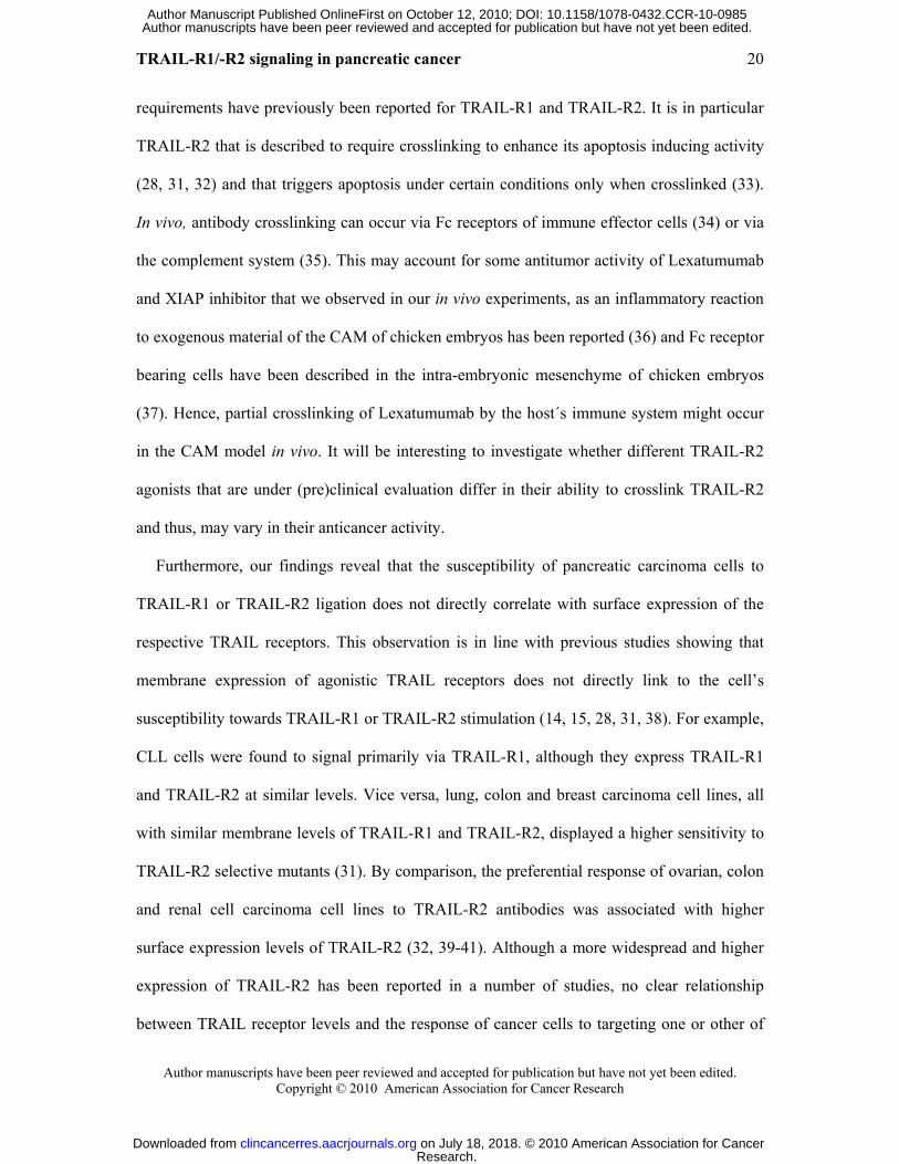
yielded negative stainings (Fig. 1A, a). Results of immunohistochemistry were scored as
“negative”, n; “weakly positive”, wp; “positive”, p; “strongly positive”, sp. In cases of
staining heterogeneity within the target cell population two modalities were allowed, e.g.
“p/n“, and not further quantified.
Chorioallantoic membrane assay
Chorionallantoic membrane (CAM) assay was done as described previously (16). Briefly,
1x10⁶ tumor cells were resuspended in 10 µl serum-free medium and 10 µl Matrigel Matrix
(BD Biosciences) and implanted on fertilized chicken eggs on day 8 of incubation. Tumors
were topically treated with 0.25 µg Mapatumumab or Lexatumumab diluted in 15 µl serum-
free medium with or without 10 µM XIAP inhibitor daily for 3 days, sampled with
surrounding CAM 4 days after seeding, fixed in 4% paraformaldehyde, paraffin embedded,
cut in 5 µm sections and hematoxylin/eosin stained. Tumor area was measured in a
Research. on July 18, 2018. © 2010 American Association for Cancerclincancerres.aacrjournals.org Downloaded from
Author manuscripts have been peer reviewed and accepted for publication but have not yet been edited. Author Manuscript Published OnlineFirst on October 12, 2010; DOI: 10.1158/1078-0432.CCR-10-0985

TRAIL-R1/-R2 signaling in pancreatic cancer
Author manuscripts have been peer reviewed and accepted for publication but have not yet been edited. Copyright © 2010 American Association for Cancer Research
11
representative picture of each tumor and the percentage of cellular area of the whole tumor
area was calculated using OPTIMAS 6.5.1 (Media Cybernetics, Bethesda, MD). In detail,
whole tumor area of each picture was marked as region of interest, including tumor cells,
matrigel and CAM tissue. Color threshold was set for viable hematoxylin/eosin stained tumor
cells and the threshold reaching area containing viable tumor cells was calculated and
expressed as percentage of region of interest.
Statistical analysis
Statistical significance was assessed by two-sided Student's t-test using Microsoft® Excel®
(Microsoft Deutschland GmbH, Unterschleißheim, Germany). Interaction between XIAP
inhibitors and TRAIL receptor antibodies was analyzed by the Combination index (CI)
method based on that described by Chou (27) using CalcuSyn software (Biosoft, Cambridge,
UK). Combination index (CI) <0.9 indicates synergism, 0.9-1.1 additivity and >1.1
antagonism.
Research. on July 18, 2018. © 2010 American Association for Cancerclincancerres.aacrjournals.org Downloaded from
Author manuscripts have been peer reviewed and accepted for publication but have not yet been edited. Author Manuscript Published OnlineFirst on October 12, 2010; DOI: 10.1158/1078-0432.CCR-10-0985

TRAIL-R1/-R2 signaling in pancreatic cancer
Author manuscripts have been peer reviewed and accepted for publication but have not yet been edited. Copyright © 2010 American Association for Cancer Research
12
Results
Recently, we reported that small molecule XIAP inhibitors synergize with TRAIL to trigger
apoptosis in pancreatic carcinoma cells in vitro and in vivo (16). Since it is not known at
present which of the two agonistic TRAIL receptors is superior as a therapeutic target in
pancreatic carcinoma, we evaluated agonistic TRAIL-R1 and -R2 specific antibodies alone
and in combination with XIAP inhibitors in the present study.
Expression of TRAIL receptors in pancreatic carcinoma
First, we explored the expression status of TRAIL receptors in human primary pancreatic
ductal adenocarcinoma samples and in normal pancreatic tissue by immunohistochemistry.
Ducts of normal, non-inflammed pancreata were consistently negative for all TRAIL
receptors (not shown), confirming our previously published data (26). Expression data of
pancreatic carcinomas are listed in Tab. 1. Of 24 carcinomas 12 were at least in part induced
for TRAIL-R1 expression compared to normal pancreatic ducts, seven of which were entirely
TRAIL-R1 positive (Fig. 1A, b). TRAIL-R2 was expressed in 18/24 carcinomas, 12 of which
were entirely TRAIL-R2 positive including one with strong TRAIL-R2 expression throughout
(Fig. 1A, c). TRAIL-R3 was expressed in 14/24 carcinomas, often only in subsets of
neoplastic cells and only once in a weak manner (Fig. 1A, e). TRAIL-R4 was most frequently
induced in pancreatic carcinomas compared to normal pancreatic tissue (22/24 samples) (Fig.
1A, f). Regarding the TRAIL-receptor expression profile, there was no entirely TRAIL-R1 to
-R4 positive nor an entirely TRAIL-R1 to -R4 negative case, however, four cases lacked both,
TRAIL-R1 and –R2 (Tab. 1). No obvious correlation of TRAIL receptor expression and grade
of differentiation was observed (Tab. 1).
Next, we examined cell surface expression of TRAIL receptors in a panel of pancreatic
carcinoma cell lines. All cell lines exhibited surface expression of the two agonistic TRAIL
Research. on July 18, 2018. © 2010 American Association for Cancerclincancerres.aacrjournals.org Downloaded from
Author manuscripts have been peer reviewed and accepted for publication but have not yet been edited. Author Manuscript Published OnlineFirst on October 12, 2010; DOI: 10.1158/1078-0432.CCR-10-0985
TRAIL-R1/-R2 signaling in pancreatic cancer
Author manuscripts have been peer reviewed and accepted for publication but have not yet been edited. Copyright © 2010 American Association for Cancer Research
13
receptors TRAIL-R1 and -R2 (Fig. 1B and (17)). TRAIL-R3 and TRAIL-R4 were expressed
at low or undetectable levels in most cell lines except PaTu8988t, T3M4, ASPC1 and PaTuII
that express considerable levels of TRAIL-R4 (Fig. 1B and (17).
XIAP inhibitor preferentially cooperates with Mapatumumab to reduce viability in
pancreatic carcinoma cells
To gain insight into the regulation of TRAIL-induced apoptosis via TRAIL-R1 and TRAIL-
R2 in pancreatic carcinoma cells, we analyzed the cytotoxicity of fully human monoclonal
antibodies specifically directed against TRAIL-R1 (Mapatumumab) and TRAIL-R2
(Lexatumumab). Interestingly, Mapatumumab was more potent to reduce cell viability than
Lexatumumab in the majority of pancreatic carcinoma cell lines (9 of 13 cell lines), while
four cell lines (MiaPaCa2, PaTu8988t, PaTu8988s, ASPC1) were more susceptible to
Lexatumumab (Fig. 2A).
Next, we assessed the effect of TRAIL receptor specific antibodies in combination with a
small molecule XIAP inhibitor that binds to the BIR3 domain of XIAP (23). Of note, the
XIAP inhibitor significantly enhanced loss of viability in combination with one of the
agonistic TRAIL receptor antibodies in all cell lines investigated (Fig. 2A). The majority of
pancreatic carcinoma cell lines (9 of 13 cell lines) were more susceptible to the combination
of Mapatumumab and the XIAP inhibitor compared to Lexatumumab (Fig. 2A). By
comparison, the four cell lines that were more responsive to treatment with Lexatumumab
alone (MiaPaCa2, PaTu8988t, PaTu8988s, ASPC1) were also more sensitive to the
combination of Lexatumumab plus XIAP inhibitor (Fig. 2A).
Moreover, we simultaneously treated cells with Mapatumumab and Lexatumumab to test
whether the concomitant stimulation of both agonistic TRAIL receptors results in additive,
synergistic or antagonistic cytotoxicity. The combined use of Mapatumumab and
Research. on July 18, 2018. © 2010 American Association for Cancerclincancerres.aacrjournals.org Downloaded from
Author manuscripts have been peer reviewed and accepted for publication but have not yet been edited. Author Manuscript Published OnlineFirst on October 12, 2010; DOI: 10.1158/1078-0432.CCR-10-0985

TRAIL-R1/-R2 signaling in pancreatic cancer
Author manuscripts have been peer reviewed and accepted for publication but have not yet been edited. Copyright © 2010 American Association for Cancer Research
14
Lexatumumab neither acted in concert to reduce viability nor antagonized each other, in the
presence or absence of the XIAP inhibitor (Fig. 2A). The only exception were MiaPaCa2
cells, where simultaneous treatment with Mapatumumab and Lexatumumab resulted in
enhanced reduction of cell viability compared to treatment with Mapatumumab and
Lexatumumab alone (Fig. 2A). Control experiments using a close structural analogue that
weakly binds to XIAP (23) showed no cooperative interaction with either of the TRAIL
receptor antibodies (Suppl. Fig. 1). Together, this set of experiments demonstrates that the
majority of pancreatic carcinoma cell lines are more susceptible to TRAIL-R1 than TRAIL-
R2 specific antibodies, either as single agents or in combination with a small molecule XIAP
inhibitor. For further studies, we selected PancTu1 and PaTuII pancreatic carcinoma cells as
prototype cell lines, which preferentially respond to Mapatumumab in combination with the
XIAP inhibitor.
Distinct XIAP inhibitors preferentially cooperate with Mapatumumab to induce
apoptosis in pancreatic carcinoma cells
To test the broader relevance of our findings, we extended our studies to additional,
structurally modified XIAP inhibitors, which all bind to the same surface groove of the BIR3
domain of XIAP (23, 24). Distinct XIAP inhibitors profoundly enhanced Mapatumumab-
induced loss of viability, while they displayed a minor cooperative interaction with
Lexatumumab (Fig. 2B, Suppl. Fig. 2). The simultaneous stimulation with Mapatumumab and
Lexatumumab resulted in a similar reduction of cell viability compared to stimulation with
Mapatumumab alone, either in the absence or in the presence of XIAP inhibitors (Fig. 2B,
Suppl. Fig. 2). Dose titration studies of Mapatumumab and XIAP inhibitor 3 revealed that the
interaction of these two agents was highly synergistic (Fig. 2C, Tab. 2).
Research. on July 18, 2018. © 2010 American Association for Cancerclincancerres.aacrjournals.org Downloaded from
Author manuscripts have been peer reviewed and accepted for publication but have not yet been edited. Author Manuscript Published OnlineFirst on October 12, 2010; DOI: 10.1158/1078-0432.CCR-10-0985
TRAIL-R1/-R2 signaling in pancreatic cancer
Author manuscripts have been peer reviewed and accepted for publication but have not yet been edited. Copyright © 2010 American Association for Cancer Research
15
To confirm that cells die by apoptotic cell death, we assessed DNA fragmentation as a
characteristic feature of apoptosis. Importantly, addition of XIAP inhibitors profoundly
increased Mapatumumab-induced apoptosis in a dose- and time-dependent manner (Fig. 3A,
3B and Suppl. Fig. 3). By comparison, no or only a slight augmentation of apoptosis was
observed, when the XIAP inhibitor was combined with Lexatumumab (Fig. 3A and 3B).
Furthermore, we performed colony assays to examine the long-term effects of the
combination treatment. The XIAP inhibitor preferentially cooperated with Mapatumumab to
suppress colony formation compared to Lexatumumab (Fig. 3C). The specificity of
Mapatumumab and Lexatumumab for TRAIL-R1 and TRAIL-R2, respectively, was
confirmed by RNAi-mediated knockdown (Suppl. Fig. 4). Together, this set of experiments
demonstrates that inhibition of XIAP preferentially sensitizes pancreatic carcinoma cells for
TRAIL-R1-mediated apoptosis, resulting in long-term suppression of clonogenic survival.
Preferential activity of TRAIL-R1 selective mutants against pancreatic carcinoma cells
To further explore the susceptibility of pancreatic carcinoma cells towards TRAIL-R1 versus
TRAIL-R2 stimulation, we used mutant forms of TRAIL that bind to TRAIL-R1 or TRAIL-
R2 with high specificity (14). In combination with the XIAP inhibitor, the TRAIL-R1
selective mutant was more potent than the TRAIL-R2 selective mutant to reduce viability of
PancTu1 and PaTuII cells (Fig. 4A). These results confirm the findings with TRAIL receptor
specific antibodies that pancreatic carcinoma cells are more susceptible to TRAIL-R1
triggering.
Crosslinking increases Mapatumumab’s activity when combined with XIAP inhibitor
Since TRAIL-R2 has previously been reported to require crosslinking for its full activity (28),
we next investigated whether a crosslinking agent augments the cytotoxicity of TRAIL
Research. on July 18, 2018. © 2010 American Association for Cancerclincancerres.aacrjournals.org Downloaded from
Author manuscripts have been peer reviewed and accepted for publication but have not yet been edited. Author Manuscript Published OnlineFirst on October 12, 2010; DOI: 10.1158/1078-0432.CCR-10-0985

TRAIL-R1/-R2 signaling in pancreatic cancer
Author manuscripts have been peer reviewed and accepted for publication but have not yet been edited. Copyright © 2010 American Association for Cancer Research
16
receptor antibodies. In the presence of the XIAP inhibitor, crosslinking of Lexatumumab
significantly increased its cytotoxicity, whereas crosslinking of Mapatumumab did not alter
its cytotoxicity (Fig. 4B). In the absence of the XIAP inhibitor however, the addition of a
crosslinker had no or a minor effect on the cytotoxicity of TRAIL receptor antibodies (Fig.
4B). These findings demonstrate that crosslinking enhances Lexatumumab-induced
cytotoxicity either alone or when XIAP is simultaneously neutralized.
XIAP inhibitor preferentially cooperates with Mapatumumab to trigger caspase
activation
To gain insight into the activation of the TRAIL signalling cascade upon triggering of
TRAIL-R1 or TRAIL-R2 and its modulation by XIAP inhibitors, we monitored cleavage of
caspases by Western blotting. In both PancTu1 and PaTuII cells, Mapatumumab alone was
more potent to induce cleavage of caspase-9 and -3 into active fragments compared to
Lexatumumab (Fig. 5A). Also, the XIAP inhibitor preferentially cooperated with
Mapatumumab to enhance caspase activation (Fig. 5A). Treatment with the XIAP inhibitor
alone did not initiate caspase cleavage (Fig. 5A), consistent with our findings that the
concentration of XIAP inhibitor used in these experiments is subtoxic and insufficient to
initiate apoptosis in the absence of an additional pro-apoptotic stimulus (Fig. 2 and 3). We
also assessed caspase activity by enzymatic caspase assays. Similarly, the XIAP inhibitor
preferentially acted in concert with Mapatumumab to increase caspase activity compared to
Lexatumumab (Fig. 5B, Suppl. Fig. 5). To test the requirement of caspase activity for
apoptosis induction we used the broad range caspase inhibitor zVAD.fmk. Addition of
zVAD.fmk almost completely rescued loss of viability upon the combination treatment with
Mapatumumab or Lexatumumab and XIAP inhibitor (Fig. 5C), demonstrating that loss of
viability occurred in a caspase-dependent manner.
Research. on July 18, 2018. © 2010 American Association for Cancerclincancerres.aacrjournals.org Downloaded from
Author manuscripts have been peer reviewed and accepted for publication but have not yet been edited. Author Manuscript Published OnlineFirst on October 12, 2010; DOI: 10.1158/1078-0432.CCR-10-0985
TRAIL-R1/-R2 signaling in pancreatic cancer
Author manuscripts have been peer reviewed and accepted for publication but have not yet been edited. Copyright © 2010 American Association for Cancer Research
17
No preferential activation of PI3K/Akt/mTOR or Raf/MEK/ERK survival pathways by
Mapatumumab or Lexatumumab
Since TRAIL has been reported to stimulate survival signalling such as the PI3K/Akt/mTOR
and Raf/MEK/ERK pathways besides the induction of apoptosis (29), we asked whether
TRAIL-R1 and TRAIL-R2 may differentially activate these survival cascades in pancreatic
carcinoma cells. To address this question we monitored the phosphorylation status of both
upstream and downstream components of the PI3K/Akt/mTOR pathway using Akt as a target
of PI3K, S6 ribosomal protein as a target of mTOR and ERK as a component of the
Raf/MEK/ERK pathway. Treatment with Mapatumumab or Lexatumumab did not increase
phosphorylation of Akt, S6 ribosomal protein or ERK compared to control cells treated with
solvent (Suppl. Fig. 6). This indicates that the reduced cytotoxicity of Lexatumumab over
Mapatumumab is not simply due to preferential activation of the PI3K/Akt/mTOR and/or
Raf/MEK/ERK pathways by Lexatumumab.
Mapatumumab shows higher activity than Lexatumumab against primary cultured
pancreatic carcinoma cells
In order to validate the results obtained in cell lines, we extended our studies to primary
cultured pancreatic carcinoma cells derived from a pancreatic adenocarcinoma specimen.
Primary cultured pancreatic carcinoma cells (ULA) express TRAIL-R1, -R2 and -R4 as well
as cIAP1, cIAP2, XIAP and survivin protein (Fig. 6A and 6B). At equimolar concentrations,
Mapatumumab was more potent than Lexatumumab to reduce cell viability of primary
cultured pancreatic carcinoma cells (Fig. 6C). Importantly, the addition of the XIAP inhibitor
further enhanced Mapatumumab-mediated loss of viability (Fig. 6C). These findings
Research. on July 18, 2018. © 2010 American Association for Cancerclincancerres.aacrjournals.org Downloaded from
Author manuscripts have been peer reviewed and accepted for publication but have not yet been edited. Author Manuscript Published OnlineFirst on October 12, 2010; DOI: 10.1158/1078-0432.CCR-10-0985

TRAIL-R1/-R2 signaling in pancreatic cancer
Author manuscripts have been peer reviewed and accepted for publication but have not yet been edited. Copyright © 2010 American Association for Cancer Research
18
demonstrate that Mapatumumab exerts higher cytotoxicity against primary cultured pancreatic
carcinoma cells compared to Lexatumumab alone or in combination with XIAP inhibitor.
Mapatumumab cooperates with XIAP inhibitor to suppress pancreatic carcinoma
growth in vivo
Finally, we extended our studies to an in vivo setting, using the CAM model as an established
in vivo tumor model that allows the assessment of antitumor activity in a three dimensional
setting (16). Mapatumumab together with XIAP inhibitor significantly reduced tumor growth
compared to untreated tumors (Fig. 6D). Also, the combined treatment with Lexatumumab
and the XIAP inhibitor exerted some antitumor activity compared to the control, although this
did not reach statistical significance (Fig. 6D). This demonstrates that although both
Mapatumumab and Lexatumumab can act in concert with XIAP inhibitor to suppress
pancreatic carcinoma growth in vivo Mapatumumab is significantly more potent than
Lexatumumab.
Research. on July 18, 2018. © 2010 American Association for Cancerclincancerres.aacrjournals.org Downloaded from
Author manuscripts have been peer reviewed and accepted for publication but have not yet been edited. Author Manuscript Published OnlineFirst on October 12, 2010; DOI: 10.1158/1078-0432.CCR-10-0985

TRAIL-R1/-R2 signaling in pancreatic cancer
Author manuscripts have been peer reviewed and accepted for publication but have not yet been edited. Copyright © 2010 American Association for Cancer Research
19
Discussion
TRAIL receptor agonists are currently evaluated in early clinical trials in a variety of tumors
including pancreatic cancers (13, 19-22, 30). There is mounting evidence that individual
cancers preferentially signal via one of the agonistic TRAIL receptors (14, 15). In pancreatic
carcinoma however, it has not yet been explored whether one of the two proapoptotic TRAIL
receptors is superior as a therapeutic target.
Here, we provide first evidence that the majority of pancreatic carcinoma cell lines and
also primary cultured pancreatic carcinoma cells are more sensitive to TRAIL-R1 over
TRAIL-R2 agonists, especially in combination with XIAP inhibitors, and that crosslinking is
required for maximal activity of the TRAIL-R2 antibody Lexatumumab. This conclusion is
supported by two distinct approaches to trigger one of the agonistic TRAIL receptors: Firstly,
fully human monoclonal antibodies that specifically bind with a 1000 times greater affinity to
either TRAIL-R1 or TRAIL-R2 (13) and secondly, TRAIL receptor-selective mutants (14).
Data obtained in a panel of pancreatic carcinoma cell lines underline the generality of the
results and experiments using an established culture of clinical tumor material confirm the
clinical relevance of the findings.
The molecular basis of the preferential signaling via one of the two agonistic TRAIL
receptors in cancer cells that express both receptors is currently not exactly known. While our
data demonstrate no differential activation of cell survival signaling, i.e. activation of the
PI3K/Akt/mTOR or Raf/MEK/ERK pathways, upon triggering of TRAIL-R1 or TRAIL-R2,
they reveal clear differences in the crosslinking requirements. Accordingly, crosslinking of
TRAIL-R2 profoundly enhances its cytotoxicity either alone or when XIAP is concomitantly
antagonized, while crosslinking of TRAIL-R1 has no or minimal additional effects. Thus, the
relatively low susceptibility of pancreatic cancer cells to TRAIL-R2 antibodies can be
overcome by increasing the crosslinking status of TRAIL-R2. Distinct crosslinking
Research. on July 18, 2018. © 2010 American Association for Cancerclincancerres.aacrjournals.org Downloaded from
Author manuscripts have been peer reviewed and accepted for publication but have not yet been edited. Author Manuscript Published OnlineFirst on October 12, 2010; DOI: 10.1158/1078-0432.CCR-10-0985
TRAIL-R1/-R2 signaling in pancreatic cancer
Author manuscripts have been peer reviewed and accepted for publication but have not yet been edited. Copyright © 2010 American Association for Cancer Research
20
requirements have previously been reported for TRAIL-R1 and TRAIL-R2. It is in particular
TRAIL-R2 that is described to require crosslinking to enhance its apoptosis inducing activity
(28, 31, 32) and that triggers apoptosis under certain conditions only when crosslinked (33).
In vivo, antibody crosslinking can occur via Fc receptors of immune effector cells (34) or via
the complement system (35). This may account for some antitumor activity of Lexatumumab
and XIAP inhibitor that we observed in our in vivo experiments, as an inflammatory reaction
to exogenous material of the CAM of chicken embryos has been reported (36) and Fc receptor
bearing cells have been described in the intra-embryonic mesenchyme of chicken embryos
(37). Hence, partial crosslinking of Lexatumumab by the host´s immune system might occur
in the CAM model in vivo. It will be interesting to investigate whether different TRAIL-R2
agonists that are under (pre)clinical evaluation differ in their ability to crosslink TRAIL-R2
and thus, may vary in their anticancer activity.
Furthermore, our findings reveal that the susceptibility of pancreatic carcinoma cells to
TRAIL-R1 or TRAIL-R2 ligation does not directly correlate with surface expression of the
respective TRAIL receptors. This observation is in line with previous studies showing that
membrane expression of agonistic TRAIL receptors does not directly link to the cell’s
susceptibility towards TRAIL-R1 or TRAIL-R2 stimulation (14, 15, 28, 31, 38). For example,
CLL cells were found to signal primarily via TRAIL-R1, although they express TRAIL-R1
and TRAIL-R2 at similar levels. Vice versa, lung, colon and breast carcinoma cell lines, all
with similar membrane levels of TRAIL-R1 and TRAIL-R2, displayed a higher sensitivity to
TRAIL-R2 selective mutants (31). By comparison, the preferential response of ovarian, colon
and renal cell carcinoma cell lines to TRAIL-R2 antibodies was associated with higher
surface expression levels of TRAIL-R2 (32, 39-41). Although a more widespread and higher
expression of TRAIL-R2 has been reported in a number of studies, no clear relationship
between TRAIL receptor levels and the response of cancer cells to targeting one or other of
Research. on July 18, 2018. © 2010 American Association for Cancerclincancerres.aacrjournals.org Downloaded from
Author manuscripts have been peer reviewed and accepted for publication but have not yet been edited. Author Manuscript Published OnlineFirst on October 12, 2010; DOI: 10.1158/1078-0432.CCR-10-0985

TRAIL-R1/-R2 signaling in pancreatic cancer
Author manuscripts have been peer reviewed and accepted for publication but have not yet been edited. Copyright © 2010 American Association for Cancer Research
21
the agonistic TRAIL receptors has been established (13). Thus, the relative contribution of
each of the agonistic TRAIL receptors to initiate apoptosis in cells that express both receptors
is not a simple consequence of surface expression levels and might be determined by
intracellular regulators of apoptosis. This highlights the importance of functional (pre)clinical
studies such as the present one to identify the TRAIL receptor subtype that may preferentially
or exquisitely transmit the apoptotic signal in a given type of cancer.
From the translational perspective of targeting TRAIL receptors in pancreatic cancer it is
important to note that neoexpression of all TRAIL receptors was found in malignant versus
non-malignant pancreatic carcinoma tissue, consistent with our previous findings showing the
absence of TRAIL receptors in normal, non-inflamed pancreata (26). A recent
immunohistochemical study similarly showed upregulation of TRAIL-R1 and TRAIL-R4 in
pancreatic carcinoma tissue compared to the normal pancreas (42). Together, these findings
suggest that the TRAIL receptor system may serve as therapeutic targets in pancreatic cancer.
Another important finding of this study is that simultaneous inhibition of XIAP enhanced
TRAIL-R1- or TRAIL-R2-induced apoptosis in a highly synergistic manner. This is
particularly relevant, since TRAIL receptor antibodies as monotherapy displayed limited
antitumor activity in the majority of the pancreatic cancer cell lines investigated, in line with
our previous results for soluble recombinant TRAIL (17). This indicates that combination
regimens to enhance the therapeutic potential of TRAIL receptor agonists are required to
ensure the success of TRAIL receptor agonists against pancreatic cancer. Previously, we
demonstrated that neutralizing XIAP either by RNA interference-mediated knockdown or by
small molecule inhibitors acted in concert with soluble recombinant TRAIL to induce
apoptosis in pancreatic cancer in vitro and in vivo (16-18). In addition, XIAP small molecule
antagonists that target the BIR2 domain of XIAP were reported to synergize with TRAIL in
pancreatic cancer (17, 43). Compared to these earlier reports that focus on soluble
Research. on July 18, 2018. © 2010 American Association for Cancerclincancerres.aacrjournals.org Downloaded from
Author manuscripts have been peer reviewed and accepted for publication but have not yet been edited. Author Manuscript Published OnlineFirst on October 12, 2010; DOI: 10.1158/1078-0432.CCR-10-0985

TRAIL-R1/-R2 signaling in pancreatic cancer
Author manuscripts have been peer reviewed and accepted for publication but have not yet been edited. Copyright © 2010 American Association for Cancer Research
22
recombinant TRAIL, the current study shows for the first time that the antitumor activity of
TRAIL receptor specific antibodies is profoundly enhanced by neutralizing XIAP in
pancreatic cancer cells. These findings support the concept that simultaneous targeting of
XIAP in combination with stimulation of the TRAIL pathway is a promising approach to
augment the antitumor activity of TRAIL receptor agonists against pancreatic cancer. They
also have important implications for the design of future clinical trials with TRAIL receptor
antibodies for the treatment of pancreatic cancer, since Mapatumumab and Lexatumumab are
already under clinical evaluation alone or in combination with chemotherapy (22). The
clinical relevance of our findings is supported by data obtained in primary cultured pancreatic
carcinoma cells that were established from a tumor specimen of a patient with pancreatic
adenocarcinoma. Although the analysis of primary material is so far restricted to one
specimen, the data provide a first proof-of-concept that the reported findings are not restricted
to established cell lines, but are also relevant for patients’ derived primary tumor cells
established in culture.
In conclusion, this preclinical evaluation of a rational combination of two novel classes of
apoptosis-targeting drugs, i.e. TRAIL receptor antibodies and XIAP inhibitors, in relevant
preclinical in vitro and in vivo models of pancreatic cancer provides the molecular basis for
the design of new combination therapies for the treatment of pancreatic cancer. This strategy
may help to overcome apoptosis resistance of pancreatic cancer, one of the cancers with the
worst prognosis.
Acknowledgements
We thank C. Hulford and M. Luzzio (Pfizer Inc., Groton, CN) for providing XIAP inhibitor,
A. Dittrich and E. Scheidhauer for excellent technical assistance and B. Welz for expert
Research. on July 18, 2018. © 2010 American Association for Cancerclincancerres.aacrjournals.org Downloaded from
Author manuscripts have been peer reviewed and accepted for publication but have not yet been edited. Author Manuscript Published OnlineFirst on October 12, 2010; DOI: 10.1158/1078-0432.CCR-10-0985

TRAIL-R1/-R2 signaling in pancreatic cancer
Author manuscripts have been peer reviewed and accepted for publication but have not yet been edited. Copyright © 2010 American Association for Cancer Research
23
secretarial work. This work has been partially supported by grants from the Deutsche
Forschungsgemeinschaft, the Deutsche Krebshilfe, the European Community (ApopTrain,
APO-SYS) and IAP6/18 (to S. F.).
Research. on July 18, 2018. © 2010 American Association for Cancerclincancerres.aacrjournals.org Downloaded from
Author manuscripts have been peer reviewed and accepted for publication but have not yet been edited. Author Manuscript Published OnlineFirst on October 12, 2010; DOI: 10.1158/1078-0432.CCR-10-0985

TRAIL-R1/-R2 signaling in pancreatic cancer
Author manuscripts have been peer reviewed and accepted for publication but have not yet been edited. Copyright © 2010 American Association for Cancer Research
24
References
1. Li D, Xie K, Wolff R, Abbruzzese JL. Pancreatic cancer. Lancet 2004;363: 1049-57.
2. Schneider G, Siveke JT, Eckel F, Schmid RM. Pancreatic cancer: basic and clinical
aspects. Gastroenterology 2005;128: 1606-25.
3. Fulda S. Apoptosis pathways and their therapeutic exploitation in pancreatic cancer. J Cell
Mol Med 2009;13: 1221-7.
4. Gukovskaya AS, Pandol SJ. Cell death pathways in pancreatitis and pancreatic cancer.
Pancreatology 2004;4: 567-86.
5. Fulda S, Debatin KM. Extrinsic versus intrinsic apoptosis pathways in anticancer
chemotherapy. Oncogene 2006;25: 4798-811.
6. Ashkenazi A. Targeting the extrinsic apoptosis pathway in cancer. Cytokine Growth Factor
Rev 2008;19: 325-31.
7. Kroemer G, Galluzzi L, Brenner C. Mitochondrial membrane permeabilization in cell
death. Physiol Rev 2007;87: 99-163.
8. Ashkenazi A. Directing cancer cells to self-destruct with pro-apoptotic receptor agonists.
Nat Rev Drug Discov 2008;7: 1001-12.
9. Fulda S. Tumor resistance to apoptosis. Int J Cancer 2009;124: 511-5.
10. Fulda S. Targeting inhibitor of apoptosis proteins (IAPs) for cancer therapy. Anticancer
Agents Med Chem 2008;8: 533-9.
11. LaCasse EC, Mahoney DJ, Cheung HH, Plenchette S, Baird S, Korneluk RG. IAP-
targeted therapies for cancer. Oncogene 2008;27: 6252-75.
12. Fairbrother WJ, Vucic D. The inhibitor of apoptosis proteins as therapeutic targets in
cancer. Clin Cancer Res 2007;13: 5995-6000.
13. Humphreys RC, Halpern W. Trail receptors: targets for cancer therapy. Adv Exp Med
Biol 2008;615: 127-58.
Research. on July 18, 2018. © 2010 American Association for Cancerclincancerres.aacrjournals.org Downloaded from
Author manuscripts have been peer reviewed and accepted for publication but have not yet been edited. Author Manuscript Published OnlineFirst on October 12, 2010; DOI: 10.1158/1078-0432.CCR-10-0985

TRAIL-R1/-R2 signaling in pancreatic cancer
Author manuscripts have been peer reviewed and accepted for publication but have not yet been edited. Copyright © 2010 American Association for Cancer Research
25
14. MacFarlane M, Kohlhaas SL, Sutcliffe MJ, Dyer MJS, Cohen GM. TRAIL receptor-
selective mutants signal to apoptosis via TRAIL-R1 in primary lymphoid malignancies.
Cancer Res 2005;65: 11265-70.
15. MacFarlane M, Inoue S, Kohlhaas SL, et al. Chronic lymphocytic leukemic cells
exhibit apoptotic signaling via TRAIL-R1. Cell Death Differ 2005;12: 773-82.
16. Vogler M, Walczak H, Stadel D, et al. Small molecule XIAP inhibitors enhance
TRAIL-induced apoptosis and antitumor activity in preclinical models of pancreatic
carcinoma. Cancer Res 2009;69: 2425-34.
17. Vogler M, Durr K, Jovanovic M, Debatin KM, Fulda S. Regulation of TRAIL-induced
apoptosis by XIAP in pancreatic carcinoma cells. Oncogene 2007;26: 248-57.
18. Vogler M, Walczak H, Stadel D, et al. Targeting XIAP bypasses Bcl-2-mediated
resistance to TRAIL and cooperates with TRAIL to suppress pancreatic cancer growth in vitro
and in vivo. Cancer Res 2008;68: 7956-65.
19. Ashkenazi A, Herbst RS. To kill a tumor cell: the potential of proapoptotic receptor
agonists. J Clin Invest 2008;118: 1979-90.
20. Moretto P, Hotte SJ. Targeting apoptosis: preclinical and early clinical experience with
mapatumumab, an agonist monoclonal antibody targeting TRAIL-R1. Expert Opin Investig
Drugs 2009;18: 311-25.
21. Hotte SJ, Hirte HW, Chen EX, et al. A Phase 1 Study of Mapatumumab (Fully Human
Monoclonal Antibody to TRAIL-R1) in Patients with Advanced Solid Malignancies. Clin
Cancer Res 2008;14: 3450-5.
22. Mom CH, Verweij J, Oldenhuis CN, et al. Mapatumumab, a fully human agonistic
monoclonal antibody that targets TRAIL-R1, in combination with gemcitabine and cisplatin:
a phase I study. Clin Cancer Res 2009;15: 5584-90.
Research. on July 18, 2018. © 2010 American Association for Cancerclincancerres.aacrjournals.org Downloaded from
Author manuscripts have been peer reviewed and accepted for publication but have not yet been edited. Author Manuscript Published OnlineFirst on October 12, 2010; DOI: 10.1158/1078-0432.CCR-10-0985

TRAIL-R1/-R2 signaling in pancreatic cancer
Author manuscripts have been peer reviewed and accepted for publication but have not yet been edited. Copyright © 2010 American Association for Cancer Research
26
23. Oost TK, Sun C, Armstrong RC, et al. Discovery of potent antagonists of the
antiapoptotic protein XIAP for the treatment of cancer. J Med Chem 2004;47: 4417-26.
24. Chao B, Deckwerth TL, Furth P, S., et al., Chao B, Deckwerth TL, Furth P, S., et
al.Chao B, Deckwerth TL, Furth P, S., et al.s; Tetrapeptide analogs. United States patent
PCT/US2005/024700. 2006 16.02.
25. Fulda S, Sieverts H, Friesen C, Herr I, Debatin KM. The CD95 (APO-1/Fas) system
mediates drug-induced apoptosis in neuroblastoma cells. Cancer Res 1997;57: 3823-9.
26. Hasel C, Durr S, Rau B, et al. In chronic pancreatitis, widespread emergence of TRAIL
receptors in epithelia coincides with neoexpression of TRAIL by pancreatic stellate cells of
early fibrotic areas. Lab Invest 2003;83: 825-36.
27. Chou TC. The median-effect principle and the combination index for quantitation of
synergism and antagonism. In: Chou TC, editor. Synergism and antagonism in chemotherapy.
San Diego, USA: Academic Press; 1991. p. 61-102.
28. Natoni A, MacFarlane M, Inoue S, et al. TRAIL signals to apoptosis in chronic
lymphocytic leukaemia cells primarily through TRAIL-R1 whereas cross-linked agonistic
TRAIL-R2 antibodies facilitate signalling via TRAIL-R2. Br J Haematol 2007;139: 568-77.
29. Di Pietro R, Zauli G. Emerging non-apoptotic functions of tumor necrosis factor-related
apoptosis-inducing ligand (TRAIL)/Apo2L. J Cell Physiol 2004;201: 331-40.
30. Greco FA, Bonomi P, Crawford J, et al. Phase 2 study of mapatumumab, a fully human
agonistic monoclonal antibody which targets and activates the TRAIL receptor-1, in patients
with advanced non-small cell lung cancer. Lung Cancer 2008;61: 82-90.
31. Kelley RF, Totpal K, Lindstrom SH, et al. Receptor-selective mutants of apoptosis-
inducing ligand 2/tumor necrosis factor-related apoptosis-inducing ligand reveal a greater
contribution of death receptor (DR) 5 than DR4 to apoptosis signaling. J Biol Chem
2005;280: 2205-12.
Research. on July 18, 2018. © 2010 American Association for Cancerclincancerres.aacrjournals.org Downloaded from
Author manuscripts have been peer reviewed and accepted for publication but have not yet been edited. Author Manuscript Published OnlineFirst on October 12, 2010; DOI: 10.1158/1078-0432.CCR-10-0985
TRAIL-R1/-R2 signaling in pancreatic cancer
Author manuscripts have been peer reviewed and accepted for publication but have not yet been edited. Copyright © 2010 American Association for Cancer Research
27
32. Zeng Y, Wu XX, Fiscella M, et al. Monoclonal antibody to tumor necrosis factor-
related apoptosis-inducing ligand receptor 2 (TRAIL-R2) induces apoptosis in primary renal
cell carcinoma cells in vitro and inhibits tumor growth in vivo. Int J Oncol 2006;28: 421-30.
33. Muhlenbeck F, Schneider P, Bodmer JL, et al. The tumor necrosis factor-related
apoptosis-inducing ligand receptors TRAIL-R1 and TRAIL-R2 have distinct cross-linking
requirements for initiation of apoptosis and are non-redundant in JNK activation. J Biol Chem
2000;275: 32208-13.
34. Motoki K, Mori E, Matsumoto A, et al. Enhanced apoptosis and tumor regression
induced by a direct agonist antibody to tumor necrosis factor-related apoptosis-inducing
ligand receptor 2. Clin Cancer Res 2005;11: 3126-35.
35. Chuntharapai A, Dodge K, Grimmer K, et al. Isotype-dependent inhibition of tumor
growth in vivo by monoclonal antibodies to death receptor 4. J Immunol 2001;166: 4891-8.
36. Valdes TI, Kreutzer D, Moussy F. The chick chorioallantoic membrane as a novel in
vivo model for the testing of biomaterials. J Biomed Mater Res 2002;62: 273-82.
37. Fleischer B. The avian immune system. Immunol Today 2001;10: 195-200.
38. Belyanskaya LL, Marti TM, Hopkins-Donaldson S, Kurtz S, Felley-Bosco E, Stahel
RA. Human agonistic TRAIL receptor antibodies Mapatumumab and Lexatumumab induce
apoptosis in malignant mesothelioma and act synergistically with cisplatin. Mol Cancer
2007;6: 66.
39. Nawrocki ST, Carew JS, Douglas L, Cleveland JL, Humphreys R, Houghton JA.
Histone deacetylase inhibitors enhance lexatumumab-induced apoptosis via a p21Cip1-
dependent decrease in survivin levels. Cancer Res 2007;67: 6987-94.
40. Saulle E, Petronelli A, Pasquini L, et al. Proteasome inhibitors sensitize ovarian cancer
cells to TRAIL induced apoptosis. Apoptosis 2007;12: 635-55.
Research. on July 18, 2018. © 2010 American Association for Cancerclincancerres.aacrjournals.org Downloaded from
Author manuscripts have been peer reviewed and accepted for publication but have not yet been edited. Author Manuscript Published OnlineFirst on October 12, 2010; DOI: 10.1158/1078-0432.CCR-10-0985

TRAIL-R1/-R2 signaling in pancreatic cancer
Author manuscripts have been peer reviewed and accepted for publication but have not yet been edited. Copyright © 2010 American Association for Cancer Research
28
41. Marini P, Denzinger S, Schiller D, et al. Combined treatment of colorectal tumours with
agonistic TRAIL receptor antibodies HGS-ETR1 and HGS-ETR2 and radiotherapy: enhanced
effects in vitro and dose-dependent growth delay in vivo. Oncogene 2006;25: 5145-54.
42. Sanlioglu AD, Dirice E, Elpek O, et al. High TRAIL death receptor 4 and decoy
receptor 2 expression correlates with significant cell death in pancreatic ductal
adenocarcinoma patients. Pancreas 2009;38: 154-60.
43. Karikari CA, Roy I, Tryggestad E, et al. Targeting the apoptotic machinery in
pancreatic cancers using small-molecule antagonists of the X-linked inhibitor of apoptosis
protein. Mol Cancer Ther 2007;6: 957-66.
Research. on July 18, 2018. © 2010 American Association for Cancerclincancerres.aacrjournals.org Downloaded from
Author manuscripts have been peer reviewed and accepted for publication but have not yet been edited. Author Manuscript Published OnlineFirst on October 12, 2010; DOI: 10.1158/1078-0432.CCR-10-0985

TRAIL-R1/-R2 signaling in pancreatic cancer
Author manuscripts have been peer reviewed and accepted for publication but have not yet been edited. Copyright © 2010 American Association for Cancer Research
29
Figure legends
Figure 1. TRAIL receptor expression in pancreatic carcinoma
A, TRAIL receptor expression was analyzed by immunohistochemisty on frozen sections in
24 samples of pancreatic ductal adenocarinoma and 4 samples of normal pancreas. Examples
of a case of a moderately differentiated (case number 11; a-c) and a poorly differentiated (case
number 15; d-f) ductal pancreatic adenocarcinoma are presented. Case 11 shows weak
TRAIL-R1 expression in all tumor cells (b) and TRAIL-R2 expression at a clearly higher
level (c) (a is the negative control). Case 15 expresses TRAIL-R1 only weakly and in a
minority of neoplastic cells (d) and has clear-cut TRAIL-3 expression in a subset of neoplastic
cells (e) while featuring strong TRAIL-R4 expression (f). (Scale bar in a, valid for a-c,
corresponds to 240 µm; scale bar in d, valid for d-f, corresponds to 120 µm).
B, Surface expression of TRAIL receptors 1-4 on pancreatic carcinoma cell lines was
determined by flow cytometry (thin line: isotype control, thick line: anti-TRAIL receptor
antibodies). Fluorescence intensity (x-axis) is blotted against cell counts (y-axis). A
representative experiment of 3 independent experiments is shown.
Figure 2. XIAP inhibitors cooperate with TRAIL receptor antibodies to reduce viability
of pancreatic carcinoma cells.
A, Cells were treated for 48h with indicated concentrations of Mapatumumab, Lexatumumab
or both Mapatumumab and Lexatumumab and/or 10 μM XIAP inhibitor 2 or DMSO. Cell
viability was determined by MTT assay and is expressed as percentage of untreated controls.
B, PancTu1 (left panels) and PaTuII (right panels) cells were treated for 48h with indicated
concentrations of Mapatumumab (upper panels), Lexatumumab (middle panels) or
Mapatumumab and Lexatumumab (lower panels) and/or a subtoxic concentration (10 μM) of
XIAP inhibitor 3 or DMSO.
Research. on July 18, 2018. © 2010 American Association for Cancerclincancerres.aacrjournals.org Downloaded from
Author manuscripts have been peer reviewed and accepted for publication but have not yet been edited. Author Manuscript Published OnlineFirst on October 12, 2010; DOI: 10.1158/1078-0432.CCR-10-0985

TRAIL-R1/-R2 signaling in pancreatic cancer
Author manuscripts have been peer reviewed and accepted for publication but have not yet been edited. Copyright © 2010 American Association for Cancer Research
30
C, Cells were treated for 48h with indicated concentrations of Mapatumumab and indicated
concentrations of XIAP inhibitor 3. Cell viability was determined by MTT assay and is
expressed as percentage of untreated controls. Mean + SEM of three independent experiments
performed in triplicate are shown; #, P<0.05; *, P<0.01 comparing XIAP inhibitors to
solvent.
Figure 3. XIAP inhibitor preferentially cooperates with Mapatumumab to induce
apoptosis and to reduce colony formation.
PancTu1 (left panel) and PaTuII (right panel) cells were treated with Mapatumumab,
Lexatumumab or both Mapatumumab and Lexatumumab at indicated concentrations (A) or 3
μg/ml (B) and/or 10 μM XIAP inhibitor 3 or DMSO. Apoptosis was determined by FACS
analysis of DNA fragmentation of propidium iodide stained nuclei. In C, colony formation
after treatment with Mapatumumab or Lexatumumab at 3 μg/ml (left panel) or 1 μg/ml (right
panel) and/or 10 μM XIAP inhibitor 3 or DMSO was assessed by crystal violet staining. A
representative experiment of three independent experiments is shown.
Figure 4. XIAP inhibitor preferentially cooperates with TRAIL-R1 selective mutant to
induce apoptosis whereas crosslinking of TRAIL-R2 potentiates apoptosis.
PancTu1 (left panels) and PaTuII (right panels) cells were treated for 48h with wildtype
soluble TRAIL, TRAIL-R1 selective mutant (R1-5) or TRAIL-R2 selective mutant (R2-6) or
with Mapatumumab and Lexatumumab and/or 10 μM XIAP inhibitor 3 or DMSO (A). In B,
Mapatumumab and Lexatumumab were used in the absence or presence of crosslinker. Cell
viability was determined by MTT assay and is expressed as percentage of untreated controls.
Mean + SEM of three independent experiments performed in triplicate are shown.
Research. on July 18, 2018. © 2010 American Association for Cancerclincancerres.aacrjournals.org Downloaded from
Author manuscripts have been peer reviewed and accepted for publication but have not yet been edited. Author Manuscript Published OnlineFirst on October 12, 2010; DOI: 10.1158/1078-0432.CCR-10-0985

TRAIL-R1/-R2 signaling in pancreatic cancer
Author manuscripts have been peer reviewed and accepted for publication but have not yet been edited. Copyright © 2010 American Association for Cancer Research
31
Figure 5. XIAP inhibitor enhances TRAIL-induced activation of caspases.
A, PancTu1 (upper panel) and PaTuII (lower panel) cells were treated for indicated times with
3 μg/ml Mapatumumab or Lexatumumab and/or 10 μM XIAP inhibitor 3 or DMSO. Caspase
activation was determined by Western blotting. Arrows indicate caspase cleavage fragments.
For pro-caspase-8, short and long exposures of the blots are presented. A representative
experiment of three independent experiments is shown.
B, PancTu1 (left panel) and PaTuII (right panel) cells were treated for indicated times with 3
μg/ml (PancTu1) or 1 μg/ml (PaTuII) Mapatumumab or Lexatumumab and/or 10 μM XIAP
inhibitor 3 or DMSO. Caspase activity was assessed by caspase assay.
C, PancTu1 (left panel) and PaTuII (right panel) cells were treated for 48h with 3 μg/ml
(PancTu1) or 1 μg/ml (PaTuII) Mapatumumab or Lexatumumab and/or 10 μM XIAP
inhibitor 3 or DMSO in the presence or absence of 25 µM zVAD.fmk. Cell viability was
determined by MTT assay and is expressed as percentage of untreated controls. Mean + SEM
of three independent experiments performed in triplicate are shown. #, P<0.05; *, P<0.01
comparing XIAP inhibitor to solvent.
Figure 6. XIAP inhibitor cooperates with TRAIL receptor antibodies to induce
apoptosis in primary pancreatic carcinoma cells.
A, Surface expression of TRAIL receptors was analyzed by flow cytometry in primary
cultured pancreatic carcinoma cells (ULA).
B, Expression levels of cIAP1, cIAP2, XIAP, survivin and β-actin was assessed by Western
blotting in primary cultured pancreatic carcinoma cells (ULA).
C, Primary cultured pancreatic carcinoma cells (ULA) were treated for 48h with indicated
concentrations of TRAIL-R1 Mapatumumab (Mapa), Lexatumumab (Lexa) or Mapatumumab
and Lexatumumab (Mapa + Lexa) and/or 10 μM XIAP inhibitor 3 or DMSO. Cell viability
Research. on July 18, 2018. © 2010 American Association for Cancerclincancerres.aacrjournals.org Downloaded from
Author manuscripts have been peer reviewed and accepted for publication but have not yet been edited. Author Manuscript Published OnlineFirst on October 12, 2010; DOI: 10.1158/1078-0432.CCR-10-0985

TRAIL-R1/-R2 signaling in pancreatic cancer
Author manuscripts have been peer reviewed and accepted for publication but have not yet been edited. Copyright © 2010 American Association for Cancer Research
32
was determined by MTT assay and is expressed as percentage of untreated controls. Mean +
SEM of three independent experiments performed in triplicate are shown; #, P<0.05; *,
P<0.01 comparing XIAP inhibitors to solvent.
D, PancTu1 cells were seeded on the CAM of chicken embryos, allowed to establish and
treated for 3 d with 0.25 µg Mapatumumab (Mapa) or Lexatumumab (Lexa) in the presence
or absence of 10 µM XIAP inhibitor 3. The CAM was excised on day 4, fixed and H&E
stained. Tumor cell area was determined as described in Materials and Methods and is
presented as percent of total tumor area. Mean + SEM of a representative experiment is
shown; #, P<0.05; *, P<0.01 comparing XIAP inhibitors to solvent.
Research. on July 18, 2018. © 2010 American Association for Cancerclincancerres.aacrjournals.org Downloaded from
Author manuscripts have been peer reviewed and accepted for publication but have not yet been edited. Author Manuscript Published OnlineFirst on October 12, 2010; DOI: 10.1158/1078-0432.CCR-10-0985
Fig. 1A
Research. on July 18, 2018. © 2010 American Association for Cancerclincancerres.aacrjournals.org Downloaded from
Author manuscripts have been peer reviewed and accepted for publication but have not yet been edited. Author Manuscript Published OnlineFirst on October 12, 2010; DOI: 10.1158/1078-0432.CCR-10-0985

TRAIL-R1 TRAIL-R2 TRAIL-R3 TRAIL-R4
Fig. 1B
PaTu8988s
MiaPaCa2
PaTu8988t
Panc1
BxPC3
cell
coun
t
Panc98
c
T3M4
PaTu8902PaTu8902
fluorescence intensity
Research. on July 18, 2018. © 2010 American Association for Cancerclincancerres.aacrjournals.org Downloaded from
Author manuscripts have been peer reviewed and accepted for publication but have not yet been edited. Author Manuscript Published OnlineFirst on October 12, 2010; DOI: 10.1158/1078-0432.CCR-10-0985
Fig. 2PancTu1
80
100
80
100
PaTuII PaTu8902
80
100
A
Colo357
0
20
40
60
0 0.03 0.1 0.3 1 3
BxPC3
0
20
40
60
0 0.3 1 3 6 10
Panc-1
0
20
40
60
0 0.3 1 3 6 10
20
40
60
80
100
20
40
60
80
100
20
40
60
80
100
lity
(%
)
DanG
60
80
100
0
0 0.01 0.03 0.1 0.3 1
0
0 0.03 0.1 0.3 1 3 6 10
0
0 0.03 0.1 0.3 1
T3M4
60
80
100
cell
viab
il
0
20
40
0 0.3 1 3 6 10
100
MiaPaCa2 PaTu8988t
100 100
ASPC1
0
20
40
0 0.1 0.3 1 3 6 10
0
20
40
60
80
100
0
20
40
60
80
100
0
20
40
60
80
100
0
0 0.3 1 3 6 10
0
0 0.3 1 3 6 10
DMSO/Mapa XIAP inh. 2/Mapa
DMSO/Lexa XIAP inh. 2/Lexa
DMSO/Mapa + Lexa XIAP inh. 2/Mapa + Lexa
0
0 0.3 1 3 6 10
PaTu8988s
60
80
100
Panc98
60
80
100
agonistic TRAIL-R mAB (µg/ml)
0
20
40
0 0.3 1 3 6 10
0
20
40
0 0.3 1 3 6 10
Research. on July 18, 2018. © 2010 American Association for Cancerclincancerres.aacrjournals.org Downloaded from
Author manuscripts have been peer reviewed and accepted for publication but have not yet been edited. Author Manuscript Published OnlineFirst on October 12, 2010; DOI: 10.1158/1078-0432.CCR-10-0985

80
100
(%)
DMSO
80
100
(%)
BPancTu1 PaTuII
** *
* *
* **
* *
0
20
40
60
0 0.03 0.1 0.3 1 3
Mapa (µg/ml)
cell
viab
ility
( DMSO
XIAP inh. 3
0
20
40
60
0 0.3 1 3 6 10
Mapa (µg/ml)
cell
viab
ility
Mapa (µg/ml) Mapa (µg/ml)
40
60
80
100
viab
ilit
y (%
)
40
60
80
100
viab
ility
(%
)
* *#
* * *
0
20
0 0.03 0.1 0.3 1 3
Lexa (µg/ml)
cell
v
0
20
0 0.3 1 3 6 10
Lexa (µg/ml)
cell
100 100* **
0
20
40
60
80
cell
viab
ility
(%
)
0
20
40
60
80
cell
viab
ility
(%
)** *
** *
*
00 0.03 0.1 0.3 1 3
Mapa + Lexa (µg/ml)
00 0.3 1 3 6 10
Mapa + Lexa (µg/ml)
PancTu1 PaTuII
2030405060708090
100
cell
viab
ility
(%
) 30 µM10 µM3 µM1 µM0.3 µM0 µM
2030405060708090
100110
cell
viab
ility
(%
)
CPancTu1 PaTuII
01020
0 0.03 0.1 0.3 1 3
Mapa (µg/ml)
c
01020
0 0.3 1 3 6 10
Mapa (µg/ml)
Research. on July 18, 2018. © 2010 American Association for Cancerclincancerres.aacrjournals.org Downloaded from
Author manuscripts have been peer reviewed and accepted for publication but have not yet been edited. Author Manuscript Published OnlineFirst on October 12, 2010; DOI: 10.1158/1078-0432.CCR-10-0985

60
70 DMSO/MapaDMSO/LexaDMSO/Mapa + Lexa 60
70
A
Fig. 3
PancTu1 PaTuII
**
**
0
10
20
30
40
50
apo
pto
sis
(%)
XIAP inh. 3/Mapap
XIAP inh. 3/LexaXIAP inh. 3/Mapa + Lexa
0
10
20
30
40
50
apo
pto
sis
(%)
A*
*
*# *
*
00 0.03 0.1 0.3 1 3
agonistic TRAIL-R mAB (µg/ml)
00 0.3 1 3 6 10
agonistic TRAIL-R mAB (µg/ml)
DMSODMSO/Mapa
PancTu1 PaTuII
pto
sis
(%)
30
40
50
60
70
30
40
50
60
70
pto
sis
(%)
XIAP inh. 3
p
XIAP inh. 3/Mapa
DMSO/Lexa
XIAP inh. 3/Lexa
DMSO/Mapa + Lexa
XIAP inh. 3/Mapa + Lexa
B
**
*
*
*
*ap
op
0
10
20
24 48 72
time (h)
0
10
20
30
24 48 72
time (h)
apo
p
C medium DMSOXIAP inh. 3
medium
medium DMSOXIAP inh. 3
medium
PancTu1 PaTuII
Mapa
Lexa
Mapa
Lexa
Research. on July 18, 2018. © 2010 American Association for Cancerclincancerres.aacrjournals.org Downloaded from
Author manuscripts have been peer reviewed and accepted for publication but have not yet been edited. Author Manuscript Published OnlineFirst on October 12, 2010; DOI: 10.1158/1078-0432.CCR-10-0985
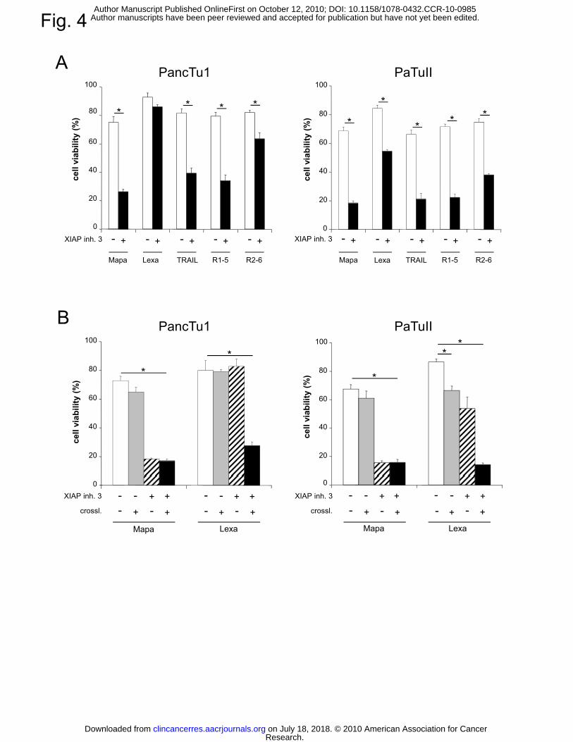
Fig. 4
A100100
PancTu1 PaTuII
40
60
80
cell
viab
ility
(%)
40
60
80
cell
viab
ility
(%)
****
*
*
** *
0
20
Mapa Lexa TRAIL R1-5 R2-6
0
20
Mapa Lexa TRAIL R1-5 R2-6
XIAP inh. 3 +- +- +- +- +- XIAP inh. 3 +- +- +- +- +-
B
80
100
80
100
PancTu1 PaTuII
**
*
*
20
40
60
80
cell
viab
ility
(%)
20
40
60
80
cell
viab
ility
(%) **
0
Mapa Lexa
+- +- +- +-
0
crossl.
Mapa Lexa
+- +- +- +-++ + +----XIAP inh. 3
crossl.
XIAP inh. 3 ++ + +----
Research. on July 18, 2018. © 2010 American Association for Cancerclincancerres.aacrjournals.org Downloaded from
Author manuscripts have been peer reviewed and accepted for publication but have not yet been edited. Author Manuscript Published OnlineFirst on October 12, 2010; DOI: 10.1158/1078-0432.CCR-10-0985
Fig. 5
A
Casp-8
Mapa Lexa
DMSO XIAP inh. 30 2 5 12 2
448
2 5 12 24
48
2 5 12 24
48
2 5 12 24
48
2 5 12 24
48
2 5 12 24
48
DMSO XIAP inh. 3 DMSO XIAP inh. 3time (h)
55/53 kD
Casp-3
Casp-8
Pan
cTu1
55/53 kD
32 kD
β-actin
Casp-9
P
48 kD
41 kD
Casp-8
Casp-8
Mapa Lexa
DMSO XIAP inh. 30 2 5 12 2
448
2 5 12 24
48
2 5 12 24
48
2 5 12 24
48
2 5 12 24
48
2 5 12 24
48
DMSO XIAP inh. 3 DMSO XIAP inh. 3time (h)
55/53 kD
55/53 kD
Casp-9
Casp-3
PaT
uII 32 kD
48 kD
β-actin
Casp 9
41 kD
Research. on July 18, 2018. © 2010 American Association for Cancerclincancerres.aacrjournals.org Downloaded from
Author manuscripts have been peer reviewed and accepted for publication but have not yet been edited. Author Manuscript Published OnlineFirst on October 12, 2010; DOI: 10.1158/1078-0432.CCR-10-0985

B Mapa/ DMSOMapa/ XIAP inh. 3Lexa/ DMSO
6
7
7
8
PancTu1 PaTuII
**
Lexa/ XIAP inh. 3
fold
indu
ctio
n of
ca
spas
e-3
activ
ity
1
2
3
4
5
6
fold
indu
ctio
n of
ca
spas
e-3
activ
ity
1
2
3
4
5
6
7
* *
* * **
*
#
#
time (h)
02 5 12
time (h)
0
1
2 5 12
CPancTu1 PaTuII
C
20
40
60
80
100
20
40
60
80
100
cell
viab
ility
(%)
20
40
60
80
100
20
40
60
80
100
cell
viab
ility
(%)
* #* *
* *
XIAP inh. 3zVAD.fmk
Mapa Lexa
0
20
0
20
+
0
20
-0
20
Mapa Lexa
+++
+ ++
--
--
--+
-+ +--+-- +
+ +--+-- +
Research. on July 18, 2018. © 2010 American Association for Cancerclincancerres.aacrjournals.org Downloaded from
Author manuscripts have been peer reviewed and accepted for publication but have not yet been edited. Author Manuscript Published OnlineFirst on October 12, 2010; DOI: 10.1158/1078-0432.CCR-10-0985

A
Fig. 6
tTRAIL-R1 TRAIL-R2 TRAIL-R3�� TRAIL-R4
fluorescence intensity
cell
cou
nt
60
80
100
y (%
)
B C
cIAP1 70 kD
0
20
40
60
cell
viab
ility
DMSO/Mapa
XIAP inh. 3/Mapa
DMSO/Lexa
XIAP inh. 3/Lexa
DMSO/Mapa + Lexa
XIAP inh. 3/Mapa + Lexa
cIAP2
XIAP
survivin
β-actin
72 kD
53 kD
16 kD
41 kD
0 0.03 0.1 0.3 1 3
agonistic TRAIL-R mAB (µg/ml)
D70
#
#
30
40
50
60
r ce
ll ar
ea (
%)
0
10
20tum
o
XIAP inh. 3
mapa+ + +
++-- -- -
-mapa
lexa+
++
+- -
- -- -- -
Research. on July 18, 2018. © 2010 American Association for Cancerclincancerres.aacrjournals.org Downloaded from
Author manuscripts have been peer reviewed and accepted for publication but have not yet been edited. Author Manuscript Published OnlineFirst on October 12, 2010; DOI: 10.1158/1078-0432.CCR-10-0985

Tab. 1. TRAIL receptor expression in primary pancreatic carcinoma samples
Results of immunohistochemistry were scored as “negative”, n; “weakly positive”, wp; “positive”, p; “strongly positive”, sp, or in cases of staining heterogeneity as p/n. dpc, ductal pancreatic carcinoma; pc, pancreatic carcinoma; c., cell.
Research. on July 18, 2018. © 2010 American Association for Cancerclincancerres.aacrjournals.org Downloaded from
Author manuscripts have been peer reviewed and accepted for publication but have not yet been edited. Author Manuscript Published OnlineFirst on October 12, 2010; DOI: 10.1158/1078-0432.CCR-10-0985
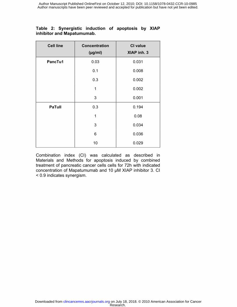
Table 2: Synergistic induction of apoptosis by XIAP inhibitor and Mapatumumab.
Cell line
Concentration
(µg/ml)
CI value
XIAP inh. 3
PancTu1 0.03 0.031
0.1 0.008
0.3 0.002
1 0.002
3 0.001
PaTuII 0.3 0.194
1 0.08
3 0.034
6 0.036
10 0.029
Combination index (CI) was calculated as described in Materials and Methods for apoptosis induced by combined treatment of pancreatic cancer cells cells for 72h with indicated concentration of Mapatumumab and 10 µM XIAP inhibitor 3. CI < 0.9 indicates synergism.
Research. on July 18, 2018. © 2010 American Association for Cancerclincancerres.aacrjournals.org Downloaded from
Author manuscripts have been peer reviewed and accepted for publication but have not yet been edited. Author Manuscript Published OnlineFirst on October 12, 2010; DOI: 10.1158/1078-0432.CCR-10-0985

Published OnlineFirst October 12, 2010.Clin Cancer Res Dominic Stadel, Caroline Ref, Marion MacFarlane, et al. enhanced by XIAP inhibitorsreceptor 1 in pancreatic carcinoma cells and profoundly TRAIL-induced apoptosis is preferentially mediated via TRAIL
Updated version
10.1158/1078-0432.CCR-10-0985doi:
Access the most recent version of this article at:
Material
Supplementary
http://clincancerres.aacrjournals.org/content/suppl/2010/12/08/1078-0432.CCR-10-0985.DC1
Access the most recent supplemental material at:
Manuscript
Authoredited. Author manuscripts have been peer reviewed and accepted for publication but have not yet been
E-mail alerts related to this article or journal.Sign up to receive free email-alerts
Subscriptions
Reprints and
To order reprints of this article or to subscribe to the journal, contact the AACR Publications
Permissions
Rightslink site. Click on "Request Permissions" which will take you to the Copyright Clearance Center's (CCC)
.http://clincancerres.aacrjournals.org/content/early/2010/10/09/1078-0432.CCR-10-0985To request permission to re-use all or part of this article, use this link
Research. on July 18, 2018. © 2010 American Association for Cancerclincancerres.aacrjournals.org Downloaded from
Author manuscripts have been peer reviewed and accepted for publication but have not yet been edited. Author Manuscript Published OnlineFirst on October 12, 2010; DOI: 10.1158/1078-0432.CCR-10-0985