Embed Size (px)
Citation preview
REVIEW Open Access
Trafficking in and to the primary ciliumYi-Chun Hsiao1,2†, Karina Tuz2† and Russell J Ferland2,3*
Abstract
Polarized vesicle trafficking is mediated by small GTPase proteins, such as Rabs and Arls/Arfs. These proteins haveessential roles in maintaining normal cellular function, in part, through regulating intracellular trafficking. Moreover,these families of proteins have recently been implicated in the formation and function of the primary cilium. Theprimary cilium, which is found on almost every cell type in vertebrates, is an organelle that protrudes from thesurface of the cell and functions as a signaling center. Interestingly, it has recently been linked to a variety ofhuman diseases, collectively referred to as ciliopathies. The primary cilium has an exceptionally high density ofreceptors on its membrane that are important for sensing and transducing extracellular stimuli. Moreover, theprimary cilium serves as a separate cellular compartment from the cytosol, providing for unique spatial andtemporal regulation of signaling molecules to initiate downstream events. Thus, functional primary cilia areessential for normal signal transduction. Rabs and Arls/Arfs play critical roles in early cilia formation but are alsoneeded for maintenance of ciliary function through their coordination with intraflagellar transport (IFT), aspecialized trafficking system in primary cilia. IFT in cilia is pivotal for the proper movement of proteins into andout of this highly regulated organelle. In this review article, we explore the involvement of polarized vesiculartrafficking in cilia formation and function, and discuss how defects in these processes could subsequently lead tothe abnormalities observed in ciliopathies.
Keywords: Primary cilium, Trafficking, Ciliopathies, Intraflagellar transport, Ciliary signaling
Primary cilia are evolutionarily conserved organelles pro-jecting from the plasma membrane in almost every verte-brate cell. In general, primary cilia serve as sensorsthrough which cells receive signals from light, chemical,or mechanical stimuli [1]. Moreover, the involvement ofprimary cilia in several signaling pathways important fordevelopment and tissue homeostasis (including the Sonichedgehog and Wnt signaling pathways) has attractedmuch interest and stimulated extensive studies on thisancient cellular structure [2-6]. A functional primarycilium is required to properly activate primary cilia-mediated cellular signaling. Therefore, any defects in pri-mary cilia could lead to cellular dysfunction. Indeed,abnormalities in primary cilia have been linked to a con-stellation of phenotypically and genetically overlappinghuman diseases, which include Bardet-Biedl syndrome,Joubert syndrome, Meckel-Gruber syndrome, nephro-nophthisis and Sensenbrenner syndrome; all now
collectively known as ciliopathies [7-10]. The clinicalmanifestations of these disorders can include brain mal-formations, skeletal abnormalities, retinal degeneration,and cystic kidney disease.The formation and function of primary cilia are tightly
regulated by polarized vesicle trafficking, not only to theprimary cilium, but also in coordination with traffickingthroughout the entire cell [5]. Although bioinformatic,proteomic and genetic studies have suggested that morethan a thousand proteins can be localized at the primarycilium, it is still unclear why and how these proteinswork together in this specialized cellular compartment[11-15]. Therefore, studying the formation and functionof the primary cilium, through investigations into thefunction of these ciliary proteins, will help to elucidatethe pathophysiological mechanisms responsible for caus-ing the ciliopathies.
Structure and function of the primary ciliumCilia are categorized into two classes: motile and non-motile. Motile cilia, such as tracheal cilia, can be numer-ous on a cell surface and have the prominent function ofmoving mucus and fluids, but are not the focus of this
* Correspondence: [email protected]† Contributed equally2Albany Medical College, Center for Neuropharmacology and Neuroscience,Albany, NY 12208, USAFull list of author information is available at the end of the article
Hsiao et al. Cilia 2012, 1:4http://www.ciliajournal.com/content/1/1/4
© 2012 Hsiao et al; licensee BioMed Central Ltd. This is an open access article distributed under the terms of the Creative CommonsAttribution License (http://creativecommons.org/licenses/by/2.0), which permits unrestricted use, distribution, and reproduction inany medium, provided the original work is properly cited.
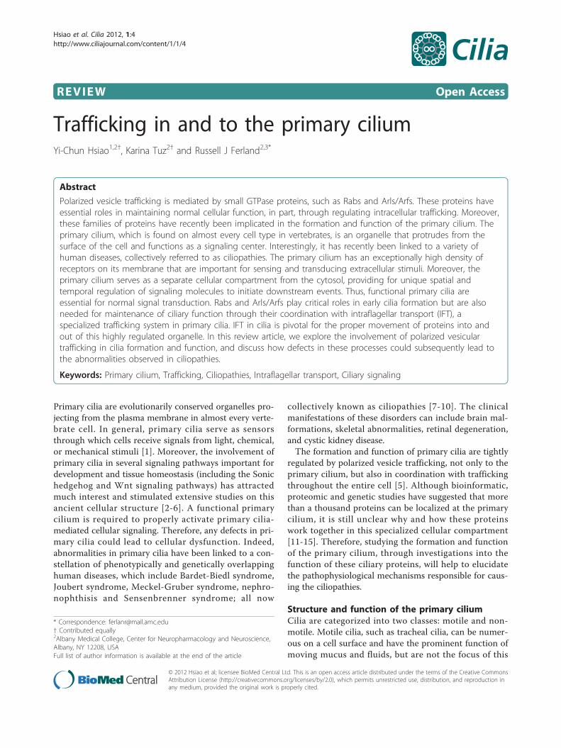
review. This review focuses on non-motile cilia, alsoreferred to more commonly as primary cilia, which aresolitary and mainly serve as a sensory organelle for thecell. Importantly, most primary cilia are non-motile,except for those present in the ventral nodes of verte-brates [1,16]. Primary cilia are polarized structures pro-truding from the surface of the cell into the extracellularspace and are present on almost every quiescent cell inthe body. The ciliary axoneme, which is composed ofmicrotubule bundles, is the core structure of the cilium[1]. For primary cilia, the ciliary axoneme consists of aradial array of nine doublet microtubules with no centralpair of singlet microtubules, and therefore is called a “9 +0” configuration. The microtubule axoneme is nucleatedat the basal body just beneath the plasma membrane(Figure 1). The basal body is a cytosolic microtubuleorganizing center that is derived from the mother cen-triole [1]. All of these structural components of the pri-mary cilium are necessary for the proper formation andfunction of this signaling structure.
Primary cilia are enriched with receptors and provide aseparate highly regulated compartment in which signal-ing events are conveyed from the extracellular space intothe cell. Sensing the extracellular environment is a majorfunction of primary cilia. For instance, the ciliated cells ofthe retina (photoreceptors) and the olfactory system(olfactory sensory neurons) receive and transduce the sti-muli of light and odorants to the cells, respectively[17,18]. Primary cilia on the epithelial cells of kidneytubules act as mechanosensors for sensing fluid flowresulting in increased intracellular calcium signaling[19-21].Recently, a vital role for primary cilia in signaling path-
ways important for embryonic development and tissuehomeostasis has been identified. While the sonic hedgehog(Shh) pathway has been long known as a critical compo-nent of neural tube closure and organ patterning duringembryonic development, it was only recently discoveredthat primary cilia are necessary for this signaling [2,6,22].In mammalian cells when Shh is absent, the Shh receptor,
Figure 1 Structure of the primary cilium. The core structure of primary cilia is composed of microtubule bundles (ciliary axoneme) extending fromthe basal body, a microtubule-based structure derived from the mother centriole. The ciliary membrane is continuous with the plasma membrane,but contains a unique protein composition, such as channels and receptors. Thus, primary cilia can function as a sensory organelle for receiving andtransducing extracellular stimuli into cells, such as fluid flow or via signaling molecules. The inset image is of ciliated murine inner medullary collectingduct (IMCD3) cells in which the basal body is labeled with a g-tubulin antibody (red) and the primary cilium is marked by an Ift88 antibody (green).
Hsiao et al. Cilia 2012, 1:4http://www.ciliajournal.com/content/1/1/4
Page 2 of 13
Patched-1 (Ptch1), is localized at the base of the primarycilium and suppresses the activity of another transmem-brane protein, Smoothened (Smo). Upon Shh stimulation,Smo is relieved from Ptch1 suppression and translocatesto the primary cilium, while Ptch1 is removed from theprimary cilium. This translocation of Smo to the primarycilium, after Shh stimulation, is required for activation ofShh-mediated downstream signaling [23-25].Importantly, the integrity of the primary cilium and the
proper functioning of the intraflagellar transport (IFT) sys-tem (the specialized transport system in the primarycilium) are both required for proper Shh signaling in mice[6,26]. For example, Ift172 deficient mouse embryos dis-play abnormal brain development resulting from defectsin cilia formation, consequently leading to reduced Shhsignaling [27]. In addition to Shh signaling, primary ciliaare also critical for Wnt signaling [3,28,29], calcium signal-ing [30,31], growth factor signaling [32-34], G-proteincoupled receptor signaling [35,36], and receptor tyrosinekinase signaling [37]. Interestingly, the range of differenttypes of signaling associated with cilia is ever expanding asmore receptors and proteins are identified at the primarycilium. Although it is unclear why receptors are enrichedat the primary cilium as compared to the plasma mem-brane or why signaling mechanisms are concentrated atthe primary cilium, it is clear that this unique and separatecellular compartment makes primary cilia an unusualstructure for receiving and transducing a variety of signals.
Mechanisms for sorting ciliary bound proteins tothe primary ciliumTo date, there have been no studies which have shownthat proteins can be produced in the cilium. Therefore,the unique density of proteins and receptors on the mem-brane around the ciliary axoneme were targeted therethrough a highly regulated importing process [25].Trafficking of proteins from the cytosol and Golgi to theprimary cilium, and for moving proteins along the ciliaryaxoneme, is regulated by polarized vesicle trafficking andintraflagellar transport, respectively. Detailed mechanismsof these two trafficking systems will be discussed later.The transition zone (TZ), a region adjacent to the basal
body, provides a selective barrier that can exclude vesicles,impede diffusion of proteins and lipids between compart-ments (cytosolic/plasma membrane versus ciliary), andcontrol the entry and exit of proteins from the primarycilium [38-40]. The TZ is composed of transitional fibers(TFs), the ciliary necklace and Y-links. TFs anchor thebasal body to the proximal ciliary membrane, forming apinwheel-like structure whose protein composition isunknown. The ciliary necklace is distal to the TFs and con-sists of rows of protein particles around the ciliary mem-brane at the base of the cilium. Y-links connect axonemalmicrotubules to the membrane at the ciliary necklace [41].
Whereas TZ components include CC2D2A, MKS1, MKS3,MKS5, MKS6, MKS1 related-1, MKS1 related-2, NPHP1,NPHP4, Septin 2, Tctn1, and Tctn3 [40-44], the Y-linksare associated with CEP290 localization [39]. Importantly,different components perform different functions at thisregion. For instance, while CEP20 functions as a gate kee-per, controlling cilia protein composition by restricting theentrance of non-ciliary proteins, CC2D2A facilitates theentry of ciliary proteins through a role in vesicle trafficking[39,42].To aid in this trafficking, ciliary proteins contain a tar-
geting sequence enabling it to efficiently localize at theprimary cilium. To date, different targeting sequenceshave been identified: rhodopsin possesses a C-terminaltargeting sequence with a VxPx motif; polycystin-2 hasan N-terminal RVxP motif; and polycystin-1 harbors aC-terminal targeting sequence, KVHPSST [45,46]. Thishas led to the suggestion of a VxP motif as a genericciliary targeting sequence (CTS). However, cystin con-tains a N-terminal CTS with a AxEGG motif [47],whereas the G-protein coupled receptors, Sstr3, Htr6,and Mchr1, present an Ax(S/A)xQ targeting sequence intheir third intracellular loop [48]. Overall, these dataindicate that there is no unique consensus CTS andfurther suggest that there is more than one molecularmechanism involved in the recognition of suchsequences.Palmitoylation and myristoylation are post-translational
modifications that provide a point of membrane associationand have been identified as a requirement for some ciliaryproteins in their proper targeting to the cilium. For exam-ple, the targeting sequence for rhodopsin contains twocysteine residues that are palmitoylated and which arenecessary for rod outer segment targeting [49]. The same istrue for fibrocystin, in which its CTS has three palmitoy-lated cysteine residues [50]. Lastly, cystin is myristoylated atits G2 residue and it has been shown that this acylation isrequired for the proper localization of cystin to the ciliarymembrane [47]. Therefore, the sorting of ciliary targetedproteins to the primary cilium not only occurs throughCTSs, but can also be accomplished through post-transla-tional modifications. It is these modifications that providethe sorting information for proteins resulting in theirproper trafficking to the specialized membrane microdo-mains of the primary cilium.Targeting of proteins to the primary cilium also
involves regulation of proteins under the control of theGDP-GTP cycle. Rab8, one such protein, interacts withthe CTS of fibrocystin, as suggested by a study using aGFP fusion of the fibrocystin CTS [50]. This GFP-CTSlocalizes to primary cilia in cells expressing Flag-Rab8and Flag-Rab8Q67L (a constitutively active form ofRab8a having decreased GTPase activity, thereby main-taining the protein in the GTP bound state), but not
Hsiao et al. Cilia 2012, 1:4http://www.ciliajournal.com/content/1/1/4
Page 3 of 13
Flag-Rab8T22N (a dominant negative form of Rab8 hav-ing a higher affinity for GDP than GTP) expressingcells. Rab8T22N bound more GFP-CTS than either thewild type or the Q67L mutant, suggesting that activationof Rab8 results in the release of the GFP-CTS fromRab8. Therefore, the regulation of Rab8 activity controlsthe localization of fibrocystin through its CTS. Whetherother ciliary localizing proteins containing CTSs interactwith Rabs through their CTS, remains to be determined.A mechanism for NPHP3 ciliary targeting has been
partially described in which myristoylation of its G2residue and regulation of GTPase activity are required.That is, NPHP3, when it is myristoylated, binds toUNC119 (HRG4). The phenylalanine residues lining thehydrophobic b-sandwich in UCN119 contribute to thisNPHP3 binding. This complex is trafficked through anunknown mechanism to the primary cilium. In thecilium, ARL3-GTP binds to its effector, UNC119, releas-ing myristoylated NPHP3 into the cilium or ciliarymembrane. RP2 then activates ARL3 GTPase activity,releasing UNC119 to reset the cycle [51]. These andother studies clearly indicate that protein sorting to theprimary cilium is a tightly regulated process that mayinvolve multiple molecular mechanisms of protein-protein recognition and protein-membrane interaction,in which the GDP-GTP state of these proteins isfundamental.
Functional primary cilia depend on polarizedtrafficking and intraflagellar transportPolarized vesicle trafficking is a highly regulated deliv-ery system in cells for transporting proteins and vesi-cles to their proper destinations in order to performand maintain normal cell function (Figure 2). In mostcases, the primary cilium is formed at the end of thecell cycle, after cell polarity is established. At the earlystage of cilium formation (ciliogenesis), a vesiclederived from the Golgi encapsulates the distal end ofthe mother centriole (the origin of the basal body) as itmigrates toward the apical plasma membrane. Afterdocking of the basal body, the primary cilium elongatesas the axoneme extends. Additional vesicles carry cili-ary membrane proteins to the cilium, which then fuseto the plasma membrane where the cilium originates[52]. Thus, ciliogenesis requires axoneme assembly,membrane biogenesis, and a proper compartmentaliza-tion of ciliary proteins in coordination with polarizedvesicle trafficking [1,53].
Polarized vesicular trafficking in ciliogenesis and ciliaryfunctionPolarized vesicle trafficking is a specialized cellulartransport mechanism used to deliver proteins andmembranes to their proper cellular compartments
(Figure 2). Perturbations in this process can have sig-nificant impacts on normal cell function [54,55]. Emer-ging evidence has shown an important link betweenpolarized vesicle trafficking and the proper formationand function of the primary cilium. Not only haveGolgi-derived vesicles been implicated in early cilio-genesis, but the constant trafficking of vesicles throughthe post-Golgi to the primary cilium has been shownto be crucial for normal ciliary structure and function[56,57]. Polarized vesicle trafficking is mediated byRabs along with Arf/Arl members of the Ras superfa-miliy of small GTPases, which assist in the recruitmentof vesicle coating complexes during vesicle budding,docking and fusion of vesicles [58,59]. Additionally, theessential roles of these small GTPase proteins in theformation and function of primary cilia have beendemonstrated in several studies (Table 1) [2,60,61].The activities of Rabs and Arf/Arl proteins are regu-lated by their nucleotide binding status. GDP-boundRab proteins, usually referred to as inactive forms, areswitched to the GTP-bound active state through cata-lyzation by guanine nucleotide exchange factors(GEFs). Then, GTP-bound Rabs can modulate severalcellular events through activation of their downstreameffectors, including vesicle sorting proteins, tetheringproteins, kinases, phosphatases and motor proteins.Conversion of active GTP-bound Rabs back to inactiveGDP-bound Rabs is through GTP hydrolysis byGTPase activating proteins (GAPs). The correct mem-brane targeting of Rab proteins is crucial and regulatedby its specific GDP dissociation inhibitors (GDIs) andmembrane-bound GDI displacement factors (GDFs)[59].Arl proteins in ciliary traffickingArl6 is exclusively expressed in ciliated organisms [62],and its human ortholog ARL6, encoded by BBS3, wasthe first identified small GTPase protein that was linkedto the human ciliopathy, Bardet-Biedl syndrome (BBS)[63]. This rare inherited disorder features dysfunction inmultiple organ systems, leading to retinal dystrophy,obesity, renal disease and cognitive impairments [64].The abnormalities associated with the loss of BBS3 inBBS are thought to arise due to dysfunctional primarycilia [63]. That is, Arl6 is localized at the distal end ofthe basal body, near or at the TFs, a region of the ciliarycompartment that controls the entry of proteins intothe primary cilium [65]. Arl6 functions as a recruiter ofthe BBSome (a basal body localizing complex composedof seven BBS disease proteins) to the basal body and asa regulator of the function of the BBSome in ciliary pro-tein trafficking [60,66]. ARL13B is another small GTPaseprotein connected to the human ciliopathy, Joubert syn-drome, an inherited neurodevelopmental disorder withmidbrain-hindbrain malformations, retinal dystrophy
Hsiao et al. Cilia 2012, 1:4http://www.ciliajournal.com/content/1/1/4
Page 4 of 13

and, occasionally, nephronophthisis [67]. Deletion ofArl13b in mice leads to the deformation of primary ciliaand defective neuronal tube development due to hyper-active Shh signaling [2,68]. Work in Caenorhabditis
elegans has demonstrated that the formation of the IFT-A and IFT-B subcomplex is critical for building the cili-ary structure and that the coordination of Arl13 andArl3 is required for stabilizing this subcomplex [69-71].
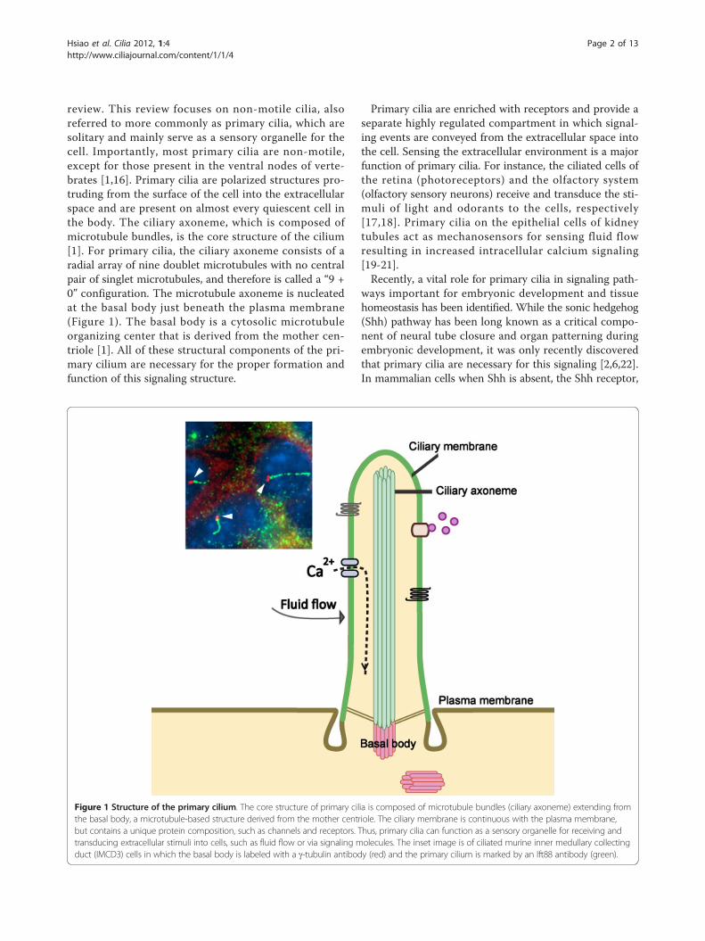
Figure 2 Polarized vesicle trafficking mediates the formation and function of primary cilia. Enriched expression of receptors and ionchannels on the ciliary membrane make the primary cilium a specialized organelle for receiving and transducing extracellular stimuli into cells.Proteins synthesized from the Golgi move to the primary cilium through polarized vesicle trafficking utilizing microtubule networks (not shown).For instance, vesicles carrying ciliary proteins leave the Golgi and move toward the basal body of the primary cilium. These vesicles can eitherbe delivered to the surface plasma membrane and then the protein cargo moves to the ciliary membrane or they can be trafficked toward thebasal body through Rab proteins, IFT20, or exocysts. Entry of protein cargo to the cilium is regulated by active forms of Rab8, a mastermodulator for the ciliary protein trafficking. Rab8 is recruited to the basal body of primary cilium, possibly mediated by Ahi1. The activities ofRab8 are then regulated by Rabin8, and its activity and basal body localization is modulated by the BBSome and Rab11. Once proteins aretransported into the primary cilium, the IFT system continues the trafficking of these proteins or membrane receptors up and down along theciliary axoneme. IFT-B (anterograde) and IFT-A (retrograde) are protein complexes associated with the molecular motors, kinesin-2 andcytoplasmic dynein, respectively.
Hsiao et al. Cilia 2012, 1:4http://www.ciliajournal.com/content/1/1/4
Page 5 of 13

Rab small GTPasesRab GTPases are the largest family of small GTPases, andtheir function and distinct localization at intracellularmembranes has been studied extensively [59]. However,the distribution of several Rab GTPases at the primarycilium was only recently discovered. Disruption of theciliary localization or the activities of these Rab GTPasesare associated with several ciliopathies due to impair-ments in cilium formation and function [72]. For exam-ple, Rab8 is a critical modulator for the formation andthe function of primary cilia in addition to having animportant role in vesicular trafficking between the trans-Golgi and the basolateral membrane [53,73]. As men-tioned previously, rhodopsin is present at the outersegments of photoreceptor cells. Rab8 is, in part, respon-sible for delivering rhodopsin-bearing post-Golgi vesiclesclose to the base of the photoreceptor connecting cilium,where these vesicles fuse, and rhodopsin is then trans-ported through the connecting cilium to the outer seg-ment [74]. Rab8 mutations that interfere with the GTP/GDP cycle of the protein have been utilized to test itsrole on ciliogenesis. Work in Xenopus laevis has shownthat Rab8T22N (a dominant negative form of Rab8) andRab8Q67L (a constitutively active form of Rab8) expres-sion disrupts rhodopsin trafficking and leads to retinaldegeneration, a common occurrence in human ciliopa-thies [75]. Expression of Rab8T22N in RPE or IMCD3cells was shown to inhibit cilia formation, whereasexpression of Rab8Q67L promoted ciliogenesis in thesecell types [50,60]. In Danio rerio, injection of Rab8T22Nresults in abnormalities in Kupffer’s Vesicle (embryonicciliated structure similar to the node of mammals), pre-sumably through defects in cilia [60]. This suggests thatthe activity of Rab8 is critical for the biogenesis of ciliarymembrane [60]. Indeed, studies of BBS and other humanciliopathies demonstrate that the ciliary localization andactivity of Rab8 is critical for cilium formation andfunction.The integrity of the BBSome is necessary for the regula-
tion of Rab8 activity and only the constitutively activeform of Rab8 is able to enter the primary cilium [60].
Therefore, mutations of any BBS disease protein in theBBSome would consequently affect the activity of Rab8 atthe primary cilium. Moreover, it has also shown that theactivity of Rab8 can also be regulated by the retinitis pig-mentosa GTPase regulator (RPGR), a ciliary protein impli-cated in X-linked retinitis pigmentosa [76]. This suggeststhat there are multiple regulatory mechanisms for Rab8activity that can be used in ciliated cells or even in differ-ent cell types.The targeting of Rab8 to the basal body is essential for
the function of Rab8 at the primary cilium. The basalbody localizing protein AHI1, when mutated, can causethe human ciliopathy Joubert syndrome [77-79], and isrequired for recruiting Rab8 to the basal body [80,81].Knockdown of Ahi1 expression in IMCD3 cells impairsciliogenesis and results in the loss of Rab8 localization tothe basal body. Interestingly, these phenotypes cannot berescued by overexpressing a constitutively activated formof Rab8, indicating the requirement for Ahi1 in theproper localization for Rab8 [80]. Also, mice with a tar-geted deletion of Ahi1 develop retinal degeneration withan accumulation of rhodopsin in the photoreceptor innersegments, possibly due to a decrease in the levels ofphotoreceptor Rab8 expression [82]. Overall, this impliesthat the localization of Ahi1 at the basal body is requiredfor the ciliary targeting of Rab8.Moreover, the central role of Rab8 in vesicle targeting
to the primary cilium has been supported by many stu-dies showing an association of Rab8 with other proteinsinvolved in vesicular trafficking to the primary cilium[50,83,84]. For instance, studies in C. elegans have shownthat Rab8 genetically interacts with Rabaptin5, an endo-cytosis regulator, and through which it forms a complexwith Elipsa, an IFT particle polypeptide binding to Ift20,thereby regulating protein trafficking to the primarycilium [84]. In ciliated sensory neurons in C. elegans,coordination of Rab8 with the vesicle coating complex,AP-1, was found to be necessary for sorting and traffick-ing of ciliary membrane [83]. In addition to regulatingproteins that participate in vesicle trafficking, Rab8 isalso directly involved in targeting proteins to the primary
Table 1 Genes/proteins involved in primary cilia trafficking
ITF particle/component IFT20, Elipsa
Rab GTPases Rab5, Rab6, Rab8, Rab10, Rab11, Rab23
Arf/Arl GTPases ARF4, ARL3, ARL6, ARL13, ARL13b
Guanosyl nucleotide exchange factor Rabin8 (Rab8)
GTPase activating protein RP2 (ARL3), ASAP1 (Arf4)
Rab effectors Rabaptin5 (Rab5), FIP3 (Rab11)
Clathrin adaptor AP-1
Exocyst Sec3, Sec5, Sec6, Sec8, Sec10, Sec15, Exo70, Exo84
BBSome BBS1, BBS2, BBS4, BBS5, BBS7, BBS8, BBS9, BBIP10
TRAPPII complex TRAPPC1, TRAPPC2, TRAPPC3, TRAPPC4, TRAPPC5, TRAPPC6A, TRAPPC6B, TRAPPC9, TRAPPC10
Hsiao et al. Cilia 2012, 1:4http://www.ciliajournal.com/content/1/1/4
Page 6 of 13

cilium through its interaction with CTSs in these pro-teins. Fibrocystin, a ciliary protein which is encoded by agene implicated in autosomal recessive polycystic kidneydisease, fails to traffic to the primary cilium when Rab8activity is inhibited in IMCD3 cells [50]. Overall, the datadiscussed above clearly support a fundamental role forRab8 in modulating vesicle targeting to the primarycilium.In addition to Rab8, several other Rab GTPases have
also been identified at the primary cilium and have beenshown to function in different ciliary trafficking pathwaysor processes. A key regulator of endosome recycling,Rab11, is enriched at the base of primary cilia, and com-promising the function of Rab11 in cells abolishes cilio-genesis [85]. The mechanism by which Rab11 is involvedin cilia formation is through the recruitment of Rabin8, aRab8 specific GEF, to the centrosome, thereby stimulat-ing centrosomal Rab8 activity during early cilium forma-tion [86]. Rab11 also participates in ciliary targeting byrecognizing conserved ciliary targeting sequences in cili-ary proteins [87].Our knowledge of the function of various Rab small
GTPases in ciliary trafficking is growing through the useof ciliary fluorescence recovery after photo-bleaching(FRAP) [88]. Consistent with previous studies that haveshown a direct role of Rab8 in controlling proteins enter-ing the primary cilium, ciliary FRAP experiments havedemonstrated that cells expressing a dominant negativeRab8 exhibit a slower fluorescent recovery rate for theGFP-bound ciliary proteins, Smo and Kim1 (an apicalmembrane receptor), indicating a disruption of ciliaryanterograde transport. In addition, results of FRAPexperiments in cells expressing a dominant negativeRab5 exhibited a slower fluorescent recovery rate onlyfor Kim1 indicating that there are likely multiple path-ways for delivering ciliary proteins. Conversely, inactiva-tion of Rab23, a protein that is essential for ciliogenesis,does not block the entry of proteins into the primarycilium, but has effects on their recycling to the primarycilium. This interpretation was based on the finding thatwhile these cells had a normal fluorescent recovery rate,they had had an increase in the fluorescence intensityrecovered in the cilium supporting a role for Rab23 inprotein recycling. However, expression of a dominantnegative Rab23 specifically influences only Smo and notKim1. As a result, Rab23 is necessary for maintaining thelevel of Smo at the primary cilium through its fundamen-tal role in Smo recycling, providing a mechanism forRab23 that is proposed to act as a negative regulator inShh signaling [89]. This further supports the essentialrole for primary cilia in Shh signal transduction.ExocystsThe exocyst is a conserved octameric protein complexconsisting of Sec3, Sec5, Sec6, Sec8, Sec10, Sec15, Exo70,
and Exo84, and it is involved in basolateral protein sortingand membrane trafficking in cells [90]. The integrity ofthe exocyst is essential for exocytosis in yeast [91] and forthe ability of Madin-Darby canine kidney (MDCK) cells toform cysts in culture [92]. The distribution of exocysts incells is not only at cell-cell junctions, but also at the pri-mary cilium in polarized cells [12,93]. Compromising thefunction of exocysts by knockdown of Sec10 results in theformation of shortened primary cilia, along with reducedlevels of Sec8, Exo70, and Ift88 [94]. This suggests a cen-tral role for Sec10 in stabilizing the exocyst complex andpossibly a role of Sec10 in trafficking Ift88 to the primarycilium. The involvement of the exocyst in early ciliogenesishas been suggested by an association of Sec8 and dishev-elled (Dvl), a protein involved in the planar cell polarity(PCP) signaling pathway [95]. During basal body dockingin multi-ciliated cells, Sec8 cannot co-localize with thebasal body when Dvl is knocked down. The exocyst,labeled by Sec6 and Sec8, that is localized at the base ofthe photoreceptor-connecting cilium, co-localizes withRab8 at the fusion sites of rhodopsin carrying vesicles,implicating a role for the exocyst as a vesicle tether at cilia[96]. In addition to Rab8, an association of exocysts withRab10 has also been demonstrated by a direct interactionand co-localization of Sec8 and Rab10 at the basal body ofa nascent primary cilium [97]. This interaction of Rab10and Sec8 suggests that coordination of the exocyst andRab10 is important in mediating the biogenesis of the cili-ary membrane [97]. Lastly, the binding of Sec15 andRab11 was found to regulate the function of exocysts inbasolateral-to-apical transcytosis [98]. Although this studydoes not describe whether primary cilia were defectivewhen Rab11 binding to Sec15 was disrupted, it does sug-gest another pathway through which the exocyst could beinvolved in regulating the primary cilium.MicrotubulesEB1 and EB3 are microtubule plus end tracking proteinsthat localize to the base of the primary cilium in humanfibroblasts and RPE cells [99]. EB1 also localizes to thebasal body in the green algae, Chlamydomonas [100].Both proteins are important for cilia formation, likelythrough a role in microtubule minus end-basal bodyanchoring activity, since the microtubule array anchoredat the basal body is disorganized in EB1 and EB3 knock-down cells having aberrant vesicle accumulation andimpaired ciliogenesis. These results demonstrate the rele-vance of microtubules in anchoring to the basal bodythereby providing a ‘roadmap’ for the targeting of vesiclescarrying ciliary proteins to the vicinity of the basal bodywhere they are exocytosed [99,101].
Intraflagellar transport in ciliogenesis and ciliary functionTransport of axonemal precursors and several ciliarymembrane proteins to the ciliary tip is mediated by IFT, a
Hsiao et al. Cilia 2012, 1:4http://www.ciliajournal.com/content/1/1/4
Page 7 of 13

process which moves cargos up and down along the ciliaryaxoneme [102,103]. Given that IFT is highly conservedamong organisms with cilia and flagella, much of our cur-rent knowledge about IFT comes from work on Chlamy-domonas. Newly synthesized ciliary proteins made in thecell body need to be transported into the cilium. Thisoccurs through the association of these ciliary proteinswith the IFT complex B (IFT-B), which then moves itscargo toward the cilium tip (anterograde transport) underthe power of the molecular motor, kinesin-2 [104]. Con-versely, retrograde transport delivers proteins back to thebase of the primary cilium through the IFT complex A(IFT-A) using the motor protein, dynein-2 [56,105]. Atleast twenty proteins have been identified to date as com-ponents of the IFT-A and IFT-B complexes in ciliatedmammalian cells [22,106]. The crucial role of IFT in theformation and function of primary cilia has been eluci-dated by genetic studies utilizing IFT mutants [6,107-109].For instance, mice with deletions of the gene encodingIft88/Polaris, a component of IFT-B, develop polycystickidney disease (PKD), hepatic fibrosis, and situs inversus,resulting from malformed or absent primary cilia[107,110]. Not only is proper functioning of IFT criticalfor cilia formation, but it is also necessary for its mainte-nance. Tubulin turns over steadily on the flagella tips inChlamydomonas, but resorption does not occur since IFTcontinuously delivers tubulin to the microtubule ends,thereby regulating flagella length [111]. Similarly, the outersegments of photoreceptors contain stacks of membranediscs, which receive and transduce light signals. Thesemembrane discs undergo rapid turnover leading to thereplacement of entire outer segments every two weeks[112]. Importantly, the link between the outer segmentand the inner segment of the photoreceptor is through aconnecting cilium, the analog to the TZ, and is critical forthe proper maintenance of photoreceptor outer segments[113,114]. Therefore, functional IFT is essential for trans-porting newly synthesized proteins and membrane to theouter segment through the connecting cilium [115,116]. Insupport of this, mice with conditionally deleted Kif3A havean abnormal distribution of rhodopsin in the photorecep-tor inner segments; an aberrant localization since rhodop-sin is rapidly transported from the inner segments to theouter segments via the connecting cilium. Interestingly, itis this aberrant localization of rhodopsin in the cell bodythat results in the subsequent loss of photoreceptors dueto increased reactive oxygen species accumulation [117].Similar phenotypes are also observed in Ift88/Polarismutant mice and other IFT mutant animals [110,118,119].IFT is also important for cilium-dependent signaling,
with the Shh pathway as the most striking example[120,121]. Studies of IFT mutant mouse models have sug-gested that functional IFT is essential for trafficking of Shhsignaling components in the primary cilium and for
activating downstream signaling [6,107,122]. For instance,patterning of the neural tube and formation of the limbsare determined by a gradient of Shh signaling. Mice withdeletion of Ift88 fail to form primary cilia and display phe-notypes resembling mice with reduced Shh signaling, suchas loss of ventral neuronal cell types and polydactyly[120,123]. Shh phenotypes have also been observed inother anterograde IFT mutant mouse models, such aswith Ift172 and Kif3A knockouts [6,124]. Furthermore,mutant mice with defective retrograde IFT usually exhibitphenotypes reminiscent of excessive Shh signaling[22,125,126]. Disruption of the retrograde IFT-A complexin mice by deleting one of its components, Ift122, resultsin defective ciliogenesis and uncontrolled Shh signaling.More specifically, Ift122 knockout mouse embryos have aventrolateral expansion of motoneurons and ectrosyndac-tyly (absence of digits), all consistent with an excess in Shhsignaling [126]. However, given the diversity of phenotypesthat are observed in IFT mutants and the involvement ofdifferent tissues, this would suggest that the regulatoryrole of IFT proteins in Shh signaling may be more compli-cated. Case in point, Ift88 deficient mice display an abnor-mal neural tube and defective limb development resultingfrom decreased Shh signaling, but these mutant mice alsoshow ectopic molar tooth development due to hyperactiveShh signaling [127]. Lastly, the function of IFT in Shh sig-naling varies depending on the model system used. Redu-cing Ift88 expression in zebrafish disrupts ciliogenesis butdoes not affect Shh signaling, unlike the defective Shh sig-naling observed in Ift88 knockout mice [128,129]. Also, inDrosophila, primary cilia are not required for proper Shhsignaling and Ift Drosophila mutants do not have defectiveShh signaling [62,130,131]. Whether the regulatory rolesthat IFT and primary cilia have in mediating Shh signaltransduction are restricted to mammals or more generallyto vertebrates still remains to be elucidated.While IFT particles principally transport cargos along
the primary cilium, they can occasionally carry vesiclescontaining ciliary proteins to the primary cilium from theGolgi. That is the case for Ift20, which is a component ofIFT-B and is also localized at the Golgi. Like other IFTmutants, reduced expression of Ift20 in cells impairs ciliaformation [132]; moreover, disrupting the Golgi localiza-tion of Ift20 through knockdown of GMAP20, a Golgianchoring protein for IFT20, results in a loss of the mem-brane protein, polycystin-2, at the primary cilium [133].These results implicate the involvement of IFT20 in thetrafficking and sorting of ciliary proteins between theGolgi and the primary cilium.
Ciliopathies result from impaired ciliarytraffickingPolarized vesicle trafficking is the foundation for functionalprimary cilia, and disruption at any step impairs the
Hsiao et al. Cilia 2012, 1:4http://www.ciliajournal.com/content/1/1/4
Page 8 of 13
formation and function of primary cilia. Indeed, studies ofthe molecular mechanisms responsible for human ciliopa-thies demonstrate that ciliary function is compromisedwhen vesicle trafficking is disrupted. BBS has been exten-sively studied as a ciliopathy model to understand how itsmutant proteins affect the primary cilium and conse-quently lead to the observed clinical pathologies. An indivi-dual with BBS displays malfunctions in multiple organsystems, including retinal degeneration, cognitive dysfunc-tion, obesity, polydactyly, and cystic kidney disease. Likemost ciliopathies, BBS is a genetically heterogeneous dis-ease that can be caused by mutations in fourteen genes[134]. Seven BBS proteins (BBS1, BBS2, BBS4, BBS5, BBS7,BBS8, and BBS9) and one novel protein BBIP10 composethe BBSome. The other BBS proteins are not considered aspart of the BBSome itself, but are involved in regulatingeither the assembly or function of the BBSome [135,136].Therefore, mutations in any of the BBS proteins in thisnetwork could affect ciliary function and result in similarclinical phenotypes in BBS [137]. To date, the major func-tion for the BBSome appears to be in modulating ciliarytrafficking. Recruitment of the BBSome to the basal body isregulated by Arl6, which is encoded by BBS3 as mentionedearlier [66]. The localization of the BBSome to the basalbody is essential for modulating the activity and the ciliaryentry of Rab8, through its association with Rabin8 viaBBS1 [60]. Moreover, the BBSome also participates in thesorting and trafficking of ciliary membrane proteins. Deple-tion of Bbs4 in mouse neurons causes a lack of ciliary loca-lization of the somatostatin receptor 3 (Sstr3) and themelanin-concentrating hormone receptor 1 (MchR1) [35].This is due to a direct interaction of the BBSome and theintracellular loop 3 of Sstr3, which contains its ciliary tar-geting sequence [66]. Given that the composition of theBBSome resembles the structure of the canonical vesiclecoat complexes and its ability to recruit lipids, it has beensuggested that the BBSome may function as the ciliarymembrane protein sorting machinery at the primary cilium[66].In addition to the BBS proteins, several ciliopathy-
related proteins have been linked to ciliary trafficking.CEP290 encodes for a centrosomal protein, mutations ofwhich have been linked to several ciliopathies, includingBBS [138], Joubert syndrome [79,139], nephronophthisis,and Leber Congenital Amaurosis [140]. It has been pro-posed that CEP290 may function as a gatekeeper for theprimary cilium to control ciliary trafficking [141]. Elec-tron microscopic examination of flagellated Chlamydo-monas has shown that cep290 is associated with theflagellar membrane and the microtubules at the transi-tion zone of flagella, a structure providing a diffusion bar-rier for selective cilium transport. Also, cep290 depletionin Chlamydomonas results in malformation of the flagellaalong with aberrant ciliary protein composition in
isolated flagella, indicative of a loss of a flagellar diffusionbarrier. While there was no effect on anterograde IFT incep290 mutants, a slight effect on retrograde IFT wasobserved [39]. These results indicate that the function ofCEP290 in ciliary trafficking is more likely at the level ofentry of proteins into the primary cilium. In further sup-port, mutations in CEP290 are also associated with BBS[138]; however, CEP290 is not part of the BBSome.Therefore, the mechanism for how CEP290 regulates cili-ary trafficking through the BBSome remains unknown.In addition to a function of Ahi1 in modulating Wnt
signaling [142-144], recent studies have now shown thatAhi1 is also linked to polarized vesicle trafficking inciliated cells [80,82]. Knockdown of Ahi1 expression inIMCD3 cells not only reduces cilium formation, but alsoaffects the endocytosis of chlorea toxin A and transferrinreceptors. Also, Ahi1 knockdown cells exhibit abnormalGolgi structure and location suggesting that polarized vesi-cle trafficking has been severely affected with loss of Ahi1[80]. However, additional studies are needed examiningwhether this abnormal Golgi structure and non-apicalpositioning occurs in cells from individuals with AHI1mutations. Consistent with the results from IMCD3 cells,neurons isolated from mice with a targeted deletion ofAhi1 fail to form primary cilia [80]. However, work inanother Ahi1 knockout mouse has shown that primarycilia are found in normal numbers, but clearly have signal-ing defects [142-144]. This discrepancy in ciliogenesis, andalso in the presence of cystic kidney disease [143] (unpub-lished observations), may be accounted for by the back-ground strain of the mice used or whether the gene wastargeted conditionally or not. Studies showing significantdecreases in ciliogenesis in Ahi1 knockout mice were on apure inbred line using a traditional knockout strategy,whereas the studies not showing differences in cilia forma-tion were on a mixed background and were conditionalknockouts. In support of the background strain hypoth-esis, all Ahi1 knockout mice on a pure C57BL6/J or C3H/HeJ background die within 12 hours following birth; how-ever, the same knockout mice on a FVB/NJ or BALB/cJbackground have a significant increase in survival, eveninto adulthood (unpublished observations). Importantly,this is not a result of maternal behavior. These observa-tions suggest that there are likely modifying genes in thevarious inbred strains of mice that may account for thedifferences in ciliogenesis observed in different knockoutmice. This raises an important consideration when com-paring results from knockout mice on different geneticbackgrounds and provides a difficult caveat for interpret-ing results demonstrating the impact of genes on cilia for-mation and function.The finding of ciliopathy-disease proteins modulating
ciliary protein trafficking clearly indicates the importanceof these processes and how disruptions in ciliary
Hsiao et al. Cilia 2012, 1:4http://www.ciliajournal.com/content/1/1/4
Page 9 of 13

trafficking could result in the developmental abnormalitiesand organ malfunction observed in ciliopathies. Moreover,given the genetic heterogeneity and phenotypic variabilitydisplayed in the ciliopathies, this would suggest that theregulatory mechanisms in primary cilia are mediated bymultiple proteins, and are likely cell-type dependent.Areas of new interest in the field, with important implica-tions for understanding the function of primary cilia, arehow ciliary proteins leave and are recycled back to the pri-mary cilium. This new avenue of research could result intherapeutic strategies for the treatment of the ciliopathies.Primary cilia, originally thought of as vestigial structures,actually have dynamic and complex vesicle trafficking reg-ulation through which signaling can be properly per-formed. Through the study of the unique roles of primarycilia in cellular function, we hope to understand better andpossibly treat the vast array of clinical symptoms thatresult when cilia are dysfunctional.
AbbreviationsBBS: Bardet-Biedl syndrome; CTS: Ciliary targeting sequence; Dvl: Disheveled;FRAP: Fluorescence recovery after photo-bleaching; GAP: GTPase activatingprotein; GDI: GDP dissociation inhibitor; GDF: GDP dissociation inhibitordisplacement factor; GEF: Guanine nucleotide exchange factor; IFT:Intraflagellar transport; IMCD: Inner medullary collecting duct; MDCK: Madin-Darby canine kidney; PCP: Planar cell polarity; PKD: Polycystic kidney disease;RPE: Retinal pigment epithelium; RPGR: Retinitis pigmentosa GTPaseregulator; TFs: Transition fibers; TZ: Transition zone.
AcknowledgementsThis work was supported in part by the National Institutes of Health[MH71801 to R.J.F.] and the March of Dimes Foundation [5-FY09-29 to R.J.F.].This work was also supported in part by the Empire State Stem Cell Fundthrough the New York State Department of Health Contract #C024324 to R.J.F. The opinions expressed here are solely those of the author and do notnecessarily reflect those of the Empire State Stem Cell Board, the New YorkState Department of Health, or the State of New York. The authors wish tothank Linda Crane Bonin for critically reading our manuscript.
Author details1Department of Biology, Rensselaer Polytechnic Institute, Troy, NY 12180,USA. 2Albany Medical College, Center for Neuropharmacology andNeuroscience, Albany, NY 12208, USA. 3Department of Neurology, AlbanyMedical College, Albany, NY 12208, USA.
Authors’ contributionsYH wrote the first draft. KT made substantial changes to the manuscript. RJFdirected the focus of the draft, in addition to commenting on and editingthe drafts. All authors read and approved the manuscript.
Competing interestsThe authors declare that they have no competing interests.
Received: 5 August 2011 Accepted: 25 April 2012Published: 25 April 2012
References1. Satir P, Christensen ST: Overview of structure and function of mammalian
cilia. Annu Rev Physiol 2007, 69:377-400.2. Caspary T, Larkins CE, Anderson KV: The graded response to Sonic
Hedgehog depends on cilia architecture. Dev Cell 2007, 12:767-778.3. Otto EA, Schermer B, Obara T, O’Toole JF, Hiller KS, Mueller AM, Ruf RG,
Hoefele J, Beekmann F, Landau D, Foreman JW, Goodship JA, Strachan T,Kispert A, Wolf MT, Gagnadoux MF, Nivet H, Antignac C, Walz G,
Drummond IA, Benzing T, Hildebrandt F: Mutations in INVS encodinginversin cause nephronophthisis type 2, linking renal cystic disease tothe function of primary cilia and left-right axis determination. Nat Genet2003, 34:413-420.
4. Singla V, Reiter JF: The primary cilium as the cell’s antenna: signaling at asensory organelle. Science 2006, 313:629-633.
5. Gerdes JM, Davis EE, Katsanis N: The vertebrate primary cilium indevelopment, homeostasis, and disease. Cell 2009, 137:32-45.
6. Huangfu D, Liu A, Rakeman AS, Murcia NS, Niswander L, Anderson KV:Hedgehog signalling in the mouse requires intraflagellar transportproteins. Nature 2003, 426:83-87.
7. Badano JL, Mitsuma N, Beales PL, Katsanis N: The ciliopathies: an emergingclass of human genetic disorders. Annu Rev Genomics Hum Genet 2006,7:125-148.
8. Fliegauf M, Benzing T, Omran H: When cilia go bad: cilia defects andciliopathies. Nat Rev Mol Cell Biol 2007, 8:880-893.
9. Bredrup C, Saunier S, Oud MM, Fiskerstrand T, Hoischen A, Brackman D,Leh SM, Midtbo M, Filhol E, Bole-Feysot C, Nitschké P, Gilissen C,Haugen OH, Sanders JS, Stolte-Dijkstra I, Mans DA, Steenbergen EJ,Hamel BC, Matignon M, Pfundt R, Jeanpierre C, Boman H, Rødahl E,Veltman JA, Knappskog PM, Knoers NV, Roepman R, Arts HH: Ciliopathieswith skeletal anomalies and renal insufficiency due to mutations in theIFT-A gene WDR19. Am J Hum Genet 2011, 89:634-643.
10. Hildebrandt F, Benzing T, Katsanis N: Ciliopathies. N Engl J Med 2011,364:1533-1543.
11. Inglis PN, Boroevich KA, Leroux MR: Piecing together a ciliome. TrendsGenet 2006, 22:491-500.
12. Liu Q, Tan G, Levenkova N, Li T, Pugh EN Jr, Rux JJ, Speicher DW, Pierce EA:The proteome of the mouse photoreceptor sensory cilium complex. MolCell Proteomics 2007, 6:1299-1317.
13. Mitchell KA, Szabo G, Otero Ade S: Methods for the isolation of sensoryand primary cilia–an overview. Methods Cell Biol 2009, 94:87-101.
14. Ostrowski LE, Blackburn K, Radde KM, Moyer MB, Schlatzer DM, Moseley A,Boucher RC: A proteomic analysis of human cilia: identification of novelcomponents. Mol Cell Proteomics 2002, 1:451-465.
15. Pazour GJ, Agrin N, Leszyk J, Witman GB: Proteomic analysis of aeukaryotic cilium. J Cell Biol 2005, 170:103-113.
16. Okada Y, Takeda S, Tanaka Y, Izpisua Belmonte JC, Hirokawa N: Mechanismof nodal flow: a conserved symmetry breaking event in left-right axisdetermination. Cell 2005, 121:633-644.
17. Glees P, Spoerri PE: Surface processes of olfactory receptors. Cell Tissue Res1978, 188:149-152.
18. Richardson TM: Cytoplasmic and ciliary connections between the innerand outer segments of mammalian visual receptors. Vision Res 1969,9:727-731.
19. Low SH, Vasanth S, Larson CH, Mukherjee S, Sharma N, Kinter MT, Kane ME,Obara T, Weimbs T: Polycystin-1, STAT6, and P100 function in a pathwaythat transduces ciliary mechanosensation and is activated in polycystickidney disease. Dev Cell 2006, 10:57-69.
20. Chauvet V, Tian X, Husson H, Grimm DH, Wang T, Hiesberger T, Igarashi P,Bennett AM, Ibraghimov-Beskrovnaya O, Somlo S, Caplan MJ: Mechanicalstimuli induce cleavage and nuclear translocation of the polycystin-1 Cterminus. J Clin Invest 2004, 114:1433-1443.
21. Hanaoka K, Qian F, Boletta A, Bhunia AK, Piontek K, Tsiokas L, Sukhatme VP,Guggino WB, Germino GG: Co-assembly of polycystin-1 and -2 producesunique cation-permeable currents. Nature 2000, 408:990-994.
22. Goetz SC, Anderson KV: The primary cilium: a signalling centre duringvertebrate development. Nat Rev Genet 2010, 11:331-344.
23. Corbit KC, Aanstad P, Singla V, Norman AR, Stainier DY, Reiter JF: VertebrateSmoothened functions at the primary cilium. Nature 2005, 437:1018-1021.
24. Rohatgi R, Milenkovic L, Scott MP: Patched1 regulates hedgehog signalingat the primary cilium. Science 2007, 317:372-376.
25. Berbari NF, O’Connor AK, Haycraft CJ, Yoder BK: The primary cilium as acomplex signaling center. Curr Biol 2009, 19:R526-R535.
26. Haycraft CJ, Banizs B, Aydin-Son Y, Zhang Q, Michaud EJ, Yoder BK: Gli2and Gli3 localize to cilia and require the intraflagellar transport proteinpolaris for processing and function. PLoS Genet 2005, 1:e53.
27. Gorivodsky M, Mukhopadhyay M, Wilsch-Braeuninger M, Phillips M, Teufel A,Kim C, Malik N, Huttner W, Westphal H: Intraflagellar transport protein 172is essential for primary cilia formation and plays a vital role inpatterning the mammalian brain. Dev Biol 2009, 325:24-32.
Hsiao et al. Cilia 2012, 1:4http://www.ciliajournal.com/content/1/1/4
Page 10 of 13

28. Cano DA, Murcia NS, Pazour GJ, Hebrok M: Orpk mouse model ofpolycystic kidney disease reveals essential role of primary cilia inpancreatic tissue organization. Development 2004, 131:3457-3467.
29. Oishi I, Kawakami Y, Raya A, Callol-Massot C, Izpisua Belmonte JC:Regulation of primary cilia formation and left-right patterning inzebrafish by a noncanonical Wnt signaling mediator, duboraya. NatGenet 2006, 38:1316-1322.
30. Nauli SM, Alenghat FJ, Luo Y, Williams E, Vassilev P, Li X, Elia AE, Lu W,Brown EM, Quinn SJ, Ingber DE, Zhou J: Polycystins 1 and 2 mediatemechanosensation in the primary cilium of kidney cells. Nat Genet 2003,33:129-137.
31. Shiba D, Takamatsu T, Yokoyama T: Primary cilia of inv/inv mouse renalepithelial cells sense physiological fluid flow: bending of primary ciliaand Ca2+ influx. Cell Struct Funct 2005, 30:93-100.
32. Schneider L, Clement CA, Teilmann SC, Pazour GJ, Hoffmann EK, Satir P,Christensen ST: PDGFRalphaalpha signaling is regulated through theprimary cilium in fibroblasts. Curr Biol 2005, 15:1861-1866.
33. Schneider L, Stock CM, Dieterich P, Jensen BH, Pedersen LB, Satir P,Schwab A, Christensen ST, Pedersen SF: The Na+/H + exchanger NHE1 isrequired for directional migration stimulated via PDGFR-alpha in theprimary cilium. J Cell Biol 2009, 185:163-176.
34. Chakravarthy B, Gaudet C, Menard M, Atkinson T, Chiarini A, Dal Pra I,Whitfield J: The p75 neurotrophin receptor is localized to primary cilia inadult murine hippocampal dentate gyrus granule cells. Biochem BiophysRes Commun 2010, 401:458-462.
35. Berbari NF, Lewis JS, Bishop GA, Askwith CC, Mykytyn K: Bardet-Biedlsyndrome proteins are required for the localization of G protein-coupledreceptors to primary cilia. Proc Natl Acad Sci USA 2008, 105:4242-4246.
36. Zhang X, Cote RH: cGMP signaling in vertebrate retinal photoreceptorcells. Front Biosci 2005, 10:1191-1204.
37. Christensen ST, Clement CA, Satir P, Pedersen LB: Primary cilia andcoordination of receptor tyrosine kinase (RTK) signalling. J Pathol 2012,226:172-184.
38. Deane JA, Cole DG, Seeley ES, Diener DR, Rosenbaum JL: Localization ofintraflagellar transport protein IFT52 identifies basal body transitionalfibers as the docking site for IFT particles. Curr Biol 2001, 11:1586-1590.
39. Craige B, Tsao CC, Diener DR, Hou Y, Lechtreck KF, Rosenbaum JL,Witman GB: CEP290 tethers flagellar transition zone microtubules to themembrane and regulates flagellar protein content. J Cell Biol 2010,190:927-940.
40. Gilula NB, Satir P: The ciliary necklace. A ciliary membrane specialization.J Cell Biol 1972, 53:494-509.
41. Williams CL, Li C, Kida K, Inglis PN, Mohan S, Semenec L, Bialas NJ,Stupay RM, Chen N, Blacque OE, Yoder BK, Leroux MR: MKS and NPHPmodules cooperate to establish basal body/transition zone membraneassociations and ciliary gate function during ciliogenesis. J Cell Biol 2011,192:1023-1041.
42. Bachmann-Gagescu R, Phelps IG, Stearns G, Link BA, Brockerhoff SE,Moens CB, Doherty D: The ciliopathy gene cc2d2a controls zebrafishphotoreceptor outer segment development through a role in Rab8-dependent vesicle trafficking. Hum Mol Genet 2011, 20:4041-4055.
43. Garcia-Gonzalo FR, Corbit KC, Sirerol-Piquer MS, Ramaswami G, Otto EA,Noriega TR, Seol AD, Robinson JF, Bennett CL, Josifova DJ, García-Verdugo JM, Katsanis N, Hildebrandt F, Reiter JF: A transition zonecomplex regulates mammalian ciliogenesis and ciliary membranecomposition. Nat Genet 2011, 43:776-784.
44. Hu Q, Milenkovic L, Jin H, Scott MP, Nachury MV, Spiliotis ET, Nelson WJ: Aseptin diffusion barrier at the base of the primary cilium maintainsciliary membrane protein distribution. Science 2010, 329:436-439.
45. Pazour GJ, Bloodgood RA: Targeting proteins to the ciliary membrane.Curr Top Dev Biol 2008, 85:115-149.
46. Ward HH, Brown-Glaberman U, Wang J, Morita Y, Alper SL, Bedrick EJ,Gattone VH, Deretic D, Wandinger-Ness A: A conserved signal and GTPasecomplex are required for the ciliary transport of polycystin-1. Mol BiolCell 2011, 22:3289-3305.
47. Tao B, Bu S, Yang Z, Siroky B, Kappes JC, Kispert A, Guay-Woodford LM:Cystin localizes to primary cilia via membrane microdomains and atargeting motif. J Am Soc Nephrol 2009, 20:2570-2580.
48. Berbari NF, Johnson AD, Lewis JS, Askwith CC, Mykytyn K: Identification ofciliary localization sequences within the third intracellular loop of Gprotein-coupled receptors. Mol Biol Cell 2008, 19:1540-1547.
49. Tam BM, Moritz OL, Hurd LB, Papermaster DS: Identification of an outersegment targeting signal in the COOH terminus of rhodopsin usingtransgenic Xenopus laevis. J Cell Biol 2000, 151:1369-1380.
50. Follit JA, Li L, Vucica Y, Pazour GJ: The cytoplasmic tail of fibrocystincontains a ciliary targeting sequence. J Cell Biol 2010, 188:21-28.
51. Wright KJ, Baye LM, Olivier-Mason A, Mukhopadhyay S, Sang L, Kwong M,Wang W, Pretorius PR, Sheffield VC, Sengupta P, Slusarski DC, Jackson PK:An ARL3-UNC119-RP2 GTPase cycle targets myristoylated NPHP3 to theprimary cilium. Genes Dev 2011, 25:2347-2360.
52. Sorokin S: Centrioles and the formation of rudimentary cilia byfibroblasts and smooth muscle cells. J Cell Biol 1962, 15:363-377.
53. Leroux MR: Taking vesicular transport to the cilium. Cell 2007,129:1041-1043.
54. Schwartz SL, Cao C, Pylypenko O, Rak A, Wandinger-Ness A: Rab GTPasesat a glance. J Cell Sci 2007, 120:3905-3910.
55. Zerial M, McBride H: Rab proteins as membrane organizers. Nat Rev MolCell Biol 2001, 2:107-117.
56. Pedersen LB, Veland IR, Schroder JM, Christensen ST: Assembly of primarycilia. Dev Dyn 2008, 237:1993-2006.
57. Sorokin SP: Reconstructions of centriole formation and ciliogenesis inmammalian lungs. J Cell Sci 1968, 3:207-230.
58. Barr FA: Rab GTPase function in Golgi trafficking. Semin Cell Dev Biol 2009,20:780-783.
59. Stenmark H: Rab GTPases as coordinators of vesicle traffic. Nat Rev MolCell Biol 2009, 10:513-525.
60. Nachury MV, Loktev AV, Zhang Q, Westlake CJ, Peranen J, Merdes A,Slusarski DC, Scheller RH, Bazan JF, Sheffield VC, Jackson PK: A corecomplex of BBS proteins cooperates with the GTPase Rab8 to promoteciliary membrane biogenesis. Cell 2007, 129:1201-1213.
61. Mukhopadhyay S, Lu Y, Shaham S, Sengupta P: Sensory signaling-dependent remodeling of olfactory cilia architecture in C. elegans. DevCell 2008, 14:762-774.
62. Avidor-Reiss T, Maer AM, Koundakjian E, Polyanovsky A, Keil T,Subramaniam S, Zuker CS: Decoding cilia function: defining specializedgenes required for compartmentalized cilia biogenesis. Cell 2004,117:527-539.
63. Fan Y, Esmail MA, Ansley SJ, Blacque OE, Boroevich K, Ross AJ, Moore SJ,Badano JL, May-Simera H, Compton DS, Green JS, Lewis RA, van Haelst MM,Parfrey PS, Baillie DL, Beales PL, Katsanis N, Davidson WS, Leroux MR:Mutations in a member of the Ras superfamily of small GTP-bindingproteins causes Bardet-Biedl syndrome. Nat Genet 2004, 36:989-993.
64. Tobin JL, Beales PL: Bardet-Biedl syndrome: beyond the cilium. PediatrNephrol 2007, 22:926-936.
65. Wiens CJ, Tong Y, Esmail MA, Oh E, Gerdes JM, Wang J, Tempel W,Rattner JB, Katsanis N, Park HW, Leroux MR: Bardet-Biedl syndrome-associated small GTPase ARL6 (BBS3) functions at or near the ciliarygate and modulates Wnt signaling. J Biol Chem 2010, 285:16218-16230.
66. Jin H, White SR, Shida T, Schulz S, Aguiar M, Gygi SP, Bazan JF, Nachury MV:The conserved Bardet-Biedl syndrome proteins assemble a coat thattraffics membrane proteins to cilia. Cell 2010, 141:1208-1219.
67. Cantagrel V, Silhavy JL, Bielas SL, Swistun D, Marsh SE, Bertrand JY,Audollent S, Attie-Bitach T, Holden KR, Dobyns WB, Traver D, Al-Gazali L,Ali BR, Lindner TH, Caspary T, Otto EA, Hildebrandt F, Glass IA, Logan CV,Johnson CA, Bennett C, Brancati F, International Joubert Syndrome RelatedDisorders Study Group, Valente EM, Woods CG, Gleeson JG: Mutations inthe cilia gene ARL13B lead to the classical form of Joubert syndrome.Am J Hum Genet 2008, 83:170-179.
68. Horner VL, Caspary T: Disrupted dorsal neural tube BMP signaling in thecilia mutant Arl13b(hnn) stems from abnormal Shh signaling. Dev Biol2011, 355:43-54.
69. Scholey JM: Intraflagellar transport motors in cilia: moving along thecell’s antenna. J Cell Biol 2008, 180:23-29.
70. Cevik S, Hori Y, Kaplan OI, Kida K, Toivenon T, Foley-Fisher C, Cottell D,Katada T, Kontani K, Blacque OE: Joubert syndrome Arl13b functions atciliary membranes and stabilizes protein transport in Caenorhabditiselegans. J Cell Biol 2010, 188:953-969.
Hsiao et al. Cilia 2012, 1:4http://www.ciliajournal.com/content/1/1/4
Page 11 of 13
71. Li Y, Wei Q, Zhang Y, Ling K, Hu J: The small GTPases ARL-13 and ARL-3coordinate intraflagellar transport and ciliogenesis. J Cell Biol 2010,189:1039-1051.
72. Yoshimura S, Egerer J, Fuchs E, Haas AK, Barr FA: Functional dissection ofRab GTPases involved in primary cilium formation. J Cell Biol 2007,178:363-369.
73. Huber LA, Pimplikar S, Parton RG, Virta H, Zerial M, Simons K: Rab8, a smallGTPase involved in vesicular traffic between the TGN and thebasolateral plasma membrane. J Cell Biol 1993, 123:35-45.
74. Deretic D, Huber LA, Ransom N, Mancini M, Simons K, Papermaster DS:rab8 in retinal photoreceptors may participate in rhodopsin transportand in rod outer segment disk morphogenesis. J Cell Sci 1995, 108(Pt1):215-224.
75. Moritz OL, Tam BM, Hurd LL, Peranen J, Deretic D, Papermaster DS: Mutantrab8 impairs docking and fusion of rhodopsin-bearing post-Golgimembranes and causes cell death of transgenic Xenopus rods. Mol BiolCell 2001, 12:2341-2351.
76. Murga-Zamalloa CA, Atkins SJ, Peranen J, Swaroop A, Khanna H: Interactionof retinitis pigmentosa GTPase regulator (RPGR) with RAB8A GTPase:implications for cilia dysfunction and photoreceptor degeneration. HumMol Genet 2010, 19:3591-3598.
77. Dixon-Salazar T, Silhavy JL, Marsh SE, Louie CM, Scott LC, Gururaj A, Al-Gazali L, Al-Tawari AA, Kayserili H, Sztriha L, Gleeson JG: Mutations in theAHI1 gene, encoding jouberin, cause Joubert syndrome with corticalpolymicrogyria. Am J Hum Genet 2004, 75:979-987.
78. Ferland RJ, Eyaid W, Collura RV, Tully LD, Hill RS, Al-Nouri D, Al-Rumayyan A,Topcu M, Gascon G, Bodell A, Shugart YY, Ruvolo M, Walsh CA: Abnormalcerebellar development and axonal decussation due to mutations inAHI1 in Joubert syndrome. Nat Genet 2004, 36:1008-1013.
79. Sayer JA, Otto EA, O’Toole JF, Nurnberg G, Kennedy MA, Becker C,Hennies HC, Helou J, Attanasio M, Fausett BV, Utsch B, Khanna H, Liu Y,Drummond I, Kawakami I, Kusakabe T, Tsuda M, Ma L, Lee H, Larson RG,Allen SJ, Wilkinson CJ, Nigg EA, Shou C, Lillo C, Williams DS, Hoppe B,Kemper MJ, Neuhaus T, Parisi MA et al: The centrosomal proteinnephrocystin-6 is mutated in Joubert syndrome and activatestranscription factor ATF4. Nat Genet 2006, 38:674-681.
80. Hsiao YC, Tong ZJ, Westfall JE, Ault JG, Page-McCaw PS, Ferland RJ: Ahi1,whose human ortholog is mutated in Joubert syndrome, is required forRab8a localization, ciliogenesis and vesicle trafficking. Hum Mol Genet2009, 18:3926-3941.
81. Kim J, Krishnaswami SR, Gleeson JG: CEP290 interacts with the centriolarsatellite component PCM-1 and is required for Rab8 localization to theprimary cilium. Hum Mol Genet 2008, 17:3796-3805.
82. Westfall JE, Hoyt C, Liu Q, Hsiao YC, Pierce EA, Page-McCaw PS, Ferland RJ:Retinal degeneration and failure of photoreceptor outer segmentformation in mice with targeted deletion of the Joubert syndrome gene,Ahi1. J Neurosci 2010, 30:8759-8768.
83. Kaplan OI, Molla-Herman A, Cevik S, Ghossoub R, Kida K, Kimura Y,Jenkins P, Martens JR, Setou M, Benmerah A, Blacque OE: The AP-1clathrin adaptor facilitates cilium formation and functions with RAB-8in C. elegans ciliary membrane transport. J Cell Sci 2010,123:3966-3977.
84. Omori Y, Zhao C, Saras A, Mukhopadhyay S, Kim W, Furukawa T,Sengupta P, Veraksa A, Malicki J: Elipsa is an early determinant ofciliogenesis that links the IFT particle to membrane-associated smallGTPase Rab8. Nat Cell Biol 2008, 10:437-444.
85. Knodler A, Feng S, Zhang J, Zhang X, Das A, Peranen J, Guo W:Coordination of Rab8 and Rab11 in primary ciliogenesis. Proc Natl AcadSci USA 2010, 107:6346-6351.
86. Westlake CJ, Baye LM, Nachury MV, Wright KJ, Ervin KE, Phu L, Chalouni C,Beck JS, Kirkpatrick DS, Slusarski DC, Sheffield VC, Scheller RH, Jackson PK:Primary cilia membrane assembly is initiated by Rab11 and transportprotein particle II (TRAPPII) complex-dependent trafficking of Rabin8 tothe centrosome. Proc Natl Acad Sci USA 2011, 108:2759-2764.
87. Mazelova J, Astuto-Gribble L, Inoue H, Tam BM, Schonteich E, Prekeris R,Moritz OL, Randazzo PA, Deretic D: Ciliary targeting motif VxPx directsassembly of a trafficking module through Arf4. EMBO J 2009, 28:183-192.
88. Boehlke C, Bashkurov M, Buescher A, Krick T, John AK, Nitschke R, Walz G,Kuehn EW: Differential role of Rab proteins in ciliary trafficking: Rab23regulates smoothened levels. J Cell Sci 2010, 123:1460-1467.
89. Eggenschwiler JT, Espinoza E, Anderson KV: Rab23 is an essential negativeregulator of the mouse Sonic hedgehog signalling pathway. Nature 2001,412:194-198.
90. Kang RS, Folsch H: An old dog learns new tricks: novel functions of theexocyst complex in polarized epithelia in animals. F1000 Biol Rep 2009, 1:83.
91. Novick P, Field C, Schekman R: Identification of 23 complementationgroups required for post-translational events in the yeast secretorypathway. Cell 1980, 21:205-215.
92. Lipschutz JH, Guo W, O’Brien LE, Nguyen YH, Novick P, Mostov KE: Exocystis involved in cystogenesis and tubulogenesis and acts by modulatingsynthesis and delivery of basolateral plasma membrane and secretoryproteins. Mol Biol Cell 2000, 11:4259-4275.
93. Rogers KK, Wilson PD, Snyder RW, Zhang X, Guo W, Burrow CR,Lipschutz JH: The exocyst localizes to the primary cilium in MDCK cells.Biochem Biophys Res Commun 2004, 319:138-143.
94. Zuo X, Guo W, Lipschutz JH: The exocyst protein Sec10 is necessary forprimary ciliogenesis and cystogenesis in vitro. Mol Biol Cell 2009,20:2522-2529.
95. Park TJ, Mitchell BJ, Abitua PB, Kintner C, Wallingford JB: Dishevelledcontrols apical docking and planar polarization of basal bodies inciliated epithelial cells. Nat Genet 2008, 40:871-879.
96. Mazelova J, Ransom N, Astuto-Gribble L, Wilson MC, Deretic D: Syntaxin 3and SNAP-25 pairing, regulated by omega-3 docosahexaenoic acid,controls the delivery of rhodopsin for the biogenesis of cilia-derivedsensory organelles, the rod outer segments. J Cell Sci 2009,122:2003-2013.
97. Babbey CM, Bacallao RL, Dunn KW: Rab10 associates with primary ciliaand the exocyst complex in renal epithelial cells. Am J Physiol RenalPhysiol 2010, 299:F495-F506.
98. Oztan A, Silvis M, Weisz OA, Bradbury NA, Hsu SC, Goldenring JR,Yeaman C, Apodaca G: Exocyst requirement for endocytic traffic directedtoward the apical and basolateral poles of polarized MDCK cells. Mol BiolCell 2007, 18:3978-3992.
99. Schroder JM, Larsen J, Komarova Y, Akhmanova A, Thorsteinsson RI,Grigoriev I, Manguso R, Christensen ST, Pedersen SF, Geimer S, Pedersen LB:EB1 and EB3 promote cilia biogenesis by several centrosome-relatedmechanisms. J Cell Sci 2011, 124:2539-2551.
100. Pedersen LB, Geimer S, Sloboda RD, Rosenbaum JL: The Microtubule plusend-tracking protein EB1 is localized to the flagellar tip and basalbodies in Chlamydomonas reinhardtii. Curr Biol 2003, 13:1969-1974.
101. Schroder JM, Schneider L, Christensen ST, Pedersen LB: EB1 is required forprimary cilia assembly in fibroblasts. Curr Biol 2007, 17:1134-1139.
102. Rosenbaum JL, Witman GB: Intraflagellar transport. Nat Rev Mol Cell Biol2002, 3:813-825.
103. Pedersen LB, Rosenbaum JL: Intraflagellar transport (IFT) role in ciliaryassembly, resorption and signalling. Curr Top Dev Biol 2008, 85:23-61.
104. Cole DG, Diener DR, Himelblau AL, Beech PL, Fuster JC, Rosenbaum JL:Chlamydomonas kinesin-II-dependent intraflagellar transport (IFT): IFTparticles contain proteins required for ciliary assembly in Caenorhabditiselegans sensory neurons. J Cell Biol 1998, 141:993-1008.
105. Pazour GJ, Wilkerson CG, Witman GB: A dynein light chain is essential forthe retrograde particle movement of intraflagellar transport (IFT). J CellBiol 1998, 141:979-992.
106. Ishikawa H, Marshall WF: Ciliogenesis: building the cell’s antenna. Nat RevMol Cell Biol 2011, 12:222-234.
107. Murcia NS, Richards WG, Yoder BK, Mucenski ML, Dunlap JR, Woychik RP:The Oak Ridge Polycystic Kidney (orpk) disease gene is required for left-right axis determination. Development 2000, 127:2347-2355.
108. Nonaka S, Tanaka Y, Okada Y, Takeda S, Harada A, Kanai Y, Kido M,Hirokawa N: Randomization of left-right asymmetry due to loss of nodalcilia generating leftward flow of extraembryonic fluid in mice lackingKIF3B motor protein. Cell 1998, 95:829-837.
109. Takeda S, Yonekawa Y, Tanaka Y, Okada Y, Nonaka S, Hirokawa N: Left-rightasymmetry and kinesin superfamily protein KIF3A: new insights indetermination of laterality and mesoderm induction by kif3A-/- miceanalysis. J Cell Biol 1999, 145:825-836.
110. Pazour GJ, Dickert BL, Vucica Y, Seeley ES, Rosenbaum JL, Witman GB,Cole DG: Chlamydomonas IFT88 and its mouse homologue, polycystickidney disease gene tg737, are required for assembly of cilia andflagella. J Cell Biol 2000, 151:709-718.
Hsiao et al. Cilia 2012, 1:4http://www.ciliajournal.com/content/1/1/4
Page 12 of 13

111. Marshall WF, Rosenbaum JL: Intraflagellar transport balances continuousturnover of outer doublet microtubules: implications for flagellar lengthcontrol. J Cell Biol 2001, 155:405-414.
112. Deretic D: A role for rhodopsin in a signal transduction cascade thatregulates membrane trafficking and photoreceptor polarity. Vision Res2006, 46:4427-4433.
113. Young RW: Passage of newly formed protein through the connectingcilium of retina rods in the frog. J Ultrastruct Res 1968, 23:462-473.
114. Young RW, Droz B: The renewal of protein in retinal rods and cones.J Cell Biol 1968, 39:169-184.
115. Pazour GJ, Baker SA, Deane JA, Cole DG, Dickert BL, Rosenbaum JL,Witman GB, Besharse JC: The intraflagellar transport protein, IFT88, isessential for vertebrate photoreceptor assembly and maintenance. J CellBiol 2002, 157:103-113.
116. Jimeno D, Feiner L, Lillo C, Teofilo K, Goldstein LS, Pierce EA, Williams DS:Analysis of kinesin-2 function in photoreceptor cells using synchronousCre-loxP knockout of Kif3a with RHO-Cre. Invest Ophthalmol Vis Sci 2006,47:5039-5046.
117. Marszalek JR, Liu X, Roberts EA, Chui D, Marth JD, Williams DS, Goldstein LS:Genetic evidence for selective transport of opsin and arrestin bykinesin-II in mammalian photoreceptors. Cell 2000, 102:175-187.
118. Krock BL, Perkins BD: The intraflagellar transport protein IFT57 is requiredfor cilia maintenance and regulates IFT-particle-kinesin-II dissociation invertebrate photoreceptors. J Cell Sci 2008, 121:1907-1915.
119. Sukumaran S, Perkins BD: Early defects in photoreceptor outer segmentmorphogenesis in zebrafish ift57, ift88 and ift172 Intraflagellar Transportmutants. Vision Res 2009, 49:479-489.
120. Haycraft CJ, Zhang Q, Song B, Jackson WS, Detloff PJ, Serra R, Yoder BK:Intraflagellar transport is essential for endochondral bone formation.Development 2007, 134:307-316.
121. Huangfu D, Anderson KV: Cilia and Hedgehog responsiveness in themouse. Proc Natl Acad Sci USA 2005, 102:11325-11330.
122. Zhang Q, Murcia NS, Chittenden LR, Richards WG, Michaud EJ, Woychik RP,Yoder BK: Loss of the Tg737 protein results in skeletal patterningdefects. Dev Dyn 2003, 227:78-90.
123. Liu A, Wang B, Niswander LA: Mouse intraflagellar transport proteinsregulate both the activator and repressor functions of Gli transcriptionfactors. Development 2005, 132:3103-3111.
124. Marszalek JR, Ruiz-Lozano P, Roberts E, Chien KR, Goldstein LS: Situsinversus and embryonic ciliary morphogenesis defects in mousemutants lacking the KIF3A subunit of kinesin-II. Proc Natl Acad Sci USA1999, 96:5043-5048.
125. Tran PV, Haycraft CJ, Besschetnova TY, Turbe-Doan A, Stottmann RW,Herron BJ, Chesebro AL, Qiu H, Scherz PJ, Shah JV, Yoder BK, Beier DR:THM1 negatively modulates mouse sonic hedgehog signal transductionand affects retrograde intraflagellar transport in cilia. Nat Genet 2008,40:403-410.
126. Cortellino S, Wang C, Wang B, Bassi MR, Caretti E, Champeval D, Calmont A,Jarnik M, Burch J, Zaret KS, Larue L, Bellacosa A: Defective ciliogenesis,embryonic lethality and severe impairment of the Sonic Hedgehogpathway caused by inactivation of the mouse complex A intraflagellartransport genes Ift122/Wdr10, partially overlapping with the DNA repairgene Med1/Mbd4. Dev Biol 2009, 325:225-237.
127. Ohazama A, Haycraft CJ, Seppala M, Blackburn J, Ghafoor S, Cobourne M,Martinelli DC, Fan CM, Peterkova R, Lesot H, Yoder BK, Sharpe PT: Primarycilia regulate Shh activity in the control of molar tooth number.Development 2009, 136:897-903.
128. Lunt SC, Haynes T, Perkins BD: Zebrafish ift57, ift88, and ift172intraflagellar transport mutants disrupt cilia but do not affect hedgehogsignaling. Dev Dyn 2009, 238:1744-1759.
129. Huang P, Schier AF: Dampened Hedgehog signaling but normal Wntsignaling in zebrafish without cilia. Development 2009, 136:3089-3098.
130. Han YG, Kwok BH, Kernan MJ: Intraflagellar transport is required inDrosophila to differentiate sensory cilia but not sperm. Curr Biol 2003,13:1679-1686.
131. Ray K, Perez SE, Yang Z, Xu J, Ritchings BW, Steller H, Goldstein LS: Kinesin-II is required for axonal transport of choline acetyltransferase inDrosophila. J Cell Biol 1999, 147:507-518.
132. Follit JA, Tuft RA, Fogarty KE, Pazour GJ: The intraflagellar transportprotein IFT20 is associated with the Golgi complex and is required forcilia assembly. Mol Biol Cell 2006, 17:3781-3792.
133. Follit JA, San Agustin JT, Xu F, Jonassen JA, Samtani R, Lo CW, Pazour GJ:The Golgin GMAP210/TRIP11 anchors IFT20 to the Golgi complex. PLoSGenet 2008, 4:e1000315.
134. Tobin JL, Beales PL: The nonmotile ciliopathies. Genet Med 2009,11:386-402.
135. Jin H, Nachury MV: The BBSome. Curr Biol 2009, 19:R472-R473.136. Seo S, Baye LM, Schulz NP, Beck JS, Zhang Q, Slusarski DC, Sheffield VC:
BBS6, BBS10, and BBS12 form a complex with CCT/TRiC familychaperonins and mediate BBSome assembly. Proc Natl Acad Sci USA 2010,107:1488-1493.
137. Zaghloul NA, Katsanis N: Mechanistic insights into Bardet-Biedl syndrome,a model ciliopathy. J Clin Invest 2009, 119:428-437.
138. Leitch CC, Zaghloul NA, Davis EE, Stoetzel C, Diaz-Font A, Rix S, Alfadhel M,Lewis RA, Eyaid W, Banin E, Dollfus H, Beales PL, Badano JL, Katsanis N:Hypomorphic mutations in syndromic encephalocele genes areassociated with Bardet-Biedl syndrome. Nat Genet 2008, 40:443-448.
139. Valente EM, Silhavy JL, Brancati F, Barrano G, Krishnaswami SR, Castori M,Lancaster MA, Boltshauser E, Boccone L, Al-Gazali L, Fazzi E, Signorini S,Louie CM, Bellacchio E, International Joubert Syndrome Related DisordersStudy Group, Bertini E, Dallapiccola B, Gleeson JG: Mutations in CEP290,which encodes a centrosomal protein, cause pleiotropic forms ofJoubert syndrome. Nat Genet 2006, 38:623-625.
140. den Hollander AI, Koenekoop RK, Yzer S, Lopez I, Arends ML, Voesenek KE,Zonneveld MN, Strom TM, Meitinger T, Brunner HG, Hoyng CB, van denBorn LI, Rohrschneider K, Cremers FP: Mutations in the CEP290 (NPHP6)gene are a frequent cause of Leber congenital amaurosis. Am J HumGenet 2006, 79:556-561.
141. Betleja E, Cole DG: Ciliary trafficking: CEP290 guards a gated community.Curr Biol 2010, 20:R928-R931.
142. Lancaster MA, Gopal DJ, Kim J, Saleem SN, Silhavy JL, Louie CM, Thacker BE,Williams Y, Zaki MS, Gleeson JG: Defective Wnt-dependent cerebellarmidline fusion in a mouse model of Joubert syndrome. Nat Med 2011,17:726-731.
143. Lancaster MA, Louie CM, Silhavy JL, Sintasath L, Decambre M, Nigam SK,Willert K, Gleeson JG: Impaired Wnt-beta-catenin signaling disrupts adultrenal homeostasis and leads to cystic kidney ciliopathy. Nat Med 2009,15:1046-1054.
144. Lancaster MA, Schroth J, Gleeson JG: Subcellular spatial regulation ofcanonical Wnt signalling at the primary cilium. Nat Cell Biol 2011,13:700-707.
doi:10.1186/2046-2530-1-4Cite this article as: Hsiao et al.: Trafficking in and to the primary cilium.Cilia 2012 1:4.
Submit your next manuscript to BioMed Centraland take full advantage of:
• Convenient online submission
• Thorough peer review
• No space constraints or color figure charges
• Immediate publication on acceptance
• Inclusion in PubMed, CAS, Scopus and Google Scholar
• Research which is freely available for redistribution
Submit your manuscript at www.biomedcentral.com/submit
Hsiao et al. Cilia 2012, 1:4http://www.ciliajournal.com/content/1/1/4
Page 13 of 13