Embed Size (px)
Citation preview

Journal of Cell Science 103, 183-190 (1992)Printed in Great Britain © The Company of Biologists Limited 1992
183
Association of myosin with the connecting cilium of rod photoreceptors
DAVID S. WILLIAMS1*, MARK A. HALLETT1 and KENTARO ARIKAWA1'2
' Department of Visual Sciences and Institute for Molecular and Cellular Biology, Indiana University, 800 E. Atwater Ave, Bloomington,Indiana 47405, USA2Department of Biology, Yokohama City University, Yokohama 236, Japan
•Author for correspondence
Summary
The cilium of a vertebrate photoreceptor cell connectsthe phototransductive outer segment of the cell to theinner segment. Previous studies have shown that, withinthe connecting cilium, there is a small cluster of actinfilaments, which play a critical role in the formation ofnew disk membranes. Here, we have detected apolypeptide in rat rod outer segments that is recognizedby myosin heavy chain antibodies and was found topossess other characteristics of conventional non-musclemyosin heavy chain: it comigrates in SDS-PAGE withnon-muscle myosin heavy chain; it associates with thecytoskeleton of rod outer segments in an ATP-sensitive
manner; and it binds to purified actin filaments in theabsence of ATP. Myosin ATPase activity was alsodetected in isolated rod outer segments. Electronimmunomicroscopy revealed that myosin is present inthe small actin-containing domain within the connectingcilium at the site of disk membrane morphogenesis.These results pose the possibility that an actin-myosincontractile mechanism functions in the formation of newphotoreceptor disk membranes.
Key words: photoreceptor, myosin, cilium, cytoskeleton,membrane turnover.
Introduction
The outer segment of a vertebrate photoreceptorconsists of a stack of phototransductive disk mem-branes, and is connected to the inner segment by amodified, non-motile cilium (Rohlich, 1975; Fig. 1).The movement of proteins through this connectingcilium is highly regulated. Most membrane proteinsappear to be restricted to either the inner or the outersegment. The Na+,K+-ATPase, for example, isexcluded from the outer segment (Schneider and Kraig,1990). Opsin is transported to the outer segment, andthen prevented from moving proximally through theconnecting cilium back into the inner segment (Paper-master et al., 1985; Spencer et al., 1988). In contrast,several soluble proteins (arrestin, transducin, and a 33kDa phosphoprotein) have been shown to movebetween the two segments in response to the onset oflight or darkness (Brann and Cohen, 1987; Philip et al.,1987; Mangini and Pepperberg, 1988; Whelan andMcGinnis, 1988).
The connecting cilium is also the site of the formationof new disk membranes. As part of the turnover ofphotoreceptor membrane, new disk membrane is addedat a rapid rate (Besharse et al., 1977), comparable tothe rate of addition of new membrane in growth conesof actively elongating neurites (Pfenninger and Maylie-Pfenninger, 1981; Papermaster et al., 1985). Disk
membrane morphogenesis occurs by an evagination ofthe ciliary plasma membrane, followed by the forma-tion of a disk rim that results in enclosure of the disk bythe plasma membrane (Steinberg et al., 1980; Arikawaet al., 1992). At present, very little is known about themolecular mechanisms underlying disk morphogenesis,or the prevention or facilitation of movements ofproteins through the connecting cilium, but the cyto-skeleton of the cilium is likely to be involved.
The connecting cilium contains a 9 + 0 array ofmicrotubules (Rohlich, 1975). Within the cilium, thereis a discrete domain of actin filaments (Chaitin et al.,1984; Chaitin and Bok, 1986; Vaughan and Fisher,1987). The filaments are concentrated in a small regionadjacent to the nascent disks, where they projectbetween the microtubule doublets, with their minusends in the center of the cilium and their plus ends at theplasma membrane. A few filaments also extend alongthe center of the axoneme, parallel to the microtubules(Arikawa and Williams, 1989; Chaitin and Burnside,1989; Fig. 1). The actin filaments appear to have a rolein the morphogenesis of disk membranes, for theirdepolymerization by cytochalasin D results in a pertur-bation of disk morphogenesis (Williams et al., 1988;Vaughan and Fisher, 1989). The mechanism by whichthe filaments function in this role remains unknown.Alpha-actinin colocalizes with the actin filaments,•suggesting that the filaments may be crosslinked

184 D. S. Williams and others
CC
IS
Fig. 1. Diagram of a rod inner(IS) and outer (OS) segmentand the connecting cilium(CC). A detailed enlargementof the connecting cilium at thesite of disk membranemorphogenesis is shown on theright, with the cytoskeletonorganized as described byArikawa and Williams (1989).Actin filaments (a) have theirminus ends near the center ofthe cilium adjacent to thenascent disks, and their plusends at the plasma membrane(except for two verticallyaligned filaments, which extendbeyond the illustrated area).Alpha-actinin (a-) is depictedcrosslinking actin filaments.The position of microtubules(t), which would be out of theplane of the diagram, is alsoshown.
(Arikawa and Williams, 1989). However, there hasbeen no clear evidence showing the presence of otheractin-associated proteins, including myosin, in theconnecting cilium. In order to elucidate how the actinfilaments of the photoreceptor-connecting cilium func-tion, a necessary early step is to determine whether amolecular motor protein is associated with them. In thisreport, we present evidence that conventional myosin(i.e., myosin II) is associated with the actin filaments ofthe connecting cilium.
Materials and methods
AnimalsLong Evans rats were bred and maintained under cyclic roomlighting (lights-on at 6 a.m., lights-off at 6 p.m.). At 40-60days of age, they were decapitated in the mid-morning, andtheir eyes were quickly enucleated and hemisected. Brainsand arterial smooth muscle were removed from some animals.
Adult white leghorn chickens were hooded and decapi-tated. Their eyes were enucleated and hemisected, and theposterior segment was then dissected so that the pecten wasexcluded. Gizzard smooth muscle was taken from someanimals.
Purification of rod outer segmentsRod outer segments were purified following the proceduredescribed by Williams et al. (1989). In brief, they were firstlybroken off from the inner segment by vortexing retinas inbuffer A (150 mM NaCl, 1 mM EGTA, 1 mM EDTA, 20 mMHEPES, pH 7.0). In some cases, 1 /iM Ep-475 was included inthe buffer (this increased the yield of sealed rod outer
segments (Williams et al. 1989), but seemed to have littleeffect on the relative amount of myosin heavy chain in the rodouter segments). After vortexing, the resulting suspension ofrod outer segments was then added to 30% to 65% Percollgradients and centrifuged. Sealed and unsealed rod outersegments were removed from the gradients and separatedfrom the Percoll by centrifugation at 100,000 £ for 1.5 hour.The purified rod outer segments, which aggregate as a loosepellet on top of the solid Percoll pellet, were then incubated inbuffered detergent (as below) or solubilized in Laemmlisample buffer with 2 mM EGTA and 0.4% 2-mercaptoethanolfor SDS-PAGE and Western blotting.
In order to extract components of the rod outer segmentsthat are soluble in Triton X-100, the purified rod outersegments from each lot of 20 retinas were incubated in 3 mlbuffer B (100 mM KC1, 5 mM MgCI2, 1 mM PMSF(phenylmethylsulfonyl fluoride), 24 ^M leupeptin, 24 /iMpepstatin, 10 jig/ml phalloidin, 1% Triton X-100, 20 mM Tris,pH 7.5) with mild agitation for 15 minutes on ice, and thencentrifuged in a Beckman TLA-100.3 rotor at 541,000 g for 25minutes. The supernatant and pellet were then separated.
PMSF, leupeptin and pepstatin were purchased fromBoehringer Mannheim Biochemicals.
Solubilization by ATP and cosedimentation with actinfilamentsThe Triton X-100-insoluble fraction of rod outer segmentsthat were purified from 40 retinas was resuspended in 600 fA ofbuffer C (200 mM KC1, 5 mM MgCl2, 1 mM dithiothreitol(DTT), 0.2 mM PMSF, 24 yM leupeptin, 24 juM pepstatin, 20mM Tris, pH 7.5). The sample was divided into threealiquots; buffer C plus ATP (5 mM final concentration) wasadded to two, and buffer C alone to the other. Afterincubation for 20 minutes on ice, during which they weregently vortexed every few minutes, the samples were

Myosin in the photoreceptor cilium 185
centrifuged in a Beckman TLA-100 rotor for 1 hour at 436,000g. The supernatant of one ATP-treated sample was dialysed toremove the ATP. It was then incubated with purified actinfilaments, and centrifuged again at 436,000 g for 1 hour. Actinfilaments were purified from an acetone powder of rabbit backskeletal muscle, following the procedures of Spudich andWatt (1971) and Pardee and Spudich (1982).
All the supernatants were removed, their proteins precipi-tated with 10% trichloroacetic acid (TCA), and then pelleted.The pellets and the TCA precipitates were solubilized inLaemmli sample buffer with 2 mM EGTA and 0.4% 2-mercaptoethanol for SDS-PAGE and Western blotting.
Myosin-ATPase assayK+-EDTA ATPase activity of the Triton X-100-insolublefraction of rat rod outer segments from 20 rats was measuredfollowing the method described by Korn et al. (1982). Thepellet was resuspended in 0.5 ml of buffer D (10 mMimidazole, pH 7.5, 0.5 M KCI, 2 mM EDTA, 1 mM ATP, 86liCi [>^32P]ATP) and incubated for 60 minutes at 21CC withoccasional agitation. A control reaction contained only bufferwith no sample. The reaction was stopped by adding 0.5 ml of2-butanol-benzene and 0.25 ml of 4% silicotungstic acid in 1.5M H2SO4. After vortexing vigorously for several seconds, 0.3ml of 10% ammonium molybdate was added and the mixturewas vortexed again. Phase separation occurred after severalminutes. The upper organic phase, containing the phospho-molybdate complex, was then removed and its radioactivitymeasured in a scintillation counter. The concentration ofprotein was determined by the bicinchoninic acid (BCA)method (Pierce).
Western blotting and antibodiesA polyclonal antibody (rabbit IgG) against chicken gizzardmyosin heavy chain was kindly provided by S.J. Singer's Lab(U.C. San Diego). It was affinity-purified and used at 1 /ig/ml.A monoclonal antibody (mouse IgG, ascites) against chickenbrain myosin heavy chain was kindly provided by G. Conradand A. Conrad (mAb 23 by their nomenclature). It has beenshown to crossreact with non-muscle myosins and chickcardiac myosins, but not with skeletal or smooth musclemyosins (Conrad et al., 1991). Ascites serum was affinity-purified and used at 1 ^g/ml. Both antibodies have beenused previously on studies of adherens junctions in thechicken retina (Williams et al., 1990). A monoclonal antibodyagainst actin was also used; it was made by Lin (1981), andprovided by the Developmental Studies Hybridoma Bank(Baltimore).
Proteins were separated in SDS-polyacrylamide slab gels,and either stained with Coomassie blue or electrophoreticallytransferred to Immobilon (Millipore). Molecular massmarkers were obtained from Sigma; they included myosinheavy chain from rabbit skeletal muscle (205 kDa). Electro-phoretic transfer was carried out overnight at 40 volts inbuffer containing 25 mM Tris, 192 mM glycine, 20%methanol, pH 8.3.
Western blots were blocked with 5% milk and 3% BSA inTBS (Tris-buffered saline), incubated with primary antibody,extensively washed, and then incubated with secondaryantibody, and washed again. Secondary antibodies, eithergoat anti-rabbit IgG or goat anti-mouse IgG, were conjugatedto alkaline phosphatase (Promega). Control lanes wereincubated with normal rabbit or mouse IgG (10 jig/ml),instead of the primary antibody.
Electron immunomicroscopyPieces of the posterior segment of each eye were fixed in 0.1%
glutaraldehyde plus 3.0% paraformaldehyde in 0.1 M phos-phate buffer, pH 7.4, for 1 hour. Tissues were thendehydrated in a graded methanol series, infiltrated, andembedded in L.R. White resin. Ultrathin sections werecollected on mesh grids covered with Formvar. They wereblocked in buffered (pH 7.4) 4% BSA, incubated withaffinity-purified primary antibody (at 10 /̂ g/ml) for 1-3 hours,and extensively washed with buffer. They were then incu-bated with buffered biotinylated goat anti-rabbit IgG orbiotinylated goat anti-mouse IgG for 30 minutes, washed,incubated with buffered streptavidin-gold (5 nm) for 30minutes, and washed again. Control sections were incubatedwith normal rabbit or mouse IgG (100 /ig/ml), instead of theprimary antibody.
The density of labelling was compared among the fourdifferent regions: the outer segment disks, the distal connect-ing cilium, the proximal connecting cilium, and the innersegment. Student's /-test was employed to determine theprobability of there being no significant difference betweenthe distal connecting cilium and each of the other regions.
Results
Western blot analysisOn Coomassie blue-stained gels of the detergent-insoluble proteins of purified rod outer segments, apolypeptide is evident as a minor band adjacent to themyosin heavy chain relative molecular mass marker(Fig. 2a). Two antibodies against myosin heavy chainwere found to recognize a single polypeptide of thesame Mr on Western blots of purified rat rod outersegments. Their labelling of rod outer segments wascompared with that of whole retina, brain and smoothmuscle (Fig. 2b, c). One of the antibodies, a polyclonal,detected at least two closely migrating polypeptides inrat retina, rat brain, and smooth muscle from chickengizzard and rat artery. In whole chicken retina that hadhad its pecten removed, so that it contained novasculature, this antibody reacted with only onepolypeptide, which comigrated with the one recognizedin rat rod outer segments. The labelled single polypep-tide in rod outer segments and chicken retina appearedto comigrate with the fastest migrating labelled poly-peptide in whole rat retina and brain (Fig. 2b).
The other antibody, a monoclonal, has been foundpreviously to crossreact with non-muscle and cardiacmuscle myosin, but not skeletal or smooth musclemyosin (Conrad et al., 1991). Consistent with thisreport, we found that the antibody did not recognizeany protein in either arterial (not shown) or gizzardsmooth muscle (Fig. 2c). In chicken retina, as well as ratbrain (not shown) and rat retina, this antibody reactedwith a single polypeptide that comigrated with thelabelled polypeptide in rat rod outer segments (Fig. 2c).The myosin heavy chain in rod outer segmentstherefore appears by one-dimensional SDS-PAGE tobe similar to the non-muscle form detected in the wholeretina and in brain; the other form(s), detected in ratretina and brain by the polyclonal antibody, are mostlikely from the smooth muscle of the vasculaturepresent in these tissues.

186 D. S. Williams and others
8 oDC CCO
CCCC
mcc
5
CC
2
o5COo
cccc
toocc cc
oT — —T
205 —
—205
116 —
97 —
—116
—97
F ——66— F
Fig. 2. Purified rat rod outer segments (ROS) and various myosin-containing tissues, immunolabelled with antibodiesagainst myosin heavy chain, (a) Coomassie blue-stained 5% gel. ROS lane contains 20 fig of detergent-insoluble protein.Molecular mass (kDa) markers (M) are on the left, (b) Western blot of a 4% to 6% gradient gel labelled with a polyclonalantibody shown to recognize both non-muscle and smooth muscle myosins. ROS lane contains 50 fig of protein,(c) Western blot of a 4% to 6% gradient gel labelled with a monoclonal antibody that recognizes non-muscle myosin, butnot smooth muscle myosin. ROS lane contains 100 fig of protein. CR, chicken retina (250 fig protein). RR, rat retina (250fig protein). RB, rat brain (250 /ig protein). RSM, rat arterial smooth muscle (15 fig protein). CSM, chicken gizzardsmooth muscle (3 fig protein). Molecular masses (kDa) from standards are indicated on the right for b and c.
Solubilization by ATP and cosedimentation with actinfilamentsA characteristic of myosins is their ability to bind toactin filaments in an ATP-sensitive manner (Lymm andTaylor, 1971). The polypeptide detected in rod outersegments by the myosin heavy chain antibodies dis-played this characteristic. After purified rod outersegments had been incubated with Triton X-100 in theabsence of ATP, and then centrifuged, the polypeptidewas shown by Western blots to be present in the pellet.Incubation of this pellet with 5 mM ATP for 20 minuteson ice resulted in the solublization of the polypeptide.The amount that was solubilized during this incubationvaried from experiment to experiment, but it was
Fig. 3. Western blot of a 5% to 12% gradient gel,illustrating the ATP-sensitive binding of myosin to the rodouter segment cytoskeleton and to purified actin filaments.The blot was double-labelled with antibodies againstmyosin heavy chain (the polyclonal) and actin. M, myosinheavy chain. A, actin. The detergent-insoluble fraction ofpurified rod outer segments was incubated with or without5 mM ATP, and then centrifuged. Samples of the resultingsupernatants (S) and pellets (P) are shown in lanes 1-4.Supernatant from the ATP sample (+ATP/S) was dialysedto remove the ATP, then incubated with purified actinfilaments, and centrifuged again. The supernatant andpellet of this last centrifugation are shown in lanes 5 and 6.Lane 7 (A) contained only the purified actin filaments, andshows that the actin was not contaminated by myosin.
+ATP/Sdialysed
•ATP -ATP • actin A
S P S P S P
M
always greater than half; Fig. 3 shows the result of anexperiment in which all became soluble. Following theremoval of the ATP from the soluble fraction bydialysis, the polypeptide was then found to cosedimentwith purified actin filaments that were added to thefraction (Fig. 3).
Myosin-ATPase activityIn order to test for the presence of myosin ATPaseactivity in rod outer segments, we attempted tomeasure ATPase activity in presence of K+ and EDTA.Under these conditions, the specific activity of thedetergent-insoluble fraction of purified rod outer
Myosin in the photoreceptor cilium 187
segments was found to be 21.3 nmol Pj/min per mgprotein.
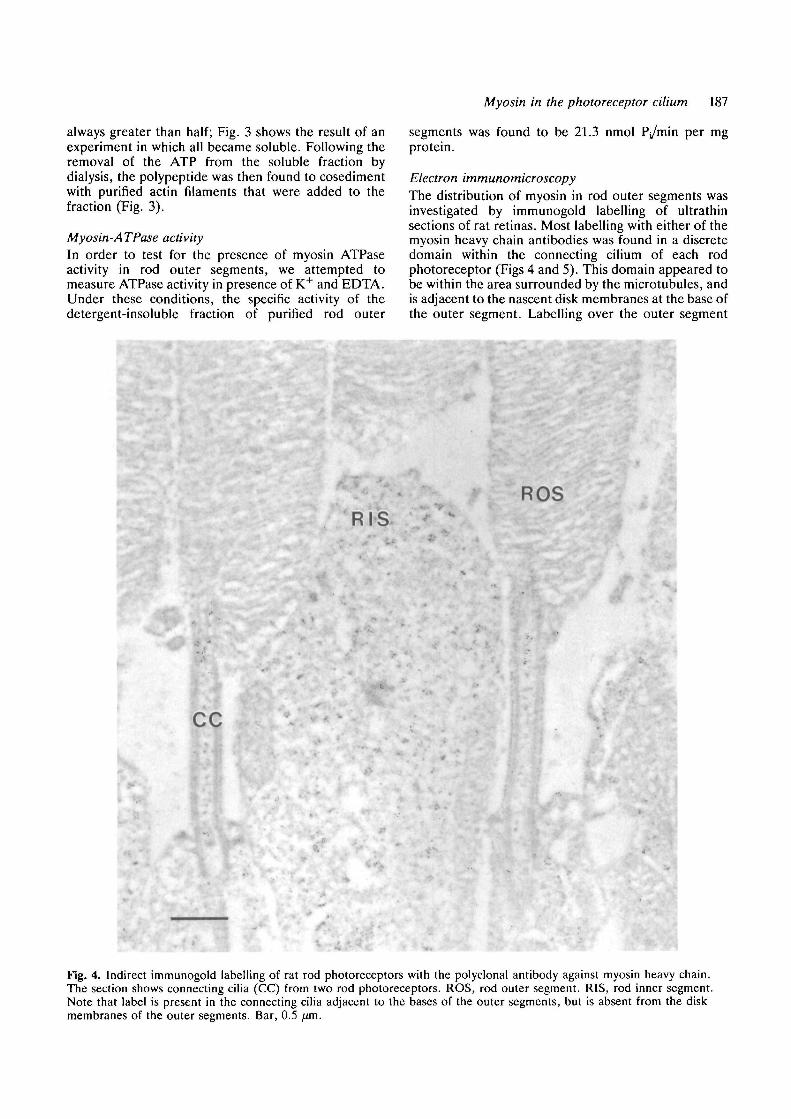
Electron immunomicroscopyThe distribution of myosin in rod outer segments wasinvestigated by immunogold labelling of ultrathinsections of rat retinas. Most labelling with either of themyosin heavy chain antibodies was found in a discretedomain within the connecting cilium of each rodphotoreceptor (Figs 4 and 5). This domain appeared tobe within the area surrounded by the microtubules, andis adjacent to the nascent disk membranes at the base ofthe outer segment. Labelling over the outer segment
RISROS
CC
Fig. 4. Indirect immunogold labelling of rat rod photoreceptors with the polyclonal antibody against myosin heavy chain.The section shows connecting cilia (CC) from two rod photoreceptors. ROS, rod outer segment. RIS, rod inner segment.Note that label is present in the connecting cilia adjacent to the bases of the outer segments, but is absent from the diskmembranes of the outer segments. Bar, 0.5 fxm.

188 D. S. Williams and others
ROS
CC
Fig. 5. Indirect immunogold labelling of the connectingcilium (CC) of two different rat rod photoreceptors withthe monoclonal antibody (left) and polyclonal antibody(right) against myosin heavy chain. ROS, rod outersegment. Both are at the same magnification; bar, 0.5 ^m
Table 1. Quantification of immunogold labelling
RegionGold particles//im2
(±s.e.m.)
DisksCC, distalCC, proximalIS
2.3±0.6110±2615±313±6
The distal connecting cilium (CC) is the area adjacent to theouter segment disks. The proximal connecting cilium is betweenthe disks and the inner segment. The inner segment includes thebasal bodies of the connecting cilium. Data were obtained from 5different cells, labelled with the polyclonal antibody againstmyosin heavy chain. The probability of there being no significantdifference between the distal connecting cilium and each of theother regions was less than 0.01 in all cases.
disk membranes was not above that of the background,but some light labelling by the myosin antibodies wasdetected in the inner segments (Table 1), where bundlesof actin filaments have been shown to be present(Drenckhahn and Groschel-Stewart, 1977; Burnside,1978; for the first reports).
Discussion
Different forms of myosin heavy chain in retinaOur Western blot analyses showed that the rat retinacontains more than one form of myosin heavy chain.One of these forms appears to be non-muscle myosinheavy chain, which was the only one detected inpurified rod outer segments. Given that the avascularchicken retina (with pecten removed) contained onlynon-muscle myosin heavy chain, the additional form(s)detected in rat retina and brain by the polyclonalantibody are most likely from the smooth muscle of thevasculature present in these tissues. Smooth muscleshave been shown to contain two forms of myosin heavychain (Rovner et al., 1986). Groschel-Stewart et al.(1989) were able to resolve three forms of myosin heavychain in porcine brain, using low percentage polyacryl-amide gels and Western blot analysis; they concludedfrom comparisons with other tissues that the fastestmigrating form was non-muscle, and the two slowermigrating forms were from smooth muscle. Morerecently, Murakami et al. (1991) reported that threeforms of non-muscle myosin heavy chain were evidentin different amounts in distinct regions of rat and bovinebrain. It is not known at present whether there is morethan one form of non-muscle myosin in the retina.
Myosin in rod outer segmentsThe presence of K+-EDTA ATPase activity (a charac-teristic of myosins) in isolated bovine rod outersegments has been reported in an abstract (Hesketh etal., 1978). However, myosin was not evident in theouter segments of retinas labelled for immunofluor-escence microscopy (Drenckhahn and Groschel-Stewart, 1977; Drenckhahn and Wagner, 1985), andresults of electron immunomicroscopic studies have sofar been inconclusive (see discussion by Chaitin andBurnside, 1989).
Here, we have detected myosin heavy chain onWestern blots of purified rod outer segments, using twodifferent antibodies. It was shown to be solubilizedfrom the rod outer segment cytoskeleton in thepresence of ATP, and to bind to purified actin filamentsin the absence of ATP, thus manifesting an importantcharacteristic of a myosin. K+-EDTA ATPase activitywas also detected in the Triton X-100-insoluble fractionof rat rod outer segments. The specific ATPase activitywas 23 times higher than that reported by Hesketh et al.(1978) from bovine rod outer segments that had notbeen fractionated; the detergent-insoluble fraction isenriched in myosin, which would account for much ofthe difference.
Results with isolated rod outer segments are clearlycompromised by any impurites. The most likely sourceof contamination comes from the inner segment, partsof which may remain attached to the connecting cilium.Electron immunomicroscopy showed, however, thatthe concentration of myosin in the inner segment isrelatively low (Table 1), so that the effect of minorcontamination from the inner segment is probablysmall. It is noteworthy that Percoll gradient isolation of
Myosin in the photoreceptor cilium 189
rod outer segments provides one of the purest prep-arations of rod outer segments (Shuster and Farber,1984; Biernbaum and Bownds 1985; authors' ownunpublished observations).
By electron immunomicroscopy we found that,within the rod outer segment, the myosin was restrictedto a small domain in the connecting cilium. Theconnecting cilium has been shown previously to containactin filaments (Chaitin et al., 1984; Vaughan andFisher, 1986; Arikawa and Williams, 1989; Chaitin andBurnside, 1989), and alpha-actinin (Arikawa andWilliams, 1989). The distribution of myosin within theconnecting cilium appears similar to that of actin in thatit is concentrated adjacent to the nascent disks.However, nearly all of the myosin labelling wasdetected within the microtubules, whereas actin fila-ments extend between the microtubules to the moreperipheral parts of the cilium (Arikawa and Williams,1989; Chaitin and Burnside, 1989).
Comparison with other axonemesThe presence of actin filaments and myosin within anaxoneme is unusual. Although actin antibodies havebeen shown to label the axonemes of various motilecilia and flagella, NBD-phallacidin (which binds onlyF-actin) evidently does not (Virtanen et al., 1984;Detmers et al., 1985; Chailley et al., 1986). Myosin hasbeen detected between the basal bodies in oviductepithelial cells, but appears to be absent from theaxonemes (Klotz et al., 1986; Sandoz et al., 1988). Itseems likely that the presence of actin filaments andmyosin in the connecting cilium of photoreceptor cellsis related to a specialized function of this cilium, such asthe regulation of protein movements through thecilium, or the formation of new phototransductive diskmembranes.
Disk membrane morphogenesisA large amount of phototransductive membrane isturned over daily (Besharse et al., 1977), so that theformation of new disk membranes at the connectingcilium represents a major undertaking by photoreceptorcells. Actin filaments have been shown to be necessaryfor normal disk membrane morphogenesis (Williams etal., 1988; Vaughan and Fisher, 1989). Colocalization ofmyosin with the actin filaments in the connecting ciliumindicates the possibility of an actin-myosin contractilemechanism functioning in disk membrane morphogen-esis. Given the organization of the actin filaments in theconnecting cilium, with their plus ends at the plasmamembrane and their minus ends in the center of theaxoneme (Arikawa and Williams, 1989; Chaitin andBurnside, 1989), myosin heads interacting with thesefilaments would move towards the ciliary plasmamembrane. The most likely consequence of thismovement would be a mechanical force on the ciliaryplasma membrane, resulting in an invagination of theplasma membrane. Perhaps bipolar myosin filamentsinteract with actin filaments that originate from op-posite sides of the cilium and thus have oppositepolarity; consistent with such an arrangement, myosin,
in comparison to actin, appears to be confined more tothe center of the cilium (cf. Figs 4 and 5 with those ofArikawa and Williams, 1989, and Chaitin and Burnside,1989).
The first stage in the morphogenesis of a photorecep-tor disk membrane appears to be a protrusion of theciliary plasma membrane, followed by a flattening ofthis protrusion into a disk (Steinberg et al., 1980). Thelatter could be effected by an invagination of the ciliaryplasma membrane at the base of the protrusion (lowerarrow in Fig. 1). Supporting this suggestion is theobservation that it is the initiation of disk membranemorphogenesis that appears to be dependent on actinfilaments; the subsequent outgrowth of a disk, onceinitiated, can occur in their absence (Williams et al.,1988).
We are grateful to S.J. Singer, G. Conrad and A. Conradfor providing the myosin antibodies, and to the Developmen-tal Studies Hybridoma Bank (supported by NICHD contractN01-HD-6-2915) for the anti-actin. Ep-475 was generouslyprovided by A. D. Blest, from part of a gift he received fromthe Taisho Pharmaceutical Company, Tokyo. A. Newtonhelped draw part of Fig. 1, and R. Nickells and C.L. Schlampmade helpful comments on the manuscript. The work wassupported by NIH grant EY 07042 to D.W., and NIH FogartyInternational fellowship TW 03929 and a Kihara MemorialYokohama Foundation grant to K.A.
References
Arikawa, K., Molday, L. L., Molday, R. S. and Williams, D. S.(1992). Localization of peripherin//-<Zs in the disk membranes ofcone and rod photoreceptors: relationship to disk membranemorphogenesis and retinal degeneration. / . Cell Biol. 116,659-667.
Arikawa, K. and Williams, D. S. (1989). Organization of actinfilaments and immunocolocalization of alpha-actinin in theconnecting cilium of rat photoreceptors. J. Comp. Neurol. 288,640-646.
Besharse, J. C , Hollyneld, J. G. and Rayborn, M. E. (1977).Turnover of rod photoreceptor outer segments. II. Membraneaddition and loss in relationship to light. J. Cell Biol. 75, 507-527.
Biernbaura, M. S. and Bownds, M. D. (1985). Frog rod outersegments with attached inner segment ellipsoids as an in vitromodel for photoreceptors of the re t ina . / Gen. Physiol.S5, 83-105.
Brann, M. R. and Cohen, L. V. (1987). Diurnal expression oftransducin mRNA and translocation of transducin in rods of ratretina. Science 235, 585-587.
Burnside, B. (1978). Thin (actin) and thick (myosinlike) filaments incone contraction in the teleost retina. J. Cell Biol. 78, 227-246.
Chailley, B., Bork, K., Gounon, P. and Sandoz, D. (1986).Immunological detection of actin in isolated ciliar from quailoviduct. Biol. Cell. 58, 43-52.
Chaitin, M. H. and Bok, D. (1986). Immunoferritin localization ofactin in retinal photoreceptors. Invest. Ophthalmol. Vis. Sci. 27,1764-1767.
Chaitin, M. H. and Burnside, B. (1989). Actin filament polarity at thesite of rod outer segment disk morphogenesis. Invest. Ophthalmol.Vis. Sci. 30, 2461-2469.
Chaitin, M. H., Schneider, B. G., Hall, M. O. and Papermaster, D. S.(1984). Actin in the photoreceptor connecting cilium:Immunocytochemical localization to the site of outer segment diskformation. J. Cell Biol. 99, 239-247.
Conrad, A. H., Clark, W. A. and Conrad, G. W. (1991). Subcellularcompartmentalization of myosin isoforms in embryonic chick heartventricle myocytes during cytokinesis. Cell Motil. Cytoskel. 19, 189-206.

190 D. S. Williams and others
Detmers, P. A., Carboni, J. M. and Condeelis, J. (1985). Localizationof actin in Chlamydomonas using antiactin and NBD-Phallacidin.Cell Motil. 5, 415-430.
Drenckbahn, D. and Groschel-Stewart, U. (1977). Localization ofmyosin and actin in ocular nonmuscle cells. Cell Tiss. Res. 181,493-503.
Drenckhahn, D. and Wagner, H. J. (1985). Relation of retinomotorresponses and contractile proteins in vertebrate retinas. Eur. J. CellBiol. 37, 156-168.
Groschel-Stewart, U., Magel, E., Elke, P. and Neidlinger, A. C.(1989). Pig brain homogenates contain smooth muscle myosin andcytoplasmic myosin isoforms. Cell Tiss. Res. 257, 137-139.
Hesketh, J., Virmaux, N. and Aunis, D. (1978). Evidence for myosinassociated with bovine photorecptor cell outer segments. Biochem.Soc. Trans. 6, 1271-1272.
Klotz, C , Bordes, N., Laine, M. C , Sandoz, D. and Bornens, M.(1986). Myosin at the apical pole of ciliated epithelial cells asrevealed by a monoclonal antibody. J. Cell Biol. 103, 613-619.
Korn, E. D., Collins, J. H. and Mamta, H. (1982). Myosins fromAcanthamoeba castellanii. Methods Enzymol. 85, 357-363.
Lin, J. J. C. (1981). Monoclonal antibodies against myofibrillarcomponents of rat skeletal muscle decorate the intermediatefilaments of cultured cells. Proc. Nat. Acad. Sci. USA 78, 2335-2339.
Lymm, R. W. and Taylor, E. W. (1971). Mechanism of adenosinetriphosphate hydrolysis by acto-myosin. Biochemistry 10, 4617-4624.
Mangini, N. J. and Pepperberg, D. R. (1988). Immunolocalization of48K in rod photoreceptors. Invest. Ophthalmol. Vis. Sci. 29, 1221-1234.
Murakami, N., Mchta, P. and Elzinga, M. (1991). Studies on thedistribution of cellular myosin with anitbodies to isoform-specificsynthetic peptides. FEBS Lett. 278, 23-25.
Papennaster, D. S., Schneider, B. G. and Besharse, J. C. (1985).Vesicular transport of newly synthesized opsin from the Golgiapparatus toward the rod outer segment. Invest. Ophthalmol. Vis.Sci. 26, 1386-1404.
Pardee, J. D. and Spudich, J. A. (1982). Purification of muscle actin.In Methods in Cell Biology, vol. 24, The Cytoskeleton, Part A (ed.L. Wilson), pp. 271-289. Academic Press, Orlando.
Pfenninger, K. H. and Maylle-Pfennlnger, M. F. (1981). Lectinlabeling of sprouting neurons. II. Relative movement andappearance of glycoconjugates during plasmalemmal expansion. J.Cell Biol. 89, 547-557.
Philip, N. J., Chang, W. and Long, K. (1987). Light-stimulatedprotein movement in rod photoreceptor cells of the rat retina.FEBS Lett. 225, 127-132.
Rohlich, P. (1975). The sensory cilium of retinal rods is analogous
to the transitional zone of motile cilia. Cell Tiss. Res. 161, 421-430.
Rovner, A. S., Murphy, R. A. and Owens, G. K. (1986). Expressionof smooth muscle and nonmuscle myosin heavy chains in culturedvascular smooth muscle cells. J. Biol. Chem. 261, 14740-14745.
Sandoz, D., Chailley, B., Botevleux-Ulrlch, E., Lemuleois, M., Laine,M. and Bautista-Harris, G. (1988). Organization and functions ofcytoskeleton in metazoan ciliated cells. Biol. Cell. 63, 183-193.
Schneider, B. G. and Kraig, E. (1990). Na+,K+-ATPase of thephotoreceptor: selective expression of a3 and t>2 isoforms. Exp. EyeRes. 51, 553-564.
Shuster, T. A. and Farber, D. B. (1984). Phosphorylation in sealedrod outer segments: effects of cyclic nucleotides. Biodiemistry 23,515-521.
Spencer, M., Detwiler, P. B. and Bunt-Milam, A. H. (1988).Distribution of membrane proteins in mechanically dissociatedretinal rods. Invest. Ophthalmol. Vis. Sci. 29, 1012-1020.
Spudich, J. A. and Watt, S. (1971). The regulation of rabbit skeletalmuscle contraction. J. Biol. Chem. 246, 4866-4871.
Steinberg, R. H., Fisher, S. K. and Anderson, D. H. (1980). Discmorphogenesis in vertebrate photoreceptors. J. Comp. Neurol.190, 501-518.
Vaughan, D. K. and Fisher, S. K. (1987). The distribution of F-actinin cells isolated from vertebrate retinas. Exp. Eye Res. 44, 393-406.
Vaughan, D. K. and Fisher, S. K. (1989). The effects of cytochalasinD in the rabbit retina. Invest. Ophthalmol. Vis. Sci. 30, 339-342.
Virtanen, I., Bradley, R. A., Paasivuo, R. and Lehto, V.-P. (1984).Distinct cytoskeletal domains revealed in sperm cells. J. Cell Biol.99, 1083-1091.
Whelan, J. P. and McGinnis, J. F. (1988). Light-dependentsubcellular movement of photoreceptor proteins. J. Neurosci. Res.20, 263-270.
Williams, D. S., Arlkawa, K. and Paallysaho, T. (1990). Cytoskeletalcomponents of the adherens junctions between the photoreceptorsand the supportive muller cells. J. Comp. Neurol. 295, 155-164.
Williams, D. S., Linberg, K. A., Vaughan, D. K., Fariss, R. N. andFisher, S. K. (1988). Disruption of microfilament organization andderegulation of disk membrane morphogenesis by cytochalasin Din rod and cone photoreceptors. J. Comp. Neurol. Til, 161-176.
Williams, D. S., Shuster, T. A., Moldrawski, M. R., Blest, A. D. andFarber, D. B. (1989). Isolation of rod outer segments on percollgradients: effect of specific protease inhibition. Exp. Eye Res. 49,439-444.
{Received 22 January 1992 - Accepted, in revised form,2 June 1992)