Embed Size (px)
Citation preview
Toxoplasmosis in Three Species of Native and Introduced Hawaiian BirdsAuthor(s): Thierry M. Work, J. Gregory Massey, David S. Lindsay, J. P. DubeySource: The Journal of Parasitology, Vol. 88, No. 5 (Oct., 2002), pp. 1040-1042Published by: The American Society of ParasitologistsStable URL: http://www.jstor.org/stable/3285558 .Accessed: 11/01/2011 21:16
Your use of the JSTOR archive indicates your acceptance of JSTOR's Terms and Conditions of Use, available at .http://www.jstor.org/page/info/about/policies/terms.jsp. JSTOR's Terms and Conditions of Use provides, in part, that unlessyou have obtained prior permission, you may not download an entire issue of a journal or multiple copies of articles, and youmay use content in the JSTOR archive only for your personal, non-commercial use.
Please contact the publisher regarding any further use of this work. Publisher contact information may be obtained at .http://www.jstor.org/action/showPublisher?publisherCode=asp. .
Each copy of any part of a JSTOR transmission must contain the same copyright notice that appears on the screen or printedpage of such transmission.
JSTOR is a not-for-profit service that helps scholars, researchers, and students discover, use, and build upon a wide range ofcontent in a trusted digital archive. We use information technology and tools to increase productivity and facilitate new formsof scholarship. For more information about JSTOR, please contact [email protected].
The American Society of Parasitologists is collaborating with JSTOR to digitize, preserve and extend access toThe Journal of Parasitology.
http://www.jstor.org
1040 THE JOURNAL OF PARASITOLOGY, VOL. 88, NO. 5, OCTOBER 2002
molecules to disease, R. C. A. Thompson, J. A. Reynoldson, and A. J. Lymbery (eds.). CAB International, Wallingford, U.K., p. 15- 37.
FAYER, R., J. M. TROUT, E. WALSH, AND R. COLE. 2000. Rotifers ingest oocysts of Cryptosporidium parvum. Journal of Eukaryotic Micro- biology 47: 161-163.
GILBERT, J. J., AND P L. STARKWEATHER. 1977. Feeding in the rotifer Brachionus calyciflorus. I. Regulatory mechanisms. Oecologia 28: 125-131.
, AND - . 1978. Feeding in the rotifer Brachionus calyciflo- rus. III. Direct observations on the effects of food type, food den- sity, and starvation on the incidence of psuedotrochal screening. Internationale Vereinigung fuer Theoretische und Angewandlte Limnologie Verhandlungen 20: 2382-2388.
KILANI, R. T, AND L. SEKLA. 1987. Purification of Cryptosporidium oocysts and sporozoites by cesium chloride and percoll gradients. American Journal of Tropical Medicine and Hygiene 36: 505-508.
KUHLE, K., AND W. KLEINOW. 1990. Glycosidases in Brachionus plica- tilis (Rotifera). Comparative Biochemistry and Physiology. B: Comparative Biochemistry 95: 393-402.
NOGRADY, T., R. L. WALLACE, AND T. W. SNELL. 1993. Rotifera. Vol. 1:
Biology, ecology, and systematics. In Guides to the identification of the microinvertebrates of the continental waters of the world, H. J. Dumont (ed.). SPB Academic Publishing, The Hague, The Neth- erlands, p. 1-142.
RESVOI, P. 1926. Observations on the feeding of rotifers. Travaux de la Societe des Naturalistes de Leningrad 56: 73-89. [In Russian.]
SCHRAMM, U. 1978. Studies of the ultrastructure of the rotifer Habro- trocha rosa Donner (Aschelminthes). The alimentary tract. Cell and Tissue Research 189: 525-535.
SEGERS, H. 2002. Contributions to the nomenclature of Rotifera: An- notated checklist of valid family- and genus-group names. Journal of Natural History 36: 631-640.
STARKWEATHER, P. L., AND J. J. GILBERT. 1977. Radiotracer determina- tion of feeding in Brachionus: The importance of gut passage times. Archiv Fur Hydrobiologie Beiheft 8: 261-263.
WALLACE, R. L., AND T W. SNELL. 2001. Rotifera. Chapter 8. In Ecology and classification of North American freshwater invertebrates, 2nd ed., J. Thorp and A. Covich (eds.). Academic Press, New York, p. 195-254.
J. Parasitol., 88(5), 2002, pp. 1040-1042 ? American Society of Parasitologists 2002
Toxoplasmosis in Three Species of Native and Introduced Hawaiian Birds
Thierry M. Work, J. Gregory Massey*, David S. Lindsayt, and J. P. Dubeyt, Hawaii Field Station, National Wildlife Health Center, U.S. Geological Survey, P.O. Box 50167, Honolulu, Hawaii 96850; *Hawaii Department of Land and Natural Resources, 2600 Piiholo Road, Makawao, Hawaii 96795; tVirginia Tech, 1410 Prices Fork Road, Blacksburg, Virginia 24061-0342; t Epidemiology and Systematics Laboratory, Parasite Biology, Animal Resources Institute, Agricultural Research Service, U.S. Department of Agriculture, Building 1001, BARC-East, Beltsville, Maryland 20507-2350. e-mail: [email protected]
ABSTRACT: Toxoplasma gondii was found in endemic Hawaiian birds, including 2 nene geese (Nesochen sandvicensis), 1 red-footed booby (Sula sula), and an introduced bird, the Erckels francolin (Francolinus erckelii). All 4 birds died of disseminated toxoplasmosis; the parasite was found in sections of many organs, and the diagnosis was confirmed by immunohistochemical staining with anti-T. gondii-specific poly- clonal antibodies. This is the first report of toxoplasmosis in these spe- cies of birds.
Toxoplasma gondii can cause mortality and subclinical infections in many species of warm-blooded animals including birds (Dubey and Beattie, 1988; Literik et al., 1992; Dubey, 2002). In tropical island ecosystems, T. gondii has been documented only on islands with feral cats, underlining the fact that felids are the only known definitive hosts (Wallace et al., 1972). In Hawaii, T. gondii has significantly affected reintroduction programs for the endangered Hawaiian crow (Work et al., 2000). This article documents acute toxoplasmosis in 3 other species of endemic and introduced Hawaiian birds, including nene goose (Ne- sochen sandvicensis), the red-footed booby (Sula sula), and Erckels francolin (Francolinus erckelii).
Birds were submitted refrigerated within 12-24 hr of death to the Hawaii Field Station or the Hawaii Department of Fish and Wildlife. Birds were weighed and examined systematically externally and inter- nally. Tissues including brain, heart, lung, skeletal muscle, kidney, spleen, adrenal, small and large intestines, trachea, and liver were fixed in 10% buffered formalin, sectioned at 5 ?pm, and stained with hema- toxylin and eosin for microscopic examination. Immunohistochemistry was done on paraffin-embedded sections using polyclonal anti-T. gondii and anti-Sarcocystis neurona antibodies and anti-BAG-1 antibodies specific for bradyzoites (Lindsay and Dubey, 1989; McAllister et al., 1996; Dubey and Hamir, 2000).
Two endangered Hawaiian goose (nene) goslings housed in a sand enclosure were found dead 3 days apart in August 1994 at a private zoo on Maui. Gross examination revealed both birds (1 male, 1 female) to be in excellent body condition with adequate fat reserves. The only significant gross lesion in the female included focal congestion of the
liver, marked splenomegaly, heavy wet lungs, and locally extensive fi- brinous exudates on the jejunal mucosa. The main gross lesions in the male included marked hemorrhage and consolidation of the left lung, marked splenomegaly, and linear brown areas of discoloration on the large intestinal mucosa.
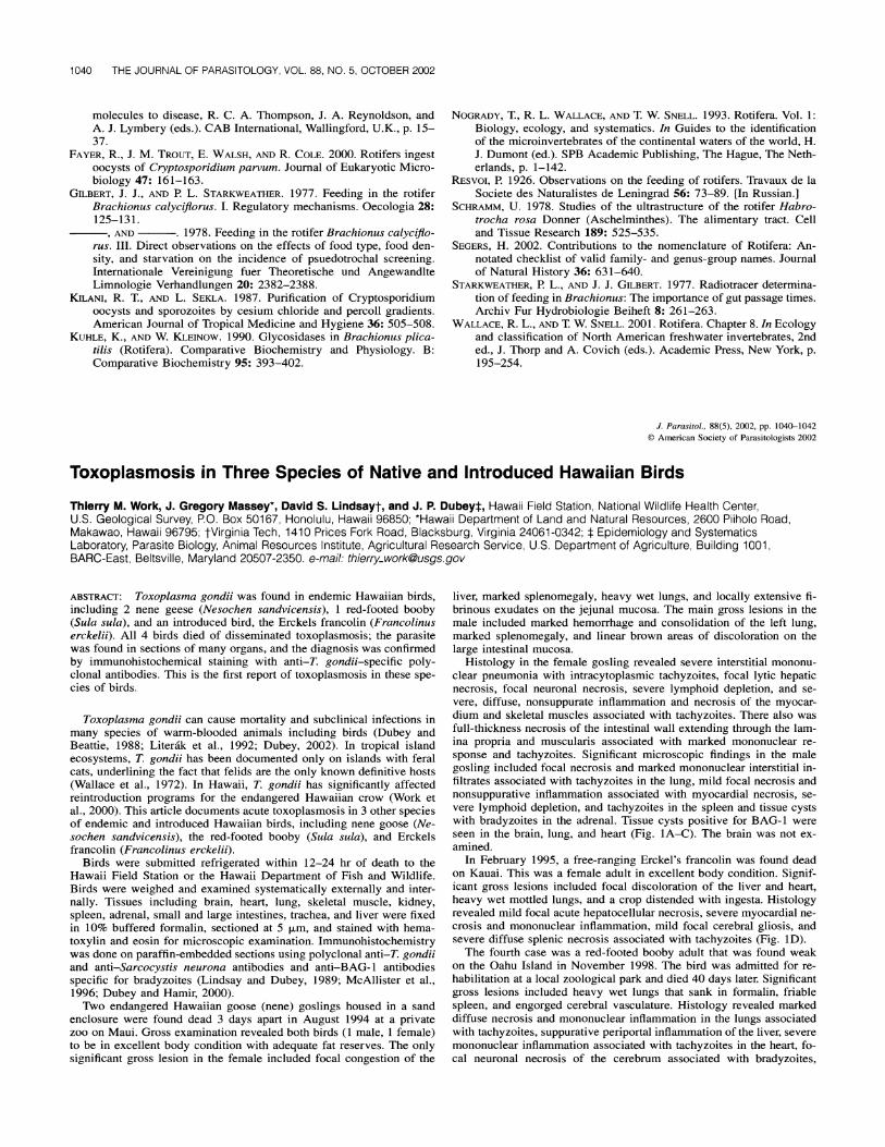
Histology in the female gosling revealed severe interstitial mononu- clear pneumonia with intracytoplasmic tachyzoites, focal lytic hepatic necrosis, focal neuronal necrosis, severe lymphoid depletion, and se- vere, diffuse, nonsuppurate inflammation and necrosis of the myocar- dium and skeletal muscles associated with tachyzoites. There also was full-thickness necrosis of the intestinal wall extending through the lam- ina propria and muscularis associated with marked mononuclear re- sponse and tachyzoites. Significant microscopic findings in the male gosling included focal necrosis and marked mononuclear interstitial in- filtrates associated with tachyzoites in the lung, mild focal necrosis and nonsuppurative inflammation associated with myocardial necrosis, se- vere lymphoid depletion, and tachyzoites in the spleen and tissue cysts with bradyzoites in the adrenal. Tissue cysts positive for BAG-1 were seen in the brain, lung, and heart (Fig. 1A-C). The brain was not ex- amined.
In February 1995, a free-ranging Erckel's francolin was found dead on Kauai. This was a female adult in excellent body condition. Signif- icant gross lesions included focal discoloration of the liver and heart, heavy wet mottled lungs, and a crop distended with ingesta. Histology revealed mild focal acute hepatocellular necrosis, severe myocardial ne- crosis and mononuclear inflammation, mild focal cerebral gliosis, and severe diffuse splenic necrosis associated with tachyzoites (Fig. 1D).
The fourth case was a red-footed booby adult that was found weak on the Oahu Island in November 1998. The bird was admitted for re- habilitation at a local zoological park and died 40 days later. Significant gross lesions included heavy wet lungs that sank in formalin, friable spleen, and engorged cerebral vasculature. Histology revealed marked diffuse necrosis and mononuclear inflammation in the lungs associated with tachyzoites, suppurative periportal inflammation of the liver, severe mononuclear inflammation associated with tachyzoites in the heart, fo- cal neuronal necrosis of the cerebrum associated with bradyzoites,

RESEARCH NOTES 1041
A' AAO m
mt
' .
"+ v...
4r*
. 0_
--. ..-0 0"
: ',% ...
low
rt.
++,+• nl- ,. m
OF it....
414
% ,
.,- 'rll Ib
?Ilk? S i .+ :JO -
~ OW
..2?; L1 ?(? ri eo
4N? 16*,, a, ~\low "Nsor.,
FIGURE 1. Toxoplasma gondii and associated lesions in birds from Hawaii. A, B, and E, hematoxylin and eosin stain; C and D, immunohis- tochemical stain with anti-T. gondii antibodies. (A-C) Lungs of nene goose. Note necrosis, infiltration by mononuclear cells, and individual tachyzoites (arrows) and groups of tachyzoites. Tachyzoites in B appear half the size of those in C. (D) Necrosis of myocardium of Erckel's francolin. Numerous tachyzoites (arrows) are in the lesion. (E) Cerebrum of red-footed booby. Note perivasculitis (arrow) and 3 tissue cysts (arrowheads).

1042 THE JOURNAL OF PARASITOLOGY, VOL. 88, NO. 5, OCTOBER 2002
prominent mononuclear cell hyperplasia in the spleen, and focal necro- sis and nonsuppurative inflammation in the adrenal (Fig. IE). Tissue cysts positive for BAG-1 were seen in intestines, lung, brain, and heart.
Protozoans in all 4 birds reacted to anti-T. gondii but not to anti-S. neurona polyclonal antibodies.
The nene acquired infection in captivity, and it is likely that their environment (sand substrate) was conducive to the spread of toxoplas- mosis by ingested feces from feral cats that were commonly observed in the vicinity. The source of infection for the francolin and the booby is less clear. The francolin may have acquired T. gondii through inges- tion of oocysts from cat feces or a transport host (Wallace, 1973). We suspect that the booby acquired infection in the wild and not in captiv- ity. Birds undergoing rehabilitation at the zoological park are caged and housed on concrete, which is cleaned daily, thus minimizing potential exposure to cat feces. On the other hand, feral cats are common around the largest red-footed booby colony on Oahu at the Kaneohe Marine Corps Air Station, and we suspect that the bird acquired the infection there.
Lesions in these birds were similar to those of other avian species infected with T. gondii (Dubey, 2002). The presence of tachyzoites in multiple organs suggested that the disease was more fulminant in the geese and francolin, and the presence of ingesta in the proventriculus of the latter suggested acute death. Presence of bradyzoites in the brain of the booby suggested a more chronic infection. In all cases, the heart and lung were the organs most commonly and severely affected.
In addition to Hawaii (Work et al., 2000), this article extends the geographic range of avian toxoplasmosis to 3 more islands (Maui, Kau- ai, and Oahu). Feral cats are present on all these islands, thereby pro- viding opportunities for infection of avian hosts. This article also re- inforces the fact that T. gondii is capable of infecting a wide variety of birds with disparate life histories, including upland game birds (Fran- colin), pelagic seabirds (Booby), and low-elevation-grazing anseriforms (nene geese). The fact that so few birds of these species have been documented with T. gondii suggests that these cases were accidental
infections. To date, we have no evidence that T. gondii poses a severe threat to populations of these birds.
LITERATURE CITED
DUBEY, J. P. 2002. A review of toxoplasmosis in wild birds. Veterinary Parasitology 106: 121-153.
, AND C. P. BEATTIE. 1988. Toxoplasmosis of animals and man. CRC Press, Boca Raton, Florida, 220 p.
, AND A. N. HAMIR. 2000. Immunohistochemical confirmation of Sarcocystis neurona infections in raccoons, mink, cat, skunk, and pony. Journal of Parasitology 86: 1150-1152.
LINDSAY, D. S., AND J. P. DUBEY. 1989. Immunohistochemical diagnosis of Neospora caninum in tissue sections. American Journal of Vet- erinary Research 50: 1981-1983.
LITERAK, I., K. HEJLICEK, J. NEZVAL, AND C. FOLK. 1992. Incidence of
Toxoplasma gondii in a population of wild birds in the Czech re- public. Avian Pathology 21: 659-665.
MCALLISTER, M. M., S. E PARMLEY, L. M. WEISS, V. J. WELCH, AND A. M. McGUIRE. 1996. An immunohistochemical method for detecting bradyzoite antigen (BAG 5) in Toxoplasma gondii-infected tissues cross-reacts with a Neospora caninum bradyzoite antigen. Journal of Parasitology 82: 354-355.
WALLACE, G. D. 1973. Intermediate and transport hosts in the natural history of Toxoplasma gondii. American Journal of Tropical Med- icine and Hygiene 22: 456-464.
.- , L. MARSHALL, AND M. MARSHALL. 1972. Cats, rats and toxo-
plasmosis on a small Pacific island. American Journal of Epide- miology 95: 475-482.
WORK, T. M., J. G. MASSEY, B. A. RIDEOUT, C. H. GARDINER, D. B.
LEDIG, O. C. H. KWOK, AND J. P. DUBEY. 2000. Fatal toxoplasmosis in free-ranging endangered 'Alala from Hawaii. Journal of Wildlife Diseases 36: 205-212.
J. Parasitol., 88(5), 2002, pp. 1042-1044 ? American Society of Parasitologists 2002
Radical Curative Efficacy of Five-Day Regimen of Primaquine for Treatment of Plasmodium vivax Malaria in India
Rajpal S. Yadav* and S. K. Ghosh, Malaria Research Centre, Field Station, Rourkela, Orissa, India. *Present address: Malaria Research Centre, Civil Hospital, Nadiad 387001, Gujarat, India. e-mail: [email protected]
ABSTRACT: For over 4 decades the antimalarial program in India has been prescribing a 5-day primaquine regimen as an antirelapse therapy to treat Plasmodium vivax malaria. In view of conflicting reports on the effectiveness of this regimen in the Indian subcontinent, and the varying prevalence of P. vivax in various ecosystems in India, the antirelapse efficacy of this regimen was evaluated in Orissa, a malaria endemic state in eastern India where P. falciparum predominates. In 723 cases of P. vivax infection treated with chloroquine alone and followed up weekly for 1 yr, the prevalence of recurrence of parasitaemia with fever was 8.6%. Among another 759 P. vivax cases treated with chloroquine and a 5-day regimen of primaquine at 15 mg/day (adult dose), the re- currence of infection was 6.5%. The difference in recurrence was not significant (P = 0.53). It is important to note that in a great majority of cases of P. vivax in this area, infection did not recur even without treatment with primaquine. This finding, that the use of the 5-day pri- maquine regimen with chloroquine had no significant advantage over the use of chloroquine alone, undermines the rationale of using pri- maquine as an antirelapse drug in forested areas with a high prevalence of P. falciparum.
Plasmodium vivax is the predominant human malarial species in most parts of India, except in the forested areas of central, eastern, and north- eastern India where P. falciparum is the main agent of infection. Plas-
modium vivax accounts for over two-thirds of the 3 million cases of malaria reported annually in India. For radical cure of P. vivax the malaria eradication program of India, started in 1958, adopted a regimen comprising 600 mg chloroquine given once and 15 mg primaquine for 5 consecutive days (adult doses). Contemporary Indian studies reported a low relapse rate in cases of P. vivax treated with chloroquine and a 5-day regimen of primaquine (Singh et al., 1954; Sharma et al., 1973). These studies, however, had the limitation of not comparing the efficacy of this regimen with a control group not treated with primaquine. An- other study in Mysore State (now Karnataka State), however, demon- strated a low relapse rate with chloroquine plus 5-day primaquine ther- apy compared with chloroquine alone (Basavaraj, 1960). In contrast, through experimental infection of nonindigenous human volunteers with a West Pakistan strain of P. vivax, Contacos et al. (1973) found that the 5-day primaquine therapy was a 100% failure. In view of variations in the prevalence of P. vivax in different ecosystems in India and conflict- ing reports on the usefulness of the 5-day primaquine therapy in the Indian subcontinent, a study was conducted to evaluate the radical cu- rative efficacy of the 5-day primaquine plus chloroquine therapy in treating P. vivax malaria in comparison with chloroquine alone.
The study was conducted in the villages of the Bisra block in a hill- forest ecosystem in Sundargarh district, Orissa State, in eastern India during 1988-1991. People live in small villages where the population