Embed Size (px)
Citation preview
DOI: 10.1590/1414-462X201600020135
Artigo de Revisão
Cad. Saúde Colet., 2016, Rio de Janeiro, 24 (2): 262-273262
Towards a personalized risk assessment for exposure of humans to toxic substances
Em direção a uma avaliação de risco personalizado para exposição de seres humanos a substâncias tóxicas
Thaís de Almeida Pedrete1, Caroline de Lima Mota1, Eline Simões Gonçalves1, Josino Costa Moreira1
1Centro de Estudos da Saúde do Trabalhador e Ecologia Humana, Escola Nacional de Saúde Pública Sérgio Arouca, Fundação Oswaldo Cruz (FIOCRUZ) - Rio de Janeiro (RJ), Brazil.Study carried out at Escola Nacional de Saúde Pública, Fundação Oswaldo Cruz (FIOCRUZ) – Rio de Janeiro (RJ), Brazil.Correspondence: Thaís de Almeida Pedrete – Rua Leopoldo Bulhões, 1480 – Manguinhos – CEP: 21041-210 – Rio de Janeiro (RJ), Brazil – Email: [email protected] support: CNPq, FAPERJ e CAPES - Bolsa de estudo – doutorado.Conflict of interests: nothing to declare.
AbstractGreat response variability caused by genetic and/or environmental factors has been observed among organisms exposed to hazardous chemicals. This subject has been a topic of intense discussion in the USA since President Obama announced support for an “era of precision medicine”, which consists in the inclusion of genetic data of patients in the treatment design, imposing a new approach to risk assessment. Personalized evaluation must consider the phenotypic factors of an individual. Among the markers that have been developed to evaluate any alteration in the structure or function of organisms, biomarkers of susceptibility are of great importance because they indicate the natural characteristics of a given organism which make it more sensitive to a specific adverse effect or disease, or more responsive to exposure to a specific chemical/drug. The ‘-omics’ technologies provide an insight into the relationship between chemical effects and molecular mechanisms of action. These technologies are the pillars for a personalized toxicology and precision medicine. Predictive toxicology requires a more comprehensive knowledge on specific individual factors or susceptibilities predisposing to diseases, enabling personalized risk assessment and adequate medical treatment.Keywords: personalized risk; susceptibility; predictive toxicology; human exposure; toxic substances.
ResumoHá uma grande variabilidade nas respostas observadas entre os organismos expostos a uma substância química perigosa. Essa variabilidade é causada por causas genéticas e / ou ambientais. Esse assunto tem sido intensamente discutido, mesmo nos Estados Unidos, desde que o presidente Obama anunciou o apoio a uma “era da medicina de precisão”, a qual consiste na inclusão de dados genéticos do paciente no projeto do tratamento, impondo uma nova abordagem para avaliação de risco. A avaliação personalizada deve considerar fatores fenotípicos de um indivíduo. Entre os biomarcadores que foram desenvolvidos para avaliar qualquer alteração da estrutura ou função do organismo, os biomarcadores de susceptibilidade têm uma grande importância, uma vez que indicam as características naturais de um dado organismo, que o tornam mais sensíveis a um efeito ou doença adversa específica ou em resposta a uma determinada exposição. As tecnologias “ômicas” permitem a compreensão da relação entre os efeitos químicos e dos mecanismos moleculares de ação. Essas tecnologias “ômicas” são os pilares para a toxicologia personalizada e para a medicina de precisão. Toxicologia preditiva exige uma melhor compreensão dos fatores ou susceptibilidades individuais específicas predisponentes a doenças, permitindo uma avaliação de riscos personalizada e um tratamento médico adequado.Palavras-chave: risco personalizado; susceptibilidade; toxicologia preditiva; exposição humana; substâncias tóxicas.
Cad. Saúde Colet., 2016, Rio de Janeiro, 24 (2): 262-273 263
Personalized risk assessment for human exposure
▄ INTRODUCTION
Life is a complex and risky process; hence, it is necessary to understand and consider its complexity and risks to improve its quality.
The word ‘risk’ can have a number of meanings and interpretations, but in this paper, it is considered as ‘the chance or probability of a person being harmed or experiencing an adverse health effect if exposed to a hazardous chemical’. To this end, a process called risk assessment (RA) was developed to decrease and/or eliminate these risks. It involves hazard identification, that is, an estimate of the probability of risk to individuals or populations associated with that hazard1.
In fact, risk can be expressed by the following equation:
Risk = Hazard x Exposure x Susceptibility (1)
According to Equation 1, absence of risk (risk = 0) requires that all components be equal to zero, which is a difficult state to be achieved2. Among the three components of this equation, the third one, susceptibility, is the most difficult to evaluate, and it is usually abandoned.
In the case of hazardous chemicals, knowledge on exposure assessment and toxicological properties of the substance is essential for a RA process. Exposure assessment requires the understanding of pathways and patterns (such as air, water, food, soil, and workstation), frequency and duration of exposure to the hazard, and evaluation of the probability of any adverse effect. It is based on cause-effect and dose-response data1. However, workers or the population in general are seldom exposed to a single substance, rather, they are usually exposed to complex mixtures which may present additive, synergistic, or antagonistic actions, increasing the complexity of risk assessment even more1,3.
Risk characterization of exposure to a xenobiotic involves hazard identification, dose-response evaluation, and exposure assessment. This procedure results in qualitative and/or quantitative description, under specific exposure conditions. It is very important to develop risk management and risk communication procedures, thereby providing a scientific basis to support decision-making in risk management. This helps determine the means of eliminating or controlling exposure to a given chemical3.
However, even under risk management policies, unexpected toxicity and adverse events can be observed, most frequently in hypersensitive individuals or in a relatively small, undetected, susceptible population group4. Assuming that the main objective of any risk assessment study is the development of methodologies to prevent or avoid any adverse effect on the entire population, these susceptible groups must be considered in order to improve the effectiveness of any risk management policy.
Once the basis of our understanding of effective risk assessment is ascertained, an important question to be answered is: How can we identify these susceptible groups?
Responding to this question is not an easy task because several of these health effects are usually complex and may present multifactorial causes.
▄ METHODS
A literature search of these issues was conducted to improve knowledge for questioning and discussion on susceptibility and personalized medicine. In addition, observation and further discussion were directed to the applications of the ‘-omics’ technologies for identification of susceptible groups and for personalized medical treatments.
A search in the PubMedTM database was conducted in the first trimester of this year to collect data. PubMedTM database is a research platform which primarily catalogs information on medical and biomedical themes. The survey was conducted using specific keywords from 1995 to 2016. The results were subsequently refined to provide information only from original scientific articles, reviews, and book chapters.
The number of publications related to personalized medicine and the ‘-omics’ technologies has increased exponentially over the past decade, and the contribution of these sciences to personalized medical treatment according to individual susceptibility is remarkable.
▄ RESULTS AND DISCUSSION
There is great variability of responses between organisms exposed to hazardous chemicals. Similar doses of the same xenobiotic can present different responses even in organisms of the same species. Even twins (mono or dizygotic) and isogenic animals may respond differently after exposure to a xenobiotic. This difference has been attributed to genetic and environmental factors5. It is apparently clear that chromosomal DNA alone cannot completely determine the susceptibility of an individual. DNA interacts with the environment and this interaction can predispose or protect the organism from a disease or other health hazards6,7.
In fact, this susceptibility results from complex interactions between several factors. In addition to genetic factors, experiments in animals reveal that 20-30% of the observed individual differences are due to environmental influences and the remaining 70-80% are attributed to a third component6,8.
It is important to state that a particular cellular environment is determined by the physiology and metabolism of the entire organism and of the cells in its immediate neighborhood. Signals from this local environment influence cellular gene expression appropriate for replication, differentiation, quiescence, or apoptotic death, depending on the cell type and developmental context7.
Cad. Saúde Colet., 2016, Rio de Janeiro, 24 (2): 262-273264
Thaís de Almeida Pedrete, Caroline de Lima Mota, Eline Simões Gonçalves, Josino Costa Moreira
In a multicellular organism, the cells are genetically homogeneous, but they can be structurally and functionally diverse owing to different gene expression. Therefore, identification of the proteins produced by these different tissues or group of cells may be an important tool to understand the complex cellular processes or even the impact of a toxicant on the organism exposed to it9.
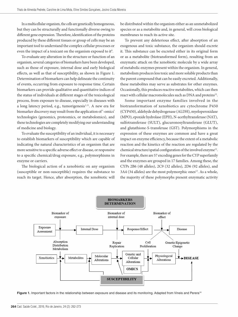
To evaluate any alteration in the structure or function of an organism, several categories of biomarkers have been developed, such as those of exposure, internal dose and early biological effects, as well as that of susceptibility, as shown in Figure 1. Determination of biomarkers can help delineate the continuity of events, occurring from exposure to response time. Certain biomarkers can provide qualitative and quantitative indices of the status of individuals at different stages of the toxicological process, from exposure to disease, especially in diseases with a long latency period, e.g., tumorigenesis1,11. A new era for biomarker discovery may result from the application of ‘-omics’ technologies (genomics, proteomics, or metabolomics), and these technologies are completely modifying our understanding of medicine and biology.
To evaluate the susceptibility of an individual, it is necessary to establish biomarkers of susceptibility which are capable of indicating the natural characteristics of an organism that are more sensitive to a specific adverse effect or disease, or responsive to a specific chemical/drug exposure, e.g., polymorphisms in enzyme or carriers.
The biological action of a xenobiotic on any organism (susceptible or non-susceptible) requires the substance to reach its target. Hence, after absorption, the xenobiotic will
be distributed within the organism either as an unmetabolized species or as a metabolite and, in general, will cross biological membranes to reach its active site.
To prevent any deleterious effect, after absorption of an exogenous and toxic substance, the organism should excrete it. This substance can be excreted either in its original form or as a metabolite (biotransformed form), resulting from an enzymatic attack on the xenobiotic molecule by a wide array of metabolic enzymes present within the organism. In general, metabolism produces less toxic and more soluble products than the parent compound that can be easily excreted. Additionally, these metabolites may serve as substrates for other enzymes. Occasionally, this produces reactive metabolites, which can then react with cellular macromolecules such as DNA and proteins12.
Some important enzyme families involved in the biotransformation of xenobiotics are cytochrome P450 (CYP450), aldehyde dehydrogenase (ALDH), myeloperoxidase (MPO), epoxide hydrolase (EPH), N-acethyltransferase (NAT), sulfotransferase (SULT), glucuronosyltransferase (GLUT), and glutathione-S-transferase (GST). Polymorphisms in the expression of these enzymes are common and have a great impact on enzyme efficiency, because the extent of a metabolic reaction and the kinetics of the reaction are regulated by the chemical structure/spatial configuration of the involved enzyme12. For example, there are 57 encoding genes for the CYP superfamily and the enzymes are grouped in 17 families. Among these, the CYPs 2B6 (48 alleles), 2C9 (32 alleles), 2D6 (92 alleles), and 3A4 (34 alleles) are the most polymorphic ones13. As a whole, the majority of these polymorphs present enzymatic activity
Figure 1. Important factors in the relationship between exposure and disease and its monitoring. Adapted from Vineis and Perera10
Cad. Saúde Colet., 2016, Rio de Janeiro, 24 (2): 262-273 265
Personalized risk assessment for human exposure
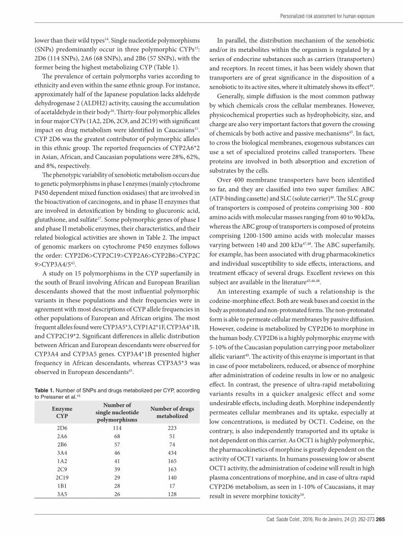
lower than their wild types14. Single nucleotide polymorphisms (SNPs) predominantly occur in three polymorphic CYPs15: 2D6 (114 SNPs), 2A6 (68 SNPs), and 2B6 (57 SNPs), with the former being the highest metabolizing CYP (Table 1).
The prevalence of certain polymorphs varies according to ethnicity and even within the same ethnic group. For instance, approximately half of the Japanese population lacks aldehyde dehydrogenase 2 (ALDH2) activity, causing the accumulation of acetaldehyde in their body16. Thirty-four polymorphic alleles in four major CYPs (1A2, 2D6, 2C9, and 2C19) with significant impact on drug metabolism were identified in Caucasians15. CYP 2D6 was the greatest contributor of polymorphic alleles in this ethnic group. The reported frequencies of CYP2A6*2 in Asian, African, and Caucasian populations were 28%, 62%, and 8%, respectively.
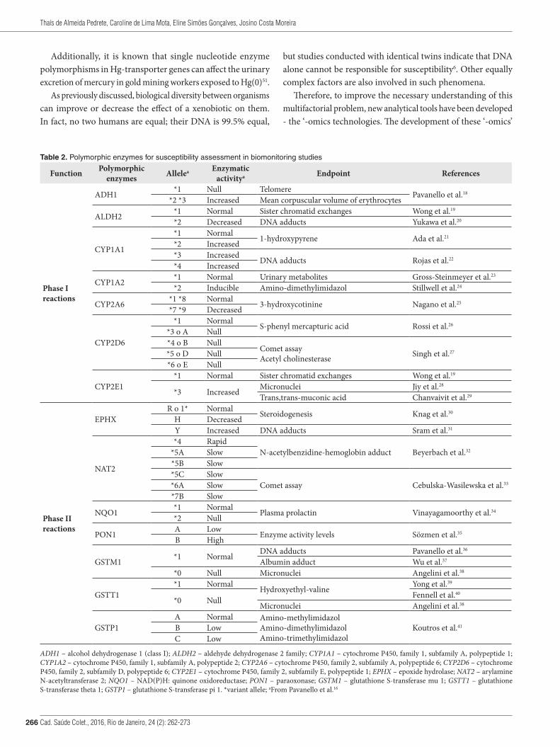
The phenotypic variability of xenobiotic metabolism occurs due to genetic polymorphisms in phase I enzymes (mainly cytochrome P450 dependent mixed function oxidases) that are involved in the bioactivation of carcinogens, and in phase II enzymes that are involved in detoxification by binding to glucuronic acid, glutathione, and sulfate17. Some polymorphic genes of phase I and phase II metabolic enzymes, their characteristics, and their related biological activities are shown in Table 2. The impact of genomic markers on cytochrome P450 enzymes follows the order: CYP2D6>CYP2C19>CYP2A6>CYP2B6>CYP2C9>CYP3A4/542.
A study on 15 polymorphisms in the CYP superfamily in the south of Brazil involving African and European Brazilian descendants showed that the most influential polymorphic variants in these populations and their frequencies were in agreement with most descriptions of CYP allele frequencies in other populations of European and African origins. The most frequent alleles found were CYP3A5*3, CYP1A2*1F, CYP3A4*1B, and CYP2C19*2. Significant differences in allelic distribution between African and European descendants were observed for CYP3A4 and CYP3A5 genes. CYP3A4*1B presented higher frequency in African descendants, whereas CYP3A5*3 was observed in European descendants43.
In parallel, the distribution mechanism of the xenobiotic and/or its metabolites within the organism is regulated by a series of endocrine substances such as carriers (transporters) and receptors. In recent times, it has been widely shown that transporters are of great significance in the disposition of a xenobiotic to its active sites, where it ultimately shows its effect44.
Generally, simple diffusion is the most common pathway by which chemicals cross the cellular membranes. However, physicochemical properties such as hydrophobicity, size, and charge are also very important factors that govern the crossing of chemicals by both active and passive mechanisms45. In fact, to cross the biological membranes, exogenous substances can use a set of specialized proteins called transporters. These proteins are involved in both absorption and excretion of substrates by the cells.
Over 400 membrane transporters have been identified so far, and they are classified into two super families: ABC (ATP-binding cassette) and SLC (solute carrier)46. The SLC group of transporters is composed of proteins comprising 300 - 800 amino acids with molecular masses ranging from 40 to 90 kDa, whereas the ABC group of transporters is composed of proteins comprising 1200-1500 amino acids with molecular masses varying between 140 and 200 kDa47,48. The ABC superfamily, for example, has been associated with drug pharmacokinetics and individual susceptibility to side effects, interactions, and treatment efficacy of several drugs. Excellent reviews on this subject are available in the literature45,46,48.
An interesting example of such a relationship is the codeine-morphine effect. Both are weak bases and coexist in the body as protonated and non-protonated forms. The non-protonated form is able to permeate cellular membranes by passive diffusion. However, codeine is metabolized by CYP2D6 to morphine in the human body. CYP2D6 is a highly polymorphic enzyme with 5-10% of the Caucasian population carrying poor metabolizer allelic variant49. The activity of this enzyme is important in that in case of poor metabolizers, reduced, or absence of morphine after administration of codeine results in low or no analgesic effect. In contrast, the presence of ultra-rapid metabolizing variants results in a quicker analgesic effect and some undesirable effects, including death. Morphine independently permeates cellular membranes and its uptake, especially at low concentrations, is mediated by OCT1. Codeine, on the contrary, is also independently transported and its uptake is not dependent on this carrier. As OCT1 is highly polymorphic, the pharmacokinetics of morphine is greatly dependent on the activity of OCT1 variants. In humans possessing low or absent OCT1 activity, the administration of codeine will result in high plasma concentrations of morphine, and in case of ultra-rapid CYP2D6 metabolism, as seen in 1-10% of Caucasians, it may result in severe morphine toxicity50.
Table 1. Number of SNPs and drugs metabolized per CYP, according to Preissner et al.15
EnzymeCYP
Number of single nucleotide polymorphisms
Number of drugs metabolized
2D6 114 2232A6 68 512B6 57 743A4 46 4341A2 41 1652C9 39 163
2C19 29 1401B1 28 173A5 26 128
Cad. Saúde Colet., 2016, Rio de Janeiro, 24 (2): 262-273266
Thaís de Almeida Pedrete, Caroline de Lima Mota, Eline Simões Gonçalves, Josino Costa Moreira
Additionally, it is known that single nucleotide enzyme polymorphisms in Hg-transporter genes can affect the urinary excretion of mercury in gold mining workers exposed to Hg(0)51.
As previously discussed, biological diversity between organisms can improve or decrease the effect of a xenobiotic on them. In fact, no two humans are equal; their DNA is 99.5% equal,
but studies conducted with identical twins indicate that DNA alone cannot be responsible for susceptibility6. Other equally complex factors are also involved in such phenomena.
Therefore, to improve the necessary understanding of this multifactorial problem, new analytical tools have been developed - the ‘-omics technologies. The development of these ‘-omics’
Table 2. Polymorphic enzymes for susceptibility assessment in biomonitoring studies
Function Polymorphic enzymes Allelea Enzymatic
activitya Endpoint References
Phase I reactions
ADH1 *1 Null Telomere Pavanello et al.18
*2 *3 Increased Mean corpuscular volume of erythrocytes
ALDH2 *1 Normal Sister chromatid exchanges Wong et al.19
*2 Decreased DNA adducts Yukawa et al.20
CYP1A1
*1 Normal 1-hydroxypyrene Ada et al.21
*2 Increased*3 Increased DNA adducts Rojas et al.22
*4 Increased
CYP1A2 *1 Normal Urinary metabolites Gross-Steinmeyer et al.23
*2 Inducible Amino-dimethylimidazol Stillwell et al.24
CYP2A6 *1 *8 Normal 3-hydroxycotinine Nagano et al.25
*7 *9 Decreased
CYP2D6
*1 Normal S-phenyl mercapturic acid Rossi et al.26
*3 o A Null*4 o B Null
Comet assayAcetyl cholinesterase Singh et al.27*5 o D Null
*6 o E Null
CYP2E1*1 Normal Sister chromatid exchanges Wong et al.19
*3 Increased Micronuclei Jiy et al.28
Trans,trans-muconic acid Chanvaivit et al.29
Phase II reactions
EPHXR o 1* Normal Steroidogenesis Knag et al.30
H DecreasedY Increased DNA adducts Sram et al.31
NAT2
*4 RapidN-acetylbenzidine-hemoglobin adduct Beyerbach et al.32*5A Slow
*5B Slow*5C Slow
Comet assay Cebulska-Wasilewska et al.33*6A Slow*7B Slow
NQO1 *1 Normal Plasma prolactin Vinayagamoorthy et al.34
*2 Null
PON1 A Low Enzyme activity levels Sözmen et al.35
B High
GSTM1 *1 Normal DNA adducts Pavanello et al.36
Albumin adduct Wu et al.37
*0 Null Micronuclei Angelini et al.38
GSTT1*1 Normal Hydroxyethyl-valine Yong et al.39
*0 Null Fennell et al.40
Micronuclei Angelini et al.38
GSTP1A Normal Amino-methylimidazol
Amino-dimethylimidazolAmino-trimethylimidazol
Koutros et al.41B LowC Low
ADH1 – alcohol dehydrogenase 1 (class I); ALDH2 – aldehyde dehydrogenase 2 family; CYP1A1 – cytochrome P450, family 1, subfamily A, polypeptide 1; CYP1A2 – cytochrome P450, family 1, subfamily A, polypeptide 2; CYP2A6 – cytochrome P450, family 2, subfamily A, polypeptide 6; CYP2D6 – cytochrome P450, family 2, subfamily D, polypeptide 6; CYP2E1 – cytochrome P450, family 2, subfamily E, polypeptide 1; EPHX – epoxide hydrolase; NAT2 – arylamine N-acetyltransferase 2; NQO1 – NAD(P)H: quinone oxidoreductase; PON1 – paraoxonase; GSTM1 – glutathione S-transferase mu 1; GSTT1 – glutathione S-transferase theta 1; GSTP1 – glutathione S-transferase pi 1. *variant allele; aFrom Pavanello et al.16
Cad. Saúde Colet., 2016, Rio de Janeiro, 24 (2): 262-273 267
Personalized risk assessment for human exposure
technologies has contributed greatly to our understanding of living organisms at a molecular level. These technologies are able to simultaneously deal with a great set of data in a short period of time, facilitating the knowledge of cellular molecular mechanisms, thus making it possible to study not only the genotypic behavior, but also and specially the phenotypic one52,53.
Thus, these technologies facilitate in-depth approaches in understanding the relationship between chemical structure-activity effects and molecular mechanisms of action. They also help in the comprehension of the differences between proteins and other cellular substances, between similar or different organisms, and the adverse effects of xenobiotics on exposed organisms54,55. Thus, these methods are generating very important sets of data which improve our understanding of cellular responses, tissue damage, and functional perturbations caused by exposure to a xenobiotic54. Moreover, these data are being used for constitution of databases containing a great variety of genetic sequence monitoring; epigenomic, proteomic, and metabolomic data; and clinical and toxicological information, thereby allowing the study of their interrelations.
It is well known that molecular profiles can be different when cells or tissues are exposed to the same toxicant, and this allows for the monitoring of biochemical homeostasis and toxic effects, simultaneously.
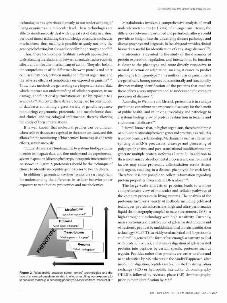
‘Omics’ datasets are fundamental to systems biology studies in order to integrate data, and thus understand the experimental system in question (disease, phenotype, therapeutic intervention)56. As shown in Figure 2, proteomics should be the technique of choice to identify susceptible groups prior to health effects.
In addition to genomics, two other ‘-omics’ are very important for understanding the differences in cellular behavior under exposure to xenobiotics: proteomics and metabolomics.
Metabolomics involves a comprehensive analysis of small molecule metabolites (< 1 kDa) of an organism. Hence, the differences between unperturbed and perturbed pathways could provide an insight into the underlying disease pathology and disease prognosis and diagnosis. In fact, this tool provides clinical biomarkers useful for identification of early-stage diseases57,58.
Proteomics is devoted to the study of the dynamics of protein expression, regulation, and interactions. Its function is closer to the phenotype and more directly responsive to natural selection or adaptation, making it easier to predict phenotype from genotype59. In a multicellular organism, cells are genetically homogeneous, but structurally and functionally diverse; making identification of the proteins that mediate these effects a very important tool to understand the complex processes of diseases1,9.
According to Wetmore and Merrick, proteomics is in a unique position to contribute to new protein discovery for the benefit of public health, and in linking toxicology and pathology to a systems biology view of protein dysfunction in toxicity and environmental disease”60.
It is well known that, in higher organisms, there is no simple one-to-one relationship between genes and proteins; as a rule, this is a one-to-many relationship. Mechanisms such as alternative splicing of mRNA precursors, cleavage and processing of polypeptide chains, and post-translational modifications may generate multiple protein isoforms (Figure 3). In addition to these mechanisms, developmental processes and environmental factors may cause proteomic differentiation across tissues and organs, resulting in a distinct phenotype for each level. Therefore, it is not possible to collect information regarding protein properties from a static DNA alone59,61.
The large-scale analysis of proteins leads to a more comprehensive view of molecular and cellular pathways of the complex processes in living systems. The analysis of the proteome involves a variety of methods including gel-based techniques, protein microarrays, high and ultra-performance liquid chromatography coupled to mass spectrometry (MS) - a high-throughput technology with high sensitivity. Currently, mass spectrometric identification of gel-separated proteins and of fractioned peptides by multidimensional protein identification technology (MudPIT) is a widely used analytical tool for proteomic studies62. In general, the former has enough sensitivity to deal with protein mixtures, and it uses a digestion of gel-separated proteins into peptides by certain specific proteases such as trypsin. Peptides rather than proteins are easier to elute and to be identified by MS. whereas in the MudPIT approach, after in-solution digestion, peptides are fractionated by strong cation exchange (SCX) or hydrophilic interaction chromatography (HILIC), followed by reversed phase (RP) chromatography prior to their identification by MS62.
Figure 2. Relationship between some ‘-omics’ technologies and the type of answered questions related to effects resulting from exposure to xenobiotics that help in decoding phenotype. Modified from Pesce et al.53

Cad. Saúde Colet., 2016, Rio de Janeiro, 24 (2): 262-273268
Thaís de Almeida Pedrete, Caroline de Lima Mota, Eline Simões Gonçalves, Josino Costa Moreira
Biomarkers of susceptibility
A biomarker of susceptibility is an indicator of an inherent or acquired limitation of an organism’s ability to respond to the challenge of exposure to a specific xenobiotic. These markers indicate individual or population differences which affect their response to that chemical substance. Such markers may include inborn differences in metabolism, variations in immunoglobulin levels, low organ-reserve capacity, or other identifiable genetically determined or environmentally induced variations in absorption, metabolism, and response to environmental agents63. Other factors that may affect individual susceptibilities include nutritional status of the organism, the role of the target site in overall body function, condition of the target tissue (present or prior to disease), and compensation by homeostatic mechanisms during and after exposure64. This means that variation in the response to xenobiotics by individuals is closely related to specific genotypes and phenotypes, which is the basis of individual clinical toxicology65.
Recently, several authors have been discussing the application of systems biology, or rather, systems toxicology to address risk assessment issues. Sauer et al.66 report a need for more predictive and accurate approaches to risk assessment, requiring a mechanistic understanding of the process by which a xenobiotic perturbs biological systems. Wetmore and Merrick60 defend the toxicoproteomics approach, which is positioned towards an
expanded understanding of protein expression during toxicity and environmental disease, for the advancement of public health. Titz et al.67 highlight the fact that protein alterations could be a close reflection of biological effects, and Sturla et al.68 discuss the identification of how biological networks are perturbed by exposure to xenobiotics and enable the development of predictive mathematical models of toxicological processes. Sauer et al.69 recommend that the use of systems toxicology with advanced analytical and computational tools should be integrated with classical toxicology. They also suggest that quantitative analysis of large networks of molecular and functional changes occurring across multiple levels of biological organization should be conducted.
Clearly, the use of systems biology/toxicology will produce a great amount of data, facilitating a more complete understanding of the overall biological processes. However, the use of these data for risk assessment in a significant number of people will have a very high cost. Therefore, the identification of susceptibility biomarkers would provide the development of inexpensive and useful tools that could be applied worldwide.
The use of such susceptibility biomarkers will allow the identification of individual responses to chemical exposure. In fact, two types of susceptibility biomarkers are possible: those used for prognosis and those used for prediction purposes. Prognostic biomarkers are biological measurements capable of
Figure 3. Phenotype formation according to the expression of mRNA and its corresponding protein, regulated by diverse mechanisms. Adapted from Diz et al.59
Cad. Saúde Colet., 2016, Rio de Janeiro, 24 (2): 262-273 269
Personalized risk assessment for human exposure
indicating an individual’s susceptibility to develop an adverse effect during a clinical treatment. Predictive biomarkers are used to indicate the possibility of an adverse effect on health when a xenobiotic is administered to an individual in a particular condition, indicating the need to avoid exposure to certain xenobiotics11. In both cases, susceptibility biomarkers are indicators of individual differences in the development of adverse effects in response to a specific chemical exposure, and are used to differentiate patients according to their degree of susceptibility70.
The recognition of such susceptible groups is very important in risk assessment studies for therapeutic or toxicological purposes.
Personalized clinical toxicology
The human health status is always a great concern worldwide. Noticeably, ‘-omics’ will contribute to improving the necessary knowledge in healthcare. The interaction of these techniques and medicine will certainly produce new diagnostic tools to determine individual risk factors and personalized medical treatments. It is clear that ‘-omics’ are the pillars of personalized clinical toxicology, which is based on knowledge of individual characteristics, and requires an understanding of the factors that influence the organic variability71. Technological development has led us to an era of individualized therapy72.
Nowadays, this subject has been a topic of intense discussion in the USA since President Obama announced support for an “era of precision medicine”, where prevention and treatment
strategies will consider individual variability, with a great potential for improving health73-75.
Predictive medicine will develop due to a better comprehension of the specific factors that cause predisposition to diseases. According to Hocquette63, it will be possible to predict the probability of developing specific diseases through scientific results and applied biotechnologies that arise from proteomics, metabolomics, transcriptomics, epigenomics, and genomics. This requires associations of gene expression patterns with diagnoses, treatments, and clinical data. Thus, preventive medicine and medical therapy will be personalized.
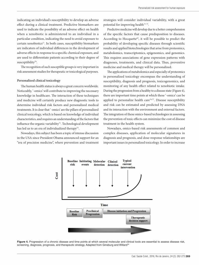
The applications of metabolomics and especially of proteomics in personalized toxicology encompass the understanding of susceptibility, diagnosis and prognosis, toxicogenomics, and monitoring of any health effect related to xenobiotic intake. During the progression from a healthy to a disease state (Figure 4), there are important time points at which these ‘-omics’ can be applied to personalize health care76,77. Disease susceptibility and risk can be estimated and predicted by assessing DNA and its interaction with the environment and external factors. The integration of these omics-based technologies in assessing the prevention of toxic effects can minimize the cost of disease treatment in the health system.
Nowadays, omics-based risk assessments of common and complex diseases, application of molecular signatures in diagnosis and prognosis, and dose-response relationships are important issues in personalized toxicology. In order to increase
Figure 4. Progression of a chronic disease and time points at which several molecular and clinical tools are essential to assess disease risk, screening, diagnosis, prognosis, and therapeutic strategy. Adapted from Ginsburg and Willard76
Cad. Saúde Colet., 2016, Rio de Janeiro, 24 (2): 262-273270
Thaís de Almeida Pedrete, Caroline de Lima Mota, Eline Simões Gonçalves, Josino Costa Moreira
their effectiveness, these techniques must be standardized and integrated to health systems77,78.
A molecular signature is a set of biomolecular features such as a DNA sequence, mRNA, and protein expression. In conjunction with a computational procedure, it can help to predict a phenotype of clinical interest52, based on single or multiple data types, for instance, the prediction of any health risk or organic response to toxic xenobiotic exposure and its physiological actions. The identification of molecular signatures from ‘-omics’ data for diverse clinical applications consists of four stages: 1) definition of the scientific and clinical context for the molecular signature; 2) data acquisition; 3) feature selection and modeling; and 4) evaluation of the molecular signature from independent datasets52,76.
The identification of genetic variants associated with individual response may help predict the occurrence of health events in susceptible individuals. In toxicokinetics, polymorphisms in encoding genes for both drug metabolizing enzymes and transporters affect drug availability at the target site, whereas in toxicodynamics, target proteins such as receptors, enzymes, and intracellular signaling proteins affect sensitivity to chemicals79. Individual attributes such as resistance, regimens, and dosing interfere in the results of toxicological trials. Genetic, phenotypic, and environmental factors should be considered in order to provide biomarkers with analytical validity and clinical utility to optimize the effectiveness of specific treatment65,78.
▄ CONCLUSIONS
Human susceptibility is one of the most problematic components of risk assessment owing to its complexity and difficulties in determination. Additionally, it is not externally
controllable as exposure. For these reasons, in general, it is not scientifically considered in risk assessment calculations. However, it is as important as the other factors.
The expectancy brought by the ‘-omics’ technologies in relation to personalized toxicology and individual health conditions is still far away from its myriad of possibilities. Indubitably, the development of genomics and the new derived ‘-omics technologies represent a major advancement to the better understanding of human organisms and their similarities and individual differences, allowing for the study of individual susceptibilities.
As proteomic is the ‘-omic’ technology closer to phenotype, proteomic biomarkers will be of great value for risk assessment studies because they involve susceptible individuals or population groups. The analytical methodologies available for separation and quantification of proteins, such as HPLC/MS/MS, electrophoresis, and protein microarray still present some limitations, and require improvements to increase the number of molecular signatures for clinical applications and personalized prediction of disease outcomes.
The applications of these new analytical tools to identify biomarkers of susceptibility or individual susceptibility will help generate data to be used in any risk assessment procedure. Such a scenario will certainly contribute to improving public health.
▄ ACKNOWLEDGEMENTS
The authors are grateful to the National Council for Scientific and Technological Development (CNPq), Research Support Foundation of Rio de Janeiro State (FAPERJ), and Coordination for the Improvement of Higher Education Personnel (CAPES) for their financial support.
▄ REFERENCES
1. Park SK, Choi J-Y. Risk assessment and pharmacogenetics in molecular and genomic epidemiology. J Prev Med Pub Health. 2009;42(6):371-6. http://dx.doi.org/10.3961/jpmph.2009.42.6.371. PMid:20009483.
2. Manno M, Viau C, Cocker J, Colosio C, Lowry L, Mutti A, et al. Biomonitoring for occupational health risk assessment (BOHRA). Toxicol Lett. 2010;192(1):3-16. http://dx.doi.org/10.1016/j.toxlet.2009.05.001. PMid:19446015.
3. DeBord DG, Burgoon L, Edwards SW, Haber LT, Kanitz MH, Kuempel E, et al. Systems biology and biomarkers of early effects for occupational exposure limit setting. J Occup Environ Hyg. 2015;12(Suppl):41-54.
4. Lin WJ, Chen JJ. Biomarker classifiers for identifying susceptible subpopulations for treatment decisions. Pharmacogenomics. 2012;13(2):147-57. http://dx.doi.org/10.2217/pgs.11.139. PMid:22188363.
5. Bell JT, Spector TD. A twin approach to unraveling epigenetics. Trends Genet. 2011;27(3):116-25. http://dx.doi.org/10.1016/j.tig.2010.12.005. PMid:21257220.
6. Wong AHC, Gottesman II, Petronis A. Phenotypic differences in genetically identical organisms: The epigenetic perspective. Hum Mol Genet. 2005;14(1):11-8.
7. Turner BM. Epigenetic responses to environmental change and their evolutionary implications. Philos Trans R Soc B Biol Sci. 2009;364(1534):3403-18.
8. Gärtner K. A third component causing random variability beside environment and genotype: a reason for the limited success of a 30 year long effort to standardize laboratory animals? Int J Epidemiol. 2012;41(2):335-41. http://dx.doi.org/10.1093/ije/dyr219. PMid:22266059.
9. Jaenisch R, Bird A. Epigenetic regulation of gene expression: how the genome integrates intrinsic and environmental signals. Nat Genet. 2003;33(3 Suppl):245-54. http://dx.doi.org/10.1038/ng1089. PMid:12610534.
10. Vineis P, Perera F. Molecular epidemiology and biomarkers in etiologic cancer research: the new in light of the old. Cancer Epidemiol Biomarkers Prev. 2007;16(10):1954-65. http://dx.doi.org/10.1158/1055-9965.EPI-07-0457. PMid:17932342.
Cad. Saúde Colet., 2016, Rio de Janeiro, 24 (2): 262-273 271
Personalized risk assessment for human exposure
11. Chen JJ, Lu T, Chen Y, Lin WJ. Predictive biomarkers for treatment selection: statistical considerations. Biomarkers Med. 2015;9(11):1121-35. http://dx.doi.org/10.2217/bmm.15.84. PMid:26507127.
12. Bozina N, Bradamante V, Lovrić M. Genetic polymorphism of metabolic enzymes P450 (CYP) as a susceptibility factor for drug response, toxicity, and cancer risk. Arh Hig Rada Toksikol. 2009;60(2):217-42. http://dx.doi.org/10.2478/10004-1254-60-2009-1885. PMid:19581216.
13. Rodriguez-Antona C, Ingelman-Sundberg M. Cytochrome P450 pharmacogenetics and cancer. Oncogene. 2006;25(11):1679-91. http://dx.doi.org/10.1038/sj.onc.1209377. PMid:16550168.
14. Thier R, Brüning T, Roos PH, Rihs H-P, Golka K, Ko Y, et al. Markers of genetic susceptibility in human environmental hygiene and toxicology: the role of selected CYP, NAT and GST genes. Int J Hyg Environ Health. 2003;206(3):149-71. http://dx.doi.org/10.1078/1438-4639-00209. PMid:12872524.
15. Preissner SC, Hoffmann MF, Preissner R, Dunkel M, Gewiess A, Preissner S. Polymorphic cytochrome P450 enzymes (CYPs) and their role in personalized therapy. PLoS One. 2013;8(12):1-12. http://dx.doi.org/10.1371/journal.pone.0082562. PMid:24340040.
16. Pavanello S, Clonfero E. Biological indicators of genotoxic risk and metabolic polymorphisms. Mutat Res Rev Mutat Res. 2000;463(3):285-308. http://dx.doi.org/10.1016/S1383-5742(00)00051-X. PMid:11018745.
17. Belitsky GA, Yakubovskaya MG. Genetic polymorphism and variability of chemical carcinogenesis. Biochem Biokhimii͡a. 2008;73(5):543-54. http://dx.doi.org/10.1134/S0006297908050076.
18. Pavanello S, Hoxha M, Dioni L, Bertazzi PA, Snenghi R, Nalesso A, et al. Shortened telomeres in individuals with abuse in alcohol consumption. Int J Cancer. 2011;129(4):983-92. http://dx.doi.org/10.1002/ijc.25999. PMid:21351086.
19. Wong R-H, Wang J-D, Hsieh L-L, Cheng T-J. XRCC1, CYP2E1 and ALDH2 genetic polymorphisms and sister chromatid exchange frequency alterations amongst vinyl chloride monomer-exposed polyvinyl chloride workers. Arch Toxicol. 2003;77(8):433-40. http://dx.doi.org/10.1007/s00204-003-0467-6. PMid:12739102.
20. Yukawa Y, Muto M, Hori K, Nagayoshi H, Yokoyama A, Chiba T, et al. Combination of ADH1B*2/ALDH2*2 polymorphisms alters acetaldehyde-derived DNA damage in the blood of Japanese alcoholics. Cancer Sci. 2012;103(9):1651-5. http://dx.doi.org/10.1111/j.1349-7006.2012.02360.x. PMid:22703580.
21. Ada AO, Yilmazer M, Suzen S, Demiroglu C, Demirbag AE, Efe S, et al. Cytochrome P450 (CYP) and glutathione S-transferases (GST) urinary levels of 1-hydroxypyrene in Turkish coke oven workers. Evaluation. 2007;519:511-9.
22. Rojas M, Cascorbi I, Alexandrov K, Kriek E, Auburtin G, Mayer L, et al. Modulation of benzo[a]pyrene diolepoxide-DNA adduct levels in human white blood cells by CYP1A1, GSTM1 and GSTT1 polymorphism. Carcinog. 2000;21(1):35-41. http://dx.doi.org/10.1093/carcin/21.1.35. PMid:10607731.
23. Gross-Steinmeyer K, Eaton DL. Dietary modulation of the biotransformation and genotoxicity of aflatoxin B1. Toxicology. 2012;299(2–3):69-79. http://dx.doi.org/10.1016/j.tox.2012.05.016. PMid:22640941.
24. Stillwell WG, Kidd LC, Wishnok JS, Tannenbaum SR, Sinha R. Urinary excretion of unmetabolized and phase II conjugates of 2-amino-1-methyl-6-phenylimidazo[4,5-b]pyridine and 2-amino-3,8-dimethylimidazo[4,5-f]quinoxaline in humans: relationship to cytochrome P4501A2 and N-acetyltransferase activity. Cancer Res. 1997;57(0008-5472):3457-64.
25. Nagano T, Yamazaki H, Shimizu M, Kiyotani K, Kamataki T, Takano R, et al. Biomonitoring of urinary cotinine concentrations associated with plasma levels of nicotine metabolites after daily cigarette smoking in a male Japanese population. Int J Environ Res Public Health. 2010;7(7):2953-64. http://dx.doi.org/10.3390/ijerph7072953. PMid:20717551.
26. Rossi AM, Guarnieri C, Rovesti S, Gobba F, Ghittori S, Vivoli G, et al. Genetic polymorphisms influence variability in benzene metabolism in humans. Pharmacogenetics. 1999;9(4):445-51. PMid:10780264.
27. Singh S, Kumar V, Vashisht K, Singh P, Banerjee BD, Rautela RS, et al. Role of genetic polymorphisms of CYP1A1, CYP3A5, CYP2C9, CYP2D6, and PON1 in the modulation of DNA damage in workers occupationally exposed to organophosphate pesticides. Toxicol Appl Pharmacol. 2011;257(1):84-92. http://dx.doi.org/10.1016/j.taap.2011.08.021. PMid:21907728.
28. Ji F, Wangy W, Xia ZL, Zheng YJ, Qiu YL, Wu F, et al. Prevalence and persistence of chromosomal damage and susceptible genotypes of metabolic and DNA repair genes in Chinese vinyl chloride-exposed workers. Carcinogenesis. 2010;31(4):648-53. http://dx.doi.org/10.1093/carcin/bgq015. PMid:20100738.
29. Chanvaivit S, Navasumrit P, Hunsonti P, Autrup H, Ruchirawat M. Exposure assessment of benzene in Thai workers, DNA-repair capacity and influence of genetic polymorphisms. Mutat Res Genet Toxicol Environ Mutagen. 2007;626(1-2):79-87. http://dx.doi.org/10.1016/j.mrgentox.2006.09.007. PMid:17095285.
30. Knag AC, Verhaegen S, Ropstad E, Mayer I, Meier S. Effects of polar oil related hydrocarbons on steroidogenesis in vitro in H295R cells. Chemosphere. 2013;92(1):106-15. http://dx.doi.org/10.1016/j.chemosphere.2013.02.046. PMid:23561572.
31. Sram RJ, Beskid O, Binkova B, Rossner P, Smerhovsky Z. Cytogenetic analysis using fluorescence in situ hybridization (FISH) to evaluate the impact of environmental exposure to PAHs. Cancer Res. 2004;64:455-6.
32. Beyerbach A, Rothman N, Bhatnagar VK, Kashyap R, Sabbioni G. Hemoglobin adducts in workers exposed to benzidine and azo dyes. Carcinogenesis. 2006;27(8):1600-6. http://dx.doi.org/10.1093/carcin/bgi362. PMid:16497705.
33. Cebulska-Wasilewska A, Binkova B, Sram RJ, Kalina I, Popov T, Farmer PB. Repair competence assay in studies of the influence of environmental exposure to c-PAHs on individual susceptibility to induction of DNA damage. Mutat Res. 2007;620(1-2):155-64. http://dx.doi.org/10.1016/j.mrfmmm.2007.03.005. PMid:17482217.
34. Vinayagamoorthy N, Krishnamurthi K, Devi SS, Naoghare PK, Biswas R, Biswas AR, et al. Genetic polymorphism of CYP2D6*2 C → T 2850, GSTM1, NQO1 genes and their correlation with biomarkers in manganese miners of Central India. Chemosphere. 2010;81(10):1286-91. http://dx.doi.org/10.1016/j.chemosphere.2010.08.047. PMid:20851451.
35. Sözmen EY, Mackness B, Sözmen B, Durrington P, Girgin FK, Aslan L, et al. Effect of organophosphate intoxication on human serum paraoxonase. Hum Exp Toxicol. 2002;21(5):247-52. http://dx.doi.org/10.1191/0960327102ht244oa. PMid:12141395.
36. Pavanello S, Pulliero A, Clonfero E. Influence of GSTM1 null and low repair XPC PAT+ on anti-B[a]PDE-DNA adduct in mononuclear white blood cells of subjects low exposed to PAHs through smoking and diet. Mutat Res. 2008;638(1-2):195-204. http://dx.doi.org/10.1016/j.mrfmmm.2007.10.004. PMid:18035379.
37. Wu H-C, Wang Q, Yang H-I, Ahsan H, Tsai W-Y, Wang L-Y, et al. Aflatoxin B(1) exposure, hepatitis B virus infection and hepatocellular carcinoma in Taiwan. Cancer Epidemiol Biomarkers Prev. 2009;18(3):846-53. http://dx.doi.org/10.1158/1055-9965.EPI-08-0697. PMid:19273485.
Cad. Saúde Colet., 2016, Rio de Janeiro, 24 (2): 262-273272
Thaís de Almeida Pedrete, Caroline de Lima Mota, Eline Simões Gonçalves, Josino Costa Moreira
38. Angelini S, Kumar R, Carbone F, Bermejo JL, Maffei F, Cantelli-Forti G, et al. Inherited susceptibility to bleomycin-induced micronuclei: correlating polymorphisms in GSTT1, GSTM1 and DNA repair genes with mutagen sensitivity. Mutat Res. 2008;638(1-2):90-7. http://dx.doi.org/10.1016/j.mrfmmm.2007.09.001. PMid:17953974.
39. Yong LC, Schulte PA, Wiencke JK, Boeniger MF, Connally LB, Walker JT, et al. Hemoglobin adducts and sister chromatid exchanges in hospital workers exposed to ethylene oxide: effects of glutathione S-transferase T1 and M1 genotypes. Cancer Epidemiol Biomarkers Prev. 2001;10(5):539-50. PMid:11352866.
40. Fennell TR, MacNeela JP, Morris RW, Watson M, Thompson CL, Bell DA. Hemoglobin adducts from acrylonitrile and ethylene oxide in cigarette smokers: effects of glutathione s-transferase T1-null and M1-null genotypes. Cancer Epidemiol Biomarkers Prev. 2000;9(7):705-12. PMid:10919741.
41. Koutros S, Berndt SI, Sinha R, Ma X, Chatterjee N, Alavanja MCR, et al. Xenobiotic metabolizing gene variants, dietary heterocyclic amine intake, and risk of prostate cancer. Cancer Res. 2009;69(5):1877-84. http://dx.doi.org/10.1158/0008-5472.CAN-08-2447. PMid:19223546.
42. Zanger UM, Schwab M. Cytochrome P450 enzymes in drug metabolism: regulation of gene expression, enzyme activities, and impact of genetic variation. Pharmacol Ther. 2013;138(1):103-41. http://dx.doi.org/10.1016/j.pharmthera.2012.12.007. PMid:23333322.
43. Kohlrausch FB, Carracedo Á, Hutz MH. Characterization of CYP1A2, CYP2C19, CYP3A4 and CYP3A5 polymorphisms in South Brazilians. Mol Biol Rep. 2014;41(3):1453-60. http://dx.doi.org/10.1007/s11033-013-2990-8. PMid:24443221.
44. Kusuhara H, Sugiyama Y. Role of transporters in the tissue-selective distribution and elimination of drugs: transporters in the liver, small intestine, brain and kidney. J Control Release. 2002;78(1-3):43-54. http://dx.doi.org/10.1016/S0168-3659(01)00480-1. PMid:11772448.
45. Klaassen CD, Aleksunes LM. Xenobiotic, bile acid, and cholesterol transporters: function and regulation. Pharmacol Rev. 2010;62(1):1-96. http://dx.doi.org/10.1124/pr.109.002014. PMid:20103563.
46. Giacomini KM, Huang S-M, Tweedie DJ, Benet LZ, Brouwer KLR, Chu X, et al. Membrane transporters in drug development. Nat Rev Drug Discov. 2010;9(3):215-36. http://dx.doi.org/10.1038/nrd3028. PMid:20190787.
47. Sai K, Saito Y, Itoda M, Fukushima-Uesaka H, Nishimaki-Mogami T, Ozawa S, et al. Genetic variations and haplotypes of ABCC2 encoding MRP2 in a Japanese population. Drug Metab Pharmacokinet. 2008;23(2):139-47. http://dx.doi.org/10.2133/dmpk.23.139. PMid:18445995.
48. Sai K, Saito Y, Maekawa K, Kim SR, Kaniwa N, Nishimaki-Mogami T, et al. Additive Effects of drug transporter genetic polymorphisms on irinotecan pharmacokinetics/pharmacodynamics in Japanese cancer patients. Cancer Chemother Pharmacol. 2010;66(1):95-105. http://dx.doi.org/10.1007/s00280-009-1138-y. PMid:19771428.
49. Samer CF, Lorenzini KI, Rollason V, Daali Y, Desmeules JA. Applications of CYP450 testing in the clinical setting. Mol Diagn Ther. 2013;17(3):165-84. http://dx.doi.org/10.1007/s40291-013-0028-5. PMid:23588782.
50. Tzvetkov MV, Dos Santos Pereira JN, Meineke I, Saadatmand AR, Stingl JC, Brockmöller J. Morphine is a substrate of the organic cation transporter OCT1 and polymorphisms in OCT1 gene affect morphine pharmacokinetics after codeine administration. Biochem Pharmacol. 2013;86(5):666-78. http://dx.doi.org/10.1016/j.bcp.2013.06.019. PMid:23835420.
51. Engström K, Ameer S, Bernaudat L, Drasch G, Baeuml J, Skerfving S, et al. Polymorphisms in genes encoding potential mercury transporters and urine
mercury concentrations in populations exposed to mercury vapor from gold Mining. Environ Health Perspect. 2013;121(1):85-91. PMid:23052037.
52. Sung J, Wang Y, Chandrasekaran S, Witten DM, Price ND. Molecular signatures from omics data: From chaos to consensus. Biotechnol J. 2012;7(8):946-57. http://dx.doi.org/10.1002/biot.201100305. PMid:22528809.
53. Pesce F, Pathan S, Schena FP. From-omics to personalized medicine in nephrology: integration is the key. Nephrol Dial Transplant. 2013;28(1):24-8. http://dx.doi.org/10.1093/ndt/gfs483. PMid:23229923.
54. Aardema MJ, MacGregor JT. Toxicology and genetic toxicology in the new era of “toxicogenomics”: impact of “-omics” technologies. Mutat Res. 2002;499(1):13-25. http://dx.doi.org/10.1016/S0027-5107(01)00292-5. PMid:11804602.
55. Waters MD, Olden K, Tennant RW. Toxicogenomic approach for assessing toxicant-related disease. Mutat Res. 2003;544(2-3):415-24. http://dx.doi.org/10.1016/j.mrrev.2003.06.014. PMid:14644344.
56. Klupczynska A, Derezinski P, Kokot ZJ. Metabolomics in medical sciences: trends, challenges and perspectives. Acta Pol Pharm Drug Res. 2015;72(4):629-41.
57. Zhang A, Sun H, Yan G, Wang P, Wang X. Metabolomics for biomarker discovery: moving to the clinic. Biomed Res Int. 2015;2015:1-6.
58. Wheelock CE, Goss VM, Balgoma D, Nicholas B, Brandsma J, Skipp PJ, et al. Application of ’omics technologies to biomarker discovery in inflammatory lung diseases. Eur Respir J. 2013;42(3):802-25. http://dx.doi.org/10.1183/09031936.00078812. PMid:23397306.
59. Diz AP, Martínez-Fernández M, Rolán-Alvarez E. Proteomics in evolutionary ecology: linking the genotype with the phenotype. Mol Ecol. 2012;21(5):1060-80. http://dx.doi.org/10.1111/j.1365-294X.2011.05426.x. PMid:22268916.
60. Wetmore B, Merrick B. Toxicoproteomics: proteomics applied to toxicology and pathology. Toxicol Pathol. 2004;32(6):619-42. http://dx.doi.org/10.1080/01926230490518244. PMid:15580702.
61. Ahmad Y, Lamond AI. A perspective on proteomics in cell biology. Trends Cell Biol. 2014;24(4):257-64. http://dx.doi.org/10.1016/j.tcb.2013.10.010. PMid:24284280.
62. Yekta RF, Koushki M, Dashatan NA. Advances in proteomics analytical techniques. J Paramed Sci. 2015;6(3):135-44.
63. Hocquette JF. Where are we in genomics? J Physiol Pharmacol. 2005;56(Suppl. 3):37-70. PMid:16077195.
64. Pielaat A, Barker GC, Hendriksen P, Hollman P, Peijnenburg A, Ter Kuile BH. A foresight study on emerging technologies: state of the art of omics technologies and potential applications in food and feed safety. REPORT 1: review on the state of art of omics technologies in risk assessment related to food and feed safety. EFSA Support Inf. 2013;EN-495:1-126.
65. Cimino G, Pan C, Henderson P. Personalized medicine for targeted and platinum-based chemotherapy of lung and bladder cancer. Bioanalysis. 2013;5(3):369-91. http://dx.doi.org/10.4155/bio.12.325. PMid:23394702.
66. Sauer JM, Hartung T, Leist M, Knudsen TB, Hoeng J, Hayes AW. Systems toxicology: the future of risk assessment. Int J Toxicol. 2015;34(4):346-8. http://dx.doi.org/10.1177/1091581815576551. PMid:25804424.
67. Titz B, Elamin A, Martin F, Schneider T, Dijon S, Ivanov NV, et al. Proteomics for systems toxicology. Comput Struct Biotechnol J. 2014;11(18):73-90. http://dx.doi.org/10.1016/j.csbj.2014.08.004. PMid:25379146.

Cad. Saúde Colet., 2016, Rio de Janeiro, 24 (2): 262-273 273
Personalized risk assessment for human exposure
68. Sturla SJ, Boobis AR, Fitzgerald RE, Hoeng J, Kavlock RJ, Schirmer K, et al. Systems toxicology: from basic research to risk assessment. Chem Res Toxicol. 2014;27(3):314-29. http://dx.doi.org/10.1021/tx400410s. PMid:24446777.
69. Sauer JM, Kleensang A, Peitsch MC, Hayes AW. Advancing risk assessment through the application of systems toxicology. Toxicol Res. 2016;32(1):5-8. http://dx.doi.org/10.5487/TR.2016.32.1.005. PMid:26977253.
70. Chen JJ, Lin W-J, Lu T-P. Biomarkers of susceptibility: pharmacogenomics and toxicogenomics. In: Gupta RC, editor. Biomarkers in toxicology. Boston: Academic Press; 2014. p. 975-82.
71. Tremblay J, Hamet P. Role of genomics on the path to personalized medicine. Metabolism. 2013;62(Suppl suppl 1):S2-5. http://dx.doi.org/10.1016/j.metabol.2012.08.023. PMid:23021037.
72. Silberring J, Ciborowski P. Biomarker discovery and clinical proteomics. TrAC - Trends Analyt Chem. 2010;29(2):128-40. http://dx.doi.org/10.1016/j.trac.2009.11.007. PMid:20174458.
73. Collins FS, Varmus H. A new initiative on precision medicine. N Engl J Med. 2015;372(9):793-5. http://dx.doi.org/10.1056/NEJMp1500523. PMid:25635347.
74. Khoury MJ, Lademarco MF, Riley WT. Precision public health for the era of precision medicine. Am J Prev Med. 2016;50(3):398-401. http://dx.doi.org/10.1016/j.amepre.2015.08.031. PMid:26547538.
75. Rubin R. Precision medicine: the future or simply politics? JAMA. 2015;313(11):1089-91. http://dx.doi.org/10.1001/jama.2015.0957. PMid:25781428.
76. Ginsburg GS, Willard HF. Genomic and personalized medicine: foundations and applications. Transl Res. 2009;154(6):277-87. http://dx.doi.org/10.1016/j.trsl.2009.09.005. PMid:19931193.
77. Hong K-W, Oh B-S. Overview of personalized medicine in the disease genomic era. BMB Rep. 2010;43(10):643-8. http://dx.doi.org/10.5483/BMBRep.2010.43.10.643. PMid:21034525.
78. Hayes DF, Markus HS, Leslie RD, Topol EJ. Personalized medicine: risk prediction, targeted therapies and mobile health technology. BMC Med. 2014;12(1):37. http://dx.doi.org/10.1186/1741-7015-12-37. PMid:24580858.
79. Low S-K, Takahashi A, Mushiroda T, Kubo M. Genome-wide association study: a useful tool to identify common genetic variants associated with drug toxicity and efficacy in cancer pharmacogenomics. Clin Cancer Res. 2014;20(10):2541-52. http://dx.doi.org/10.1158/1078-0432.CCR-13-2755. PMid:24831277.
Received on: Apr. 22, 2016 Accepted on: June 16, 2016