Embed Size (px)
Citation preview
ÓPTICA PURA Y APLICADA. www.sedoptica.es
Opt. Pura Apl. 45 (2) 87‐95 (2012) ‐ 87 ‐ © Sociedad Española de Óptica
Sección Especial: Optoel’11 / Special Section: Optoel’11
Towards a complete Lab‐On‐Chip system using integrated Mach‐Zehnder interferometers
Hacia un sistema completo Lab‐on‐Chip basado en interferómetros de Mach‐Zehnder integrados
Stefania Dante(1,*), Daphné Duval(1), Ana Belen González Guerrero(1), Johann Osmond(2), Kirill Zinoviev(3), Borja Sepúlveda(1), Carlos Domínguez(3), Laura M. Lechuga(1)
1. Nanobiosensors and Bioanalytical Application Group, Research Center on Nanoscience and Nanotechnology (CIN2), CSIC‐ICN, Campus UAB, 08193 Bellaterra, Spain.
2. Institute of Photonic Sciences (ICFO), 08860 Castelldefels, Spain 3. Instituto de Microelectrónica de Barcelona (IMB‐CNM, CSIC) Campus UAB, 08193 Bellaterra, Spain
(*) Email: [email protected]
Recibido / Received: 29/09/2011. Revisado / Revised: 14/11/2011. Aceptado / Accepted: 16/11/2011.
ABSTRACT:
This article deals with our last steps towards the implementation of a Lab‐On‐Chip (LOC) platform which could be used as a portable testing device for diagnostics. As sensing elements we employ integrated Mach‐Zehnder Interferometers (MZI) realized with Si/SiO2/Si3N4 waveguides of micro/nano dimensions, based on Total Internal Reflection (TIR) propagation. MZI sensors, that have already shown their high sensitivity for biosensing applications, are currently being implemented in a multiplexed configuration. We present here the first results of the design and simulation of a multiplexed MZI platform with 4 channels for the simultaneous detection of different analytes. Moreover, we describe a novel phase modulation technique which solves some of the problems inherent to standard MZI sensor. This modulation technique does not require additional fabrication process and will directly provide a linear phase response. Concerning the light coupling into the waveguides, it is achieved by the use of diffraction grating couplers, favoring the passage from bulky laboratory equipment to delocalized applications. We also describe an optimized process for surface biofunctionalization to bind the bioreceptors for target recognition and its validation with an immunoassay.
Key words: Photonic Biosensor, Mach‐Zehnder Interferometer, Lab‐On‐Chip, Microfluidics, Grating Coupler, Phase Modulation, Biofunctionalization.
RESUMEN:
En este trabajo presentamos los progresos realizados en el desarrollo de una plataforma Lab‐on‐Chip (LOC) para ser utilizada como dispositivo de diagnóstico portátil. Como elemento transductor utilizamos interferómetros Mach‐Zehnder (MZI), fabricados con guías ópticas de dimensiones micro/nanométricas en Si/SiO2/Si3N4 y basados en propagación por reflexión interna total. Estos transductores interferómetricos han demostrado una alta sensibilidad para aplicaciones biosensoras y están dispuestos en una configuración multiplexada. En este trabajo presentamos los primeros resultados del diseño y de la simulación de una plataforma multiplexada en la que se han diseñado cuatro canales para la detección simultánea de diferentes analitos. Además, presentamos un nuevo sistema de modulación de fase para solucionar algunos de los problemas característicos de la señal interferometrica del MZI. La implementación de esta técnica óptica de modulación permite obtener directamente una señal lineal sin necesidad de incluir más pasos de fabricación. El acoplamiento de la luz en las guías de onda se lleva a cabo mediante redes de difracción que proporcionan al dispositivo un nivel mayor de integración y lo acercan al objetivo final de pasar de los costosos análisis de laboratorio a un análisis rápido, de bajo coste y portátil. Presentamos también un protocolo optimizado de biofuncionalizacion, empleado para inmovilizar los elementos bioreceptores específicos al analito de interés y su validación mediante un inmunoensayo.
Palabras clave: Biosensor Fotónico, Interferómetro Mach‐Zehnder, Lab‐on‐Chip, Microfluídica, Redes de Difracción, Modulación de Fase, Biofuncionalización.

ÓPTICA PURA Y APLICADA. www.sedoptica.es.
Opt. Pura Apl. 45 (2) 87‐95 (2012) ‐ 88 ‐ © Sociedad Española de Óptica
REFERENCIAS Y ENLACES / REFERENCES AND LINKS
[1]. B. Kuswandi, Nuriman, J. Huskens, W. Verboom, “Optical sensing systems for microfluidic devices: A review”, Anal. Chim. Acta 601, 141‐155 (2007).
[2]. H. C. Hunt, J. S. Wilkinson, “Optofluidic integration for microanalysis”, Microfluid. Nanofluid. 4, 53‐79 (2008).
[3]. F. S. Ligler, “Perspective on optical biosensors and integrated sensor systems”, Anal. Chem. 81, 519‐526 (2008).
[4]. D. D. Kim, M. A. Brooke, N. M. Jokerst, J. J. Lillie, M. A. Thomas, S. E. Ralph, “Data acquisition sensitivity determination of a sensor‐on‐a‐chip integrated microsystem”, P. IEEE Sensors 3, 1297‐ 1300 (2004).
[5]. S. Balslev, B. Bilenberg, D. Nilsson, A. M. Jorgensen, A. Kristensen, O. Geschke, J. P. Kutter, K. B. Mogensen, D. Snakenborg, “Fully integrated optical systems for lab‐on‐a‐chip applications”, Proc. SPIE 5730, 211‐217 (2005).
[6]. P. V. Lambeck, “Integrated optical sensors for the chemical domain”, Meas. Sci. Technol. 17, R93‐R116 (2006).
[7]. X. Fan, I. M. White, S. I. Shopova, H. Zhu, J. D. Suter, Y. Sun, “Sensitive optical biosensors for unlabeled targets: a review”, Anal. Chim. Acta 620, 8‐26 (2008).
[8]. M. C. Estevez, M. Álvarez, L. M. Lechuga, “Integrated optical devices for lab‐on‐a‐chip biosensing applications”, Laser Photonics Rev. DOI 10.1002/lpor.201100025 (2011).
[9]. B. J. Luff, J. S. Wilkinson, J. Piehler, U. Hollenbach, J. Ingenhoff, N. Fabricius, “Integrated optical Mach‐Zehnder biosensor”, IEEE J. Lightwave Technol. 16, 583‐592 (1998).
[10]. G. Heideman, P. V. Lambeck, “Remote opto‐chemical sensing with extreme sensitivity: design, fabrication and performance of a pigtailed integrated optical phase‐modulated Mach–Zehnder interferometer system”, Sens. Actuat. B‐Chem. 61,100–127 (1999).
[11]. B. Sepúlveda, J. Sanchez Del Rio, M. Moreno, F. J. Blanco, K. Mayora, C. Dominguez, L. M. Lechuga, “Optical biosensor micro‐systems based on the integration of highly sensitive Mach‐Zehnder interferometer devices”, J. Opt. A ‐ Pure Appl. Opt. 8, S561‐S566 (2006).
[12]. R. Bruck, E. Melnik, P. Muellner, R. Hainberger, M. Lammer‐Hofer, “Integrated polymer‐based Mach‐Zehnder interferometer label‐free streptavidin biosensor compatible with injection molding”, Biosens. Bioelectron. 26, 3832‐3837 (2011).
[13]. M. Kitsara, K. Misiakos, I. Raptis, E. Makarona, “Integrated optical frequency‐resolved Mach‐Zehnder interferometers for label‐free affinity sensing”, Opt. Express 18, 8193‐8206 (2010).
[14]. F. Prieto, B. Sepúlveda, A. Calle, A. Llobera, C. Domínguez, A. Abad, A. Montoya, L. M. Lechuga, “An integrated optical interferometric nanodevice based on silicon technology for biosensor applications”, Nanotechnology 14, 907–12 (2003).
[15]. B. Sepúlveda, G. Armelles, L. M.Lechuga, “Magneto‐optical phase modulation in integrated Mach‐Zehnder interferometric sensors”, Sens. Actuat. A‐Phys. 134, 339‐347 (2007).
[16]. A. Dandridge, A. B. Tveten, “Phase compensation in interferometric fiber‐optic sensors”, Opt. Lett. 7, 279‐281 (1982).
[17]. L. M. Lechuga, K. Zinoviev, O. Hidalgo, L. Fernández, J. Elizalde, C. Domínguez, “Biosensing microsystem platforms based on the integration of Si Mach‐Zehnder interferometer, microfluidics and grating couplers”, Proc. SPIE 7220, 72200L (2009).
[18]. J. Diao, D. Ren, J. R. Engstrom, K. H. Lee, “A surface modification strategy on silicon nitride for developing biosensors”, Anal. Biochem. 343, 322‐328 (2005).
[19]. Y. Chen, P. Xu, M. Liu, X. Li,“Bio/chemical detection in liquid with self‐sensing Pr‐Oxi‐Lever (piezo‐resistive SiO2 cantilever) sensors”, Microelectron. Eng. 87, 2468‐2474 (2010).
[20]. T. Gao, L. J. Rothberg, “Label‐free sensing of binding to microarrays using Brewster angle straddle interferometry”, Anal. Chem. 79, 7589‐7595 (2007).
[21]. F. J. Blanco, M. Agirregabiria, J. Berganzo, K. Mayora, J. Elizalde, A. Calle, C. Dominguez, L. M. Lechuga, “Microfluidic‐optical integrated CMOS compatible devices for label‐free biochemical sensing”, J. Micromech. Microeng. 16, 1006‐1016 (2006).
ÓPTICA PURA Y APLICADA. www.sedoptica.es
Opt. Pura Apl. 45 (2) 87‐95 (2012) ‐ 89 ‐ © Sociedad Española de Óptica
1. Introduction
Nowadays, most clinical tests are time‐
consuming, expensive and have to be performed
by specialized technicians in laboratory
environments. These sophisticated techniques
typically require labeling of the samples or
reagents with fluorescent or radioactive tags.
Therefore, there is an unmet need of having
reliable diagnostic tools that ensure a sensitive,
rapid, affordable and simple analysis,
particularly in the clinical practice. These new
methods could enable the decentralization of
clinical diagnostics to point‐of‐care settings,
allowing tests in primary care facilities and
outpatient clinics, in hospital units, workplaces
and homes, among others.
The last decade has seen an increasing
number of works devoted to the realization of an
integrated system trying to fulfill these
requirements [1,2]. The advantages offered by
micro/nano systems rely in fact on the potential
offered by nanoscale approach, leading to
smaller, faster and cheaper devices [3].
Reactions can take place in reduced volume
chambers, meaning a lower consumption of
reagents and shorter analysis time. Furthermore
silicon technology, matured mainly in the
microelectronics field, can be adopted and
adapted to the field of biosensing and life science
applications. The basic idea of an integrated
system is to gather in the same platform all the
different operations necessary to perform an
assay: from sample preparation (filtration,
homogenization) to sensing and data processing.
Even if the technology for each component
fabrication is mature, their integration is still a
compelling challenge and must be carefully
designed since it is not merely the sum of the
basic units. Some examples involving the
integration of optics, microfluidics, detection and
signal processing units can be found in
references [4] and [5].
Integrated optical biosensors and, in
particular, those based on evanescent field
sensing, are very attractive for integration
purposes, especially because they allow for
compactness, sensitive, real‐time and label‐free
on‐site measurements [6‐8]. In this field, many
research activities focus on Mach‐Zehnder
Interferometer (MZI) [9‐13] which combines the
possibility of developing biosensor microarrays
with very high sensitivity.
Our main aim is to develop a highly sensitive
platform based on integrated MZI for the real‐
time detection of substances at the
pico/femtomolar level without using fluorescent
labels. Such system is fabricated with low‐cost
technologies of polymer and silicon, through
Complementary Metal Oxide Semiconductor
(CMOS) compatible processes, meeting the
requirements of disposability and portability.
Fig. 1 shows a schematic view of our envisioned
Lab‐On‐Chip (LOC). The biosensor microsystem
would be assembled by integrating the following
parts: i) the MZI sensors in a multiplexed
configuration, ii) the flow cells and the flow
delivery system (microfluidics), iii) a phase
modulation system to convert the periodic
interferometric signals into direct phase
measurements, iv) the surface functionalization,
immobilization and regeneration protocols for
the receptor, v) the light sources and
photodetectors, and vi) the processing
electronics and final packaging.
In the following, we will deal with the sensor
working principle, its optical characterization,
the implementation of a multiplexed structure, a
phase modulation system, the light in‐coupling
through grating couplers and a optimized
protocol for surface biofunctionalization.
Fig. 1. Scheme of our envisioned Lab‐On‐Chip platform.
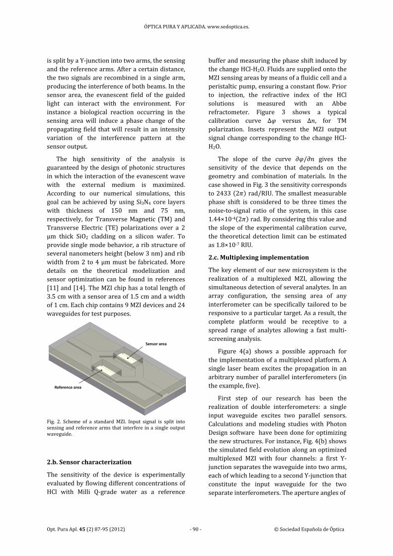
2. Mach‐Zehnder interferometer sensor
2.a. Sensor working principle
In the standard MZI configuration (Fig. 2), a
monochromatic and polarized light (λ=633 nm)
ÓPTICA PURA Y APLICADA. www.sedoptica.es.
Opt. Pura Apl. 45 (2) 87‐95 (2012) ‐ 90 ‐ © Sociedad Española de Óptica
is split by a Y‐junction into two arms, the sensing
and the reference arms. After a certain distance,
the two signals are recombined in a single arm,
producing the interference of both beams. In the
sensor area, the evanescent field of the guided
light can interact with the environment. For
instance a biological reaction occurring in the
sensing area will induce a phase change of the
propagating field that will result in an intensity
variation of the interference pattern at the
sensor output.
The high sensitivity of the analysis is
guaranteed by the design of photonic structures
in which the interaction of the evanescent wave
with the external medium is maximized.
According to our numerical simulations, this
goal can be achieved by using Si3N4 core layers
with thickness of 150 nm and 75 nm,
respectively, for Transverse Magnetic (TM) and
Transverse Electric (TE) polarizations over a 2
µm thick SiO2 cladding on a silicon wafer. To
provide single mode behavior, a rib structure of
several nanometers height (below 3 nm) and rib
width from 2 to 4 µm must be fabricated. More
details on the theoretical modelization and
sensor optimization can be found in references
[11] and [14]. The MZI chip has a total length of
3.5 cm with a sensor area of 1.5 cm and a width
of 1 cm. Each chip contains 9 MZI devices and 24
waveguides for test purposes.
Fig. 2. Scheme of a standard MZI. Input signal is split into sensing and reference arms that interfere in a single output waveguide.
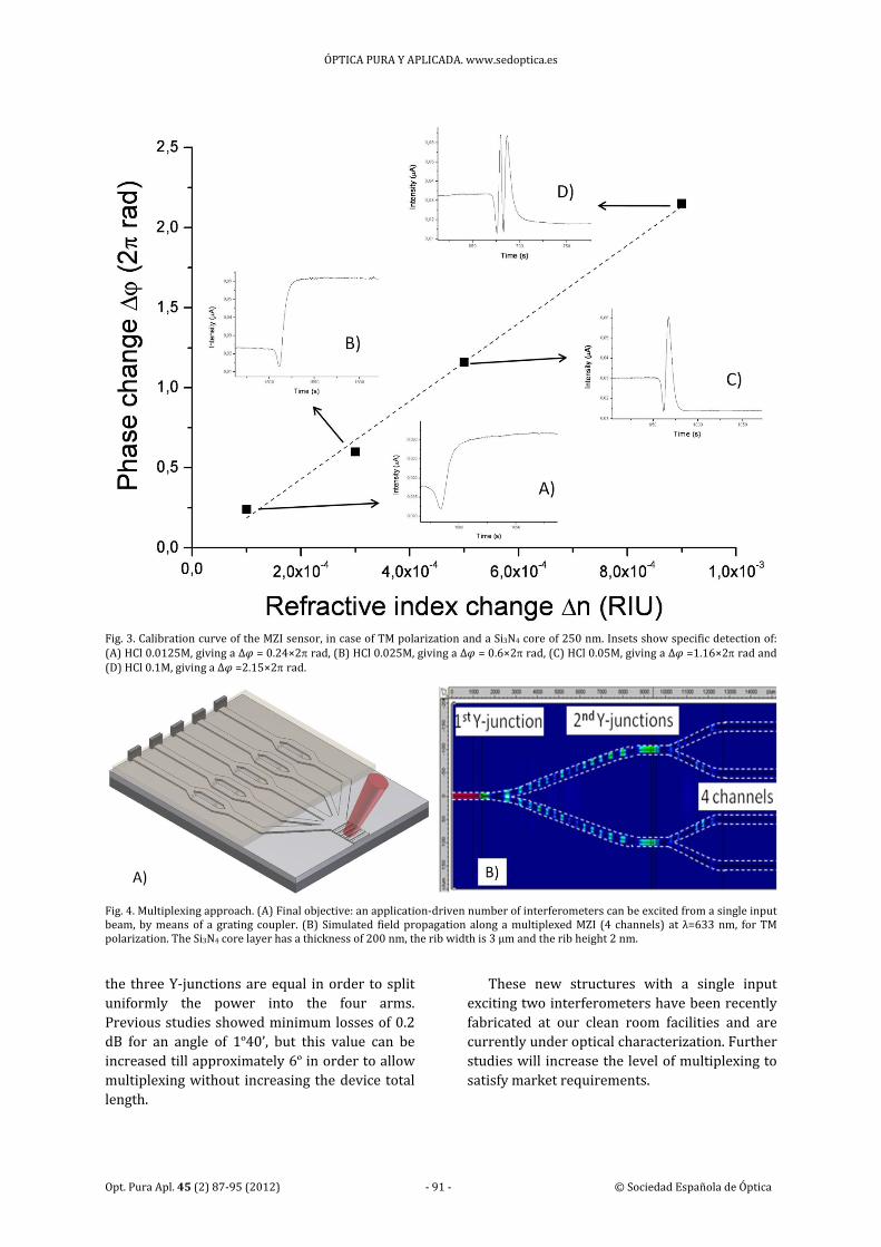
2.b. Sensor characterization
The sensitivity of the device is experimentally
evaluated by flowing different concentrations of
HCl with Milli Q‐grade water as a reference
buffer and measuring the phase shift induced by
the change HCl‐H2O. Fluids are supplied onto the
MZI sensing areas by means of a fluidic cell and a
peristaltic pump, ensuring a constant flow. Prior
to injection, the refractive index of the HCl
solutions is measured with an Abbe
refractometer. Figure 3 shows a typical
calibration curve ∆ versus ∆ , for TM
polarization. Insets represent the MZI output
signal change corresponding to the change HCl‐
H2O.
The slope of the curve / gives the
sensitivity of the device that depends on the
geometry and combination of materials. In the
case showed in Fig. 3 the sensitivity corresponds
to 2433 2 rad/RIU. The smallest measurable
phase shift is considered to be three times the
noise‐to‐signal ratio of the system, in this case
1.44×10‐4 2 rad. By considering this value and
the slope of the experimental calibration curve,
the theoretical detection limit can be estimated
as 1.8×10‐7 RIU.
2.c. Multiplexing implementation
The key element of our new microsystem is the
realization of a multiplexed MZI, allowing the
simultaneous detection of several analytes. In an
array configuration, the sensing area of any
interferometer can be specifically tailored to be
responsive to a particular target. As a result, the
complete platform would be receptive to a
spread range of analytes allowing a fast multi‐
screening analysis.
Figure 4(a) shows a possible approach for
the implementation of a multiplexed platform. A
single laser beam excites the propagation in an
arbitrary number of parallel interferometers (in
the example, five).
First step of our research has been the
realization of double interferometers: a single
input waveguide excites two parallel sensors.
Calculations and modeling studies with Photon
Design software have been done for optimizing
the new structures. For instance, Fig. 4(b) shows
the simulated field evolution along an optimized
multiplexed MZI with four channels: a first Y‐
junction separates the waveguide into two arms,
each of which leading to a second Y‐junction that
constitute the input waveguide for the two
separate interferometers. The aperture angles of
ÓPTICA PURA Y APLICADA. www.sedoptica.es
Opt. Pura Apl. 45 (2) 87‐95 (2012) ‐ 91 ‐ © Sociedad Española de Óptica
Fig. 3. Calibration curve of the MZI sensor, in case of TM polarization and a Si3N4 core of 250 nm. Insets show specific detection of: (A) HCl 0.0125M, giving a ∆ = 0.24×2rad, (B) HCl 0.025M, giving a ∆ = 0.6×2rad, (C) HCl 0.05M, giving a ∆ =1.16×2rad and (D) HCl 0.1M, giving a ∆ =2.15×2rad.
Fig. 4. Multiplexing approach. (A) Final objective: an application‐driven number of interferometers can be excited from a single input beam, by means of a grating coupler. (B) Simulated field propagation along a multiplexed MZI (4 channels) at λ=633 nm, for TM polarization. The Si3N4 core layer has a thickness of 200 nm, the rib width is 3 µm and the rib height 2 nm.
the three Y‐junctions are equal in order to split
uniformly the power into the four arms.
Previous studies showed minimum losses of 0.2
dB for an angle of 1º40’, but this value can be increased till approximately 6º in order to allow multiplexing without increasing the device total
length.
These new structures with a single input
exciting two interferometers have been recently
fabricated at our clean room facilities and are
currently under optical characterization. Further
studies will increase the level of multiplexing to
satisfy market requirements.
ÓPTICA PURA Y APLICADA. www.sedoptica.es.
Opt. Pura Apl. 45 (2) 87‐95 (2012) ‐ 92 ‐ © Sociedad Española de Óptica
3. Wavelength modulation system
MZI biosensors could give false positive reading
due to some problems related to the periodic
nature of the output signal. There are mainly
three issues: (i) signal ambiguities, (ii) intensity
variations and (iii) sensitivity fading. These
disadvantages can be solved by tuning the phase
difference between the arms of the
interferometer by using a phase modulation
system [10]. A simple way to modulate the phase
difference between the two beams (reference
and sensor arms) of a MZI is to modulate the
wavelength of the guided light [15]. Indeed,
depends of the wavelength :
2, 1
where is the length of the sensor area,
and are the effective refractive indices of
the sensor and reference arm, respectively. If the
variation in wavelength can induce a shift of 2
radians in the output signal, we could be at any
time in the optimum measurement conditions.
With our system (15 mm for the length of the
sensor area and 3% difference in effective
refractive indexes), a wavelength variation
lower than 2 nm will be sufficient. To obtain
such variation, we can take advantage of a well‐
known semiconductor laser drawback: the
wavelength modification due to optical power
variation [16]. With the commercial laser diode
we use, the ML101J27 (Thorlabs), a wavelength
modulation of 2 nm can be achieved by varying
the driving current of about 40 mA. The power
variation of the laser can be constantly
monitored thanks to the extraction of a
reference signal.
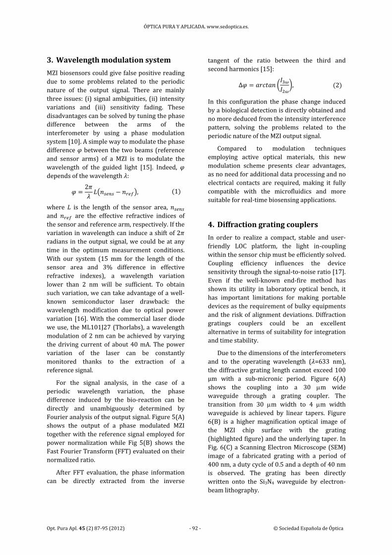
For the signal analysis, in the case of a
periodic wavelength variation, the phase
difference induced by the bio‐reaction can be
directly and unambiguously determined by
Fourier analysis of the output signal. Figure 5(A)
shows the output of a phase modulated MZI
together with the reference signal employed for
power normalization while Fig 5(B) shows the
Fast Fourier Transform (FFT) evaluated on their
normalized ratio.
After FFT evaluation, the phase information
can be directly extracted from the inverse
tangent of the ratio between the third and
second harmonics [15]:
Δ , 2
In this configuration the phase change induced
by a biological detection is directly obtained and
no more deduced from the intensity interference
pattern, solving the problems related to the
periodic nature of the MZI output signal.
Compared to modulation techniques
employing active optical materials, this new
modulation scheme presents clear advantages,
as no need for additional data processing and no
electrical contacts are required, making it fully
compatible with the microfluidics and more
suitable for real‐time biosensing applications.
4. Diffraction grating couplers
In order to realize a compact, stable and user‐
friendly LOC platform, the light in‐coupling
within the sensor chip must be efficiently solved.
Coupling efficiency influences the device
sensitivity through the signal‐to‐noise ratio [17].
Even if the well‐known end‐fire method has
shown its utility in laboratory optical bench, it
has important limitations for making portable
devices as the requirement of bulky equipments
and the risk of alignment deviations. Diffraction
gratings couplers could be an excellent
alternative in terms of suitability for integration
and time stability.
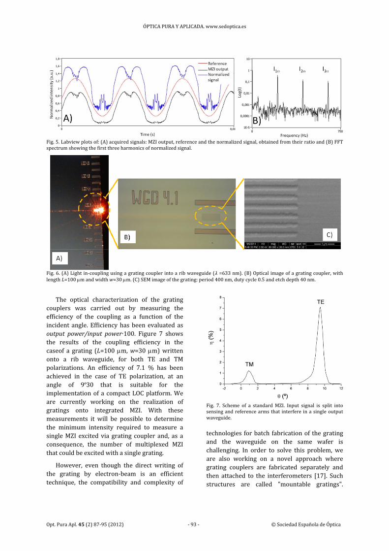
Due to the dimensions of the interferometers
and to the operating wavelength ( =633 nm),
the diffractive grating length cannot exceed 100
µm with a sub‐micronic period. Figure 6(A)
shows the coupling into a 30 m wide waveguide through a grating coupler. The
transition from 30 m width to 4 m width waveguide is achieved by linear tapers. Figure
6(B) is a higher magnification optical image of
the MZI chip surface with the grating
(highlighted figure) and the underlying taper. In
Fig. 6(C) a Scanning Electron Microscope (SEM)
image of a fabricated grating with a period of
400 nm, a duty cycle of 0.5 and a depth of 40 nm
is observed. The grating has been directly
written onto the Si3N4 waveguide by electron‐
beam lithography.
ÓPTICA PURA Y APLICADA. www.sedoptica.es
Opt. Pura Apl. 45 (2) 87‐95 (2012) ‐ 93 ‐ © Sociedad Española de Óptica
Fig. 5. Labview plots of: (A) acquired signals: MZI output, reference and the normalized signal, obtained from their ratio and (B) FFT spectrum showing the first three harmonics of normalized signal.
Fig. 6. (A) Light in‐coupling using a grating coupler into a rib waveguide ( =633 nm). (B) Optical image of a grating coupler, with length L=100 m and width w=30 m. (C) SEM image of the grating: period 400 nm, duty cycle 0.5 and etch depth 40 nm.
The optical characterization of the grating
couplers was carried out by measuring the
efficiency of the coupling as a function of the
incident angle. Efficiency has been evaluated as
output power/input power∙100. Figure 7 shows
the results of the coupling efficiency in the
caseof a grating (L=100 m, w=30 m) written onto a rib waveguide, for both TE and TM
polarizations. An efficiency of 7.1 % has been
achieved in the case of TE polarization, at an
angle of 9º30 that is suitable for the
implementation of a compact LOC platform. We
are currently working on the realization of
gratings onto integrated MZI. With these
measurements it will be possible to determine
the minimum intensity required to measure a
single MZI excited via grating coupler and, as a
consequence, the number of multiplexed MZI
that could be excited with a single grating.
However, even though the direct writing of
the grating by electron‐beam is an efficient
technique, the compatibility and complexity of
Fig. 7. Scheme of a standard MZI. Input signal is split into sensing and reference arms that interfere in a single output waveguide.
technologies for batch fabrication of the grating
and the waveguide on the same wafer is
challenging. In order to solve this problem, we
are also working on a novel approach where
grating couplers are fabricated separately and
then attached to the interferometers [17]. Such
structures are called "mountable gratings".
ÓPTICA PURA Y APLICADA. www.sedoptica.es.
Opt. Pura Apl. 45 (2) 87‐95 (2012) ‐ 94 ‐ © Sociedad Española de Óptica
Simulations are being performed to determine
the optimum grating parameters but also to
estimate the tolerance in fabrication to keep a
good grating efficiency. The grating structure is
made by electron‐beam lithography on a Si3N4
layer and then covered by a polydimethyl‐
siloxane (PDMS) film. The gratings are optically
characterized as a single element and then stuck
with the PDMS film to the photonic structures.
5. Surface functionalization
When developing a LOC system for biosensing
applications, sensitivity, selectivity and stability
of the bioreceptor are fundamental aspects to be
dealt with. Different immobilization strategies
can be adopted, according to the particular
application, i.e. the nature of the receptor to be
immobilized. Covalent binding has
demonstrated to be a valid strategy [8], ensuring
stable union and applicability to a spread family
of biomolecules. For instance, in the case of
proteomics applications, the immobilization of
the specific receptor onto the sensing area of the
MZI can be performed via amino group of the
protein.
Prior to the biofunctionalization step a
chemical activation of the surface is mandatory
[18]. In our case, the transducer surface is made
out of silicon nitride and can be functionalized
with the well‐known silane chemistry. The
silicon nitride surface is initially treated with
ozone plasma and then oxidised with a solution
of HNO3 at 10%. Carboxyethylsilanetriol, sodium
salt (CTES) is employed as silane agent and the
ended carboxylic groups of the silane are
afterwards activated through the N‐(3‐
dimethylaminopropyl)‐N′‐ethylcarbodiimide
(EDC)/ N‐Hydroxysuccinimide (NHS) chemistry
[19,20]. The receptor protein is afterwards
immobilized by covalent bonding on the sensing
area. A schematic view of the surface treatment
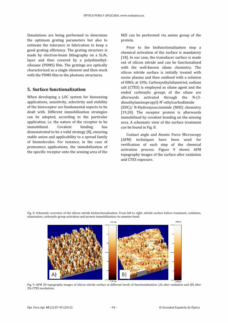
can be found in Fig. 8.
Contact angle and Atomic Force Microscopy
(AFM) techniques have been used for
verification of each step of the chemical
activation process. Figure 9 shows AFM
topography images of the surface after oxidation
and CTES exposure.
Fig. 8. Schematic overview of the silicon nitride biofunctionalization. From left to right: nitride surface before treatment, oxidation, silanization, carboxylic group activation and protein immobilization via ammine bond.
Fig. 9. AFM 3D topography images of silicon nitride surface at different levels of functionalization: (A) after oxidation and (B) after 2% CTES incubation.
ÓPTICA PURA Y APLICADA. www.sedoptica.es
Opt. Pura Apl. 45 (2) 87‐95 (2012) ‐ 95 ‐ © Sociedad Española de Óptica
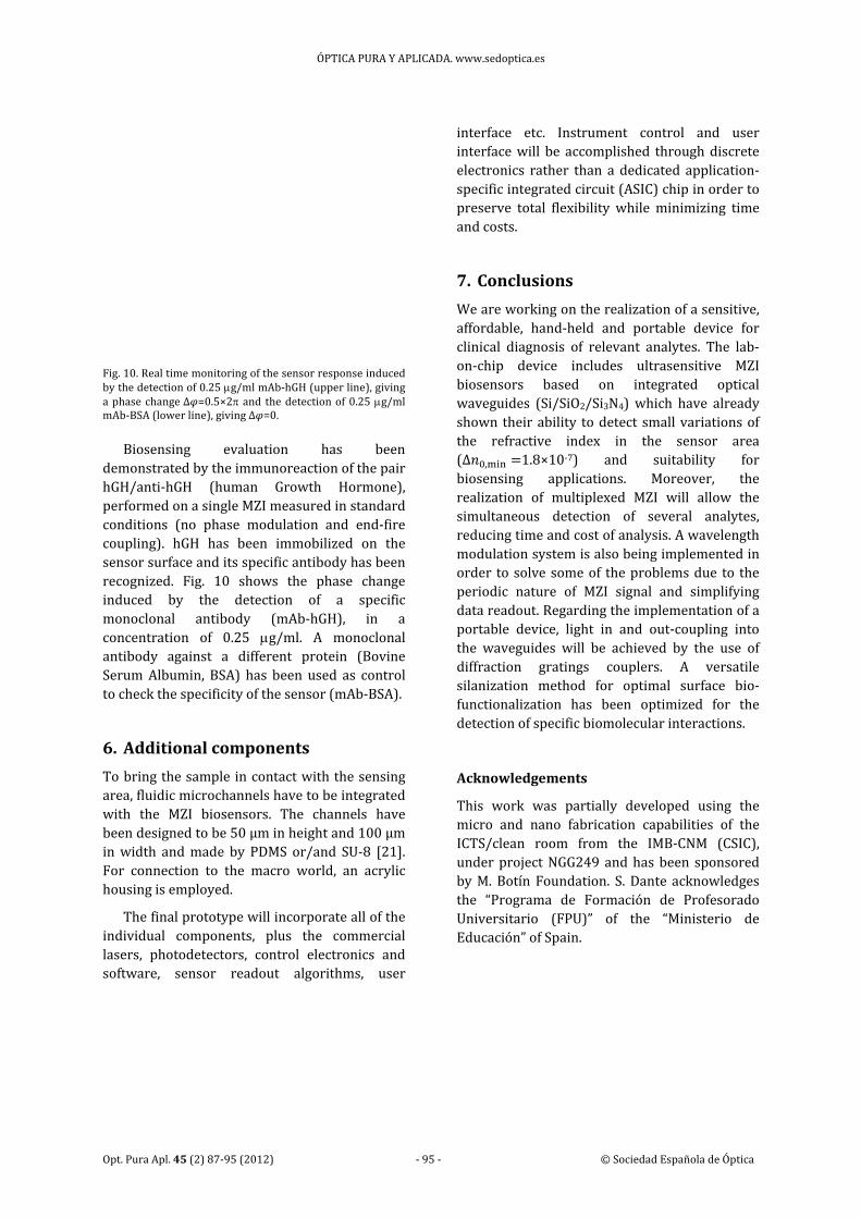
Fig. 10. Real time monitoring of the sensor response induced by the detection of 0.25 g/ml mAb‐hGH (upper line), giving a phase change∆ =0.5×2 and the detection of 0.25 g/ml mAb‐BSA (lower line), giving ∆ =0.
Biosensing evaluation has been
demonstrated by the immunoreaction of the pair
hGH/anti‐hGH (human Growth Hormone),
performed on a single MZI measured in standard
conditions (no phase modulation and end‐fire
coupling). hGH has been immobilized on the
sensor surface and its specific antibody has been
recognized. Fig. 10 shows the phase change
induced by the detection of a specific
monoclonal antibody (mAb‐hGH), in a
concentration of 0.25 g/ml. A monoclonal antibody against a different protein (Bovine
Serum Albumin, BSA) has been used as control
to check the specificity of the sensor (mAb‐BSA).
6. Additional components
To bring the sample in contact with the sensing
area, fluidic microchannels have to be integrated
with the MZI biosensors. The channels have
been designed to be 50 µm in height and 100 µm
in width and made by PDMS or/and SU‐8 [21].
For connection to the macro world, an acrylic
housing is employed.
The final prototype will incorporate all of the
individual components, plus the commercial
lasers, photodetectors, control electronics and
software, sensor readout algorithms, user
interface etc. Instrument control and user
interface will be accomplished through discrete
electronics rather than a dedicated application‐
specific integrated circuit (ASIC) chip in order to
preserve total flexibility while minimizing time
and costs.
7. Conclusions
We are working on the realization of a sensitive,
affordable, hand‐held and portable device for
clinical diagnosis of relevant analytes. The lab‐
on‐chip device includes ultrasensitive MZI
biosensors based on integrated optical
waveguides (Si/SiO2/Si3N4) which have already
shown their ability to detect small variations of
the refractive index in the sensor area
(Δ , 1.8×10‐7) and suitability for
biosensing applications. Moreover, the
realization of multiplexed MZI will allow the
simultaneous detection of several analytes,
reducing time and cost of analysis. A wavelength
modulation system is also being implemented in
order to solve some of the problems due to the
periodic nature of MZI signal and simplifying
data readout. Regarding the implementation of a
portable device, light in and out‐coupling into
the waveguides will be achieved by the use of
diffraction gratings couplers. A versatile
silanization method for optimal surface bio‐
functionalization has been optimized for the
detection of specific biomolecular interactions.
Acknowledgements
This work was partially developed using the
micro and nano fabrication capabilities of the
ICTS/clean room from the IMB‐CNM (CSIC),
under project NGG249 and has been sponsored
by M. Botín Foundation. S. Dante acknowledges
the “Programa de Formación de Profesorado
Universitario (FPU)” of the “Ministerio de
Educación” of Spain.


![Mach number P w,test [bar] P model [bar] 1.8 -0.45 -0.20 0 ...ae342/18/lab2/lab2data.pdf · Mach 2.0 Snapshot . Mach 1.8 Snapshot . Mach 2.3 Snapshot Mach 2.2 Snapshot . P w,test](https://img.pdfslide.us/doc/110x75/5fb4e5220b26be1bae0aea08/mach-number-p-wtest-bar-p-model-bar-18-045-020-0-ae34218lab2-.jpg)