Embed Size (px)
Citation preview
Toward Antibody-directed Enzyme Prodrug Therapy with theT268G Mutant of Human Carboxypeptidase A1 and Novel in VivoStable Prodrugs of Methotrexate*
(Received for publication, March 4, 1997, and in revised form, April 7, 1997)
Gary K. Smith‡, Sheila Banks, Todd A. Blumenkopf§, Michael Cory, Joan Humphreys,Ronald M. Laethem, John Miller, Cary P. Moxham, Robert Mullin, Paul H. Ray,Leslie M. Walton, and Lawrence A. Wolfe III
From GlaxoWellcome Inc., Research Triangle Park, North Carolina 27709
Antibody-directed enzyme prodrug therapy (ADEPT)has the potential of greatly enhancing antitumor selec-tivity of cancer therapy by synthesizing chemothera-peutic agents selectively at tumor sites. This therapy isbased upon targeting a prodrug-activating enzyme to atumor by attaching the enzyme to a tumor-selective an-tibody and dosing the enzyme-antibody conjugate sys-temically. After the enzyme-antibody conjugate is local-ized to the tumor, the prodrug is then also dosedsystemically, and the previously targeted enzyme con-verts it to the active drug selectively at the tumor. Un-fortunately, most enzymes capable of this specific, tu-mor site generation of drugs are foreign to the humanbody and as such are expected to raise an immune re-sponse when injected, which will limit their repeatedadministration. We reasoned that with the power ofcrystallography, molecular modeling and site-directedmutagenesis, this problem could be addressed throughthe development of a human enzyme that is capable ofcatalyzing a reaction that is otherwise not carried out inthe human body. This would then allow use of prodrugsthat are otherwise stable in vivo but that are substratesfor a tumor-targeted mutant human enzyme. We reporthere the first test of this concept using the human en-zyme carboxypeptidase A1 (hCPA1) and prodrugs ofmethotrexate (MTX). Based upon a computer model ofthe human enzyme built from the well known crystalstructure of bovine carboxypeptidase A, we have de-signed and synthesized novel bulky phenylalanine- andtyrosine-based prodrugs of MTX that are metabolicallystable in vivo and are not substrates for wild type humancarboxypeptidases A. Two of these analogs are MTX-a-3-cyclobutylphenylalanine and MTX-a-3-cyclopentylty-rosine. Also based upon the computer model, we havedesigned and produced a mutant of human carboxypep-tidase A1, changed at position 268 from the wild typethreonine to a glycine (hCPA1-T268G). This novel en-zyme is capable of using the in vivo stable prodrugs,which are not substrates for the wild type hCPA1, asefficiently as the wild type hCPA1 uses its best sub-
strates (i.e. MTX-a-phenylalanine). Thus, the kcat/Kmvalue for the wild type hCPA1 with MTX-a-phenylala-nine is 0.44 mM21 s21, and kcat/Km values for hCPA1-T268G with MTX-a-3-cyclobutylphenylalanine and MTX-a-3-cyclopentyltyrosine are 1.8 and 0.16 mM21 s21,respectively. The cytotoxic efficiency of hCPA1–268Gwas tested in an in vitro ADEPT model. For this exper-iment, hCPA1-T268G was chemically conjugated toING-1, an antibody that binds to the tumor antigen Ep-Cam, or to Campath-1H, an antibody that binds to the Tand B cell antigen CDw52. These conjugates were thenincubated with HT-29 human colon adenocarcinomacells (which express Ep-Cam but not the Campath 1Hantigen) followed by incubation of the cells with the invivo stable prodrugs. The results showed that the tar-geted ING-1:hCPA1-T268G conjugate produced excel-lent activation of the MTX prodrugs to kill HT-29 cells asefficiently as MTX itself. By contrast, the enzyme-Cam-path 1H conjugate was without effect. These datastrongly support the feasibility of ADEPT using a mu-tated human enzyme with a single amino acid change.
A current major challenge to cancer therapy is to increaseantitumor selectivity. One approach to realizing this goal is touse the exquisite selectivity of the antibody:antigen reaction totarget therapeutic entities specifically to tumors. While abso-lutely tumor-specific antibodies are not known, many antibod-ies are available that can deliver tumor-selective targeting (forreview, see Ref. 1).
One investigational therapy that makes use of this principleof antibody targeting is antibody-directed enzyme prodrugtherapy (ADEPT).1 ADEPT is a powerful strategy with thepotential for tumor-specific long-term delivery of chemotherapy(2–7). The premise of ADEPT is to target an enzyme of interestspecifically to tumor cells by coupling it to a tumor-specificantibody. This conjugate is delivered to the patient systemi-cally and then allowed to bind to the antigen-expressing targetcells. Unbound conjugate is allowed to clear from circulation,and when the circulating levels of conjugate are sufficientlylow, a prodrug is administered, also systemically, that can beconverted to a toxic chemotherapeutic drug by the targetedenzyme-antibody conjugate. The action of the enzyme-antibodyconjugate on the prodrug then ideally generates lethal levels ofdrug specifically at the tumor site. For this therapy to be
* The costs of publication of this article were defrayed in part by thepayment of page charges. This article must therefore be hereby marked“advertisement” in accordance with 18 U.S.C. Section 1734 solely toindicate this fact.
Parts of this work have been reported in the patent literature (40) andin abstract form (41, 42).
‡ To whom correspondence should be addressed: Dept. of MolecularBiochemistry, Glaxo Wellcome Research and Development, 5 MooreDr., Research Triangle Park, NC 27709. Tel.: 919-483-1502; Fax: 919-483-4320; E-mail: [email protected].
§ Current address: Pfizer Central Research, Eastern Point Rd., Gro-ton, CT 06340.
1 The abbreviations used are: ADEPT, antibody-directed enzyme pro-drug therapy; CPA, carboxypeptidase; hCPA, human CPA; bCPA, bo-vine CPA; MTX, methotrexate; HPLC, high pressure liquid chromatog-raphy; WT, wild type; Sulfo-SMCC, sulfosuccinimidyl 4-(N-maleimido-methyl)cyclohexane-1-carboxylate.
THE JOURNAL OF BIOLOGICAL CHEMISTRY Vol. 272, No. 25, Issue of June 20, pp. 15804–15816, 1997© 1997 by The American Society for Biochemistry and Molecular Biology, Inc. Printed in U.S.A.
This paper is available on line at http://www.jbc.org15804
by guest on February 8, 2018http://w
ww
.jbc.org/D
ownloaded from
selective, however, nonspecific activation of the prodrug atsites distant to the tumor must be minimized. This is generallyaccomplished by using a conjugate enzyme with an activity notendogenous to the host or at least not accessible to the prodrug(2–29).
A number of ADEPT strategies have been reported (2–29).The concept has been shown to be effective both in in vitro andin vivo models, and at least one ADEPT strategy is currentlyundergoing clinical evaluation (27–29). ADEPT in vitro efficacyhas been demonstrated with enzyme-antibody conjugates of (a)carboxypeptidase G2 along with several nitrogen mustards(14–16), (b) alkaline phosphatase with phosphorylated pro-drugs of mitomycin, a phenol mustard, and etoposide (3, 10,11), (c) b-lactamase with lactam prodrugs of doxorubicin, vincaalkaloid analogs, and a nitrogen mustard (7, 12, 13, 17, 18), (d)penicillin-G amidase with prodrugs of palytoxin, doxorubicinand melphalan (8, 9), (e) penicillin-V amidase with a prodrug ofdoxorubicin (2, 7), (f) human or Escherichia coli b-glucuroni-dase with glucuronide prodrugs of epirubicin, doxorubicin anda nitrogen mustard (22–24), (g) cytosine deaminase and 5-fluo-rocytosine (25, 26), and (h) bovine carboxypeptidase A anda-amino acid prodrugs of MTX (19–21). Further, in vivo anti-tumor efficacy has been shown in a number of systems usingthe enzymes alkaline phosphatase, carboxypeptidase G2, b-lac-tamase, and b-glucuronidase (2, 3, 7, 10–13, 15–18, 24).
An inherent problem with antibody-targeted therapies is theimmune response mounted by the host to the foreign proteinsand other antigens used in the therapy (1, 28). For example,monoclonal antibodies used in antibody targeting-based ther-apies are in general rodent in origin and as such recognized bythe immune system (1, 28). ADEPT has the additional problemthat the enzyme used to generate the site-specific drug synthe-sis can also be immunogenic, especially when a foreign enzymeis used. The immunogenicity associated with these foreignantibody or enzyme proteins decreases the utility of the anti-body-targeting strategies by decreasing the ability of the phy-sician to perform multiple dosing regimens.
Attempts are being made to overcome the immune responseto the rodent antibodies through “humanization” of the anti-bodies (30). In this strategy, much of the sequence of the mousemonoclonal antibody is replaced with corresponding humanantibody sequence. Only selected residues at the antigen com-bining site are left intact, leaving relatively few “rodent resi-dues” remaining in the antibody.
We reasoned that the imunogenicity associated with the useof enzymes of nonhuman origin might be circumvented througha similar strategy. However, we chose not to precisely followthe strategy of antibody humanization, which commences theprocess with the binding site of a foreign protein. Rather, ourapproach to generate a composite human/nonhuman enzymewas to start with a fully human enzyme and change the “activesite” at one or two residues to produce a .99.5% human en-zyme capable of efficiently performing a non-human reaction.This human enzyme with non-human specificity, along withhumanized antibodies, should then facilitate the production ofenzyme:antibody conjugates having lower immunogenicity andbenefit the development of multiple dosing regimen ADEPTstrategies.
Our initial target to generate a human enzyme with non-human specificity was human pancreatic carboxypeptidase A,recently cloned and expressed in our laboratory (31). Pancre-atic carboxypeptidase A is a zinc-containing exopeptidase re-leased into the small intestine from the pancreas as a zymogen(32, 33). The pancreatic CPA has two further subclasses, CPA1and CPA2, in both humans (31, 34, 35) and rats (36, 37). Whilethe amino acid sequences of CPA1 and CPA2 active sites are
similar and both enzymes prefer aromatic C-terminal aminoacids, CPA2 enzyme prefers bulkier aromatic C-terminalamino acids (31, 37). This was shown for the rat enzyme withdi- and tripeptide substrates and for the human with aminoacid prodrugs of MTX (31, 37). High resolution crystal struc-tures for bCPA have been determined (32). Of the nine activesite residues within 4.5 Å of the bound substrate, only threevary among bovine CPA (32), rat CPA1 (36), rCPA2 (37),hCPA1 (31, 34), and hCPA2 (31, 35). These changes, at residues253, 254, and 268, describe a larger binding pocket for CPA2than CPA1 and provide a rationale for the substratespecificities.
Huennekens and co-workers (19–21) developed an in vitroADEPT approach using MTX prodrugs in which MTX wasmodified at the a-carbon of the glutamate moiety with one ofseveral natural amino acids. These prodrugs were relativelynontoxic in cell culture but could be activated by bovine CPA orcarboxypeptidase B to form MTX. The most successful of theseprodrugs was MTX-Phe, which was found to be an excellentsubstrate for bovine CPA (21). This system was effective undercell culture conditions where the IC50 of MTX-Phe againstL1210 cells changed from 2.2 3 1026 to 6.3 3 1028 M, indistin-guishable from MTX itself, in the presence of bovine CPA or abovine CPA-antibody conjugate (21). An important positiveaspect of this system is its use of MTX with its well knownefficacy and toxicity profile (38, 39). Thus, MTX maximumtolerated doses are due to well understood gut and bone mar-row toxicities. Therefore, specific generation of high concentra-tions of MTX at tumor sites distal from these sites of knowntoxicity should not have major side effects. Two potential lim-itations to the use of the bCPA/MTX-Phe system in humans,however, are the possible background activation of prodrug byendogenous hCPA to liberate MTX systemically and the im-mune response elicited by the bovine protein.
We sought to improve upon the MTX-Phe/bCPA system intwo ways. First, we sought to use the human rather thanbovine CPA. Second, we sought to change the catalytic speci-ficity of the human enzyme(s) to accommodate MTX prodrugsthat are not substrates for the endogenous wild type car-boxypeptidases and would therefore be expected to be stable invivo. The strategy chosen to accomplish this goal exploitedparallel computer-aided design of novel active sites and of newMTX-a-amino acid prodrugs. Specifically, wild type hCPA andnovel hCPA mutant active sites were designed using proteinhomology model building from the well known high resolutioncrystal structure of bCPA (40). Then these computer-generatedactive site mutants and similarly generated modified sub-strates were evaluated together and compared with wild typeenzyme-substrate complexes. Favorable mutants and modifiedsubstrates were prepared and tested in vitro and in vivo. Wereport here MTX prodrugs that are stable in vivo, a one-aminoacid mutant of hCPA1 that can efficiently use these in vivostable prodrugs, and the use of these prodrugs along with amutant hCPA enzyme-antibody conjugate for antigen-specificcytotoxicity in vitro. The work reported demonstrates proof ofprinciple for the methodology for developing a very efficientmutant human enzyme/prodrug combination for use inADEPT.
EXPERIMENTAL PROCEDURES
Materials
Human CPAs were obtained as described previously (31). Cell lineswere obtained from ATCC (Rockville, MD) and grown in 90% RPMI1640, 10% fetal calf serum at 37 °C under 5% CO2. HT-29 cells for invitro ADEPT experiments in human serum were taken from this me-dium and grown for 3 weeks in 95% RPMI 1640, 5% human serum at37 °C under 5% CO2. Growth rates and MTX cytotoxicity were similarunder the two conditions.
T268G Mutant of hCPA1 Hydrolyzes Novel MTX Prodrugs 15805
by guest on February 8, 2018http://w
ww
.jbc.org/D
ownloaded from
MethodsSynthesis of MTX Prodrugs
The synthesis of the MTX prodrugs shown in Table I is describedelsewhere (40).
Mutagenesis of hCPA1
pMP36HCPA1 (31) containing pro-hCPA1-WT cDNA (as a fusionwith yeast a factor leader, described below) was restricted with NcoIand SalI to liberate a 481-base pair cDNA fragment. This fragmentbegins at nucleotide 893, proceeds to the 39-end of the hCPA1 cDNA,and encodes amino acids 186–309 of mature hCPA1.2 The CPA1 NcoI-SalI fragment was ligated into the NcoI and SalI cloning sites ofpGEM5zf(2) (Promega) to generate pHCPAINS. pHCPAINS was re-stricted with SphI and SalI to liberate the hCPA1 NcoI-SalI fragment,and an additional nine base pairs of pGEM5zf(2) sequence. This SphI-SalI fragment was cloned into M13mp19 (Life Technologies, Inc.) usingits SphI and SalI cloning sites to generate M13mp19HCPA1.
Single-stranded M13mp19HCPA1 DNA was used as template foroligonucleotide-directed mutagenesis using the T7-GEN in vitro mu-tagenesis kit (U.S. Biochemical Corp.). The following mutagenic oligo-nucleotide primers (Oligos Etc.) were used to mutate residues Ile255
(AAT) and Thr268 (ACC) either separately or in tandem (mutageniccodons underlined): I255A, 59-ggT CCA gTC AgC AgT gCT TCC-39(Ala 5 gCT); T268A, 59-gAg CTC gAA ggC gAA ggA gTA-39 (Ala 5 gCC);T268G, 59-gAg CTC gAA gCC gAA ggA gTA-39 (Gly 5 ggC). Using theseoligonucleotide primers, the following hCPA1 mutants were formed:T268A, T268G, I255A, and I255A/T268G. Each of the mutagenizedhCPA1 cassettes was sequenced to verify that only the desired DNAmutations were produced.
Expression of hCPA Enzymes in Yeast
Expression of hCPA enzymes in Saccharomyces cerevisiae was per-formed according to the strategy of Gardell et al. (43) as described byLaethem et al. (31). The cDNAs for pro-hCPA1 or pro-hCPA2 werecloned into the pMP36 vector and fused in frame to the yeast a factor
leader of this vector using a polymerase chain reaction approach (44).pMP36HCPA1 was restricted with NcoI to liberate a 1.2-kilobase pair
fragment that was cloned into the NcoI site of the M13mp19HCPA1mutants. The correct orientation of the NcoI fragment withinM13mp19pro-hCPA1 mutants was verified, and the DNAs were re-stricted with HindIII and SalI liberating a 1.2-kilobase pair cDNAencoding the entire pro-hCPA1 mutant enzyme. This fragment wasligated into the HindIII and SalI sites of pMP36 yielding pMPHCPA1mutants. These DNAs were restricted sequentially with BamHI, SalI,and SspI with intervening purifications by either phenol extractions oruse of Promega Magic Mini Columns with manufacturer supplied pro-cedures. Following SspI restriction, the 2.8-kilobase pair band, contain-ing hCPA1 mutant with the yeast a factor leader in frame, was gel-purified from a 1% low melting agarose gel. The BamHI-SalI fragmentwas ligated into the pBS24.1 shuttle vector overnight at 16 °C. Thisvector (pBSHCPA1-mutant) was electroporated into DH5a, and plas-mid DNA was isolated using the Wizard Miniprep Kit (Promega).
Approximately 500–2000 ng of pBSHCPA1 mutant DNA was elec-troporated into 40 ml of electrocompetent DLM101a S. cerevisiae. Oneml of 1 M sorbitol was added immediately after electroporation to rescuethe cells. 100-ml samples were plated out on dishes of yeast nitrogenbroth-uracil selection medium and were incubated at 30 °C for 2–3 days(31). Positive colonies were picked and grown in yeast nitrogen broth-leucine selection medium containing 8% glucose at 30 °C for 48 h. Thisculture was used to seed 350 liters of YP, 1% glucose in a 500-liter NewBrunswick fermenter.
Mutagenesis and Expression of hCPA2
Mutagenesis of hCPA2 was analogous to CPA1 described above. Thefollowing mutagenic oligonucleotide primer (Oligos Etc.) was used tomutate residue Ala268 (gCC): A268G, 59-CAg TTC AAA gCC AAA TgAgTA-39 (Gly 5 ggC). Using this oligonucleotide primer, the followinghCPA2 mutant A268G was formed. The mutagenized hCPA2 cassettewas sequenced to verify that only the desired DNA mutations wereproduced.
The hCPA2-A268G mutant was subcloned into pMP36, then intopBS, and expressed in yeast (described above).
Purification of Expressed hCPA1 and hCPA2 Enzymes
The wild type and mutant hCPA1 and hCPA2 enzymes were purifiedto electrophoretic homogeneity using a combination of hydrophobic andion exchange chromatography as described previously (31).
Spectrophotometric Enzymatic Assays
Enzymatic activity was determined in one of two ways. Hippuryl-L-phenylalanine and hippuryl-DL-phenyllactate were determined spectro-photometrically at 255 nm as described (45). Reactions contained either0.5 mM hippuryl-L-phenylalanine or 1.0 mM hippuryl-DL-phenyllactatein 25 mM Tris-HCl (pH 7.4), 100 mM NaCl. Reactions were initiated bythe addition of enzyme and were monitored by the change in absorbanceat 255 nm at 25 °C. Enzyme kinetic rates were determined from initialvelocities using e 5 390 M21.
Hydrolysis of MTX prodrugs was measured using a modification ofthe coupled assay described by Kuefner et al. (19). Reactions werecarried out in 1 ml of 25 mM Tris-HCl (pH 7.4), 100 mM NaCl at 25 °C.Buffer was added to the cuvette along with 0.026 units of carboxypep-tidase G; then prodrug was added, and the absorbance was determinedto calculate the concentration. Reactions were initiated by adding aknown amount of CPA enzyme, and the decrease in absorbance at 315nm was monitored. Enzyme kinetic rates were determined from initialvelocities using e 5 9.57 mM21 for MTX. One unit of enzyme activity isdefined as the hydrolysis of 1 mmol of substrate/min at 25 °C.
Thermal Stability Studies
Thermal inactivation of the enzymes was determined spectrophoto-metrically using the CPA assay described above. Inactivation reactionswere carried out by incubating 0.1 mg/ml enzyme in Dulbecco’s phos-phate-buffered saline at the desired temperature. Aliquots were sam-pled at various times and assayed with hippuryl-DL-phenyllactate.
Stability of Prodrugs in Pancreatic Juice
This parameter was determined by incubating the prodrugs in tryp-sin activated human pancreatic juice. The percentage of conversion toMTX was determined as a function of time from linear conversionversus time plots. Pancreatic juice was a generous gift of Dr. T. Pappasof Duke University Medical School (Durham, NC). As obtained, thematerial had little or no CPA activity and did not metabolize MTX
2 The numbering convention used in this paper is based on the num-bering of hCPA1 by Catasus et al. (34) to facilitate the comparison ofthis work with that of others.
TABLE IStructures of MTX-a-R prodrugs
Compound 2Ra Abbreviation
GW1311 Phenylalanine MTX-PheNegatively charged prodrugs
GW2310 Glutamate MTX-GluGW3347 Aspartate MTX-AspGW3855 2-Carboxyphenylalanine MTX-2-carboxy-PheGW3199 3-Carboxytyrosine MTX-3-carboxy-TyrGW4694 3-Carboxyphenylalanine MTX-3-carboxy-Phe
Bulky aromatic prodrugs (phenylalanine-based)GW4160 2-Iodophenylalanine MTX-2-iodo-PheGW1667 1-Naphthylalanine MTX-naphthyl-AlaGW250 2-Cyclopentylphenylalanine MTX-2-cyclopentyl-PheGW1442 2-Cyclohexylphenylalanine MTX-2-cyclohexyl-PheGW3352 3-Cyclobutylphenylalanine MTX-3-cyclobutyl-PheGW1834 3-t-Butylphenylalanine MTX-3-t-butyl-PheGW637 3-Cyclopentylphenylalanine MTX-3-cyclopentyl-PheGW827 3-(3-n-Pentyl)phenylalanine MTX-3-n-pentyl-Phe
Bulky aromatic prodrugs (tyrosine-based)GW1867 3,5-Diiodotyrosine MTX-3,5-diiodo-TyrGW2159 2-Cyclopentyltyrosine MTX-2-cyclopentyl-TyrGW5798 3-Cyclobutyltyrosine MTX-3-cyclobutyl-TyrGW3335 3-t-Butyltyrosine MTX-3-t-butyl-TyrGW5755 3-Cyclopentyltyrosine MTX-3-cyclopentyl-Tyra All R-groups are L-amino acids attached to the a-carboxyl of MTX
via a standard peptide bond.
T268G Mutant of hCPA1 Hydrolyzes Novel MTX Prodrugs15806
by guest on February 8, 2018http://w
ww
.jbc.org/D
ownloaded from
prodrugs. The pancreatic juice was activated fresh for each experimentby trypsinization with 1 mg/ml trypsin for 10 min at 37 °C. (This highconcentration of trypsin was required to overcome endogenous trypsininhibitors.) This activated solution was used directly for stability testsof the prodrugs. Activated pancreatic juice was diluted 1:40 to 1:2000into 25 mM Tris-HCl, 100 mM NaCl, pH 7.5. Prodrug was then added toa final concentration of 50 mM. The solution was then incubated at 25 °Cfor up to 24 h. During this incubation period, aliquots were removed andanalyzed by HPLC for prodrug and MTX. HPLC conditions were asfollows. Chromatography was performed on a Waters C-18 Nova Pakcolumn with a flow rate of 1 ml/min. Mobile phase conditions were astep gradient system composed of 0.1% trifluoroacetic acid in water(component A) or in acetonitrile (component B). For chromatography,the column was equilibrated with 82% A, 18% B. At 3 min, conditionswere switched to 50:50 A:B. Then the column was reequilibrated start-ing at 7 min for another 10 min with 82:18 A:B. Under these conditions,MTX eluted at 4 min, and the prodrugs eluted at 6–8 min, dependingupon hydrophobicity. Elution was monitored at 310 nm. No significantconversion of prodrug to MTX occurred when a suitable dilution oftrypsin replaced activated pancreatic juice, indicating that conversionwas due solely to materials contained in the pancreatic juice.
In Vivo Stability of Prodrugs
This parameter was measured as described elsewhere (42). Briefly,animals were dosed with prodrug intravenously. At specified times,plasma and tissues were collected and snap-frozen. The samples werethen homogenized in 0.1 M HCl and extracted with a mix of 2 volumesof the HCl homogenate and 5 volumes of 220 °C acetonitrile. Thenprodrug and MTX were measured by HPLC as described (42).
Conjugation of Mutant hCPA to Antibody ING-1 or Campath-1H
Enzyme Modification—The enzyme was modified at free amines withSulfo-SMCC. This bifunctional reagent has both an amine-reactiveN-hydroxysulfosuccinimide group and a thiol-reactive maleimidegroup. Since carboxypeptidase has no free thiols, the compound reactswith enzyme amines to place a maleimide on the enzyme for subsequentreaction/coupling with free thiols on the antibody.
Four mg of mutant or wild type carboxypeptidase A were combinedwith 0.15 mg of Sulfo-SMCC (Pierce) in 400 ml of Dulbecco’s phosphate-buffered saline. The resulting solution was stirred for 45 min at 25 °C.The modified enzyme then resolved from reagent through a 1 3 13-cmG-25 medium column equilibrated with Dulbecco’s phosphate-bufferedsaline.
Maleimide Content of the Activated Enzyme—Maleimide content wasdetermined as follows. A 0.5-ml aliquot of a 6.3 mM solution of modifiedenzyme was mixed with 6 ml of 1 mM mercaptoethanolamine. Themixture was allowed to sit for 30 min to permit the enzyme-boundmaleimide to react with the mercaptoethanolamine. Then 20 ml of 4mg/ml Ellman’s reagent was added to react with the remaining freemercaptoethanolamine. After another 20 min, absorbance at 412 nmwas determined. The amount of enzyme-bound maleimide was inferredfrom the amount of mercaptoethanolamine that was consumed duringreaction with the enzyme. Based upon a molar absorptivity of 13.6mM21, the purified product was typically found to contain 1 maleimide/enzyme molecule. The derivatization had no effect upon enzyme activitymeasured with hippurylphenylalanine.
Antibody Modification—The antibody was modified with the amine-reactive reagent 2-iminothiolane (Traut’s Reagent; Pierce), which re-places amines with free thiols. The thiols generated on the antibody bythis procedure were then used for subsequent coupling to carboxypep-tidase through the enzyme-bound, thiol-reactive, maleimide.
1.8 mg of ING-1 (46) or Campath-1H (47) were combined with 0.22mg of 2-iminothiolane in 1.3 ml of a 50:50 mixture of Dulbecco’s phos-
phate-buffered saline and 0.1 M triethanolamine-HCl, 2 mM EDTA, pH8.0, under anaerobic conditions. This was allowed to react with stirringfor 1 h and 45 min. The modified antibody was purified through a 1 313-cm G-25 medium column equilibrated with 0.02 M sodium acetate,0.1 M NaCl, pH 5.8, bubbled with and maintained under a heliumatmosphere.
Coupling of Antibody with Enzyme—The modified antibody was col-lected directly from the G-25 column into the solution of the modifiedcarboxypeptidase A. The resulting mixture was then carefully adjustedto pH 7.4 with NaOH, made anaerobic by repeated N2 gassing andevacuation, and allowed to react with stirring at 4 °C for 18 h. Excessfree maleimide groups were removed by reacting the solution with 0.3mM mercaptoethanolamine at room temperature for 1 h, and the solu-tion was then concentrated to 1 ml. The resulting concentrated conju-gate was purified from aggregates, unreacted enzyme, and small mol-ecules by chromatography on Superose 12 HR 10/30.
Cell Culture ADEPT Experiments
IC50 values for drugs and prodrugs alone were determined as de-scribed previously (48). For ADEPT experiments, HT-29 cells wereseeded at 7500 cells/well in 96-well plates containing 200 ml of 95%RPMI 1640/5% human serum (human growth medium). The cells wereallowed to grow for 24 h at 37 °C. Then medium was removed, and 50 mlof conjugate composed of either ING-1zhCPA1-T268G or Campath-1HzhCPA1-T268G in fresh human growth medium was added to tripli-cate wells. Conjugate concentrations of 0, 2, 10, and 50 mg/ml were used.After 1 h at 37 °C in a 5% CO2 incubator, conjugate was removed, andthe plates were washed three times with fresh human growth mediumwithout conjugate. Finally, prodrugs at 0, 0.01, 0.03, 0.1, 0.3, 1, 3, and30 mM were added in 200 ml of fresh human growth medium, and cellswere allowed to grow for 72 h. The plates were then stained with3-(4,5-dimethylthiazol-2-yl)-2,5-diphenyltetrazolium to assay for pro-liferation as described (48). MTX was included in separate wells in allexperiments as an internal control for IC50 reproducibility.
RESULTS
Lack of in Vivo Stability of MTX-Phe—Vitols et al. (21) re-ported that MTX-Phe is a good substrate for bovine CPA. Onlyone pancreatic CPA isozyme has been found in this species;however, in rodents and humans two isozymes exist. We re-ported previously that the compound is a good substrate forboth human isozymes, hCPA1 and hCPA2 (31). Thus, an hCPA-antibody conjugate should effectively hydrolyze MTX-Phe forhuman enzyme-based ADEPT as had been shown previouslyfor a bovine CPA-antibody conjugate (21).
For ADEPT, it is desirable that the active agent (MTX) isgenerated selectively at the site of the tumor by the action of atargeted enzyme (CPA). Therefore, the prodrugs used must notbe converted to the active agent systemically by the host. Thein vivo conversion of MTX-Phe to MTX was tested in mice afterintravenous administration. After administration of 50 mg/kgprodrug, animals were killed at 0.5 and 2 h, and tissues andblood plasma were collected and analyzed for both MTX-Pheand MTX. Table II shows the results of this experiment, wherethe data are presented as the percentage of sample found asMTX-Phe and as absolute levels of both MTX and MTX-Phe.Unfortunately, even at the 30-min time point, 69% of the sam-ple in circulation was found as MTX (31% as prodrug), and thisincreased to 97% by 2 h (3% as prodrug). The liver was the site
TABLE IIIn vivo stability and biodistribution of MTX-Phe (GW1311) upon injection of 50 mg/kg to CD-1 nu/nu mice
Time Tissue Plasma Liver Kidney Lg. int. Sm. int./panc.a Spleen
30 min % recovered as MTX-Pheb 31 95 100 65 17 81MTX-phe level, nmol/gb 17.8 335 26.9 19.3 17.3 19.9MTX level, nmol/gb 39.6 18 0 10.4 84.5 4.7
120 min % recovered as MTX-Pheb 3 8 0 22 0 0MTX-Phe level, nmol/gb 1.1 0.6 0 1.1 0 0MTX level, nmol/gb 35.7 7.1 0 3.9 203 1.4
a Small intestine and pancreas were analyzed as one tissue due to the difficulty of surgically resolving them.b Each tissue was analyzed for MTX-phe and MTX. Percentage (%) recovered as MTX-phe 5 100(MTX-Phe)/(MTX-Phe 1 MTX) for the particular
tissue analyzed. Limit of detection 5 0.5–1 nmol/g 5 3 3 background.
T268G Mutant of hCPA1 Hydrolyzes Novel MTX Prodrugs 15807
by guest on February 8, 2018http://w
ww
.jbc.org/D
ownloaded from
of the largest accumulation of prodrug at 30 min (335 nmol/g,and 95% of the sample found in the liver was prodrug); how-ever, by 2 h, MTX predominated in this tissue as well (0.6nmol/g and 8% prodrug). The site of most rapid accumulation ofMTX was the small intestine and pancreas, which was found toto have 84.5 nmol/g MTX and 17.3 nmol/g MTX-Phe at 30 min.By 2 h, MTX levels in this tissue were 203 nmol/g, and prodrugwas not detectable. Overall the data show that MTX-Phe wasrapidly converted to MTX in the mice, and significant amountsof the generated MTX were then observed in circulation. Thisrapid systemic generation of large amounts of MTX appeared tous to make MTX-Phe unsuitable for ADEPT.
In Vitro Stability of MTX-Phe in Human PancreaticJuice—To overcome the in vivo metabolism problem, the sourceof MTX-Phe metabolism was investigated. In vitro tissue me-tabolism experiments showed that the compound was stable inplasma and only slowly metabolized in Ref. 42. We reasonedthat other potential sources of this metabolism are the pancre-atic CPAs that are secreted into the small intestine. This rea-soning is consistent with the large MTX accumulation we ob-served in the small intestine and pancreas (Table II). Thesepancreatic enzymes enter the small intestine through the du-odenum in the solution known as pancreatic juice. The materialcan be obtained from patients with pancreatic fistulae and usedas a source of the human enzymes; indeed, human CPA wasoriginally purified from this source (33). We used this materialintact as a source of all pancreatic enzymes secreted into thesmall intestine to predict the stability of MTX-Phe in the smallintestine. MTX-Phe was rapidly hydrolyzed to MTX by thismaterial. A 1:100 dilution of pancreatic juice, hydrolyzed 50%of 50 mM MTX-Phe in 17 min at 25 °C. MTX-Phe is not a goodsubstrate for trypsin, chymotrypsin, or carboxypeptidase B,and since it is a good substrate for both hCPA1 and hCPA2 (31),the most likely sources of metabolism of MTX-Phe in pancreaticjuice are the CPAs. Since this experiment suggested that hu-man pancreatic juice is at least one source of in vivo metabo-lism of MTX-Phe, the stabilities of the novel prodrugs weredetermined in this material and compared with the stability of
MTX-Phe. The comparison was then used as the primary test ofnew prodrugs designed and synthesized in the current programto be more stable in vivo (Table III). The results from thestability in pancreatic juice were then confirmed with subse-quent in vivo experiments (42).
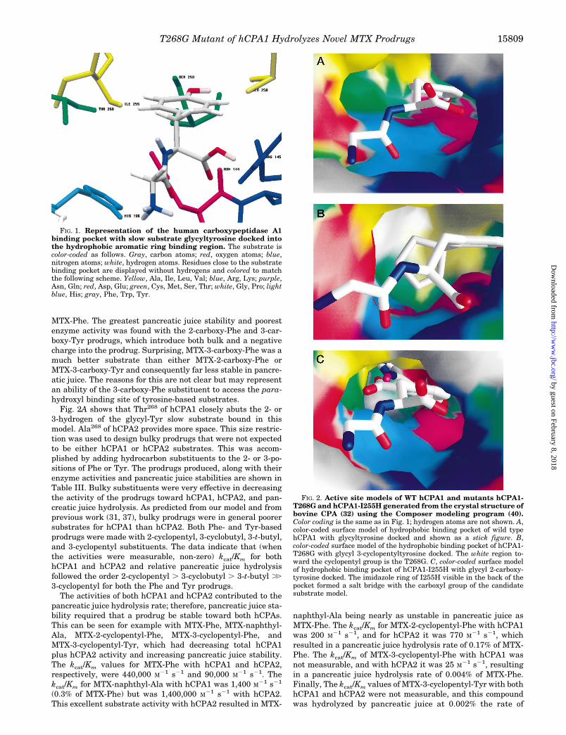
Design and Testing of MTX Prodrugs Predicted to Be Stablein Vivo—The active site model of hCPA1 was generated asdescribed elsewhere (40) from the crystal structure of bovineCPA (32) using the Composer modeling program. This model isshown in Figs. 1 and 2A. The hCPA2 model is similar. Theexceptions to this are a Gly in place of Ser at 253, Ser in placeof Thr at 254, and an Ala in place of Thr at residue 268 (31).These substitutions participate to make a larger binding pocketin hCPA2 compared with hCPA1 and are presumably respon-sible for the hydrolysis of bulkier substrates by hCPA2 versushCPA1 as has been reported for both the murine and humanenzymes (31, 37).
The models of hCPA1 and hCPA2 were used to design MTXprodrugs that would not be substrates for either enzyme. Suit-ability of the designed and synthesized prodrugs for ADEPTwas assessed by assaying them with WT hCPA1 and hCPA2and testing their stability in pancreatic juice (and in mice; Ref.42). Three general types of structural modification were ex-plored for enhancement of prodrug stability. These included (a)the introduction of a negative charge to the prodrug amino acidmoiety, (b) the addition of bulk to Phe- or Tyr-based prodrugs,and (c) the addition of both a charge and bulk.
Fig. 2 shows that the active sites of hCPA1 and hCPA2 arehighly hydrophobic. Therefore, MTX-Glu, MTX-Asp, MTX-2-carboxy-Phe, MTX-3-carboxy-Phe, and MTX-3-carboxy-Tyrwere produced. All of these compounds would introduce a neg-atively charged carboxyl group into the hydrophobic pocket andas such were not expected to be good hCPA substrates. Theactivities of these compounds with hCPAs and their relativestabilities in human pancreatic juice (compared with MTX-Phewith a defined rate of 100%) are shown in Table III. All of thesecharged compounds are poor substrates for both of the CPAsand are considerably more stable in pancreatic juice than is
TABLE IIIComparison of rate of prodrug pancreatic juice hydrolysis with CPA enzyme kinetics
CompoundPancreatic juice(% of MTX-Phehydrolysis rate)
hCPA1a hCPA2a
Vmax Km kcat/Km Vmax Km kcat/Km
mmol/min/mg mM 1/M(s) mmol/min/mg mM 1/M(s)
MTX-Phe 100 3.24 0.0043 440,000 8.06 0.056 90,000Negatively charged prodrugs
MTX-Glu 0.12 0.094 0.1 170 0 NDb 0MTX-Asp 0.06 0.063 0.5 80 0 ND 0MTX-2-carboxy-Phe 0 0.003 0.7 3 ,0.0005@50 mM ND NDMTX-3-carboxy-Tyr 0.008 0.009 0.5 10 0.01 0.2 40MTX-3-carboxy-Phe 0.27 0.08 30 1550 0.63 120 3100
Bulky aromatic prodrugs (phenylalanine based)MTX-2-iodo-Phe ND 3.42 0.017 120,000 36 0.05 420,000MTX-naphthyl-Ala 67 0.16 0.065 1400 36.9 0.016 1,400,000MTX-2-cyclopentyl-Phe 0.17 0.021 0.06 200 0.11 0.081 770MTX-2-cyclohexyl-Phe 0.0067 0 ND 0 0 ND 0MTX-3-cyclobutyl-Phe 0.053 0 ND 0 0.086 0.2 260MTX-3-t-butyl-Phe 0.02 0 ND 0 0.04 0.3 80MTX-3-cyclopentyl-Phe 0.004 0 ND 0 0.012 0.3 25MTX-3-n-pentyl-Phe 0 0 ND 0 0 ND 0
Bulky aromatic prodrugs (tyrosine-based)MTX-3,5-diiodo-Tyr 0.093 0 ND 0 0.31 0.3 610MTX-2-cyclopentyl-Tyr 0.14 0.43 0.4 575 1 0.6 910MTX-3-cyclobutyl-Tyr 0.0067 0 ND 0 0 ND 0MTX-3-t-butyl-Tyr 0.0013 0 ND 0 0 ND 0MTX-3-cyclopentyl-Tyr 0.002 0 ND 0 0 ND 0
a Limit of detection for Vo was 0.0006 mmol/min/mg for hCPA1 and 0.002 mmol/min/mg for hCPA2. Assuming a Km of ;50 mM, the limit of kcat/Kmdetection 5 5 and 20/(M)(s), respectively.
b ND, not determined.
T268G Mutant of hCPA1 Hydrolyzes Novel MTX Prodrugs15808
by guest on February 8, 2018http://w
ww
.jbc.org/D
ownloaded from
MTX-Phe. The greatest pancreatic juice stability and poorestenzyme activity was found with the 2-carboxy-Phe and 3-car-boxy-Tyr prodrugs, which introduce both bulk and a negativecharge into the prodrug. Surprising, MTX-3-carboxy-Phe was amuch better substrate than either MTX-2-carboxy-Phe orMTX-3-carboxy-Tyr and consequently far less stable in pancre-atic juice. The reasons for this are not clear but may representan ability of the 3-carboxy-Phe substituent to access the para-hydroxyl binding site of tyrosine-based substrates.
Fig. 2A shows that Thr268 of hCPA1 closely abuts the 2- or3-hydrogen of the glycyl-Tyr slow substrate bound in thismodel. Ala268 of hCPA2 provides more space. This size restric-tion was used to design bulky prodrugs that were not expectedto be either hCPA1 or hCPA2 substrates. This was accom-plished by adding hydrocarbon substituents to the 2- or 3-po-sitions of Phe or Tyr. The prodrugs produced, along with theirenzyme activities and pancreatic juice stabilities are shown inTable III. Bulky substituents were very effective in decreasingthe activity of the prodrugs toward hCPA1, hCPA2, and pan-creatic juice hydrolysis. As predicted from our model and fromprevious work (31, 37), bulky prodrugs were in general poorersubstrates for hCPA1 than hCPA2. Both Phe- and Tyr-basedprodrugs were made with 2-cyclopentyl, 3-cyclobutyl, 3-t-butyl,and 3-cyclopentyl substituents. The data indicate that (whenthe activities were measurable, non-zero) kcat/Km for bothhCPA1 and hCPA2 and relative pancreatic juice hydrolysisfollowed the order 2-cyclopentyl . 3-cyclobutyl . 3-t-butyl ..3-cyclopentyl for both the Phe and Tyr prodrugs.
The activities of both hCPA1 and hCPA2 contributed to thepancreatic juice hydrolysis rate; therefore, pancreatic juice sta-bility required that a prodrug be stable toward both hCPAs.This can be seen for example with MTX-Phe, MTX-naphthyl-Ala, MTX-2-cyclopentyl-Phe, MTX-3-cyclopentyl-Phe, andMTX-3-cyclopentyl-Tyr, which had decreasing total hCPA1plus hCPA2 activity and increasing pancreatic juice stability.The kcat/Km values for MTX-Phe with hCPA1 and hCPA2,respectively, were 440,000 M21 s21 and 90,000 M21 s21. Thekcat/Km for MTX-naphthyl-Ala with hCPA1 was 1,400 M21 s21
(0.3% of MTX-Phe) but was 1,400,000 M21 s21 with hCPA2.This excellent substrate activity with hCPA2 resulted in MTX-
naphthyl-Ala being nearly as unstable in pancreatic juice asMTX-Phe. The kcat/Km for MTX-2-cyclopentyl-Phe with hCPA1was 200 M21 s21, and for hCPA2 it was 770 M21 s21, whichresulted in a pancreatic juice hydrolysis rate of 0.17% of MTX-Phe. The kcat/Km of MTX-3-cyclopentyl-Phe with hCPA1 wasnot measurable, and with hCPA2 it was 25 M21 s21, resultingin a pancreatic juice hydrolysis rate of 0.004% of MTX-Phe.Finally, The kcat/Km values of MTX-3-cyclopentyl-Tyr with bothhCPA1 and hCPA2 were not measurable, and this compoundwas hydrolyzed by pancreatic juice at 0.002% the rate of
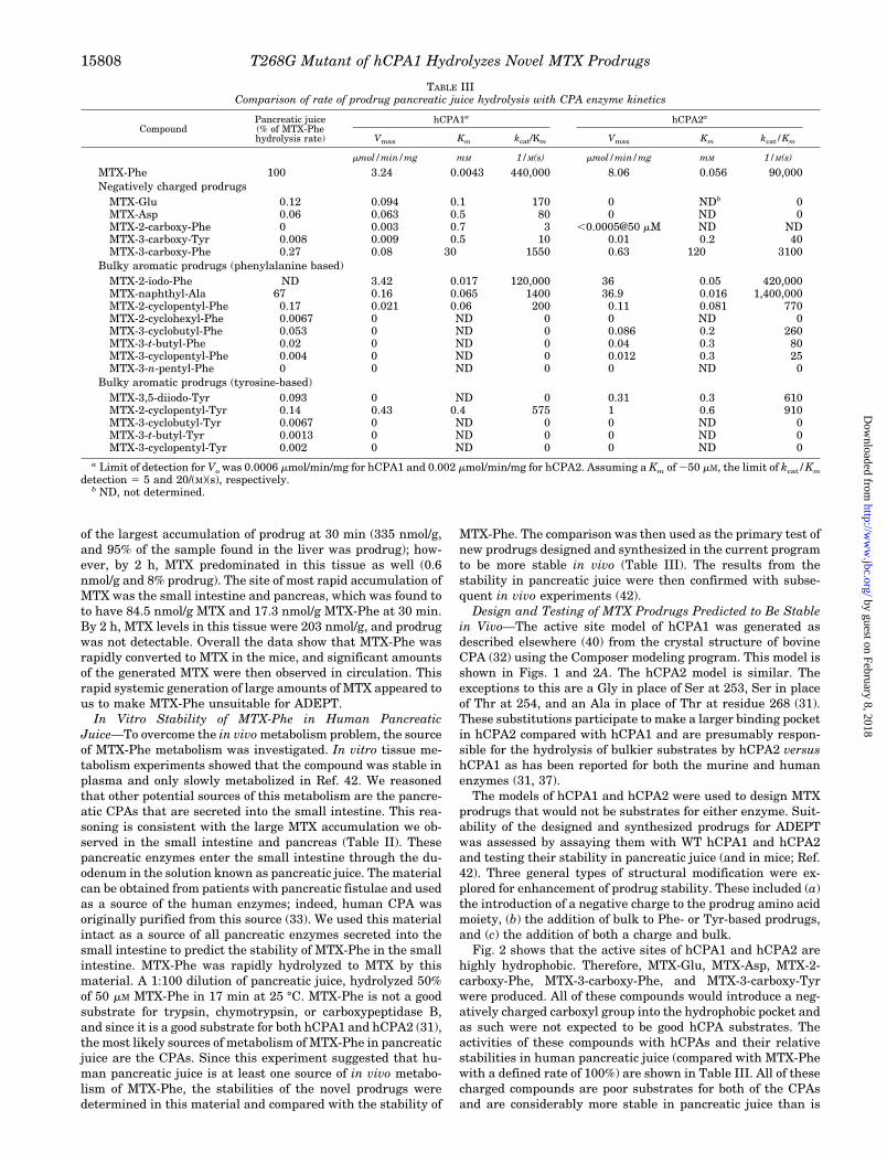
FIG. 1. Representation of the human carboxypeptidase A1binding pocket with slow substrate glycyltyrosine docked intothe hydrophobic aromatic ring binding region. The substrate iscolor-coded as follows. Gray, carbon atoms; red, oxygen atoms; blue,nitrogen atoms; white, hydrogen atoms. Residues close to the substratebinding pocket are displayed without hydrogens and colored to matchthe following scheme. Yellow, Ala, Ile, Leu, Val; blue, Arg, Lys; purple,Asn, Gln; red, Asp, Glu; green, Cys, Met, Ser, Thr; white, Gly, Pro; lightblue, His; gray, Phe, Trp, Tyr.
FIG. 2. Active site models of WT hCPA1 and mutants hCPA1-T268G and hCPA1-I255H generated from the crystal structure ofbovine CPA (32) using the Composer modeling program (40).Color coding is the same as in Fig. 1; hydrogen atoms are not shown. A,color-coded surface model of hydrophobic binding pocket of wild typehCPA1 with glycyltyrosine docked and shown as a stick figure. B,color-coded surface model of the hydrophobic binding pocket of hCPA1-T268G with glycyl 3-cyclopentyltyrosine docked. The white region to-ward the cyclopentyl group is the T268G. C, color-coded surface modelof hydrophobic binding pocket of hCPA1-I255H with glycyl 2-carboxy-tyrosine docked. The imidazole ring of I255H visible in the back of thepocket formed a salt bridge with the carboxyl group of the candidatesubstrate model.
T268G Mutant of hCPA1 Hydrolyzes Novel MTX Prodrugs 15809
by guest on February 8, 2018http://w
ww
.jbc.org/D
ownloaded from

MTX-Phe. As shown elsewhere (42), compounds with pancreaticjuice hydrolysis rates in the range of 0.01% of MTX-Phe or lesshad excellent in vivo stability.
Design and Testing of Mutants of hCPA Predicted to Effi-ciently Hydrolyze Prodrugs That Are Stable in Vivo—Our nextgoal was to utilize our structural models of hCPA1 and hCPA2to model mutant hCPAs into which the “stable prodrugs” couldbe docked. Fig. 2, B and C, shows models of two such mutantsof hCPA1, I255H and T268G. The former was designed tointroduce a positive charge into the active site to bind thecarboxyl-containing prodrugs. The latter was designed to pro-duce an active site that would bind Phe- or Tyr-based prodrugssubstituted at the 2- or 3-position. I255K and I255H/T268Gwere also designed for the charged prodrugs, and T268A,I255A, and I255A/T268A were also designed to accept bulk.The A268G mutant of hCPA2 was also predicted and made forthe bulky prodrugs. Expression and isolation of the hCPA1mutants T268A, T268G, I255A, and T268A/I255A and thehCPA2 mutant A268G were accomplished using the yeast ex-pression system described under “Experimental Procedures.”All of these mutants were synthesized and secreted by theyeast as catalytically active enzymes. Unfortunately, no hCPAprotein or enzymatic activity could be found in yeast expressingI255K, I255H, or I255H/T268G. Presumably, these three pro-teins misfolded and were degraded prior to secretion.
Enzyme Stability—Several of the WT and mutant enzymeswere characterized for their stability in phosphate-bufferedsaline at 4, 25, 37, and 50 °C. The results from these experi-ments are shown in Table IV. All enzymes are quite stable at4 °C. The I255A mutants were the least stable, since theyexhibited half-lives of 8 h at 25 °C while all other mutants werestable for several days at this temperature. At 37 °C, WThCPAs as well as hCPA1-T268A and hCPA2-A268G lost littleactivity after 2 days, and while hCPA1-T268G was less stableit also had a half-life of 24 h. At 50 °C, differential stabilitieswere clearer. At this temperature, the order of stability was WThCPA2 . hCPA2-A268G . hCPA1-T268A . WT hCPA1 .hCPA1-T268G. In summary, these data show that the mutantsof hCPA are active and stable.
Enzyme Kinetics—Specific activities of these enzymes withthe model substrates hippuryl-Phe and hippuryl-Phe lactateare shown in Table V. As can be seen, all of these enzymeshydrolyzed these substrates efficiently. Interestingly, WTCPA2 and its 268G mutant had very large esterase hydrolyticrates.
Charged Prodrugs—Kinetics of hCPA1-T268G, hCPA1-T268A, and hCPA2-A268G with the MTX prodrugs are shownin Tables VI and VII. Table VI shows the activity of the mutanthCPAs with the charged prodrugs from Table I. As indicatedabove, the hCPA mutants designed to hydrolyze these prodrugs
(I255H, I255K, and I255H/T268G) could not be isolated. ThehCPA mutants designed for the bulky hydrophobic prodrugswere not expected to efficiently hydrolyze charged prodrugs.However, mutation of hCPA1 to T268G produced a 5-fold en-hancement in kcat/Km for the hydrolysis of MTX-3-carboxy-Phe,a 50-fold enhancement in the kcat/Km for hydrolysis of MTX-3-carboxy-Tyr and a 250-fold enhancement in the kcat/Km forhydrolysis of MTX-2-carboxy-Phe. The A268G mutation ofhCPA2 was without effect on any of the charged prodrugsmeasured. The mutation of hCPA1 to T268A produced a mod-est enhancement in the hydrolysis of two of these prodrugsintermediate between the WT and T268G rates. These rateenhancements are likely due to the additional tolerance in theT268G and T268A active sites for bulk at the 2- and 3-positionsof the substrate aromatic ring (as described below). The en-hancements by hCPA1-T268G are probably inadequate for AD-EPT, however, since the absolute kcat/Km values for these re-actions were all quite small compared with that for hCPA1with MTX-Phe (approximately 1000-fold less).
Bulky Aromatic Prodrugs—Table VII shows the activity ofthe hCPA mutants with the bulky hydrophobic prodrugs fromTable I. The 268G mutants were designed to remove unfavor-able interaction between the enzyme and 2- or 3-substitutedMTX-Phe or MTX-Tyr, allowing their efficient binding andhydrolysis. As shown in Table VII, this goal was realized; themutant hCPA1-T268G proved to efficiently hydrolyze a varietyof substrates with bulky 2- or 3-position substituents (kcat/Km
$ 100,000 M21 s21). In contrast, the T268A mutant is a muchpoorer enzyme with all of these substrates. Thus, the minimalreplacement of methyl with hydrogen at residue 268 produceddramatic rate enhancements. The hCPA2-A268G mutant alsoprovided dramatic rate enhancements for some prodrugs; how-ever, kcat/Km for the mutant with none of the stable prodrugs(pancreatic juice hydrolysis rate #0.01% of MTX-Phe) exhib-ited kcat/Km approaching 100,000 M21 s21. Therefore, this en-zyme was not as efficient as hCPA1-T268G with thesecompounds.
Since the active site of the WT hCPA2 has Ala at position268, the activity of the hCPA1-T268A mutant was expected toparallel that of WT hCPA2. Data in Tables VI and VII demon-strate that this was observed, since the activity of hCPA1-T268A followed that of WT hCPA2 for most prodrugs assayed.
The I255A and I255A/T268A mutants of hCPA1 did notprove useful for the hydrolysis of any of the bulky prodrugs(data not shown). Interestingly, the activity of the I255A mu-tants appeared in general to be dictated by the identity of theamino acid at position 268. Thus, substrate specificity of I255Awas similar to that of WT hCPA1, and the substrate specificity
TABLE IVThermal stability of WT and mutant hCPAs
Samplet1⁄2
4 °C 25 °C 37 °C 50°C
hWT hCPA1 Longa .170b 140 0.77hCPA1-T268A Long .170b .96b 0.93hCPA1-T268G Long 80 24 0.17hCPA1-I255A Long 8 1hCPA1-I255A/T268A Long 8 1WT hCPA2 Long .170b .170b 8.75hCPA2-A268G Long .170b 140 5.1
a At 4 °C, all the mutants were stable for at least 2 months. Inaddition, both WT enzymes, the 268G mutants, and the T268A mutanthave been stored at this temperature for 2 years with little or no loss.
b Less than 10% decrease in activity of the enzyme over the statedtime period.
TABLE VSpecific activity of WT and mutant hCPAs with hippuryl-Phe and
hippuryl-Phe lactateAssay conditions were as follows: 0.5 mM L-hippuryl-Phe or 1 mM
DL-hippuryl-Phe lactate, at 25 °C in 25 mM Tris-HCl, 100 mM NaCl, pH7.4.
EnzymeSpecific acitvity
Hippuryl-Phe Hippuryl-Phe lactate
mmol/min/mg
WT hCPA1 7.2 790hCPA1-T268A 7.4 160hCPA1-T268G 20 60hCPA1-I255A 8 350hCPA1-I255A/T268A 12 50WT hCPA2 8.7 2800hCPA2-A268G 4.9 1900Bovine CPA 24.1 310Rat CPA1 11 560
T268G Mutant of hCPA1 Hydrolyzes Novel MTX Prodrugs15810
by guest on February 8, 2018http://w
ww
.jbc.org/D
ownloaded from
of I255A/T268A was similar to that of T268A. This suggeststhat position 255 has little impact on substrate specificity inthis class of aromatic substrates.
Overall the most useful mutant was hCPA1-T268G, since itwas able to efficiently metabolize several of the bulky, stableMTX prodrugs. The most useful substrates for the enzyme,based upon large kcat/Km for the mutant and good pancreaticjuice stability, were MTX-3-t-butyl-Phe, MTX-3-cyclobutyl-Phe, MTX-3-cyclopentyl-Phe, MTX-3-cyclopentyl-Tyr, andMTX-3-cyclobutyl-Tyr. All were approximately 4000-fold ormore stable in pancreatic juice than MTX-Phe, and all werehydrolyzed (at 25 °C) with second order rate constants of orgreater than 105 M21 s21.
Conjugation of Mutant hCPA1-T268G to an Antibody—Totest the utility of the enzyme prodrug combination for ADEPT,hCPA1-T268G was conjugated to two antibodies. ING-1 bindsto Epcam, a molecule expressed on a variety of epithelial celltumors (46). Campath-1H binds to CDw52, which is expressedon a variety of T and B cells but not on most epithelial cells ortumors (30, 47). To couple hCPA1-T268G to these antibodies, amaleimide was placed on the enzyme with Sulfo-SMCC; thisprocess had no effect on enzyme activity. A free thiol was thenplaced on the antibody with 2-iminothiolane. The hCPA1-T268Gzantibody conjugate was then generated upon combina-tion of the activated proteins under nitrogen. Conjugate waspurified from the small amount of protein aggregate formed inthe reaction and from free hCPA1-T268G by gel filtration onPharmacia Superose 12 HR 10/30.
The product was assayed for its carboxypeptidase activityand ability to bind to antigen. Table VIII shows the specific
activity of conjugates toward hippuryl-Phe. Based upon theenzyme activity measured in this way, 1–1.5 mol of hCPA1-T268G appeared to be routinely associated with each mol ofantibody. Table VIII also shows the percentage of the enzymeactivity that bound to the relevant antigen along with thebinding affinity as determined according to Lindmo (49). Bothantibody conjugates expressed enzyme activity and bound toantigen. The percentage of conjugate that bound to antigenwas 75–85%, and the Kd values are in agreement with liter-ature indicating that the antigen combining region of the an-tibody was not adversely effected by the conjugation process(30, 46, 47).
Use of Conjugates in a Cell Culture Model of ADEPT—Theconjugates were used in a series of in vitro ADEPT experi-ments. For these experiments, HT-29 cells were grown in 5%human serum. Preliminary experiments had shown that some
TABLE VIIIEnzyme activity and antigen binding affinity for conjugates
Conjugate Specific activityaPercentagebound toantigenb
Kd
mmol/min/mgtotal protein
mmol/min/mghCPA1c
% nM
ING-1zhCPA1-T268G 4.2 22.2 85 0.4C-1HzhCPA1-T268G 3.8 20 74 2.2
a Assayed with hippuryl-phenylalanine.b For ING-1 conjugate, binding was performed on fresh, trypsininzed
HT-29 cells. For C-1H conjugate, binding was performed with formal-dehyde-fixed Wein-133 cells.
c Specific activity calculation assuming 1 mol of CPA/mol of antibody.
TABLE VIKinetics of charged prodrugs with hCPA1-T268G, hCPA1-T268A, and hCPA2-A268G
CompoundhCPA1-T268G hCPA1-T268A hCPA2-A268G
Vmax Kmkcat/Km)
(kcat/Km)mut/(kcat/Km)wt
Vmax Kmkcat/Km
(kcat/Km)mut/(kcat/Km)wt
Vmax Kmkcat/Km
(kcat/Km)mut/(kcat/Km)wt
mmol/min/mg
mM 1/M(s) mmol/min/mg
mM 1/M(s) mmol/min/mg
mM 1/M(s)
MTX-Glu 0.012 0.06 120 0.7 NDa ND ND ND 0 ND NDMTX-ASP 0.024 0.25 60 0.75 ND ND ND ND 0.12 0.9 76MTX-2-carboxy-Phe 0.1 0.09 670 250 0.007 0.06 60 20 ,0.002@50 mM ND NDMTX-3-carboxy-Tyr 0.06 0.07 500 50 0.01 0.1 60 6 0.02 0.2 70 2MTX-3-carboxy-Phe 0.87 0.07 7200 4.6 0.11 0.05 1300 0.8 1.1 0.25 2500 0.8
a ND, not determined. Detection limits are as in Table III.
TABLE VIIKinetics of bulky aromatic prodrugs with hCPA1-T268G, hCPA1-T268A, and hCPA2-A268G
CompoundhCPA1-T268G hCPA1-T268A hCPA2-A268G
Vmax Kmkcat/Km)
(kcat/Km)mut/(kcat/Km)wt
Vmax Kmkcat/Km
(kcat/Km)mut/(kcat/Km)wt
Vmax Kmkcat/Km
(kcat/Km)mut/(kcat/Km)wt
mmol/min/mg
mM 1/M(s) mmol/min/mg
mM 1/M(s) mmol/min/mg
mM 1/M(s)
Phenylalanine-based compoundsMTX-Phe 3.74 0.0003 7,350,000 17 3.46 0.0009 2,250,000 5.1 3.1 0.045 41,000 0.45MTX-2-iodo-Phe 13.7 0.0004 2.7E107 230 7.56 0.009 480,000 4.1 NDa ND ND NDMTX-naphthyl-Ala 1.15 0.0005 1,400,000 1,000 10.8 0.01 640,000 460 21.5 0.002 5,500,000 4MTX-2-cyclopentyl-Phe 4.25 0.016 160,000 790 0.15 0.06 1,600 8 2.6 0.008 190,000 250MTX-2-cyclohexyl-Phe 0.094 0.08 700 .100b 0.009 0.06 100 .5b 0.3 0.19 900 .50b
MTX-3-cyclobutyl-Phe 5.2 0.002 1,800,000 .400,000b 0.21 0.16 750 .40b 0.163 0.047 2100 8MTX-3-t-butyl-Phe 1.3 0.002 380,000 .80,000b 0 ND ND ND 0.1 0.022 2700 34MTX-3-cyclopentyl-Phe 2.35 0.015 92,000 .20,000b 0.002 0.03 30 0.02 0.013 1000 40MTX-3-n-pentyl-Phe 0.18 0.008 14,000 .3,000b 0 ND ND ND 0 ND ND ND
Tyrosine-based compoundsMTX-3,5-diiodo-Tyr 2.73 0.033 53,000 .10,000 ND ND ND ND 2.7 0.0001 520,000 850MTX-2-cyclopentyl-Tyr 1.82 0.026 41,000 70 ND ND ND ND 3.6 0.008 260,000 280MTX-3-cyclobutyl-Tyr 5 0.012 280,000 .60,000b 0.036 0.05 450 .20b 0.017 0.002 5200 .300b
MTX-3-t-butyl-Tyr 0.2 0.001 110,000 .20,000b ND ND ND ND 0.049 0.003 9000 .400b
MTX-3-cyclopentyl-Tyr 5.04 0.019 160,000 .30,000b 0 ND ND ND 0.023 0.002 7000 .400b
a ND 5 not determined. Detection limits are as in Table III.b See Table III, footnote a.
T268G Mutant of hCPA1 Hydrolyzes Novel MTX Prodrugs 15811
by guest on February 8, 2018http://w
ww
.jbc.org/D
ownloaded from
component in fetal bovine serum was a competitive inhibitor ofhCPA1 and hCPA1-T268G. This inhibitor greatly decreasedthe efficacy of both enzymes, resulting in the requirement forhigh concentrations of conjugate to produce an in vitro ADEPTresponse. Human serum also inhibited the enzymes, but theinhibition was less severe and most closely fit an uncompetitivekinetic model (data not shown). Since ADEPT was to be tar-geted to humans, the human serum conditions were consideredthe more relevant.
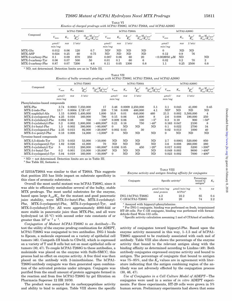
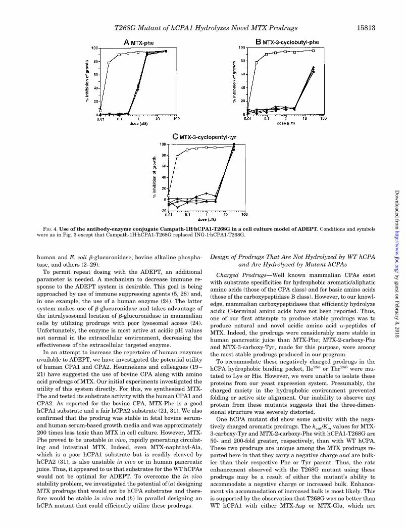
HT-29 cells, which were found to express Epcam but notsignificant amounts of CDw52, were seeded and allowed toadhere for 24 h. The cells on the plate were then incubated at37 °C for 1 h with 0, 2, 10, or 50 mg/ml conjugate consisting ofhCPA1-T268G coupled to either ING-1 or Campath-1H. Cellswere then washed free of unbound antibody and allowed togrow for 72 h in the presence of varying concentrations ofMTX-Phe, MTX-3-cyclobutyl-Phe, or MTX-3-cyclopentyl-Tyr.The results of these experiments are shown in Figs. 3 and 4 forthe ING-1 and Campath-1H conjugates, respectively. The IC50
for MTX in these cells under these conditions was 10 nM. In theabsence of conjugate, all three prodrugs were considerably lesstoxic than MTX. The stable prodrugs, MTX-3-cyclobutyl-Pheand MTX-3-cyclopentyl-Tyr, were 800 times less toxic thanMTX (IC50 values for the two prodrugs were 8.5 and 7.8 mM,respectively). MTX-Phe was 200 times less toxic than MTXunder these conditions.
When the cells were preincubated with the Epcam-specificconjugate ING-1zhCPA1-T268G, as expected all three prodrugsbecame more potent. Even the lowest dose of conjugate, 2mg/ml, increased the toxicity of all three compounds. At thisdose, the IC50 values became 0.01, 0.066, and 0.16 for MTX-Phe, MTX-3-cyclobutyl-Phe, and MTX-3-cyclopentyl-Tyr, re-spectively, in good agreement with the relative activities of the
T268G mutant enzyme with these prodrugs shown in TableVII. At 10 and 50 mg/ml conjugate, the IC50 values for all threeprodrugs approached that of MTX itself, indicating excellentconversion of all three prodrugs to the drug at these conjugatedoses.
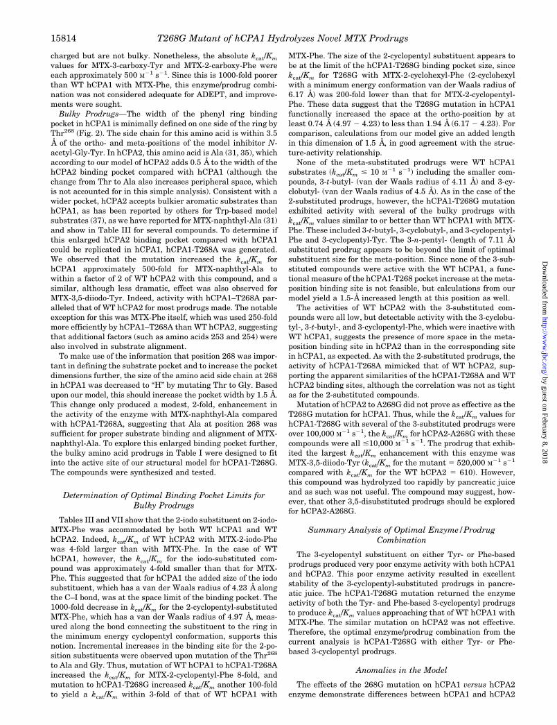
In contrast, when the cells were preincubated with the con-trol conjugate Campath-1HzhCPA1-T268G, the IC50 values didnot change with increasing conjugate. Thus, in the presence of50 mg/ml Campath-1HzhCPA1-T268G conjugate, IC50 valuesfor the three prodrugs were 2.5, 13, and 15 mM, respectively, notdifferent from the prodrugs in the absence of conjugate. There-fore, activity of the ING-1zhCPA1-T268G conjugate was immu-nospecific. In summary, the data demonstrate that an immunespecific conjugate of the hCPA mutant hCPA1-T268G, can con-vert in vivo stable MTX prodrugs to MTX in a cell cultureADEPT experiment.
DISCUSSION
ADEPT has the potential to greatly enhance the tumor se-lectivity of cancer chemotherapy by generating a means fortumor-specific synthesis of chemotherapeutic or other toxicagents. This is accomplished through staged, systemic deliveryof components that localize to tumor sites and selectively gen-erate active drug(s) at the tumor (2–7). System requirementsfor a successful ADEPT strategy include (a) a prodrug that isnot activated to drug in vivo by host endogenous enzymes, (b)an enzyme capable of producing the drug from the prodrug at arate sufficient to produce toxic levels of the drug at the specificsite, and (c) an antibody capable of targeting and localizing theenzyme to the tumor. A number of enzymes have been reportedfor use in ADEPT. These include bovine carboxypeptidase A,Pseudomonas carboxypeptidase G, several bacterial b-lactama-ses, E. coli penicillin G amidase, yeast cytosine deaminase,
FIG. 3. Use of the antibody-enzyme conjugate ING-1zhCPA1-T268G in a cell culture model of ADEPT. HT-29 cells were grown in 95%RPMI 1640, 5% human serum. Then 50 ml of conjugate ING-1zhCPA1-T268G in fresh human growth medium was added to triplicate wells.Conjugate concentrations of 0 (filled circles), 2 (filled triangles), 10 (filled diamonds), and 50 (filled squares) mg/ml were used. After 1 h at 37 °Cin a 5% CO2 incubator, conjugate was removed, and the plates were washed three times with growth medium without conjugate. Finally, prodrugat 0, 0.01, 0.03, 0.1, 0.3, 1, 3, and 30 mM was added in 200 ml of fresh human growth medium, and cells were allowed to grow for 72 h. Prodrugsused were as follows. A, MTX-Phe; B, MTX-3-cyclobutyl-Phe; C, MTX-3-cyclopentyl-Tyr. In control experiments (open squares), MTX replacedconjugate plus prodrug.
T268G Mutant of hCPA1 Hydrolyzes Novel MTX Prodrugs15812
by guest on February 8, 2018http://w
ww
.jbc.org/D
ownloaded from
human and E. coli b-glucuronidase, bovine alkaline phospha-tase, and others (2–29).
To permit repeat dosing with the ADEPT, an additionalparameter is needed. A mechanism to decrease immune re-sponse to the ADEPT system is desirable. This goal is beingapproached by use of immune suppressing agents (5, 28) and,in one example, the use of a human enzyme (24). The lattersystem makes use of b-glucuronidase and takes advantage ofthe intralysosomal location of b-glucuronidase in mammaliancells by utilizing prodrugs with poor lysosomal access (24).Unfortunately, the enzyme is most active at acidic pH valuesnot normal in the extracellular environment, decreasing theeffectiveness of the extracellular targeted enzyme.
In an attempt to increase the repertoire of human enzymesavailable to ADEPT, we have investigated the potential utilityof human CPA1 and CPA2. Heunnekens and colleagues (19–21) have suggested the use of bovine CPA along with aminoacid prodrugs of MTX. Our initial experiments investigated theutility of this system directly. For this, we synthesized MTX-Phe and tested its substrate activity with the human CPA1 andCPA2. As reported for the bovine CPA, MTX-Phe is a goodhCPA1 substrate and a fair hCPA2 substrate (21, 31). We alsoconfirmed that the prodrug was stable in fetal bovine serum-and human serum-based growth media and was approximately200 times less toxic than MTX in cell culture. However, MTX-Phe proved to be unstable in vivo, rapidly generating circulat-ing and intestinal MTX. Indeed, even MTX-naphthyl-Ala,which is a poor hCPA1 substrate but is readily cleaved byhCPA2 (31), is also unstable in vivo or in human pancreaticjuice. Thus, it appeared to us that substrates for the WT hCPAswould not be optimal for ADEPT. To overcome the in vivostability problem, we investigated the potential of (a) designingMTX prodrugs that would not be hCPA substrates and there-fore would be stable in vivo and (b) in parallel designing anhCPA mutant that could efficiently utilize these prodrugs.
Design of Prodrugs That Are Not Hydrolyzed by WT hCPAand Are Hydrolyzed by Mutant hCPAs
Charged Prodrugs—Well known mammalian CPAs existwith substrate specificities for hydrophobic aromatic/aliphaticamino acids (those of the CPA class) and for basic amino acids(those of the carboxypeptidase B class). However, to our knowl-edge, mammalian carboxypeptidases that efficiently hydrolyzeacidic C-terminal amino acids have not been reported. Thus,one of our first attempts to produce stable prodrugs was toproduce natural and novel acidic amino acid a-peptides ofMTX. Indeed, the prodrugs were considerably more stable inhuman pancreatic juice than MTX-Phe; MTX-2-carboxy-Pheand MTX-3-carboxy-Tyr, made for this purpose, were amongthe most stable prodrugs produced in our program.
To accommodate these negatively charged prodrugs in thehCPA hydrophobic binding pocket, Ile255 or Thr268 were mu-tated to Lys or His. However, we were unable to isolate theseproteins from our yeast expression system. Presumably, thecharged moiety in the hydrophobic environment preventedfolding or active site alignment. Our inability to observe anyprotein from these mutants suggests that the three-dimen-sional structure was severely distorted.
One hCPA mutant did show some activity with the nega-tively charged aromatic prodrugs. The kcat/Km values for MTX-3-carboxy-Tyr and MTX-2-carboxy-Phe with hCPA1-T268G are50- and 200-fold greater, respectively, than with WT hCPA.These two prodrugs are unique among the MTX prodrugs re-ported here in that they carry a negative charge and are bulk-ier than their respective Phe or Tyr parent. Thus, the rateenhancement observed with the T268G mutant using theseprodrugs may be a result of either the mutant’s ability toaccommodate a negative charge or increased bulk. Enhance-ment via accommodation of increased bulk is most likely. Thisis supported by the observation that T268G was no better thanWT hCPA1 with either MTX-Asp or MTX-Glu, which are
FIG. 4. Use of the antibody-enzyme conjugate Campath-1HzhCPA1-T268G in a cell culture model of ADEPT. Conditions and symbolswere as in Fig. 3 except that Campath-1HzhCPA1-T268G replaced ING-1zhCPA1-T268G.
T268G Mutant of hCPA1 Hydrolyzes Novel MTX Prodrugs 15813
by guest on February 8, 2018http://w
ww
.jbc.org/D
ownloaded from
charged but are not bulky. Nonetheless, the absolute kcat/Km
values for MTX-3-carboxy-Tyr and MTX-2-carboxy-Phe wereeach approximately 500 M21 s21. Since this is 1000-fold poorerthan WT hCPA1 with MTX-Phe, this enzyme/prodrug combi-nation was not considered adequate for ADEPT, and improve-ments were sought.
Bulky Prodrugs—The width of the phenyl ring bindingpocket in hCPA1 is minimally defined on one side of the ring byThr268 (Fig. 2). The side chain for this amino acid is within 3.5Å of the ortho- and meta-positions of the model inhibitor N-acetyl-Gly-Tyr. In hCPA2, this amino acid is Ala (31, 35), whichaccording to our model of hCPA2 adds 0.5 Å to the width of thehCPA2 binding pocket compared with hCPA1 (although thechange from Thr to Ala also increases peripheral space, whichis not accounted for in this simple analysis). Consistent with awider pocket, hCPA2 accepts bulkier aromatic substrates thanhCPA1, as has been reported by others for Trp-based modelsubstrates (37), as we have reported for MTX-naphthyl-Ala (31)and show in Table III for several compounds. To determine ifthis enlarged hCPA2 binding pocket compared with hCPA1could be replicated in hCPA1, hCPA1-T268A was generated.We observed that the mutation increased the kcat/Km forhCPA1 approximately 500-fold for MTX-naphthyl-Ala towithin a factor of 2 of WT hCPA2 with this compound, and asimilar, although less dramatic, effect was also observed forMTX-3,5-diiodo-Tyr. Indeed, activity with hCPA1–T268A par-alleled that of WT hCPA2 for most prodrugs made. The notableexception for this was MTX-Phe itself, which was used 250-foldmore efficiently by hCPA1–T268A than WT hCPA2, suggestingthat additional factors (such as amino acids 253 and 254) werealso involved in substrate alignment.
To make use of the information that position 268 was impor-tant in defining the substrate pocket and to increase the pocketdimensions further, the size of the amino acid side chain at 268in hCPA1 was decreased to “H” by mutating Thr to Gly. Basedupon our model, this should increase the pocket width by 1.5 Å.This change only produced a modest, 2-fold, enhancement inthe activity of the enzyme with MTX-naphthyl-Ala comparedwith hCPA1-T268A, suggesting that Ala at position 268 wassufficient for proper substrate binding and alignment of MTX-naphthyl-Ala. To explore this enlarged binding pocket further,the bulky amino acid prodrugs in Table I were designed to fitinto the active site of our structural model for hCPA1-T268G.The compounds were synthesized and tested.
Determination of Optimal Binding Pocket Limits forBulky Prodrugs
Tables III and VII show that the 2-iodo substituent on 2-iodo-MTX-Phe was accommodated by both WT hCPA1 and WThCPA2. Indeed, kcat/Km of WT hCPA2 with MTX-2-iodo-Phewas 4-fold larger than with MTX-Phe. In the case of WThCPA1, however, the kcat/Km for the iodo-substituted com-pound was approximately 4-fold smaller than that for MTX-Phe. This suggested that for hCPA1 the added size of the iodosubstituent, which has a van der Waals radius of 4.23 Å alongthe C–I bond, was at the space limit of the binding pocket. The1000-fold decrease in kcat/Km for the 2-cyclopentyl-substitutedMTX-Phe, which has a van der Waals radius of 4.97 Å, meas-ured along the bond connecting the substituent to the ring inthe minimum energy cyclopentyl conformation, supports thisnotion. Incremental increases in the binding site for the 2-po-sition substituents were observed upon mutation of the Thr268
to Ala and Gly. Thus, mutation of WT hCPA1 to hCPA1-T268Aincreased the kcat/Km for MTX-2-cyclopentyl-Phe 8-fold, andmutation to hCPA1-T268G increased kcat/Km another 100-foldto yield a kcat/Km within 3-fold of that of WT hCPA1 with
MTX-Phe. The size of the 2-cyclopentyl substituent appears tobe at the limit of the hCPA1-T268G binding pocket size, sincekcat/Km for T268G with MTX-2-cyclohexyl-Phe (2-cyclohexylwith a minimum energy conformation van der Waals radius of6.17 Å) was 200-fold lower than that for MTX-2-cyclopentyl-Phe. These data suggest that the T268G mutation in hCPA1functionally increased the space at the ortho-position by atleast 0.74 Å (4.97 2 4.23) to less than 1.94 Å (6.17 2 4.23). Forcomparison, calculations from our model give an added lengthin this dimension of 1.5 Å, in good agreement with the struc-ture-activity relationship.
None of the meta-substituted prodrugs were WT hCPA1substrates (kcat/Km # 10 M21 s21) including the smaller com-pounds, 3-t-butyl- (van der Waals radius of 4.11 Å) and 3-cy-clobutyl- (van der Waals radius of 4.5 Å). As in the case of the2-substituted prodrugs, however, the hCPA1-T268G mutationexhibited activity with several of the bulky prodrugs withkcat/Km values similar to or better than WT hCPA1 with MTX-Phe. These included 3-t-butyl-, 3-cyclobutyl-, and 3-cyclopentyl-Phe and 3-cyclopentyl-Tyr. The 3-n-pentyl- (length of 7.11 Å)substituted prodrug appears to be beyond the limit of optimalsubstituent size for the meta-position. Since none of the 3-sub-stituted compounds were active with the WT hCPA1, a func-tional measure of the hCPA1-T268 pocket increase at the meta-position binding site is not feasible, but calculations from ourmodel yield a 1.5-Å increased length at this position as well.
The activities of WT hCPA2 with the 3-substituted com-pounds were all low, but detectable activity with the 3-cyclobu-tyl-, 3-t-butyl-, and 3-cyclopentyl-Phe, which were inactive withWT hCPA1, suggests the presence of more space in the meta-position binding site in hCPA2 than in the corresponding sitein hCPA1, as expected. As with the 2-substituted prodrugs, theactivity of hCPA1-T268A mimicked that of WT hCPA2, sup-porting the apparent similarities of the hCPA1-T268A and WThCPA2 binding sites, although the correlation was not as tightas for the 2-substituted compounds.
Mutation of hCPA2 to A268G did not prove as effective as theT268G mutation for hCPA1. Thus, while the kcat/Km values forhCPA1-T268G with several of the 3-substituted prodrugs wereover 100,000 M21 s21, the kcat/Km for hCPA2-A268G with thesecompounds were all #10,000 M21 s21. The prodrug that exhib-ited the largest kcat/Km enhancement with this enzyme wasMTX-3,5-diiodo-Tyr (kcat/Km for the mutant 5 520,000 M21 s21
compared with kcat/Km for the WT hCPA2 5 610). However,this compound was hydrolyzed too rapidly by pancreatic juiceand as such was not useful. The compound may suggest, how-ever, that other 3,5-disubstituted prodrugs should be exploredfor hCPA2-A268G.
Summary Analysis of Optimal Enzyme/ProdrugCombination
The 3-cyclopentyl substituent on either Tyr- or Phe-basedprodrugs produced very poor enzyme activity with both hCPA1and hCPA2. This poor enzyme activity resulted in excellentstability of the 3-cyclopentyl-substituted prodrugs in pancre-atic juice. The hCPA1-T268G mutation returned the enzymeactivity of both the Tyr- and Phe-based 3-cyclopentyl prodrugsto produce kcat/Km values approaching that of WT hCPA1 withMTX-Phe. The similar mutation on hCPA2 was not effective.Therefore, the optimal enzyme/prodrug combination from thecurrent analysis is hCPA1-T268G with either Tyr- or Phe-based 3-cyclopentyl prodrugs.
Anomalies in the Model
The effects of the 268G mutation on hCPA1 versus hCPA2enzyme demonstrate differences between hCPA1 and hCPA2
T268G Mutant of hCPA1 Hydrolyzes Novel MTX Prodrugs15814
by guest on February 8, 2018http://w
ww
.jbc.org/D
ownloaded from

that cannot be accounted for in our model. Thus, the kcat/Km ofhCPA1-T268G with the 3-cyclobutyl prodrug was at least100,000 times greater than that of WT hCPA1 with this pro-drug and actually 4 times better than WT hCPA1 with MTX-Phe. In contrast, mutation of hCPA2 to hCPA2-A268G en-hanced the activity with this compound less than 10 times, andthe absolute kcat/Km for the mutant was about 40 times poorerthan WT hCPA2 with MTX-Phe. Similarly, the 3-cyclopentyl-Phe, 3-n-pentyl-Phe, 3-t-butyl-Phe, 3-cyclopentyl-Tyr, 3-cy-clobutyl-Tyr, and 3-t-butyl-Tyr prodrugs were all 10–1000-foldbetter substrates for hCPA1-T268G than for hCPA2-A268G.These differences define some as yet undetermined deficiencyin our model and stress the importance of result-directed modelrefinement, as used here, for this type of enzyme and prodrugdesign process.
Cell Culture ADEPT
For the purposes of ADEPT, the kinetic data indicate thathCPA1-T268G is the best suited enzyme. This conclusion isbased on the excellent stability of 3-substituted Phe and Tyrprodrugs in pancreatic juice and their equally excellent hydro-lytic rates with the hCPA1-T268G. This conclusion led us totest the combinations further in a series of in vitro ADEPTexperiments. In these experiments we showed that only anti-gen-specific binding of conjugate to tumor cells (i.e. the ING-1zhCPA1-T268G conjugate with HT-29 cells) was able to gen-erate an ADEPT response. Importantly, the data indicate thatthe tumor cell-bound enzyme is capable of generating MTXfrom otherwise in vivo stable prodrugs at relevant conjugateand prodrug concentrations. Indeed, in the presence of suffi-cient conjugate, the IC50 of the prodrugs tested approached thatof MTX itself. Since MTX efficacy is dependent upon both theconcentration and time of exposure, the IC50 data indicate thatMTX was rapidly and quantitatively generated in these cases.
The relative efficiency at which ING-1zhCPA1-T268G usedthe prodrugs tested in the cell culture experiments can beassessed by comparing the amount of conjugate required todrop the prodrug IC50 to that of MTX itself. For MTX-3-cyclo-pentyl-Tyr, the amount of conjugate needed was 10 mg/ml. Incomparison, for MTX-Phe the amount needed was less than 2mg/ml, since at all conjugate doses tested, MTX-Phe was asactive as MTX itself. This is consistent with the relative kcat/Km
values for these compounds with hCPA1-T268G (0.16 versus7.35 3 106 M21 s21 for MTX-3-cyclopentyl-Tyr and MTX-Phe,respectively).
The conjugate doses of 2–10 mg/ml required here can becompared with those reported for other enzyme prodrug sys-tems. Thus, others have reported the requirement of 0.25–10mg/ml for antibody-enzyme conjugates using b-lactamases (12,13), 1–10 mg/ml for E. coli b-glucuronidase (22, 23), 10–50mg/ml for bovine alkaline phosphatase, and 10–100 mg/ml forpenicillin-G amidase (8, 9). This comparison suggests the effi-ciency of the current system is comparable with that of mostother systems.
The b-lactamase system with a doxorubicin prodrug appearsto be more efficient than that reported here. However, theselectivity of the current system appears to be greater thanthat of the b-lactamase/doxorubicin system, since in the ab-sence of enzyme, the doxorubicin prodrug was only 7-fold lesstoxic than doxorubicin itself, while the MTX prodrugs were800-fold less toxic than MTX (13). Nonetheless, the greaterefficiency of the b-lactamase/doxorubicin system suggests thatfurther efficiency refinements may be warranted in the currentenzyme/prodrug system. Additional refinement also may besuggested by the inhibition of both WT and mutant hCPAs byboth bovine and human serum. While inhibition by bovine
serum was the more severe, inhibition of these enzymes byhuman serum was significant. Further site-directed or randommutagenesis experiments may permit these refinements. Theimportant advantage of the current system in using a humanenzyme warrants this additional refinement.
In summary, we have generated a novel human enzyme/prodrug combination for MTX (and potentially other antifo-lates) prodrugs. This system was realized through structure-driven site-directed mutagenesis of hCPA1 in parallel withstructure-driven design of in vivo stable prodrugs of MTX. Thework provides the first steps for the concept of generating invivo stable prodrugs of MTX and a human enzyme capable ofhydrolyzing them. The utility of this concept may extend wellbeyond the current enzyme prodrug system and provide a gen-eral methodology to make use of the wealth of structural datanow available on a variety of human enzymes to design other invivo stable prodrugs and mutate these enzymes to accept newprodrugs. The advantage that the current system may have forclinical use is the likely low immunogenicity of the mutanthCPA1 along with the known toxicity profile of MTX.
REFERENCES
1. Sell, S., and Reisfeld, R. A. (1985) Monoclonal Antibodies in Cancer, HumanaPress, Clifton, NJ
2. Bagshawe, K. D. (1989) Br. J. Cancer 60, 275–2813. Senter, P. D. (1990) FASEB 4, 188–1934. Senter, P. D., Wallace, P. M., Svensson, H. P., Vrudhula, V. M., Kerr, D. E.,
Hellstrom, I., and Hellstrom, K. E. (1993) Bioconjugate Chem. 4, 3–95. Melton, R. G., and Sherwood, R. F. (1996) J. Natl. Cancer Inst. 88, 153–1656. Huennekens, F. M. (1994) Trends Biotechnol. 12, 234–2397. Jungheim, L. N. and Shepherd, T. A. (1994) Chem. Rev. 94, 1553–15668. Bignami, G. S., Senter, P. D., Grothaus, P. G., Fischer, K. J., Humphreys, T.,
and Wallace, P. M. (1992) Cancer Res. 52, 5759–57649. Vrudhula, V. M., Senter, P. D., Fischer, K. J., and Wallace, P. M. (1993) J. Med.
Chem. 36, 919–92310. Wallace, P. M., and Senter, P. D. (1991) Bioconjugate Chem. 2, 349–35211. Senter, P. D., Schreiber, G. J., Hirschberg, D. L, Ashe, S. A., Hellstrom, K. E.,
and Hellstrom, I. (1989) Cancer Res. 49, 5789–579212. Svensson, H. P., Vrudhula, V. M., Emswiler, J. E., MacMaster, J. F., Cosand,
W. L., Senter, P. D., and Wallace, P. M. (1995) Cancer Res. 55, 2357–236513. Kerr, D. E., Schreiber, G. J., Vrudhula, V. M., Svensson, H. P., Hellstrom, I.,
Hellstrom, K. E., and Senter, P. D. (1995) Cancer Res. 55, 3558–356314. Bagshawe, K. D., Springer, C. J., Searle, F., Antoniw, P., Sharma, S. K.,
Melton, R. G., and Sherwood, R. F. (1988) Br. J. Cancer 58, 700–70315. Eccles, S. A., Court, W. J., Box, G. A., Dean, C. J., Melton, R. G., and Springer,
C. J. (1994) Cancer Res. 54, 5171–517716. Blakey, D. C., Burke, P. J., Davies, D. H., Dowell, R. I., East, S. J., Eckersley,
K. P., Fitton, J. E., McDaid, J., Melton, R. G., Niculescu-Duvaz, I. A.,Pinder, P. E., Sharma, S. K., Wright, A. F., and Springer, C. J. (1996)Cancer Res. 56, 3287–3292
17. Meyer, D. L., Jungheim, L. N., Law, K. L, Mikolajczyk, S. D., Shepherd, T. A.,Mackensen, D. G., Briggs, S. L., and Starling, J. J. (1993) Cancer Res. 53,3956–3963
18. Meyer, D. L., Law, K. L., Payne, J. K., Mikolajczyk, S. D., Zarrinmayen, H.,Jungheim, L. N., Kling, J. K., Shepherd, T. A., and Starling, J. J. (1995)Bioconjugate Chem. 6, 440–446
19. Kuefner, U., Lohrmann, U., Montejano, Y. D., Vitols, K. S., and Huennekens,F. M. (1989) Biochemistry 28, 2288–2297
20. Haenseler, E., Esswein, A., Vitols, K. S., Montejano, Y., Mueller, B. M.,Reisfeld, R. A., and Huennekens, F. M. (1992) Biochemistry 31, 891–897
21. Vitols, K. S., Haag-Zeino, B., Baer, T., Montejano, Y. D., and Huennekens, F.M. (1995) Cancer Res. 55, 478–481
22. Haisma, H. J., Boven, E., van Muijen, M., de Jong, J., van der Vigh, W. J. F.,and Pinedo, H. M. (1992) Br. J. Cancer 66, 474–478
23. Wang, S-M., Chern, J-W., Yeh, M-Y., Ng, J. C., Tung, E., and Roffler, S. R.(1992) Cancer Res. 52, 4484–4491
24. Bosslet, K., Czeh, J., and Hoffmann, D. (1994) Cancer Res. 54, 2151–215925. Senter, P. D., Su, P. C., Katsuragi, T., Sakai, T., Cosand, W. L., Hellstrom, I.,
and Hellstrom, K. E. (1991) Bioconjugate Chem. 2, 447–45126. Kerr, D. E., Garrigues, U. S., Wallace, P. M., Hellstrom, K. E., Hellstrom, I.,
and Senter, P. D. (1993) Bioconjugate Chem. 4, 353–35727. Ledermann, J. A., Begent, R. H. J., Massof, C., Kelly, A. M. B., Adam, T., and
Bagshawe, K. D. (1991) Int. J. Cancer 47, 659–66428. Sharma, S. K., Bagshawe, K. D., Melton, R. G., and Sherwood, R. F. (1992) Cell
Biophys. 21, 109–12029. Springer, C. J., Poon, G. K., Sharma, S. K., and Bagshawe, K. D. (1993) Cell
Biophys. 22, 9–2630. Reichmann, L., Clark, M., Waldmann, H., and Winter, G. (1988) Nature 332,
323–32731. Laethem, R. M., Blumenkopf, T. A., Cory, M., Elwell, L., Moxham, C. P., Ray,
P. H., Walton, L. M., and Smith, G. K. (1996) Arch. Biochem. Biophys. 332,8–18
32. Christianson, D. W., and Lipscomb, W. N. (1989) Acc. Chem. Res. 22, 62–6933. Peterson, L. M., Sokolovsky, M., and Vallee, B. L. (1976) Biochemistry 15,
2501–2508
T268G Mutant of hCPA1 Hydrolyzes Novel MTX Prodrugs 15815
by guest on February 8, 2018http://w
ww
.jbc.org/D
ownloaded from
34. Catasus, L., Villegas, V., Pascual, R., Aviles, F. X., Wicker-Planquart, C., andPuigserver, A. (1992) Biochem. J. 287, 299–303
35. Catasus, L, Vendrell, J., Aviles, F. X., Carreira, S., Puigserver, A., and Billeter,M. (1995) J. Biol. Chem. 270, 6651–6657
36. Quinto, C., Quiroga, M., Swain, W. F., Nikovitis, W. C., Jr., Standring, D. N.,Pictet, R. L., Valenzuela, P., and Rutter, W. J. (1982) Proc. Natl. Acad. Sci.U. S. A. 79, 31–35
37. Gardell, S. J., Craik, C. S., Clauser, E., Goldsmith, E. J., Stewart, C-B., Graf,M., and Rutter, W. J. (1988) J. Biol. Chem. 263, 17828–17836
38. Tattersall, M. H. N. (1984) in Folate Antagonists as Therapeutic Agents, Vol. 2(Sirotnak, F. M., Burchall, J. J., Ensminger, W. D., and Montgomery, J. A.,eds) pp. 166–189, Academic Press, Inc., Orlando, FL
39. Ensminger, W. D. (1984) in Folate Antagonists as Therapeutic Agents, Vol. 2(Sirotnak, F. M., Burchall, J. J., Ensminger, W. D., and Montgomery, J. A.,eds) pp. 133–165, Academic Press, Inc., Orlando, FL
40. Smith, G. K., Blumenkopf, T. A., Cory, M. (1995) International PatentApplication PCT/GB 94/02483; International Publication WO 95/13095
41. Laethem, R. M., Banks, S. D., Blumenkopf, T. A., Cory, M., Miller, J., Moxham,C. P., Walton, L., and Smith, G. K. (1996) Proc. Am. Assoc. Cancer Res. 37,
467 (Abstr. 3188)42. Wolfe, L. A., Mullin, R. J., Laethem, R. M., Blumenkopf, T. A., Cory, M., Miller,
J. F., Keith, B. R., Humphreys, J. and Smith, G. K. (1996) Proc. Am. Assoc.Cancer Res. 37, 467 (Abstr. 3189)
43. Gardell, S. J., Craik, C. S., Hilvert, D., Urdea, M. S., and Rutter, W. J. (1985)Nature 317, 551–555
44. Phillips, M. A., Fletterick, R., and Rutter, W. J. (1990) J. Biol. Chem. 265,20692–20698
45. Folk, J. E., and Schirmer, E. W. (1963) J. Biol. Chem. 238, 3884–389446. Liao, S. K., Horton, L., Flahart, R. E., O’Rear, L., Crumpacker, D., Imbaratto,
J. W., Yannelli, J. R., Robinson, R. R., and Oldham, R. K. (1990) Hum.Antibodies Hybridomas 1, 66–76
47. Page, M. J., and Sydenham, M. A. (1991) Bio/Technology 9, 64–6848. Duch, D. S., Banks, S., Dev, I. K., Dickerson, S. H., Ferone, R., Heath, L. S.,
Humphreys, J., Knick, V., Pendergast, W., Singer, S., Smith, G. K., Waters,K., and Wilson, H. R. (1993) Cancer Res. 53, 810–818
49. Lindmo, T., Boven, E., Cuttitta, F., Fedorko, J., Bunn, P. A., Jr. (1984)J. Immunol. Methods 72, 77–89
T268G Mutant of hCPA1 Hydrolyzes Novel MTX Prodrugs15816
by guest on February 8, 2018http://w
ww
.jbc.org/D
ownloaded from

Walton and Lawrence A. Wolfe IIIM.Ronald M. Laethem, John Miller, Cary P. Moxham, Robert Mullin, Paul H. Ray, Leslie
Gary K. Smith, Sheila Banks, Todd A. Blumenkopf, Michael Cory, Joan Humphreys,Stable Prodrugs of Methotrexate in VivoHuman Carboxypeptidase A1 and Novel
Toward Antibody-directed Enzyme Prodrug Therapy with the T268G Mutant of
doi: 10.1074/jbc.272.25.158041997, 272:15804-15816.J. Biol. Chem.
http://www.jbc.org/content/272/25/15804Access the most updated version of this article at
Alerts:
When a correction for this article is posted•
When this article is cited•
to choose from all of JBC's e-mail alertsClick here
http://www.jbc.org/content/272/25/15804.full.html#ref-list-1
This article cites 47 references, 18 of which can be accessed free at
by guest on February 8, 2018http://w
ww
.jbc.org/D
ownloaded from