Embed Size (px)
Citation preview

ORIGINAL ARTICLE
Torsional Anomalous Retinal CorrespondenceEffectively Expands the Visual Field
in Hemianopia
PremNandhini Satgunam* and Eli Peli†
ABSTRACTPurpose. Exotropia in congenital homonymous hemianopia has been reported to provide field expansion that is moreuseful when accompanied with harmonious anomalous retinal correspondence (HARC). Torsional strabismus with HARCprovides a similar functional advantage. In a subject with hemianopia demonstrating a field expansion consistent withtorsion, we documented torsional strabismus and torsional HARC.Methods. Monocular visual fields under binocular fixation conditions were plotted using a custom dichoptic visual fieldperimeter. The dichoptic visual field was also modified to measure perceived visual directions under dissociated andassociated conditions across the central 50° diameter field. The field expansion and retinal correspondence of a subjectwith torsional strabismus (along with exotropia and right hypertropia) with congenital homonymous hemianopia wascompared with that of another exotropic subject with acquired homonymous hemianopia without torsion and to a controlsubject with minimal phoria. Torsional rotations of the eyes were calculated from fundus photographs and perimetry.Results. Torsional anomalous retinal correspondence documented in the subject with congenital homonymous hemian-opia provided a functional binocular field expansion up to 18°. Normal retinal correspondence was mapped for the full50° visual field in the control subject and for the seeing field of the acquired homonymous hemianopia subject, limitingthe functional field expansion benefit.Conclusions. Torsional strabismus with anomalous retinal correspondence, when occurring with homonymous hemian-opia provides useful field expansion in the lower and upper fields. Dichoptic perimetry permits documentation of ocularalignment (lateral, vertical, and torsional) and perceived visual direction under binocular and monocular viewingconditions. Evaluating patients with congenital or early strabismus for HARC is useful when considering surgicalcorrection, particularly in the presence of congenital homonymous hemianopia.(Optom Vis Sci 2012;89:E1353–E1363)
Key Words: cyclotropia, exotropia, low vision, field loss, abnormal retinal correspondence
Homonymous hemianopia can be acquired or congeni-tal.1,2 Congenital homonymous hemianopia is fre-quently diagnosed only in early adulthood, with the
patient and family having no prior knowledge of the visual fielddefect.3–5 In some cases of congenital or presumed congenitalhomonymous hemianopia, exotropia of the eye ipsilateral to thevisual field loss has been reported.6–11 For example, in right hom-
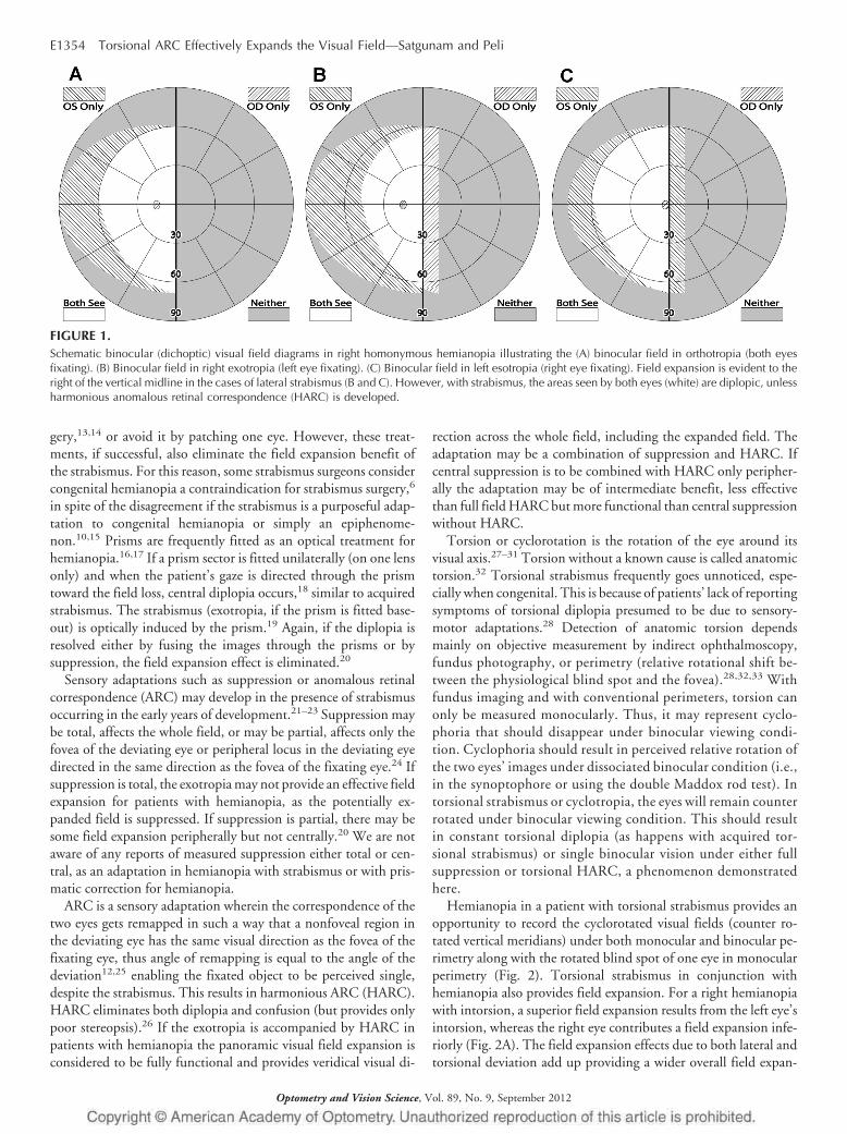
onymous hemianopia with right exotropia, the right eye coversmore of the right field of view (Fig. 1B) than in orthotropia (Fig.1A). A left esotropia would provide a similar field expansion effect,but with some reduction of the left temporal visual field (Fig. 1C).Reports of esotropia in congenital homonymous hemianopia arerelatively unknown. We found only one such probable occurrencein the literature.4
As a result of strabismus, patients may experience diplopia (twoimages of the same object with different visual directions) andvisual confusion (two different objects seen in the same visualdirection).12 Usually only diplopia is reported spontaneously. Thefield expansion illustrated in Fig. 1 may be less beneficial if accom-panied by diplopia, as central diplopia can be disturbing and illtolerated. It is possible to correct the diplopia with prisms or sur-
*BS(Opt), MS, PhD†MSc, OD, FAAOSchepens Eye Research Institute, Massachusetts Eye and Ear, and Department
of Ophthalmology, Harvard Medical School, Boston, Massachusetts (PNS, EP).Supplemental digital contents are available for this article. Direct URL citations
appear in the printed text and are provided in the HTML and PDF versions of thisarticle on the journal’s Web site (www.optvissci.com).
1040-5488/12/8909-1353/0 VOL. 89, NO. 9, PP. E1353–E1363OPTOMETRY AND VISION SCIENCECopyright © 2012 American Academy of Optometry
Optometry and Vision Science, Vol. 89, No. 9, September 2012
gery,13,14 or avoid it by patching one eye. However, these treat-ments, if successful, also eliminate the field expansion benefit ofthe strabismus. For this reason, some strabismus surgeons considercongenital hemianopia a contraindication for strabismus surgery,6
in spite of the disagreement if the strabismus is a purposeful adap-tation to congenital hemianopia or simply an epiphenome-non.10,15 Prisms are frequently fitted as an optical treatment forhemianopia.16,17 If a prism sector is fitted unilaterally (on one lensonly) and when the patient’s gaze is directed through the prismtoward the field loss, central diplopia occurs,18 similar to acquiredstrabismus. The strabismus (exotropia, if the prism is fitted base-out) is optically induced by the prism.19 Again, if the diplopia isresolved either by fusing the images through the prisms or bysuppression, the field expansion effect is eliminated.20
Sensory adaptations such as suppression or anomalous retinalcorrespondence (ARC) may develop in the presence of strabismusoccurring in the early years of development.21–23 Suppression maybe total, affects the whole field, or may be partial, affects only thefovea of the deviating eye or peripheral locus in the deviating eyedirected in the same direction as the fovea of the fixating eye.24 Ifsuppression is total, the exotropia may not provide an effective fieldexpansion for patients with hemianopia, as the potentially ex-panded field is suppressed. If suppression is partial, there may besome field expansion peripherally but not centrally.20 We are notaware of any reports of measured suppression either total or cen-tral, as an adaptation in hemianopia with strabismus or with pris-matic correction for hemianopia.
ARC is a sensory adaptation wherein the correspondence of thetwo eyes gets remapped in such a way that a nonfoveal region inthe deviating eye has the same visual direction as the fovea of thefixating eye, thus angle of remapping is equal to the angle of thedeviation12,25 enabling the fixated object to be perceived single,despite the strabismus. This results in harmonious ARC (HARC).HARC eliminates both diplopia and confusion (but provides onlypoor stereopsis).26 If the exotropia is accompanied by HARC inpatients with hemianopia the panoramic visual field expansion isconsidered to be fully functional and provides veridical visual di-
rection across the whole field, including the expanded field. Theadaptation may be a combination of suppression and HARC. Ifcentral suppression is to be combined with HARC only peripher-ally the adaptation may be of intermediate benefit, less effectivethan full field HARC but more functional than central suppressionwithout HARC.
Torsion or cyclorotation is the rotation of the eye around itsvisual axis.27–31 Torsion without a known cause is called anatomictorsion.32 Torsional strabismus frequently goes unnoticed, espe-cially when congenital. This is because of patients’ lack of reportingsymptoms of torsional diplopia presumed to be due to sensory-motor adaptations.28 Detection of anatomic torsion dependsmainly on objective measurement by indirect ophthalmoscopy,fundus photography, or perimetry (relative rotational shift be-tween the physiological blind spot and the fovea).28,32,33 Withfundus imaging and with conventional perimeters, torsion canonly be measured monocularly. Thus, it may represent cyclo-phoria that should disappear under binocular viewing condi-tion. Cyclophoria should result in perceived relative rotation ofthe two eyes’ images under dissociated binocular condition (i.e.,in the synoptophore or using the double Maddox rod test). Intorsional strabismus or cyclotropia, the eyes will remain counterrotated under binocular viewing condition. This should resultin constant torsional diplopia (as happens with acquired tor-sional strabismus) or single binocular vision under either fullsuppression or torsional HARC, a phenomenon demonstratedhere.
Hemianopia in a patient with torsional strabismus provides anopportunity to record the cyclorotated visual fields (counter ro-tated vertical meridians) under both monocular and binocular pe-rimetry along with the rotated blind spot of one eye in monocularperimetry (Fig. 2). Torsional strabismus in conjunction withhemianopia also provides field expansion. For a right hemianopiawith intorsion, a superior field expansion results from the left eye’sintorsion, whereas the right eye contributes a field expansion infe-riorly (Fig. 2A). The field expansion effects due to both lateral andtorsional deviation add up providing a wider overall field expan-
FIGURE 1.Schematic binocular (dichoptic) visual field diagrams in right homonymous hemianopia illustrating the (A) binocular field in orthotropia (both eyesfixating). (B) Binocular field in right exotropia (left eye fixating). (C) Binocular field in left esotropia (right eye fixating). Field expansion is evident to theright of the vertical midline in the cases of lateral strabismus (B and C). However, with strabismus, the areas seen by both eyes (white) are diplopic, unlessharmonious anomalous retinal correspondence (HARC) is developed.
E1354 Torsional ARC Effectively Expands the Visual Field—Satgunam and Peli
Optometry and Vision Science, Vol. 89, No. 9, September 2012
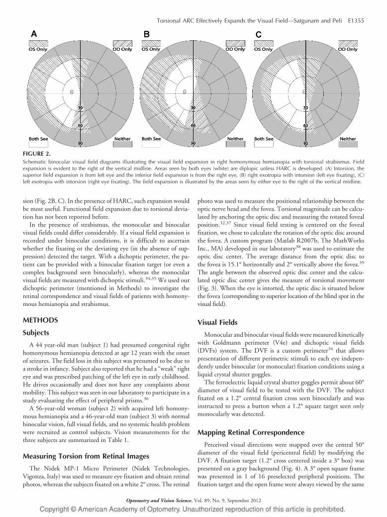
sion (Fig. 2B, C). In the presence of HARC, such expansion wouldbe most useful. Functional field expansion due to torsional devia-tion has not been reported before.
In the presence of strabismus, the monocular and binocularvisual fields could differ considerably. If a visual field expansion isrecorded under binocular conditions, it is difficult to ascertainwhether the fixating or the deviating eye (in the absence of sup-pression) detected the target. With a dichoptic perimeter, the pa-tient can be provided with a binocular fixation target (or even acomplex background seen binocularly), whereas the monocularvisual fields are measured with dichoptic stimuli.34,35 We used ourdichoptic perimeter (mentioned in Methods) to investigate theretinal correspondence and visual fields of patients with homony-mous hemianopia and strabismus.
METHODS
Subjects
A 44 year-old man (subject 1) had presumed congenital righthomonymous hemianopia detected at age 12 years with the onsetof seizures. The field loss in this subject was presumed to be due toa stroke in infancy. Subject also reported that he had a “weak” righteye and was prescribed patching of the left eye in early childhood.He drives occasionally and does not have any complaints aboutmobility. This subject was seen in our laboratory to participate in astudy evaluating the effect of peripheral prisms.36
A 56-year-old woman (subject 2) with acquired left homony-mous hemianopia and a 46-year-old man (subject 3) with normalbinocular vision, full visual fields, and no systemic health problemwere recruited as control subjects. Vision measurements for thethree subjects are summarized in Table 1.
Measuring Torsion from Retinal Images
The Nidek MP-1 Micro Perimeter (Nidek Technologies,Vigonza, Italy) was used to measure eye fixation and obtain retinalphotos, whereas the subjects fixated on a white 2° cross. The retinal
photo was used to measure the positional relationship between theoptic nerve head and the fovea. Torsional magnitude can be calcu-lated by anchoring the optic disc and measuring the rotated fovealposition.32,37 Since visual field testing is centered on the fovealfixation, we chose to calculate the rotation of the optic disc aroundthe fovea. A custom program (Matlab R2007b, The MathWorksInc., MA) developed in our laboratory38 was used to estimate theoptic disc center. The average distance from the optic disc tothe fovea is 15.1° horizontally and 2° vertically above the fovea.39
The angle between the observed optic disc center and the calcu-lated optic disc center gives the measure of torsional movement(Fig. 3). When the eye is intorted, the optic disc is situated belowthe fovea (corresponding to superior location of the blind spot in thevisual field).
Visual Fields
Monocular and binocular visual fields were measured kineticallywith Goldmann perimeter (V4e) and dichoptic visual fields(DVFs) system. The DVF is a custom perimeter34 that allowspresentation of different perimetric stimuli to each eye indepen-dently under binocular (or monocular) fixation conditions using aliquid crystal shutter goggles.
The ferroelectric liquid crystal shutter goggles permit about 60°diameter of visual field to be tested with the DVF. The subjectfixated on a 1.2° central fixation cross seen binocularly and wasinstructed to press a button when a 1.2° square target seen onlymonocularly was detected.
Mapping Retinal Correspondence
Perceived visual directions were mapped over the central 50°diameter of the visual field (pericentral field) by modifying theDVF. A fixation target (1.2° cross centered inside a 3° box) waspresented on a gray background (Fig. 4). A 3° open square framewas presented in 1 of 16 preselected peripheral positions. Thefixation target and the open frame were always viewed by the same
FIGURE 2.Schematic binocular visual field diagrams illustrating the visual field expansion in right homonymous hemianopia with torsional strabismus. Fieldexpansion is evident to the right of the vertical midline. Areas seen by both eyes (white) are diplopic unless HARC is developed. (A) Intorsion, thesuperior field expansion is from left eye and the inferior field expansion is from the right eye, (B) right exotropia with intorsion (left eye fixating), (C)left esotropia with intorsion (right eye fixating). The field expansion is illustrated by the areas seen by either eye to the right of the vertical midline.
Torsional ARC Effectively Expands the Visual Field—Satgunam and Peli E1355
Optometry and Vision Science, Vol. 89, No. 9, September 2012
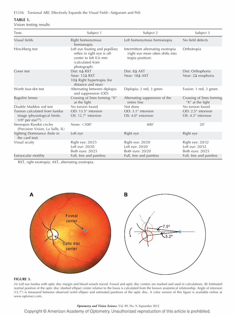
FIGURE 3.(A) Left eye fundus with optic disc margin and blood vessels traced. Foveal and optic disc centers are marked and used in calculations. (B) Estimatednormal position of the optic disc (dashed ellipse) center relative to the fovea is calculated from the known anatomical relationship. Angle of intorsion(12.7°) is measured between observed (solid ellipse) and estimated positions of the optic disc. A color version of this figure is available online atwww.optvissci.com.
TABLE 1.Vision testing results
Tests Subject 1 Subject 2 Subject 3
Visual fields Right homonymoushemianopia
Left homonymous hemianopia No field defects
Hirschberg test Left eye fixating and pupillaryreflex in right eye is off-center to left 0.6 mm(calculated fromphotograph)
Intermittent alternating exotropia(right eye more often drifts intotropia position)
Orthotropia
Cover test Dist: 6� RXT Dist: 8� AXT Dist: OrthophoriaNear: 12� RXT Near: 18� AXT Near: 2� exophoria10� Right hypertropia (for
distance and near)Worth four-dot test Alternating between diplopia
and suppression (OD)Diplopia: 2 red, 3 green Fusion: 1 red, 3 green
Bagolini lenses Crossing of lines forming “X”at the light
Alternating suppression of theentire line
Crossing of lines forming“X” at the light
Double Maddox rod test No torsion found Not done No torsion foundTorsion calculated from fundus
image (physiological limits:�9° per eye32)
OD: 13.5° intorsion OD: 3.1° intorsion OD: 2.5° intorsionOS: 12.7° intorsion OS: 4.0° extorsion OS: 4.3° intorison
Stereopsis Randot circles(Precision Vision, La Salle, IL)
None: �500� 400� 20�
Sighting Dominance (hole inthe card test)
Left eye Right eye Right eye
Visual acuity Right eye: 20/25 Right eye: 20/20 Right eye: 20/32Left eye: 20/20 Left eye: 20/20 Left eye: 20/32Both eyes: 20/25 Both eyes: 20/20 Both eyes: 20/25
Extraocular motility Full, free and painless Full, free and painless Full, free and painless
RXT, right exotropia; AXT, alternating exotropia.
E1356 Torsional ARC Effectively Expands the Visual Field—Satgunam and Peli
Optometry and Vision Science, Vol. 89, No. 9, September 2012
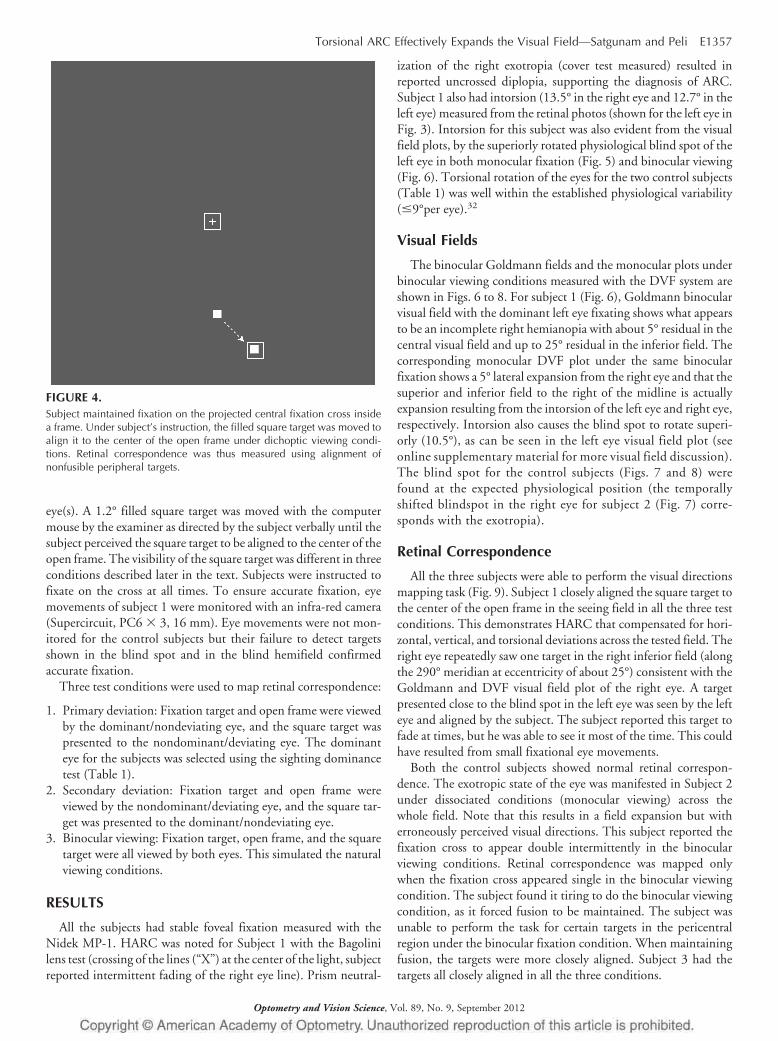
eye(s). A 1.2° filled square target was moved with the computermouse by the examiner as directed by the subject verbally until thesubject perceived the square target to be aligned to the center of theopen frame. The visibility of the square target was different in threeconditions described later in the text. Subjects were instructed tofixate on the cross at all times. To ensure accurate fixation, eyemovements of subject 1 were monitored with an infra-red camera(Supercircuit, PC6 � 3, 16 mm). Eye movements were not mon-itored for the control subjects but their failure to detect targetsshown in the blind spot and in the blind hemifield confirmedaccurate fixation.
Three test conditions were used to map retinal correspondence:
1. Primary deviation: Fixation target and open frame were viewedby the dominant/nondeviating eye, and the square target waspresented to the nondominant/deviating eye. The dominanteye for the subjects was selected using the sighting dominancetest (Table 1).
2. Secondary deviation: Fixation target and open frame wereviewed by the nondominant/deviating eye, and the square tar-get was presented to the dominant/nondeviating eye.
3. Binocular viewing: Fixation target, open frame, and the squaretarget were all viewed by both eyes. This simulated the naturalviewing conditions.
RESULTS
All the subjects had stable foveal fixation measured with theNidek MP-1. HARC was noted for Subject 1 with the Bagolinilens test (crossing of the lines (“X”) at the center of the light, subjectreported intermittent fading of the right eye line). Prism neutral-
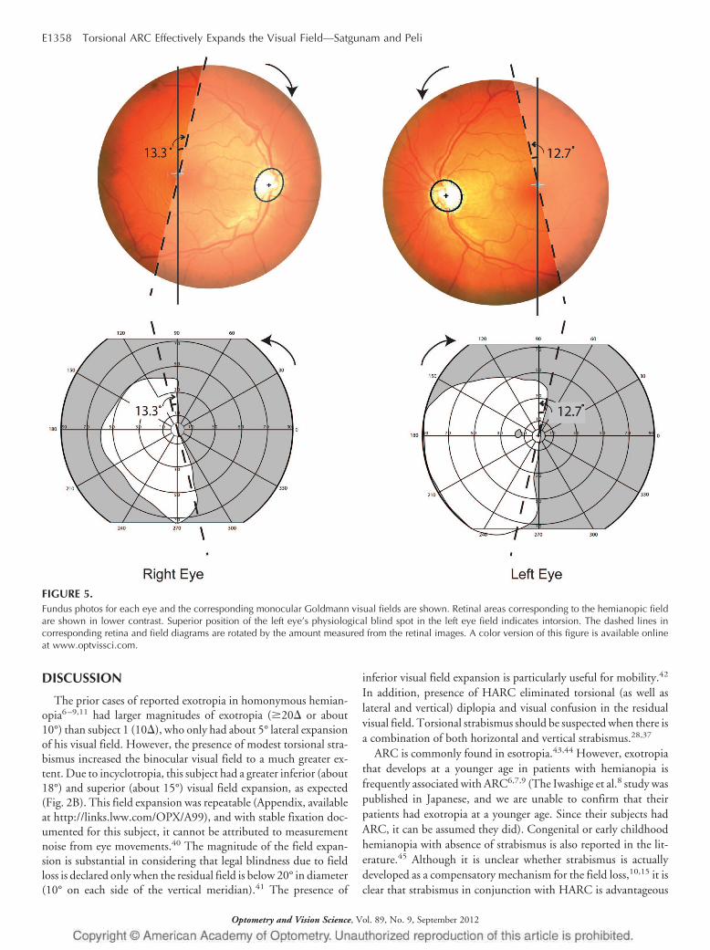
ization of the right exotropia (cover test measured) resulted inreported uncrossed diplopia, supporting the diagnosis of ARC.Subject 1 also had intorsion (13.5° in the right eye and 12.7° in theleft eye) measured from the retinal photos (shown for the left eye inFig. 3). Intorsion for this subject was also evident from the visualfield plots, by the superiorly rotated physiological blind spot of theleft eye in both monocular fixation (Fig. 5) and binocular viewing(Fig. 6). Torsional rotation of the eyes for the two control subjects(Table 1) was well within the established physiological variability(�9°per eye).32
Visual Fields
The binocular Goldmann fields and the monocular plots underbinocular viewing conditions measured with the DVF system areshown in Figs. 6 to 8. For subject 1 (Fig. 6), Goldmann binocularvisual field with the dominant left eye fixating shows what appearsto be an incomplete right hemianopia with about 5° residual in thecentral visual field and up to 25° residual in the inferior field. Thecorresponding monocular DVF plot under the same binocularfixation shows a 5° lateral expansion from the right eye and that thesuperior and inferior field to the right of the midline is actuallyexpansion resulting from the intorsion of the left eye and right eye,respectively. Intorsion also causes the blind spot to rotate superi-orly (10.5°), as can be seen in the left eye visual field plot (seeonline supplementary material for more visual field discussion).The blind spot for the control subjects (Figs. 7 and 8) werefound at the expected physiological position (the temporallyshifted blindspot in the right eye for subject 2 (Fig. 7) corre-sponds with the exotropia).
Retinal Correspondence
All the three subjects were able to perform the visual directionsmapping task (Fig. 9). Subject 1 closely aligned the square target tothe center of the open frame in the seeing field in all the three testconditions. This demonstrates HARC that compensated for hori-zontal, vertical, and torsional deviations across the tested field. Theright eye repeatedly saw one target in the right inferior field (alongthe 290° meridian at eccentricity of about 25°) consistent with theGoldmann and DVF visual field plot of the right eye. A targetpresented close to the blind spot in the left eye was seen by the lefteye and aligned by the subject. The subject reported this target tofade at times, but he was able to see it most of the time. This couldhave resulted from small fixational eye movements.
Both the control subjects showed normal retinal correspon-dence. The exotropic state of the eye was manifested in Subject 2under dissociated conditions (monocular viewing) across thewhole field. Note that this results in a field expansion but witherroneously perceived visual directions. This subject reported thefixation cross to appear double intermittently in the binocularviewing conditions. Retinal correspondence was mapped onlywhen the fixation cross appeared single in the binocular viewingcondition. The subject found it tiring to do the binocular viewingcondition, as it forced fusion to be maintained. The subject wasunable to perform the task for certain targets in the pericentralregion under the binocular fixation condition. When maintainingfusion, the targets were more closely aligned. Subject 3 had thetargets all closely aligned in all the three conditions.
FIGURE 4.Subject maintained fixation on the projected central fixation cross insidea frame. Under subject’s instruction, the filled square target was moved toalign it to the center of the open frame under dichoptic viewing condi-tions. Retinal correspondence was thus measured using alignment ofnonfusible peripheral targets.
Torsional ARC Effectively Expands the Visual Field—Satgunam and Peli E1357
Optometry and Vision Science, Vol. 89, No. 9, September 2012
DISCUSSION
The prior cases of reported exotropia in homonymous hemian-opia6–9,11 had larger magnitudes of exotropia (�20� or about10°) than subject 1 (10�), who only had about 5° lateral expansionof his visual field. However, the presence of modest torsional stra-bismus increased the binocular visual field to a much greater ex-tent. Due to incyclotropia, this subject had a greater inferior (about18°) and superior (about 15°) visual field expansion, as expected(Fig. 2B). This field expansion was repeatable (Appendix, availableat http://links.lww.com/OPX/A99), and with stable fixation doc-umented for this subject, it cannot be attributed to measurementnoise from eye movements.40 The magnitude of the field expan-sion is substantial in considering that legal blindness due to fieldloss is declared only when the residual field is below 20° in diameter(10° on each side of the vertical meridian).41 The presence of
inferior visual field expansion is particularly useful for mobility.42
In addition, presence of HARC eliminated torsional (as well aslateral and vertical) diplopia and visual confusion in the residualvisual field. Torsional strabismus should be suspected when there isa combination of both horizontal and vertical strabismus.28,37
ARC is commonly found in esotropia.43,44 However, exotropiathat develops at a younger age in patients with hemianopia isfrequently associated with ARC6,7,9 (The Iwashige et al.8 study waspublished in Japanese, and we are unable to confirm that theirpatients had exotropia at a younger age. Since their subjects hadARC, it can be assumed they did). Congenital or early childhoodhemianopia with absence of strabismus is also reported in the lit-erature.45 Although it is unclear whether strabismus is actuallydeveloped as a compensatory mechanism for the field loss,10,15 it isclear that strabismus in conjunction with HARC is advantageous
FIGURE 5.Fundus photos for each eye and the corresponding monocular Goldmann visual fields are shown. Retinal areas corresponding to the hemianopic fieldare shown in lower contrast. Superior position of the left eye’s physiological blind spot in the left eye field indicates intorsion. The dashed lines incorresponding retina and field diagrams are rotated by the amount measured from the retinal images. A color version of this figure is available onlineat www.optvissci.com.
E1358 Torsional ARC Effectively Expands the Visual Field—Satgunam and Peli
Optometry and Vision Science, Vol. 89, No. 9, September 2012

for congenital or early childhood hemianopia. Unfortunately, ithas not been proved possible to induce lateral strabismus withHARC in adult onset of hemianopia. Peli19 proposed a peripheralprism device that effectively induced “peripheral exotropia” as a
treatment for such patients. The technique has been found to besuccessful and provides expanded field shown to aid in obstacledetection.36,46,47 However, an HARC-like adaptation to the pe-ripheral prism postulated by Peli19 has not been seen.20
FIGURE 6.Binocular Goldmann visual field plots (left) and dichoptic visual field (DVF) plots (right) are shown for subject 1. DVF fields are restricted by the gogglesto only 60°. DVF plots were mapped under binocular viewing condition (central binocular fixation) while presenting monocular targets. Monoculartargets detected by the right eye are shown as filled red triangles pointing to right and those detected by the left eye are shown as open blue trianglespointing to left. Rotation of the vertical meridian due to measured intorsion (from Nidek images) are marked by the dashed lines (red and blue) for rightand left eye, respectively. The apparent restricted nasal fields are artifacts due to the mismatch between the subjects’ and the goggles’ pupillarydistances. A color version of this figure is available online at www.optvissci.com.
FIGURE 7.Binocular Goldmann visual field plots (left) and dichoptic visual field (DVF) plots (right) are shown for subject 2. Figure follows the same conventionsas Fig. 6. A color version of this figure is available online at www.optvissci.com.
Torsional ARC Effectively Expands the Visual Field—Satgunam and Peli E1359
Optometry and Vision Science, Vol. 89, No. 9, September 2012

Torsional HARC is interpreted clinically from mere absence oftorsional diplopia in the presence of monocularly measured tor-sion.28 Lack of complaints of torsional diplopia could be because oftorsional motor fusion or from the masking by large horizontal orvertical deviations that might interfere with the patient’s ability toperceive torsional diplopia.27 However, it is also possible thatHARC could develop with torsional strabismus because distancesbetween corresponding directions on both retinas increases grad-ually with retinal eccentricity in conjunction with the increase inPanum’s fusional areas.48 This suggests that if torsional strabismuscan be induced in adult onset hemianopes either surgically or op-tically, it could provide much better field expansion, particularlythe lower field needed for safe mobility as well as the overhead fieldprotecting from obstacles such as tree branches. With better direc-tional adaptation possibly with HARC-like adaptation, such a fieldexpansion would be beneficial. Optical rotation is currently possi-ble with either Dove prisms, which in addition to rotation alsoinvert the image and are too large and heavy to be used in spectaclelenses, or with twisted fiber optic bundle used in night visiondevices, which is also not suitable for this application. It may bepossible to create an image-rotating lens using some novel opticaltechniques (and a lens of that sort may provide an improved ap-proach to optical correction for hemianopia). Such lens may berestricted to the periphery, as in the Peli peripheral prisms,19 main-taining central single binocular vision but increasing field expan-sion with eccentricity. The image rotation may be introducedgradually, thus facilitating more tolerable cyclofusion in the largerperipheral Panum’s area, as the development of ARC in adults arenot documented. Testing of this concept may be initiated using abinocular head-mounted display in which the image in one eye isrotated computationally. Such a device would make it possible todetermine the viability of the concept, and if successful, motivateefforts to develop the required optical elements.
ARC is more easily observed in testing conditions that closelyresemble real-world targets or natural viewing tests such as Bagolinistriated glasses, when compared with other dissociating tests suchas the Worth four-dot test.44,49 The DVF system used in this studycan provide a natural viewing state as well as dissociated conditionsin the same instrument and with the same targets. In the presentstudy, we did not use real-world images with the DVF resulting ina dissociated (or weakly associated) conditions. The presence ofARC under this condition is a stronger indication of HARC as seenin subject 1.
Retinal correspondence has been reported to vary between thecentral and peripheral visual fields on some strabismic patients50
without any visual field defects. One study noted the correspon-dence to be normal centrally and more anomalous in the peripheralvisual field,51 whereas another study reported the opposite find-ings.52 With the DVF system we found that subject 1 maintainedHARC both centrally and pericentrally (within the tested 50°visual field diameter) and under both primary and secondary de-viations. Invariant level of HARC would be more useful as anadaptation for hemianopia. We believe that our method of directmeasure of visual directions at different eccentricities provides abetter estimate for retinal correspondence.
Lateral and vertical strabismus is manifested only in binocularviewing. Under monocular viewing, each eye takes up foveal fixa-tion even with ARC (except in some paretic strabismus and eccen-tric fixation conditions). The difference in the strabismic eyeposition between monocular and binocular viewing (i.e., cover-uncover test) establishes the diagnosis (i.e., phoria/tropia). Inpurely torsional deviation (phoria/tropia), the eye’s torsionalmovement is much harder to detect with direct observation undercover-uncover test. Most objective tests such as fundus photogra-phy indirect ophthalmoscopy and blind spot perimetry for mea-suring torsion are essentially monocular. The double Maddox rod
FIGURE 8.Binocular Goldmann visual field plots (left) and dichoptic visual field (DVF) plots (right) are shown for subject 3. Figure follows the same conventionsas Fig. 6. A color version of this figure is available online at www.optvissci.com.
E1360 Torsional ARC Effectively Expands the Visual Field—Satgunam and Peli
Optometry and Vision Science, Vol. 89, No. 9, September 2012

subjective test though viewed with both eyes dissociates binocularvision (in particular torsional fusion) and does not differentiatebetween a phoria and a tropia, similar to regular Maddox rod testfor lateral and vertical deviation. Torsional rotation only underdissociated condition is indicative of cyclophoria and lack of dip-lopia under associated condition could result from torsional fusion(cyclofusion). The presence of homonymous hemianopia as in oursubject 1 enables documentation with the DVF of the rotation of
the vertical meridian (as well as the physiological scotoma’s rota-tion) under associated binocular fixation, thus establishing the tor-sion to be tropic. Such observation is not possible in standardperimetry without hemianopia. Further, the standard perimetry isonly weakly binocularly associated in regard to torsion as the cen-tral fixation target while providing a sound fusional stimulus forlateral/vertical phoria provides minimal stimulus for torsional pho-ria. Additional peripheral targets are necessary to facilitate torsional
FIGURE 9.Measurement of retinal correspondence with the DVF for each subject (subject 1-top row, subject 2-middle row, and subject 3-bottom row). Primarydeviation (left panel): sighting dominant eye fixates (left eye for subject 1 and right eye for subjects 2 and 3) and the nondominant eye sees filled squaretargets to be aligned with the center of the open frame seen by the fixating eye. Secondary deviation (middle panel): nondominant eye now fixates (righteye for subject 1 and left eye for subjects 2 and 3), and the dominant eye sees the square targets to be aligned with the center of the open frame nowseen by the nondominant eye. Binocular fixation (right panel): fixation target, square target, and the open frame (marked in the figure) were seenbinocularly. Three-sided frames are used in the figure to aid in eye identification. A color version of this figure is available online at www.optvissci.com.
Torsional ARC Effectively Expands the Visual Field—Satgunam and Peli E1361
Optometry and Vision Science, Vol. 89, No. 9, September 2012

fusion. With dichoptic perimetry system, rotation of the physio-logical blind spot can be measured (without hemianopia) and com-pared under both monocular (not shown) and binocular fixationconditions (Fig. 6). The DVF can also present visually rich back-ground for the perimetry that can serve as stimulus for torsionalfusion. This enables differentiation of the torsional posture underassociated and dissociated binocular conditions (with a tropic eyeremaining in the torsional position under both conditions). Thus,a dichoptic perimeter with the capabilities of the DVF provides aunique testing environment for a more complete evaluation oftorsion and of torsional HARC. In our patient, despite the lessoptimal blank background, HARC was demonstrated indicating astronger adaptation.
To our knowledge, this is the first reported torsional HARC ina patient with homonymous hemianopia.
ACKNOWLEDGMENTS
Supported in part by NIH grants EY07957 and EY12890 andP30EY003790. Henry Apfelbaum helped prepare the illustrations.
Received December 5, 2011; accepted June 4, 2012.
APPENDIX
The appendix is available online at http://links.lww.com/OPX/A99.
REFERENCES
1. Huber A. Homonymous hemianopia. Neuroophthalmology 1992;12:351–66.
2. Zhang X, Kedar S, Lynn MJ, Newman NJ, Biousse V. Homonymoushemianopias: clinical-anatomic correlations in 904 cases. Neurology2006;66:906–10.
3. Bosley TM, Kiyosawa M, Moster M, Harbour R, Zimmerman R,Savino PJ, Sergott RC, Alavi A, Reivich M. Neuro-imaging and pos-itron emission tomography of congenital homonymous hemianopias.Am J Ophthalmol 1991;111:413–8.
4. Shinder R, Wolansky L, Turbin RE. Congenital homonymous hemi-anopia and cortical migration abnormalities in a young adult. J Pedi-atr Ophthalmol Strabismus 2009;46:38–41.
5. Kedar S, Zhang X, Lynn MJ, Newman NJ, Biousse V. Pediatrichomonymous hemianopia. J AAPOS 2006;10:249–52.
6. Herzau V, Bleher I, Joos-Kratsch E. Infantile exotropia with homon-ymous hemianopia: a rare contraindication for strabismus surgery.Graefes Arch Clin Exp Ophthalmol 1988;226:148–9.
7. Gote H, Gregersen E, Rindziunski E. Exotropia and panoramic vi-sion compensating for an occult congenital homonymoushemianopia: a case report. Binocul Vis Eye Muscle Surg Q 1993;8:129–32.
8. Iwashige H, Hirose O, Usui C, Shoda S, Miyasaka H, Maruo T.Surgical and botulinum toxin treatment in two cases of abnormalretinal correspondence-exotropia with congenital homonymoushemianopsia [in Japanese]. Nihon Ganka Gakkai Zasshi 1995;99:1036–44.
9. Levy Y, Turetz J, Krakowski D, Hartmann B, Nemet P. Develop-ment of compensating exotropia with anomalous retinal correspon-dence after early infancy in congenital homonymous hemianopia.J Pediatr Ophthalmol Strabismus 1995;32:236–8.
10. Donahue SP, Haun AK. Exotropia and face turn in children withhomonymous hemianopia. J Neuroophthalmol 2007;27:304–7.
11. Gamio S, Melek N. When the patient says no. Management of exo-
tropia with hemianopic visual field defects. Binocul Vis Strabismus Q2003;18:167–70.
12. Duke-Elder S, Wybar K. System of Ophthalmology. London, UK:H. Kimpton; 1973.
13. Phillips PH. Treatment of diplopia. Semin Neurol 2007;27:288–98.14. Rucker JC, Tomsak RL. Binocular diplopia. A practical approach.
Neurologist 2005;11:98–110.15. Hoyt CS, Good WV. Ocular motor adaptations to congenital hemi-
anopia. Binocul Vis Eye Muscle Surgery Q 1993;8:125–6.16. Smith JL, Weiner IG, Lucero AJ. Hemianopic Fresnel prisms. J Clin
Neuroophthalmol 1982;2:19–22.17. Cohen JM, Waiss B. Visual field remediation. In: Cole RG,
Rosenthal BP, eds. Remediation and Management of Low Vision. St.Louis, MO: Mosby; 1996:1–25.
18. Gottlieb DD. Living with Vision Loss. Atlanta, GA: St. BarthelemyPress, Ltd.; 1996.
19. Peli E. Field expansion for homonymous hemianopia by opticallyinduced peripheral exotropia. Optom Vis Sci 2000;77:453–64.
20. Giorgi RG, Woods RL, Peli E. Clinical and laboratory evaluation ofperipheral prism glasses for hemianopia. Optom Vis Sci 2009;86:492–502.
21. Pickwell D. Binocular Vision Anomalies: Investigation and Treat-ment, 2nd ed. London, UK: Butterworths; 1989.
22. Daw NW. Visual Development. New York, NY: Plenum Press;1995.
23. Verma A. Anomalous adaptive conditions associated with strabismus.Ann Ophthalmol (Skokie) 2007;39:95–106.
24. Rutstein RP, Daum KM. Anomalies of Binocular Vision: Diagnosisand Management. St. Louis, MO: Mosby; 1998.
25. Kirschen DG. Understanding sensory evaluation. In: RosenbaumAL, Santiago AP, eds. Clinical Strabismus Management: Principlesand Surgical Techniques. Philadelphia, PA: Saunders; 1999:22–36.
26. Harley RD, Nelson LB, Olitsky SE. Harley’s Pediatric Ophthalmol-ogy, 5th ed. Philadelphia, PA: Lippincott Williams & Wilkins; 2005.
27. Woo SJ, Seo JM, Hwang JM. Clinical characteristics of cyclodevia-tion. Eye (Lond) 2005;19:873–8.
28. Philips PH, Hunter DG. Evaluation of ocular torsion and principlesof management. In: Rosenbaum AL, Santiago AP, eds. Clinical Stra-bismus Management: Principles and Surgical Techniques. Philadel-phia, PA: Saunders; 1999:52–72.
29. Guyton DL. Strabismus complications from local anesthetics. SeminOphthalmol 2008;23:298–301.
30. Holgado S, Enyedi LB, Toth CA, Freedman SF. Extraocular musclesurgery for extorsion after macular translocation surgery new surgicaltechnique and clinical management. Ophthalmology 2006;113:63–9.
31. Dieterich M, Brandt T. Ocular torsion and perceived vertical in oc-ulomotor, trochlear, and abducens nerve palsies. Brain 1993;116(Pt.5):1095–104.
32. Guyton DL. Clinical assessment of ocular torsion. Am Orthoptic J1983;33:7–15.
33. Morton GV, Lucchese N, Kushner BJ. The role of funduscopy andfundus photography in strabismus diagnosis. Ophthalmology 1983;90:1186–91.
34. Woods RL, Apfelbaum HL, Peli E. DLP-based dichoptic vision testsystem. J Biomed Opt 2010;15:016011.
35. Doherty AL, Bowers AR, Luo G, Peli E. Object detection in the ringscotoma of a monocular bioptic telescope. Arch Ophthalmol 2011;129:611–7.
36. Bowers AR, Keeney K, Peli E. Community-based trial of a peripheralprism visual field expansion device for hemianopia. Arch Ophthalmol2008;126:657–64.
E1362 Torsional ARC Effectively Expands the Visual Field—Satgunam and Peli
Optometry and Vision Science, Vol. 89, No. 9, September 2012

37. Kushner BJ, Hariharan L. Observations about objective and subjec-tive ocular torsion. Ophthalmology 2009;116:2001–10.
38. Nugent AK, Keswani RN, Woods RL, Peli E. Contour integration inperipheral vision reduces gradually with eccentricity. Vision Res2003;43:2427–37.
39. Hu SY, Schuchard RA, Fletcher DC, Sabates FN. Physiological blindspot characteristics and position relative to retinal locus for fixation bySLO testing. Invest Ophthalmol Vis Sci 1994;35:S1527.
40. Jamara RJ, Van De Velde F, Peli E. Scanning eye movements inhomonymous hemianopia documented by scanning laser ophthal-moscope retinal perimetry. Optom Vis Sci 2003;80:495–504.
41. Colenbrander A. Visual standards, aspects and ranges of vision losswith emphasis on population surveys. Report prepared for the Inter-national Council of Ophthalmology at the 29th International Con-gress of Ophthalmology. Sydney, Australia: International Council ofOphthalmology; 2002:1–33.
42. Lovie-Kitchin J, Mainstone J, Robinson J, Brown B. What areas ofthe visual field are important for mobility in low vision patients? ClinVis Sci 1990;5:249–63.
43. Katsumi O, Tanaka Y, Uemura Y. Anomalous retinal correspondencein esotropia. Jpn J Ophthalmol 1982;26:166–74.
44. Rutstein RP, Daum KM, Eskridge JB. Clinical characteristics ofanomalous correspondence. Optom Vis Sci 1989;66:420–5.
45. Zangemeister WH, Meienberg O, Stark L, Hoyt WF. Eye-head co-ordination in homonymous hemianopia. J Neurol 1982;226:243–54.
46. Bowers AR, Keeney K, Apfelbaum DH, Peli E. Randomized con-trolled trial of oblique and horizontal peripheral prism glasses forhemianopia. Optom Vis Sci 2010;87:E-abstract 100960.
47. O’Neill EC, Connell PP, O’Connor JC, Brady J, Reid I, Logan P.Prism therapy and visual rehabilitation in homonymous visual fieldloss. Optom Vis Sci 2011;88:263–8.
48. Guyton DL. Ocular torsion: sensorimotor principles. Graefes ArchClin Exp Ophthalmol 1988;226:241–5.
49. Nelson JI. A neurophysiological model for anomalous correspon-dence based on mechanisms of sensory fusion. Doc Ophthalmol1981;51:3–100.
50. Sireteanu R, Fronius M. Different patterns of retinal correspondencein the central and peripheral visual field of strabismics. Invest Oph-thalmol Vis Sci 1989;30:2023–33.
51. Haase W, Lung KH. Correspondence of foveal and peripheral areasin subjects with intact binocular vision and patients with strabismus[in German]. Klin Monbl Augenheilkd 1984;184:32–6.
52. Hallden U. The longitudinal horopter in a case of concomitant stra-bismus with anomalous correspondence. Acta Ophthalmol (Copenh)1973;51:1–11.
Eli PeliSchepens Eye Research Institute
20 Staniford StreetBoston, MA 02114-2500
e-mail: [email protected]
Torsional ARC Effectively Expands the Visual Field—Satgunam and Peli E1363
Optometry and Vision Science, Vol. 89, No. 9, September 2012