Embed Size (px)
Citation preview

Zurich Open Repository andArchiveUniversity of ZurichMain LibraryStrickhofstrasse 39CH-8057 Zurichwww.zora.uzh.ch
Year: 2017
Torque differences according to tooth morphology and bracket placement: afinite element study
Papageorgiou, Spyridon N ; Sifakakis, Iosif ; Keilig, Ludger ; Patcas, Raphael ; Affolter, Stefan ;Eliades, Theodore ; Bourauel, Christoph
Abstract: INTRODUCTION Torque of the maxillary incisors is essential in esthetics and proper occlu-sion, while torque expression is influenced by many factors. The aim of this finite element study wasto assess the relative effect of tooth morphology, bracket prescription, and bracket positioning on toothdisplacement and developed stresses/strains after torque application. METHODS A three-dimensionalupper right central incisor with its periodontal ligament (PDL) and alveolus was modelled. The toothvaried in the crown-root angle (CRA) between 156°, 170°, and 184°. An 0.018-inch slot discovery® (Den-taurum, Ispringen, Germany) bracket with a rectangular 0.018 × 0.025-inch �-titanium wire was modelled.Bracket torque prescription varied between 0°, 12°, and 22°, with bracket placement at the centre of themiddle, gingival or incisal third of the crown. A total of 27 models were generated and a buccal roottorque of 30° was applied. Afterwards, crown and apex displacement, strains in the PDL, and stresses inthe bracket were calculated and analysed statistically. RESULTS The palatal crown displacement wassignificantly affected by bracket positioning (up to 94 per cent), while the buccal apex displacement wassignificantly affected by bracket prescription (up to 42 per cent) and bracket positioning (up to 23 percent). Strains in the PDL were affected mainly by CRA (up to 54 per cent), followed by bracket posi-tioning (up to 45 per cent). Finally, bracket prescription considerably affected the stresses in the bracket(up to 144 per cent). LIMITATIONS These in silico results need to be validated in vivo before theycan be clinically extrapolated. CONCLUSION Tooth anatomy and the characteristics of the orthodonticappliance should be considered during torque application.
DOI: https://doi.org/10.1093/ejo/cjw074
Posted at the Zurich Open Repository and Archive, University of ZurichZORA URL: https://doi.org/10.5167/uzh-132888Journal ArticleAccepted Version
Originally published at:Papageorgiou, Spyridon N; Sifakakis, Iosif; Keilig, Ludger; Patcas, Raphael; Affolter, Stefan; Eliades,Theodore; Bourauel, Christoph (2017). Torque differences according to tooth morphology and bracketplacement: a finite element study. European Journal of Orthodontics, 39(4):411-418.DOI: https://doi.org/10.1093/ejo/cjw074

1
Title Page
Torque differences according to tooth morphology and bracket placement: a
finite element study
Spyridon N. Papageorgiou1,*, Iosif Sifakakis2, Ludger Keilig3,4, Raphael Patcas1, Stefan Affolter1,
Theodore Eliades1, Christoph Bourauel3
1Clinic of Orthodontics and Pediatric Dentistry, Center of Dental Medicine, Faculty of Medicine,
University of Zurich, Plattenstrasse 11, Zurich 8032, Switzerland
2Department of Orthodontics, School of Dentistry, National and Kapodistrian University of Athens,
Athens, Greece
3Department of Oral Technology, School of Dentistry, University of Bonn, Welschnonnenstr. 17,
53111, Bonn, Germany
4Department of Prosthetic Dentistry, Preclinical Education and Materials Science, School of Dentistry,
University of Bonn, Bonn, Germany
Running title: Torque according to tooth and bracket differences
*Corresponding author: Spyridon N. Papageorgiou Clinic of Orthodontics and Pediatric Dentistry,
Center of Dental Medicine, University of Zurich, Plattenstrasse 11, Zurich 8032, Switzerland. Tel.:+41
44 634 32 87, Fax: +41 44 634 43 35; E-mail: [email protected]
Words in abstract: 253
Words in text: 2972
Keywords: orthodontics, fixed appliances, tooth movement, torque, treatment efficiency, tooth root,
finite element method
Conflicts of interest: none.
Acknowledgements: The authors would like to thank Dentaurum (Ispringen, Germany) for providing
the CAD/CAM data of the three bracket models used.
Funding: This work was supported by a scholarship from Forschungsgemeinschaft Dental (FGD) to
the first author and by a scholarship from the German Academic Exchange Service (DAAD; research
grant no. A/14/01558) to the second author.

2
Manuscript
Torque differences according to tooth morphology and bracket placement: a
finite element study
Summary
Introduction: Torque of the maxillary incisors is essential in esthetics and proper occlusion, while
torque expression is influenced by many factors. The aim of this finite element study was to assess
the relative effect of tooth morphology, bracket prescription, and bracket positioning on tooth
displacement and developed stresses/strains after torque application.
Methods: A three-dimensional upper right central incisor with its Periodontal Ligament (PDL) and
alveolus was modeled. The tooth varied in the crown-to-root angle between 156°, 170°, and 184°. An
0.018-inch slot discovery® (Dentaurum, Ispringen, Germany) bracket with a rectangular 0.018 x
0.025-inch β-titanium wire was modeled. Bracket torque prescription varied between 0°, 12°,and 22°,
with bracket placement at the center of the middle, gingival or incisal third of the crown. A total of 27
models were generated and a buccal root torque of 30° was applied. Afterwards, crown and apex
displacement, strains in the PDL, and stresses in the bracket were calculated and analyzed
statistically.
Results: The palatal crown displacement was significantly affected by bracket positioning (up to 94%),
while the buccal apex displacement was significantly affected by bracket prescription (up to 42%) and
bracket positioning (up to 23%). Strains in the PDL were affected mainly by crown-to-root angle (up to
54%), followed by bracket positioning (up to 45%). Finally, bracket prescription considerable affected
the stresses in the bracket (up to 144%).
Limitations: These in silico results need to be validated in vivo before they can be clinically
extrapolated.
Conclusion: Tooth anatomy and the characteristics of the orthodontic appliance should be considered
during torque application.

3
Introduction
Orthodontic tooth movement is based on the ability of surrounding bone and periodontal ligament (PDL) to react
to a mechanical stimulus with remodelling processes. Application of an orthodontic force system to a tooth
causes displacement, stresses, and strains in the structures involved (1, 2), while mechanotransductory processes
are translated to cell-to-cell signalling (3). Studies on rats and monkeys indicate that there is a close relationship
between calculated stress/strain values in the PDL and the distribution or activity of osteoclasts (2, 4, 5). As
force magnitude plays a pivotal role in determining whether physiologic remodelling phenomena or necrotizing
phenomena take place in the PDL, quantification of strains developed in the ligament and alveolar bone can
provide indications of favourable or unfavourable tooth movement (6-8).
Proper buccolingual inclination of both posterior and anterior teeth is essential to providing stability and
proper occlusal relationship in orthodontic treatment. Torque of the maxillary incisors is particularly critical in
establishing an esthetic smile line, proper anterior guidance, and a solid Class I relationship, because
undertorqued anterior teeth can preclude the retraction of the anterior maxillary dentition. Suboptimal torque of
the incisors can deprive the dental arch of space (9), while suboptimal torque of the posterior teeth might not
allow appropriate cusp-to-fossa relationships between the maxillary and mandibular teeth (10).
Therefore, optimal treatment outcomes require effective torque expression, which is sensitive to
irregularities in tooth anatomy, the size, morphology, and engagement of the archwire in the bracket, as well as
the position, material properties, and slot size of the bracket (11–17). For example, although 0.22-inch brackets
might be outperform 0.018-inch in some clinical aspects like sliding mechanics, they are inferior in torque
expression (18, 19). This is the case even with slot-filling archwires, which have limited compliance and range in
torsion and often torquing auxiliaries or smaller rectangular Titanium Molybdenum Alloy (TMA) wires with
increased activations are used.
In order to obtain proper tooth positioning in the three dimensions in the preadjusted appliance
without any wire bending, two conditions have to be fulfilled (20). First, the brackets have to be accurately
placed in a specific position on the labial or buccal surface of each tooth, attempting to express the desired
amount of torque and tip. The labial contour of the crown surface differs at different heights on the crown of the
same tooth. Therefore, an archwire, fully engaged into a bracket, will produce a different axial inclination of the
tooth (21). Second, this will only be the case when the tooth–crown morphology (tooth surface curvature and
crown–root angle; CRA) lies within the average region for each tooth (22). Several authors have reported that a
considerable variation exists in CRA, which can take values between 156° and 195° (23–25). The CRA may
limit the degree to which the roots can be torqued palatally due to an increased proximity to the cortical plate of
4
the alveolar process (22, 26). Taking into consideration that a root, moved against the cortical plate, is at higher
risk for root resorption (27–29), care should be taken to torque a tooth with a large CRA (20). Finally, bracket
prescription has a direct influence on the amount of torque attained (10, 20).
The finite element (FE) method has been suggested as a solution for complex biomechanical questions
and has been applied in several cases in orthodontics (30, 31) in order to assess the centre of resistance (32–34),
various biomechanical aspects of tooth movement (35, 36), different bracket (25, 37), anchorage (38–42) or
surgical (43, 44) treatment modalities, debonding (45–47), and retention procedures (48). The reliability of FE
analyses is dependent not only on the loading configuration, but also on the geometry of the structure and the
material properties (30, 49).
The objective of the present in silico study was to assess the influence of tooth anatomy, bracket
placement, and bracket prescription on the biomechanics of torque application. Part of the research question has
been covered by a previous FE study (50), in which however other slot size was used for another tooth
movement (extrusion), while no bilinear nature was adopted to accurately model the PDL and no other factors
were assessed.
Materials and methods
A three-dimensional (3D) solid model was constructed including a maxillary right central incisor with its PDL
and alveolus, and a uniform thickness of 0.2 mm and 0.5 mm, respectively (Figure S1). The base geometry of the
tooth model was derived on a commercial three-dimensional dataset, based on a larger survey of caucasian
patients (“teeth with roots and gum”, Viewpoint Data Labs, now Digimation Inc., Lake Mary, FL, USA). The
CRA of the incisor varied between 156°, 170°, and 184°, based on the values found by Delivanis and Kuftinec
(23) (Figure 1). A partial orthodontic fixed appliance was constructed with a composite resin adhesive layer
(mean thickness 0.2 mm) and a stainless steel bracket on the incisor tooth, while a rectangular 0.46 x 0.64 mm
(0.018 x 0.025 inch) TMA wire was passively inserted into the bracket slot and ligated with two steel ligatures
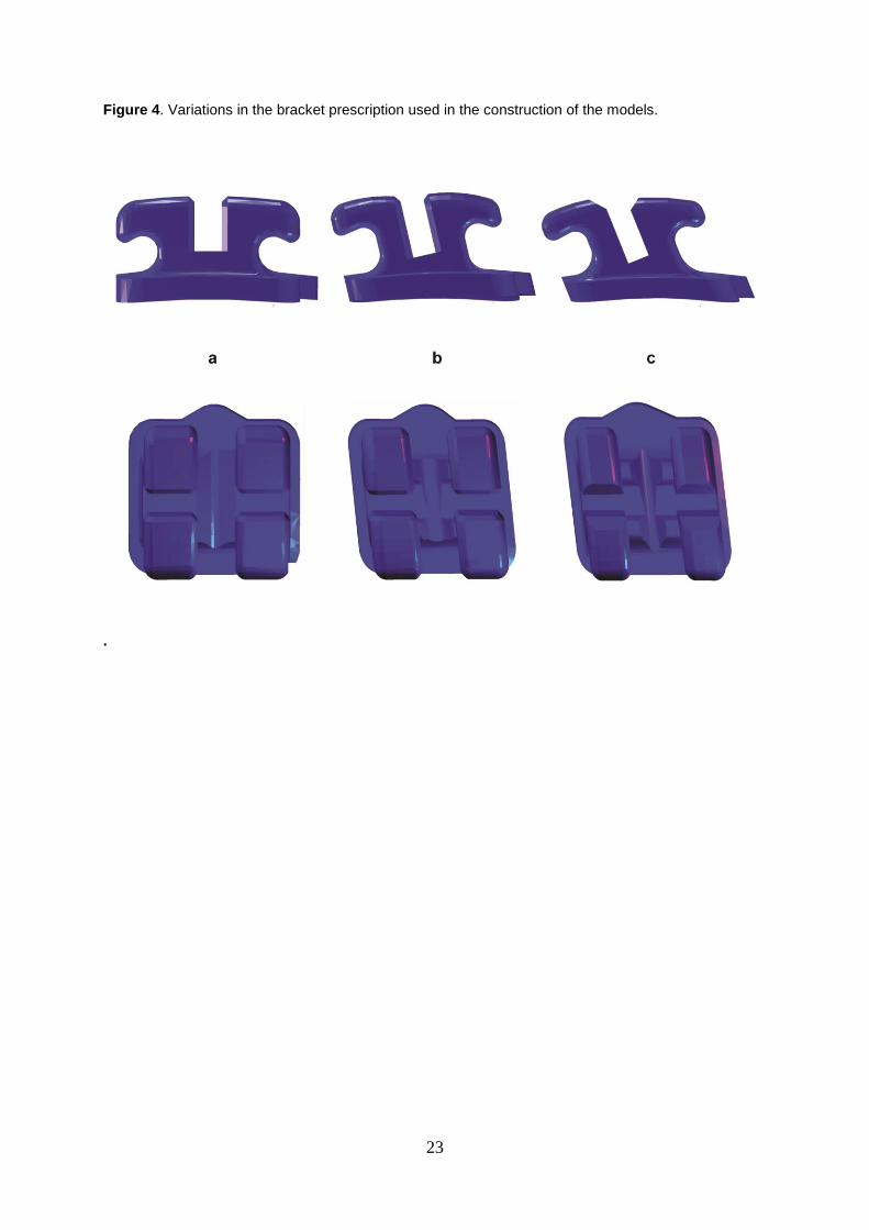
(Figures 2, 3). For all models the same bracket was used, based on computer-aided design and computer-aided
manufacturing (CAD/CAM) data of the discovery® (Dentaurum, Ispringen, Germany) bracket, provided by the
manufacturer in 0.46 mm (0.018 inch)-slot, as this is more efficient in torque expression (51, 52). The same
bracket was acquired in three torque prescriptions: (i) no torque (Standard Edgewise 18; 0°), (ii) medium torque
(Roth 18; +12°), and (iii) high torque (Ricketts ® Universal 18; +22°) (Figure 4; Table 1). The vertical positions
of the bracket on the labial surface of the incisor’s crown chosen were: (i) at the center of the middle third of the
crown, (ii) at the center of the apical third of the crown, and (iii) at the center of the incisal third of the crown
5
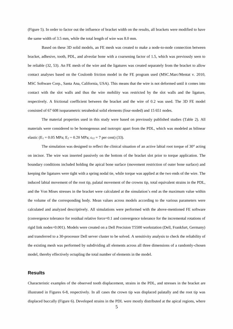
(Figure 5). In order to factor out the influence of bracket width on the results, all brackets were modified to have
the same width of 3.5 mm, while the total length of wire was 8.0 mm.
Based on these 3D solid models, an FE mesh was created to make a node-to-node connection between
bracket, adhesive, tooth, PDL, and alveolar bone with a coarsening factor of 1.5, which was previously seen to
be reliable (32, 53). An FE mesh of the wire and the ligatures was created separately from the bracket to allow
contact analyses based on the Coulomb friction model in the FE program used (MSC.Marc/Mentat v. 2010,
MSC Software Corp., Santa Ana, California, USA). This means that the wire is not deformed until it comes into
contact with the slot walls and thus the wire mobility was restricted by the slot walls and the ligature,
respectively. A frictional coefficient between the bracket and the wire of 0.2 was used. The 3D FE model
consisted of 67 608 isoparametric tetrahedral solid elements (four-noded) and 15 651 nodes.
The material properties used in this study were based on previously published studies (Table 2). All
materials were considered to be homogenous and isotropic apart from the PDL, which was modeled as bilinear
elastic (E1 = 0.05 MPa; E2 = 0.20 MPa; ε12 = 7 per cent) (33).
The simulation was designed to reflect the clinical situation of an active labial root torque of 30° acting
on incisor. The wire was inserted passively on the bottom of the bracket slot prior to torque application. The
boundary conditions included holding the apical bone surface (movement restriction of outer bone surface) and
keeping the ligatures were tight with a spring nodal tie, while torque was applied at the two ends of the wire. The
induced labial movement of the root tip, palatal movement of the crowns tip, total equivalent strains in the PDL,
and the Von Mises stresses in the bracket were calculated at the simulation’s end as the maximum value within
the volume of the corresponding body. Mean values across models according to the various parameters were
calculated and analyzed descriptively. All simulations were performed with the above-mentioned FE software
(convergence tolerance for residual relative force=0.1 and convergence tolerance for the incremental rotations of
rigid link nodes=0.001). Models were created on a Dell Precision T5500 workstation (Dell, Frankfurt, Germany)
and transferred to a 30-processor Dell server cluster to be solved. A sensitivity analysis to check the reliability of
the existing mesh was performed by subdividing all elements across all three dimensions of a randomly-chosen
model, thereby effectively octupling the total number of elements in the model.
Results
Characteristic examples of the observed tooth displacement, strains in the PDL, and stresses in the bracket are
illustrated in Figures 6-8, respectively. In all cases the crown tip was displaced palatally and the root tip was
displaced buccally (Figure 6). Developed strains in the PDL were mostly distributed at the apical regions, where

6
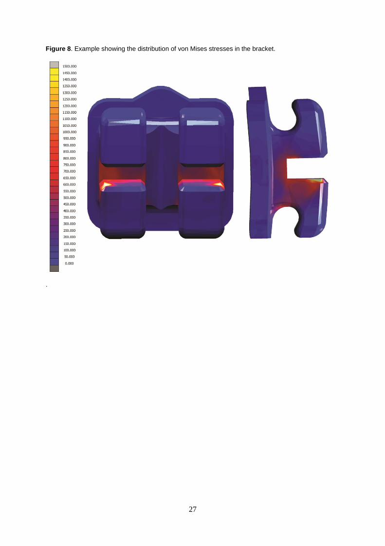
root tip was displaced buccally (Figure S2). Conversely, stresses at the bracket were mostly concentrated at the
bracket wall, where the wire’s edge came in contact with the bracket (Figure 8). The effect of the CRA, bracket
prescription, and bracket position on tooth displacement, developed strains in the PDL, and developed stresses in
the bracket can be seen in Table 3,
The average across models of the maximum palatal displacement of the crown tip was 0.14 mm. No
significant differences were seen among the models, except from a statistically significantly higher crown
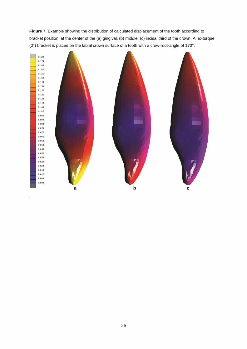
displacement when the bracket was positioned at the center of the apical third of the tooth (Figure 7).
The average across models of the maximum buccal displacement of the root apex was 0.15 mm.
Significantly higher movement of the tooth apex was seen with high-torque brackets. Moreover, bracket
positioning had a significant influence on the movement of the tooth apex, where incisal bracket placement was
associated with less apex movement and apical bracket placement was associated with more apex movement
compared to bracket placement at the middle of the crown.
The average across models of the maximum strains in the PDL were 1.24, while tooth anatomy was
significantly associated with the developed strains in the PDL. The magnitude of developed strains decreased
considerably, as the inclination of the root to the tooth increased.
Finally, the average across models of the maximum induced stresses in the bracket were 4306.91 MPa.
No significant effect on the stresses developed in the bracket was seen, except from bracket prescription.
The performed sensitivity analysis (Supplementary material) indicating that the sharp increase in the
total number of elements did not have a considerable influence on the results, as all deviations were in the level
of 5%-25% , which is in the range of FE analyses in general.
Discussion
In this study the relative contribution of the tooth’s anatomy, the bracket’s prescription, and the bracket’s
positioning on the labial tooth surface to the attained tooth displacement and the developed stresses and strains in
the PDL and the bracket were investigated in silico. It was observed that the displacement of the crown and the
root were mainly affected by the bracket positioning and prescription, while the strains induced at the PDL level
were affected mainly by the CRA, followed by the bracket positioning. Finally, the bracket prescription had a
considerable effect on the developed stresses at the bracket level.
The finite element method enables us to answer complex biomechanical questions in the field of
orthodontics via simulation; moreover, it enables investigators to predict the behavior of biological structures in
many specific situations. However, any solutions obtained via FE simulation will be numerical approximations.
7
Although many measurements cannot be taken in vivo, they can nevertheless contribute useful information to
clinical investigations.
Orthodontically-induced root resorption is a multifaceted phenomenon with complex etiopathology.
Although the duration of treatment with heavy rectangular wires and excessive torque application might be
regarded as risk factors, no single mechanical factor can fully predict treatment-induced resorption of the root.
An additional detrimental factor for the development of root resorption might be the iatrogenic approximation of
anterior tooth roots towards the cortical plate, which was found to be significantly associated with the amount of
resorption (28, 56). This might play a role, since existing data indicate that considerable variation exists in the
alveolar thickness buccal and lingual of the upper incisors according to tooth type and facial type (26). In the
present study both bracket prescription and especially bracket positioning had a considerable effect on the
displacement of the root apex and the crown tip (Figure 7) and therefore might possibly affect the displacement
of the root’s apex. The fact that apex displacement seemed to be unaffected by increased CRA values should be
appropriately considered, as labial uprighting of such palatally torqued crowns might be limited due to
anatomical reasons (24).
Tooth anatomy, judged by variation in the CRA and bracket positioning had a profound effect on the
developed strains in the PDL. It is therefore important to take these factors into account when making clinical
decisions in orthodontics, as the developed strains in the PDL are directly associated with the biological
processes of tooth movement (2, 4, 5, 55 57). There is some evidence that, unlike light forces, heavy forces
might cause necrosis (hyalinization) of the PDL, undermining bone resorption, and play a role in root resorption
(58, 59).
The strengths of this study include the bilinear modeling of the PDL, which is more accurate than the
commonly used simplified linear modeling of the PDL (60, 61). All material properties used were based on
previous studies. To reduce the systematic error, no absolute values were used to draw any conclusions, and only
the differences between the simulations were considered. Since all simulations were affected by the
simplification effects to the same extent, the analysis of the differences resulted in an additional increase of
validity.
Comparisons with other studies are limited, due to the absence of the studies with similar scope and
outcomes. There are additional factors that might influence the biomechanical behavior of fixed appliances.
Significant differences in the tie-wing tensile fracture strength of semi-twin and true-twin brackets have been
reported (62). Likewise, all brackets modeled consisted from a single material phase and no different materials
were used for the tie-wings and base of the bracket, as is sometimes done for metallic brackets (63).

8
Several considerations should be taken into account, when interpreting the results of this study. As the
scope was to investigate the net effective torque on the tooth and the surrounding structures, full wire-bracket
engagement was modeled with idealized bracket and wire dimensions. In reality, smaller wire dimensions, the
use of a 0.022-inch bracket system or the reported dimensional inaccuracy of wires and brackets (10, 64) would
most likely introduce additional wire play (35) and thereby decrease effective torque application. Also, a sum of
30° might indeed be applied to a tooth clinically during the treatment’s duration, but it is usually not appliaed as
once, as was done in our study. What is more, a labial root torque was applied in our study, which although it
might be used in cases of heavily protruded teeth or in the treatment of maxillary skeletal retrusion with severe
arch length discrepancy (65, 66), it is not seen clinically as often as palatal root torque. Additionally, our
findings are based on the average CRA values in the literature. The actual torque performance might be more
difficult to predict on an individual basis. Furthermore, the present study assesses relative contributions of
various factors to the initial force system applied singularly to an upper central incisor, which might not directly
reflect clinical scenarios with full archwire engagement. To reduce the number of equations to be solved, the
teeth were not differentiated into enamel, dentine, pulp, and cementum but were provided uniformly with the
elasticity parameters of dentine. In view of the minor forces applied, the influence of this simplification is
negligible because no substantial deformation of the dental hard tissue was to be expected. For the same reason,
the bone was not differentiated into cancellous and cortical bone (53, 67).
As far as clinical implications are concerned, careful consideration of bracket prescription and bracket
positioning is warranted, especially in cases of limited palatal or buccal alveolar thickness. Although, third-order
recommendations for upper incisors seem to be unaffected by tooth-crown morphology, the magnitude of
applied moments and the corresponding strains in PDL seems to be directly affected and should be considered in
cases of deviant CRAs to avoid unwanted side-effects. A common “one-size-fits-all” fully-prescribed straight-
wire appliance might not be appropriate to every single patient, whereas individualized treatment planning for
orthodontic mechanotherapy might be favorable.
Despite the big variation noted with respect to moments and root apex movement among various crown-
root angulations, the extent of the effect of root-crown angulation on biomechanical configurations and tooth
bucco-lingual inclination seems to be somehow limited. This can be primarily attributed to the fact that the
complex appliance-adhesive-tooth can be regarded as a unique solid body, and therefore, it can be assumed that
the entirety of the applied moment is transferred on the root. In actual conditions, the play between the bracket
slot walls and the archwire effectively minimizes the moment applied in wires of cross-section smaller than the
terminal. Considering that a 0.016 x 0.022-inch archwire has a play or clearance which is exceeds the order of

9
magnitude of the Roth prescription for the maxillary anteriors, care should be exercised in transferring the results
of this investigation to the clinical situation. The values reported in this study correspond to the moment or root
movement variants in cases of play minimization by the use of terminal sized or excessively torqued archwires,
which should counteract the play.
Conclusions
According to this in silico study, the following conclusions can be drawn:
The magnitude of the displacement of the crown tip or apex seems to be significantly influenced from the
bracket position on the labial crown surface (up to 94% variation) and the bracket’s prescription (up to 42%
variation).
The tooth’s anatomy, based on its crown-to-root angle, as well as the bracket’s positioning seem to play an
important role in the developed strains in the PDL after torque application (with variation up to 54% and
45%, respectively).
The stresses developed within the bracket seem to be mainly influenced by the brackets built-in prescription
(up to 144% variation).
As a result, these factors should be taken into consideration for each separate case andthe careful consideration
of the individual tooth anatomy and the orthodontic appliance used is warranted, when applying torque on upper
incisors. However, clinical studies are needed to verify these findings.

10
Conflicts of interest
None.
Acknowledgements
The authors would like to thank Dentaurum (Ispringen, Germany) for providing the CAD/CAM data of the three
bracket models used.
Funding
This work was supported by a scholarship from Forschungsgemeinschaft Dental (FGD) to the first author and by
a scholarship from the German Academic Exchange Service (DAAD; research grant no. A/14/01558) to the
second author.

11
References
1. Davidovitch, Z., Finkelson, M.D., Steigman, S., Shanfeld, J.L., Montgomery, P.C. and Korostoff, E. (1980)
Electric currents, bone remodeling and orthodontic tooth movement. II. Increase in rate of tooth movement
and periodontal cyclic nucleotide levels by combined force and electric current. American Journal of
Orthodontics, 77, 33–47.
2. Melsen, B. (2001) Tissue reaction to orthodontic tooth movement – a new paradigm. European Journal of
Orthodontics, 23, 671–681.
3. Turner, C.H. and Pavalko, F.M. (1998) Mechanotransduction and functional response of the skeleton to
physical stress: the mechanisms and mechanics of bone adaptation. Journal of Orthopaedic Science, 3,
346–355.
4. Kawarizadeh, A., Bourauel, C., Zhang, D., Götz, W. and Jäger, A. (2004) Correlation of stress and strain
profiles and the distribution of osteoclastic cells induced by orthodontic loading in rat. European Journal of
Oral Sciences, 112, 140–147.
5. Cossetin, E., Nóbrega, S.H.S. and Carvalho, M.G.F. (2012) Study of tension in the periodontal ligament
using the finite elements method. Dental Press Journal of Orthodontics, 17, 47.e1–e8.
6. Toms, S.R. and Eberhardt, A.W. (2003) A nonlinear finite element analysis of the periodontal ligament
under orthodontic tooth loading. American Journal of Orthodontics and Dentofacial Orthopedics, 123,
657–665.
7. Khouw, F.E. and Goldhaber, P. (1970) Changes in vasculature of the periodontium associated with tooth
movement in the rhesus monkey and dog. Archives of Oral Biology, 15, 1125–1132.
8. Quinn, R.S. and Yoshikawa, D.K. (1985) A reassessment of force magnitude in orthodontics. American
Journal of Orthodontics, 88, 252–260.
9. O’Higgins, E.A., Kirschen, R.H. and Lee, R.T. (1999) The influence of maxillary incisor inclination on
arch length. British Journal of Orthodontics, 26, 97–102.
10. Gioka, C. and Eliades, T. (2004) Materials-induced variation in the torque expression of preadjusted
appliances. American Journal of Orthodontics and Dentofacial Orthopedics, 125, 323–8.
11. Sebanc, J., Brantley, W.A., Pincsak, J.J. and Conover, J.P. (1984) Variability of effective torque as a
function of edge bevel on orthodontic arch wires. American Journal of Orthodontics, 86, 43–51.
12. Germane, N., Bentley, Jr. B.E. and Isaacson, R.J. (1989) Three biologic variables modifying faciolingual
tooth angulation by straight-wire appliances. American Journal of Orthodontics and Dentofacial
Orthopedics, 96, 312–9.

12
13. Miethke, R.R. and Melsen, B. (1999) Effect of variation in tooth morphology and bracket position on first
and third order correction with preadjusted appliances. American Journal of Orthodontics, 116, 329–35.
14. Morina, E., Eliades, T., Pandis, N., Jäger, A. and Bourauel, C. (2008) Torque expression of self-ligating
brackets compared with conventional metallic, ceramic, and plastic brackets. European Journal of
Orthodontics, 30, 233–8.
16. Archambault, A., Major, T.W., Carey, J.P., Heo, G., Badawi, H. and Major, P.W. (2010) A comparison of
torque expression between stainless steel, titanium molybdenum alloy, and copper nickel titanium wires in
metallic self-ligating brackets. The Angle Orthodontist, 80, 884–9.
17. Papageorgiou, S.N., Konstantinidis, I., Papadopoulou, K., Jäger, A. and Bourauel, C. (2014) Clinical
effects of pre-adjusted edgewise orthodontic brackets: a systematic review and meta-analysis. European
Journal of Orthodontics, 36, 350–63.
18. Amditis, C. and Smith, L.F. (2000) The duration of fixed orthodontic treatment: a comparison of two
groups of patients treated using edgewise brackets with 0.018” and 0.022” slots. Australian Orthodontic
Journal, 16, 34–9.
19. Detterline, D.A., Isikbay, S.C., Brizendine, E.J. and Kula, K.S. (2010) Clinical outcomes of 0.018-inch and
0.022-inch bracket slot using the ABO objective grading system. The Angle Orthodontist, 80, 528–32.
20. van Loenen, M., Degrieck, J., De Pauw, G. and Dermaut, L. (2005) Anterior tooth morphology and its
effect on torque. European Journal of Orthodontics, 27, 258–62.
21. Germane, N., Bentley, B., Isaacson, R.J. and Revere, J.H. (1986) The morphology of canines in relation to
preadjusted appliances. The Angle Orthodontist, 60, 49–54.
22. Bryant, R.M., Sadowsky, P.L. and Hazelrig, J.B. (1984) Variability in three morphologic features of the
permanent maxillary central incisor. American Journal of Orthodontics, 86, 25–32.
23. Delivanis, H.P. and Kuftinec, M.M. (1980) Variation in morphology of the maxillary central incisors found
in class II, division 2 malocclusions. American Journal of Orthodontics, 78, 438–43.
24. Harris, E.F., Hassankiadeh, S. and Harris, J.T. (1993) Maxillary incisor crown-root relationships in
different angle malocclusions. American Journal of Orthodontics and Dentofacial Orthopedics, 103, 48–
53.
25. Huang, Y., Keilig, L., Rahimi, A., Reimann, S., Eliades, T., Jäger, A. and Bourauel, C. (2009) Numeric
modeling of torque capabilities of self-ligating and conventional brackets. American Journal of
Orthodontics and Dentofacial Orthopedics, 136, 638–43.

13
25. Knösel, M., Jung, K., Attin, T., Engelke, W., Kubein-Meesenburg, D., Gripp-Rudolph, L. and Attin, R.
(2009) On the interaction between incisor crown-root morphology and third-order angulation. The Angle
Orthodontist, 79, 454–61.
26. Gracco, A., Lombardo, L., Mancuso, G., Gravina, V. and Siciliani, G. (2009) Upper incisor position and
bony support in untreated patients as seen on CBCT. The Angle Orthodontist, 79, 692–702.
27. Hall, A. (1978) Upper incisor root resorption during Stage II of the Begg technique. British Journal of
Orthodontics, 5, 47–50.
28. Kaley, J. and Phillips, C. (1991) Factors related to root resorption in edgewise practice. The Angle
Orthodontist, 61, 125–32.
29. Weltman, B., Vig, K.W., Fields, H.W., Shanker, S. and Kaizar, E.E. (2010) Root resorption associated with
orthodontic tooth movement: a systematic review. American Journal of Orthodontics and Dentofacial
Orthopedics, 137, 462–76.
30. Cattaneo, P.M., Dalstra, M. and Melsen, B. (2005) The finite element method: a tool to study orthodontic
tooth movement. Journal of Dental Research, 84, 428–433.
31. Bourauel, C., Keilig, L., Rahimi, A., Reimann, S., Ziegler, A. and Jäger, A. (2007) Computer-aided
analysis of the biomechanics of tooth movements. International Journal of Computerized Dentistry, 10,
25–40.
32. Reimann, S., Keilig, L., Jäger, A. and Bourauel, C. (2007) Biomechanical finite-element investigation of
the position of the centre of resistance of the upper incisors. European Journal of Orthodontics, 29, 219–
224.
33. Kettenbeil, A., Reimann, S., Reichert, C., Keilig, L., Jäger, A. and Bourauel, C. (2013) Numerical
simulation and biomechanical analysis of an orthodontically treated periodontally damaged dentition.
Journal of Orofacial Orthopedics, 74, 480–493.
34. Viecilli, R.F., Budiman, A. and Burstone, C.J. (2013) Axes of resistance for tooth movement: does the
center of resistance exist in 3-dimensional space? American Journal of Orthodontics and Dentofacial
Orthopedics, 143, 163–172.
35. Tominaga, J.Y., Chiang, P.C., Ozaki, H., Tanaka, M., Koga, Y., Bourauel, C. and Yoshida, N. (2012)
Effect of play between bracket and archwire on anterior tooth movement in sliding mechanics: a three-
dimensional finite element study. Journal of Dental Biomechanics, 3, 1758736012461269.
36. Tominaga, J.Y., Ozaki, H., Chiang, P.C., Sumi, M., Tanaka, M., Koga, Y., Bourauel, C. and Yoshida, N.
(2014) Effect of bracket slot and archwire dimensions on anterior tooth movement during space closure in
14
sliding mechanics: a 3-dimensional finite element study. American Journal of Orthodontics and
Dentofacial Orthopedics, 146, 166–174.
37. Huang, Y., Keilig, L., Rahimi, A., Reimann, S. and Bourauel, C. (2012) Torque capabilities of self-ligating
and conventional brackets under the effect of bracket width and free wire length. Orthodontics & and
Craniofacial Research, 15, 255–262.
38. Reimann, S., Keilig, L., Jäger, A., Brosh, T., Shpinko, Y., Vardimon, A.D. and Bourauel, C. (2009)
Numerical and clinical study of the biomechanical behaviour of teeth under orthodontic loading using a
headgear appliance. Medical Engineering & Physics, 31, 539–546.
39. Stahl, E., Keilig, L., Abdelgader, I., Jäger, A. and Bourauel, C. (2009) Numerical analyses of
biomechanical behavior of various orthodontic anchorage implants. Journal of Orofacial Orthopedics, 70,
115–127.
40. Chatzigianni, A., Keilig, L., Duschner, H., Götz, H., Eliades, T. and Bourauel, C. (2011) Comparative
analysis of numerical and experimental data of orthodontic mini-implants. European Journal of
Orthodontics, 33, 468–475.
41. Largura, L.Z., Argenta, M.A., Sakima, M.T., Camargo, E.S., Guariza-Filho, O. and Tanaka, O.M. (2014)
Bone stress and strain after use of a miniplate for molar protraction and uprighting: a 3-dimensional finite
element analysis. American Journal of Orthodontics and Dentofacial Orthopedics, 146, 198–206.
42. Papageorgiou, S.N., Keilig, L., Hasan, I., Jäger, A. and Bourauel, C. (2016) Effect of material variation on
the biomechanical behaviour of orthodontic fixed appliances: a finite element analysis. European Journal
of Orthodontics, 38, 300–7.
43. MacGinnis, M., Chu, H., Youssef, G., Wu, K.W., Machado, A.W. and Moon, W. (2014) The effects of
micro-implant assisted rapid palatal expansion (MARPE) on the nasomaxillary complex--a finite element
method (FEM) analysis. Progress in Orthodontics, 15, 52.
44. Kim, K.Y., Bayome, M., Park, J.H., Kim, K.B., Mo, S.S. and Kook, Y.A. (2015) Displacement and stress
distribution of the maxillofacial complex during maxillary protraction with buccal versus palatal plates:
finite element analysis. European Journal of Orthodontics, 37, 275–283.
45. Algera, T.J., Feilzer, A.J., Prahl-Andersen, B. and Kleverlaan, C.J. (2011) A comparison of finite element
analysis with in vitro bond strength tests of the bracket-cement-enamel system. European Journal of
Orthodontics, 33, 608–612.

15
46. Holberg, C., Rudzki-Janson, I., Wichelhaus, A. and Winterhalder, P. (2014) Periodontal ligament strain
induced by different orthodontic bracket removal techniques: nonlinear finite-element comparison study.
Journal of Orofacial Orthopedics, 75, 287–298.
47. Hioki, M., Shin-Ya, A., Nakahara, R., Vallittu, P.K., Nakasone, Y. and Shin-Ya, A. (2007) Shear bond
strength and FEM of a resin-modified glass ionomer cement--effects of tooth enamel shape and orthodontic
bracket base configuration. Dental Materials Journal, 26, 700–707.
48. Jahanbin, A., Abtahi, M., Heravi, F., Hoseini, M. and Shafaee, H. (2014) Analysis of different positions of
fiber-reinforced composite retainers versus multistrand wire retainers using the finite element method.
International Journal of Biomaterials, 2014, 581029.
49. Huiskes, R. and Chao, E.Y. (1983) A survey of finite element analysis in orthopedic biomechanics: the first
decade. Journal of Biomechanics, 16, 385–409.
50. Sardarian, A., Danaei, S.M., Shahidi, S., Boushehri, S.G. and Geramy, A. (2014) The effect of vertical
bracket positioning on torque and the resultant stress in the periodontal ligament--a finite element study.
Progress in Orthodontics, 15, 50.
51. Sifakakis, I., Pandis, N., Makou, M., Eliades, T., Katsaros, C. and Bourauel, C. (2013) Torque expression
of 0.018 and 0.022 inch conventional brackets. European Journal of Orthodontics, 35, 610–4.
52. Papageorgiou, S.N., Sifakakis, I., Doulis, I., Eliades, T. and Bourauel, C. (2016) Torque efficiency of
square and rectangular archwires into 0.018 and 0.022 in. conventional brackets. Progress in Orthodontics,
17, 5.
53. Vollmer, D., Bourauel, C., Maier, K. and Jäger, A. (1999) Determination of the centre of resistance in an
upper human canine and idealized tooth model. European Journal of Orthodontics, 21, 633–648.
54. Lin, L., Huang, S.F., Tsai, H.C. and Chang, W.J. (2011) Finite element submodeling analyses of damage to
enamel at the incisor enamel/adhesive interface upon de-bonding for different orthodontic bracket bases.
Journal of Biomechanics, 44, 134–142.
55. Brantley, W. and Eliades, T. (eds.). (2001) Orthodontic Materials: Scientific and Clinical Aspects, Thieme,
Stuttgart.
56. Horiuchi, A., Hotokezaka, H. and Kobayashi, K. (1998) Correlation between cortical plate proximity and
apical root resorption. American Journal of Orthodontics and Dentofacial Orthopedics, 114, 311–8.
57. Burstone, C.J. (1981) Variable-modulus orthodontics. American Journal of Orthodontics, 80, 1–16.
58. Reitan, K. (1957) Some factors determining the evaluation of force is orthodontics. American Journal of
Orthodontics, 43, 32–45.

16
59. Krishnan, V. and Davidovitch, Z. (2006) Cellular, molecular, and tissue-level reactions to orthodontic
force. American Journal of Orthodontics and Dentofacial Orthopedics, 129, 469.e1–32.
60. Ziegler, A., Keilig, L., Kawarizadeh, A., Jäger, A. and Bourauel, C. (2005) Numerical simulation of the
biomechanical behaviour of multi-rooted teeth. European Journal of Orthodontics, 27, 333–339.
61. Dong-Xu, L., Hong-Ning, W., Chun-Ling, W., Hong, L., Ping, S. and Xiao, Y. (2011) Modulus of elasticity
of human periodontal ligament by optical measurement and numerical simulation. The Angle Orthodontist,
81, 229–236.
62. Johnson, G., Walker, M.P. and Kula, K. (2005) Fracture strength of ceramic bracket tie wings subjected to
tension. The Angle Orthodontist, 75: , 95–100.
63. Zinelis, S., Annousaki, O., Eliades, T. and Makou, M. (2004) Elemental composition of brazing alloys in
metallic orthodontic brackets. The Angle Orthodontist, 74, 394–9.
64. Joch, A., Pichelmayer, M. and Weiland, F. (2010) Bracket slot and archwire dimensions: manufacturing
precision and third order clearance. Journal of Orthodontics, 37, 241–9.
65. Subtelny, J.D. (1980) Oral respiration: facial maldevelopment and corrective dentofacial orthopedics. The
Angle Orthodontist, 50, 147–64.
66. Goldin, B. (1989) Labial root torque: effect on the maxilla and incisor root apex. American Journal of
Orthodontics and Dentofacial Orthopedics, 95, 208–19.
67. Bourauel, C., Freudenreich, D., Vollmer, D., Kobe, D. and Drescher, D. (1999) Simulation of orthodontic
tooth movements-a comparison of numerical models. Journal of Orofacial Orthopedics, 60, 136–151.

17
TABLE
Table 1. Details of the different bracket prescriptions used in the present study according to the manufacturer. FDI, Fédération Dentaire Internationale.
Nr Name Slot (inch) REF FDI Torque (°) Angulation (°) In/out (mm) Width (mm)
- bracket
Width (mm) -
basis
1 Roth 18 0.018 x 0.030 790-163-00 11 +12 +5 0.7 3.4 3.9
2 Ricketts ® Universal 18 0.018 x 0.030 790-130-00 11 +22 +5 0.7 3.8 4.3
3 Standard Edgewise 18 0.018 x 0.030 718-205-11 11 0 0 0.7 3.0 3.5
.

18
Table 2. Material properties used in this study. TMA, Titanium Molybdenum Alloy.
Material Young’s modulus (MPa) Poisson’s ratio
Bone (26) 2,000 0.30
Periodontal ligament (33) bilinear: 0.05/0.20
ultimate strain ε12: 7.0% 0.30
Tooth (26) 20,000 0.30
Adhesive – composite resin (54) 8,823 0.25
Bracket & ligatures – stainless steel (25) 200,000 0.30
Wire – TMA (55) 65,000 0.30
.

19
Table 3. Calculated crown or apex displacement, strains in the Periodontal Ligament (PDL), and stresses in the bracket according to the various model
configurations. Ref, reference.
Crown tip
displacement
(mm)
Apex
displacement
(mm)
Strains in
PDL
Stresses in
bracket (MPa)
% change % change % change % change
Root-crown inclination 156° Ref: 0.162 Ref: 0.154 Ref: 1.852 Ref: 4859.359
170° -22% -1% -45% -20%
184° -23% -2% -54% -15%
Prescription 0° Ref: 0.140 Ref: 0.135 Ref: 1.179 Ref: 3281.498
12° -21% -4% -3% -50%
22° 16% 42% 19% 144%
Position Middle Ref: 0.117 Ref: 0.149 Ref: 1.513 Ref: 4303.877
Gingivally 94% 23% -9% 11%
Incisally -45% -17% -45% -11%
.

20
Figure Legends
Figure 1. Variations in the Crown–Root Angle (CRA) used in the construction of the models.
.

21
Figure 2. Details of the constructed model with its components, including cortical bone layer,
periodontal ligament, tooth, adhesive layer, bracket, wire, and ligatures. Red markers indicate that the
outer bone surface was held (boundary condition: fixed node in all three axes).
.

22
Figure 3. Details of the modeled bracket, wire, and ligatures.
.
23
Figure 4. Variations in the bracket prescription used in the construction of the models.
.

24
Figure 5. Variations in the bracket positioning on the labial crown surface used in the construction of
the models.
.

25
Figure 6. Example showing the distribution of calculated displacement of the tooth according to
bracket crown-root-angle: (a) 156°, (b) 170°, (c) 184°. A no-torque (0°) bracket is placed at the center
of the middle third of the crown’s surface.
.
26
Figure 7. Example showing the distribution of calculated displacement of the tooth according to
bracket position: at the center of the (a) gingival, (b) middle, (c) incisal third of the crown. A no-torque
(0°) bracket is placed on the labial crown surface of a tooth with a crow-root-angle of 170°.
.
27
Figure 8. Example showing the distribution of von Mises stresses in the bracket.
.

Torque differences according to tooth morphology and bracket placement: a finite element study
Supplementary material. Results of the sensitivity analysis performed by subdividing all elements across all three dimensions.
Original model Sensitivity analysis Sensitivity:original
Solid body Volume of solid body Elements Volume / element Elements Volume / element Ratio
Bracket 11.743862 24928 2123 199424 16981 8
Bone 129.224270 6884 53 55072 426 8
Adhesive 3.719721 1331 358 10648 2863 8
Left ligature 0.002172 450 207216 3600 1657726 8
Right ligature 0.002174 457 210247 3656 1681973 8
Periodontal ligament 45.630184 5644 124 45152 990 8
Tooth 499.312400 10523 21 84184 169 8
Wire 2.403810 7294 3034 58352 24275 8
Outcome % change in the sensitivity analysis
Crown tip displacement -18.4%
Apex displacement +5.3%
Strains in periodontal ligament
+16.2%
Stresses in bracket -24.7%
.
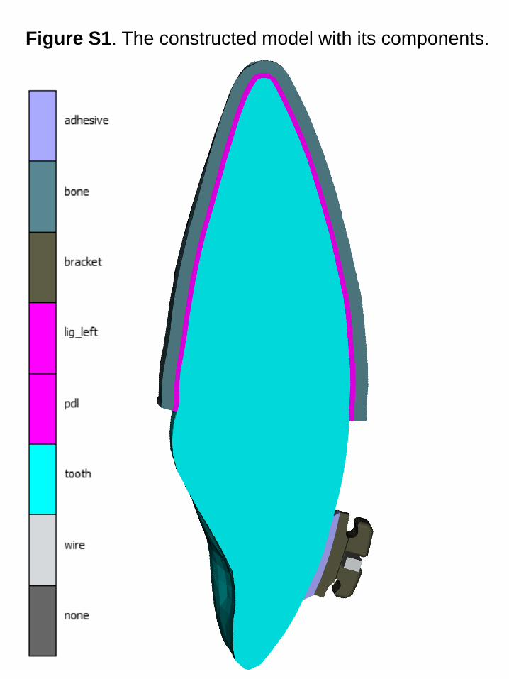
Figure S1. The constructed model with its components.

Figure S2. Example showing the distribution of displacement
(left) and strains (right) within the model. A low-torque (0°)
bracket is placed at the middle of the crown’s surface.