Embed Size (px)
Citation preview
Topic 14. The Shoot SystemIntroduction. This is the second of two lab topics that focus on the three plantorgans (root, stem, leaf). In these labs we want you to recognize how tissues areorganized in each of the three different plant organs; and to understand how thisorganization relates to the function of each organ.
In most plants, stems serve to a support the leaves which act as solar collectors toproduce food. The the stem must conduct water up to and photosynthate downfrom the leaves. Stems and leaves are tightly integrated. Together they constitutethe shoot system. Selective pressure in certain plant groups have resulted inmodified stems and leaves that serve a number of different functions including foodstorage and defense. In some plants (cacti are examples), the stem is the primaryphotosynthetic organ and the leaves are greatly reduced.
I. Coleus Shoot Tip.Take a prepared slide of a longitudinal section of a Coleus shoot tip, and survey theslide at 40x. Relate your preliminary observations to the living Coleus plant nearestyou on your bench.
Are the leaves of the plant opposite or alternate?
How are the pairs of leaves at each node oriented relative to the leaves at the nodesabove and below?
Again view the slide of Coleus. Based on what you learned from your observationsof the living plant, can you determine how many nodes are represented on yourslide?
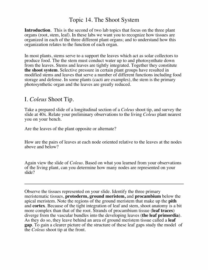
Observe the tissues represented on your slide. Identify the three primarymeristematic tissues, protoderm, ground meristem, and procambium below theapical meristem. Note the regions of the ground meristem that make up the pithand cortex. Because of the tight integration of leaf and stem, shoot anatomy is a bitmore complex than that of the root. Strands of procambium tissue (leaf traces)diverge from the vascular bundles into the developing leaves (the leaf primordia).As they do so, they leave behind an area of ground meristem tissue called a leafgap. To gain a clearer picture of the structure of these leaf gaps study the model ofthe Coleus shoot tip at the front.
Label the Figure:. A - C refer to the three primary meristems. Other labelsindicate the following. leaf primordium, node, leaf trace, and internode.
A.
B.
C.
D.
E.
F.
G.
Detail of a leaf Gap
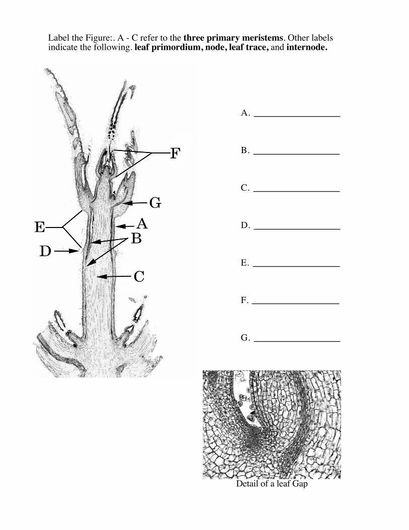
II. Growth Response in Shoots - Negative Gravitropism
The Effect of Seedling Age on Positive Gravitropism
(one group to do this per section)
As in roots, shoots respond to environmental stimuli through growth. To survive, aplant’s shoot must grow upwards towards the light. This response is due todifferential elongation of the cells in the region of elongation. In a horizontal stem,the cells on the bottom will tend to elongate more than those on the upper side,resulting in the tip of the shoot tip bending upwards. How gravity is “felt” by theplant, and how this signal is translated, isn’t completely understood, but the responseis mediated by auxin.
Procedure. Take two seedling tomatoes of different ages, and measure theirlength. Lay them both on their sides in one large saucer. Next classmeeting, measure exactly where on each plant the stem began to curveupwards, and record your observations
Plant Older Younger
Distance from the soil tothe point where bendingstarts
Proportion of the total*stem above the pointwhere bending starts
*Length bending starts to the tip ÷ Total length of the plant
III. Growth Response in Shoots to Dark Conditions(three groups to do this per section)
Plant three bean seeds in each of two paper cups. Label each cup with your sectionand table numbers. Place one cup on the window sill in each room. Place the otherin an enclosed box on the side bench. When you observe the first foliage leaves ofyour light-grown seedlings emerging, remove your dark-grown seedlings fromtheir enclosure. Compare the growth form of the beans subject to each treatment.
What is the average length of the dark grown seedlings?
________________
What is the average length of the light grown seedlings?
________________
Describe other differences between the dark and light grown seedlings.
______________________________________________________________
______________________________________________________________
How is the growth form of the dark-grown plant adaptive?
______________________________________________________________
______________________________________________________________
Place the dark-grown plants on the window sill. Next lab, again observe the twoplants. What has happened to the dark-grown plants during the time they wereexposed to light?
_____________________________________________________________
_____________________________________________________________
_____________________________________________________________
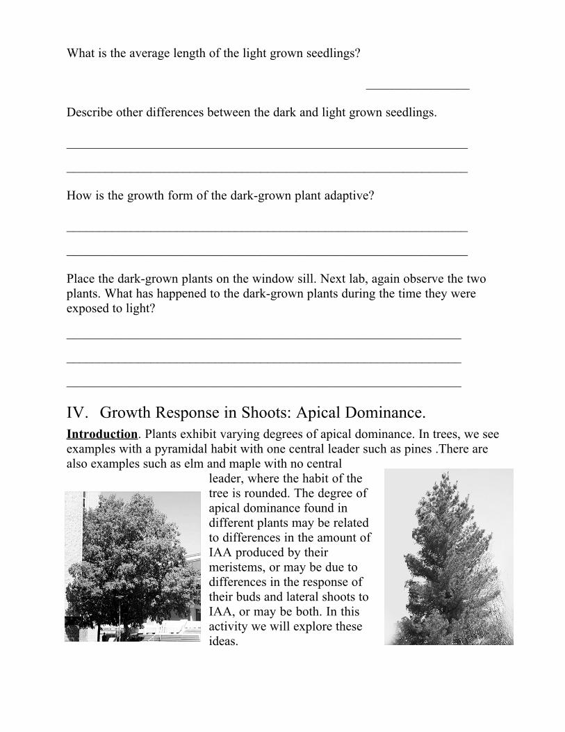
IV. Growth Response in Shoots: Apical Dominance.Introduction. Plants exhibit varying degrees of apical dominance. In trees, we seeexamples with a pyramidal habit with one central leader such as pines .There arealso examples such as elm and maple with no central
leader, where the habit of thetree is rounded. The degree ofapical dominance found indifferent plants may be relatedto differences in the amount ofIAA produced by theirmeristems, or may be due todifferences in the response oftheir buds and lateral shoots toIAA, or may be both. In thisactivity we will explore theseideas.
Each table of students will do a separate experiment. One group will usesunflowers. Commercial sunflowers (Helianthus annuus) manifest a high degreeof apical dominance.Their lateral buds remain totally dormant and their apicalmeristem eventually produces a terminal inflorescence marking the end of growthof the plant. The hypothesis we will consider here is....
Apical dominance is absolute, the lateral buds cannot grow under any range ofauxin concentrations.
Another group will use tomatoes, Lycopersicon esculentum. Lycopersicon displaysapical dominance but not to the degree of sunflower. The hypothesis we willconsider here is....
Apical dominance is caused by the production of auxins by the apical bud.
A third group will use Coleus. In Coleus, there is little or no apical dominance asthe buds begin to grow immediately behind the apex of the plant. There are twoalternative hypotheses we can consider here ....
Apical dominance is not manifested because the apical bud fails to producesufficient auxin.
and
Apical dominance is not manifested because the lateral buds do dot respond toauxin.
Procedure.
For sunflower and tomato, take a pot with three plants. Remove the apical budwith any tightly clustered nodes at the apex from two of these plants. Add IAA inlanolin to one of these two by simply placing the capsule with the lanolin mixtureover the cut stump.
For Coleus, take a pot with three plants. Add IAA in lanolin to one by simplyapplying the lanolin mixture liberally over the intact apex. Remove the apical budof the second, and conduct no further treatment on the third.
Report. After two weeks an oral report will be due from your group on theresults of your experiment.
V. Growth Response in Shoots. Effect of Gibberellic Acid onthe Development of Genetically Dwarf Peas.
Gibberellic acid is a plant hormone associated with cell elongation.Dwarf peas either have lost the capability to synthesize GA, or else,the ability to respond to GA.
Procedure. One group will treat a pot of genetically dwarf plants with GA. Thereare many plants in each pot. Vary the dose applied to each plant using one, two orthree drops. Each drop contains roughly 2.5 x 10⁻⁴ grams of GA. Tie differentlycolored string to each plant according to its dose. To treat a plant simply applydrops of GA anywhere on the shoot. Allow the plants to dry; label the pot withyour section number, and place your pot on the window sill in your room. Overthe next two weeks note the difference in the growth of the treated plants with thevarious doses and the untreated control.
Report. After two weeks an oral report will be due from your group on theresults of your experiment. Do your observations support the idea that the dwarfplants have lost the ability to produce GA, or have the plants lost the ability torespond to GA.
VI. Stem Anatomy of Herbaceous Eudicots in Cross SectionSurvey a slide of a cross section of a Medicago stem at 40x with your microscope.Note the arrangement of the vascular bundles dividing the ground tissue intopith, and cortex. This is a typical arrangement found in many eudicots. These sameplants commonly have roots with tissues arranged like that of Ranunculus studiedearlier with the vascular tissue in the center.
Structurally, how might this arrangement of the vascular bundles be adaptive to theplant?
__________________________________________________________
__________________________________________________________
__________________________________________________________
_________________________________________________________
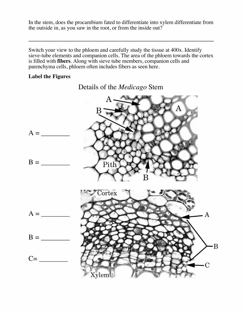
Switch to 100x and carefully study the xylem in one vascular bundle. Note that itconsists of both red stained vessel elements and parenchyma cells. Also notice thatthe vessels become smaller towards the pith. Carefully survey the vessel elements tosee which have incomplete secondary walls. These are protoxylem vessel elements.
In the stem, does the procambium fated to differentiate into xylem differentiate fromthe outside in, as you saw in the root, or from the inside out?
Switch your view to the phloem and carefully study the tissue at 400x. Identifysieve-tube elements and companion cells. The area of the phloem towards the cortexis filled with fibers. Along with sieve tube members, companion cells andparenchyma cells, phloem often includes fibers as seen here.
Label the Figures
Details of the Medicago Stem
A = ________
B = ________
A = ________
B = ________
C= ________
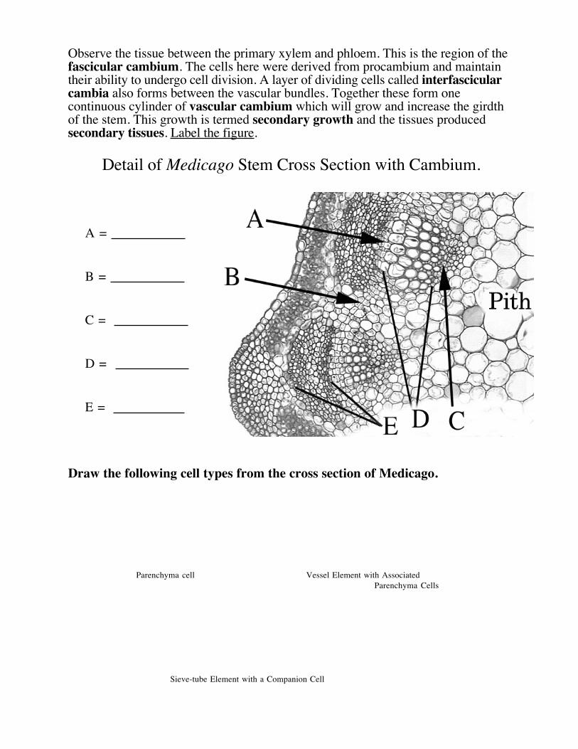
Observe the tissue between the primary xylem and phloem. This is the region of thefascicular cambium. The cells here were derived from procambium and maintaintheir ability to undergo cell division. A layer of dividing cells called interfascicularcambia also forms between the vascular bundles. Together these form onecontinuous cylinder of vascular cambium which will grow and increase the girdthof the stem. This growth is termed secondary growth and the tissues producedsecondary tissues. Label the figure.
Detail of Medicago Stem Cross Section with Cambium.
A =
B =
C =
D =
E =
Draw the following cell types from the cross section of Medicago.
Parenchyma cell Vessel Element with AssociatedParenchyma Cells
Sieve-tube Element with a Companion Cell
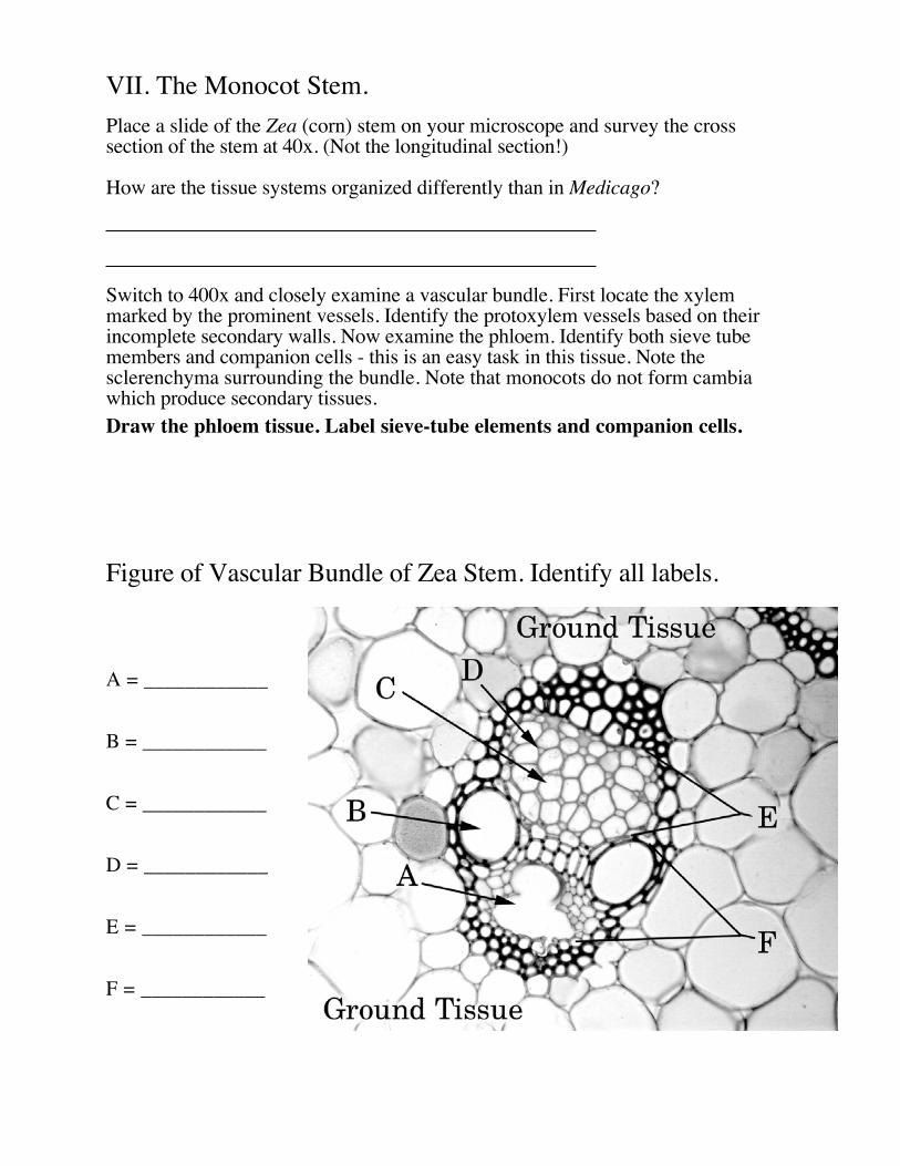
VII. The Monocot Stem.Place a slide of the Zea (corn) stem on your microscope and survey the crosssection of the stem at 40x. (Not the longitudinal section!)
How are the tissue systems organized differently than in Medicago?
Switch to 400x and closely examine a vascular bundle. First locate the xylemmarked by the prominent vessels. Identify the protoxylem vessels based on theirincomplete secondary walls. Now examine the phloem. Identify both sieve tubemembers and companion cells - this is an easy task in this tissue. Note thesclerenchyma surrounding the bundle. Note that monocots do not form cambiawhich produce secondary tissues.Draw the phloem tissue. Label sieve-tube elements and companion cells.
Figure of Vascular Bundle of Zea Stem. Identify all labels.
A = ____________
B = ____________
C = ____________
D = ____________
E = ____________
F = ____________
VIII. Leaves.
Typically, leaves have determinate growth. They grow to maturity and then allgrowth stops - forever. As we saw with the Coleus shoot tip, new leaves areproduced from the apical meristem of the shoot. As covered in our first lab, leavesare associated with axillary bud which forms branches in the shoot
The Lilac (Syringa) LeafCross Section. Survey the prepared slide of a cross section of Syringa leaf at 40x.Note how the three tissue systems are organized. Switch to 400x and carefullystudy an area of the blade away from the midvein. Observe the upper and lowerepidermis and carefully note any differences between these tissue layers. Theground tissue of the leaf is called the mesophyll, and is divided into an upper layerof vertically arranged cells (the palisade parenchyma) and a lower layer ofhorizontally arranged cells (the spongy parenchyma). Veins make up the vasculartissue. Each vein is encased in a layer of parenchyma cells called a bundle sheath.Every cell of the mesophyll is in close proximity to a minor vein. This is criticalbecause the veins move water into and photosynthate out of the leaf. To gain anappreciation of how pervading this network of veins is, observe the demonstrationslide of a cleared leaf available at the side bench.
Things to consider while viewing the cross section of the blade.
1. How might it be adaptive to have the palisade parenchyma arrangedvertically?
2. Would you expect to find more stomata on the upper or on the lowerepidermis?
3. How do materials move to and from the minor veins to the cells of the groundtissue (palisade parenchyma + spongy parenchyma)?
4. What structure controls the movement of materials to and from the veins?
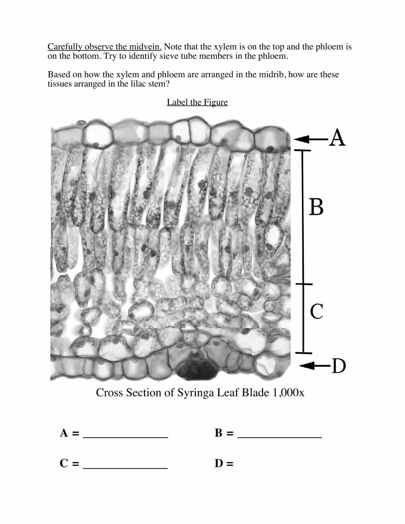
Carefully observe the midvein. Note that the xylem is on the top and the phloem ison the bottom. Try to identify sieve tube members in the phloem.
Based on how the xylem and phloem are arranged in the midrib, how are thesetissues arranged in the lilac stem?
Label the Figure
Cross Section of Syringa Leaf Blade 1,000x
A = B =
C = D =
Lilac Leaf Paradermal Section. Place a prepared slide of a Syringa leaf in paradermal section onto your stage. Firstidentify the upper epidermis by its concentration of stomata. Progressively identifyand study the palisade parenchyma bordering the upper epidermis, the spongyparenchyma bordering the palisade parenchyma, and the lower epidermis.
- Note the amount of intercellular space in both regions of mesophyll.
- Note the tight arrangement of the basal epidermal cells.
Draw a minor vein. Label vessel elements and the bundle sheath.
Draw a region of the lower epidermis. Label guard cells, basal cells of theepidermis, and the cuticle.
Anatomy of Leaves Adapted for Dry or Wet EnvironmentsSyringa is a plant adapted to moist conditions. In your slide box are examples oftwo different other leaves adapted to different moisture levels. Both of these leaveshave the same basic anatomy as Syringa. Each, however, has major differences.
Observe the leaves of Nerium and Nymphaea. Write down at least two ways theyare different from Syringa, reflecting how they are modified to their specificenvironment. Exchange ideas with your classmates and with your TA.
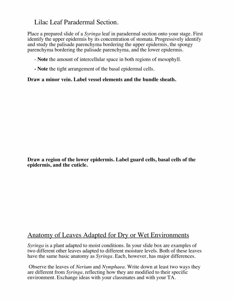
Notes for Nerium oleander. A plant that lives in a dry environment.
Cross Section of Leaf of Nerium oleander
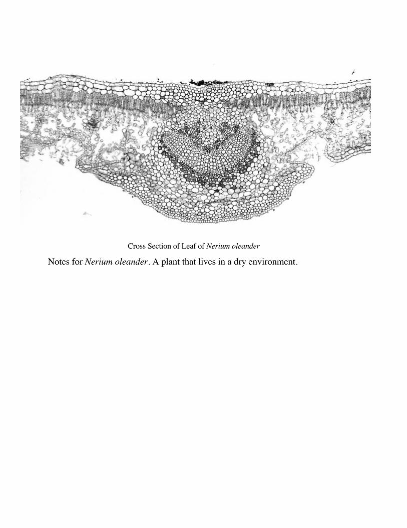
Notes &Sketches forNymphaea
oderata, a plant that lives in the water.
Cross section of Nymphaea oderata
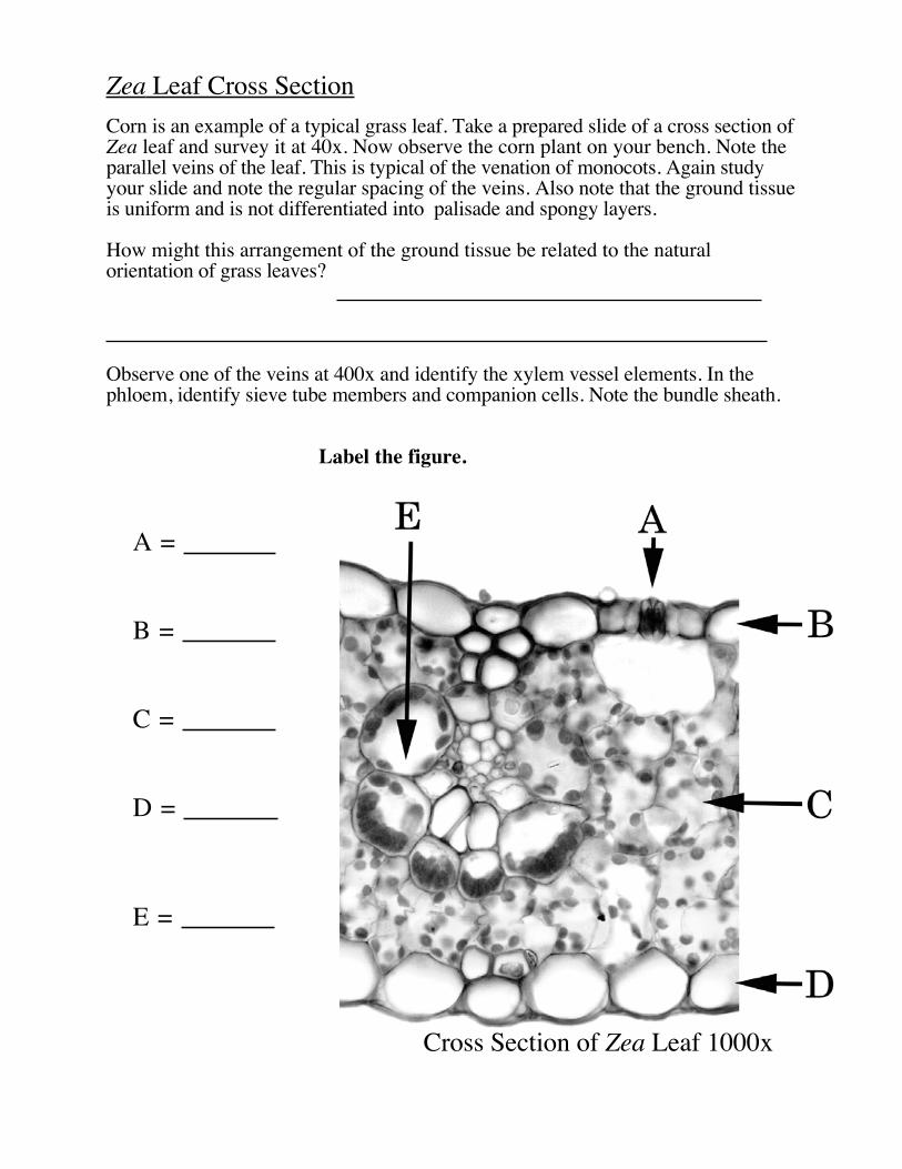
Zea Leaf Cross SectionCorn is an example of a typical grass leaf. Take a prepared slide of a cross section ofZea leaf and survey it at 40x. Now observe the corn plant on your bench. Note theparallel veins of the leaf. This is typical of the venation of monocots. Again studyyour slide and note the regular spacing of the veins. Also note that the ground tissueis uniform and is not differentiated into palisade and spongy layers.
How might this arrangement of the ground tissue be related to the naturalorientation of grass leaves?
Observe one of the veins at 400x and identify the xylem vessel elements. In thephloem, identify sieve tube members and companion cells. Note the bundle sheath.
Label the figure.
Cross Section of Zea Leaf 1000x
A =
B =
C =
D =
E =
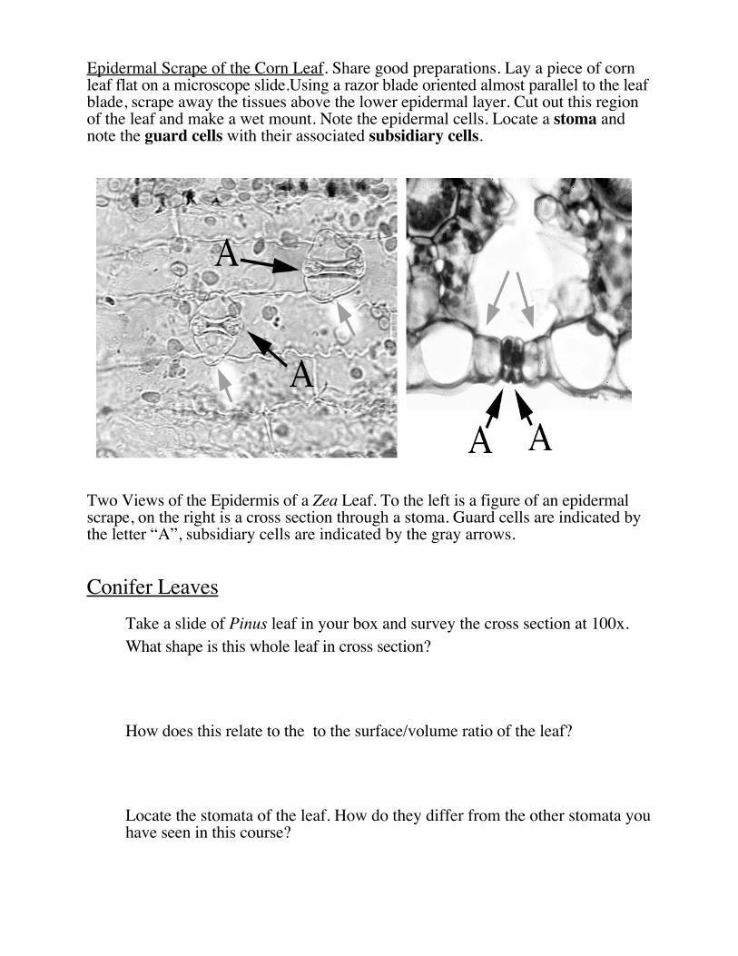
Epidermal Scrape of the Corn Leaf. Share good preparations. Lay a piece of cornleaf flat on a microscope slide.Using a razor blade oriented almost parallel to the leafblade, scrape away the tissues above the lower epidermal layer. Cut out this regionof the leaf and make a wet mount. Note the epidermal cells. Locate a stoma andnote the guard cells with their associated subsidiary cells.
Two Views of the Epidermis of a Zea Leaf. To the left is a figure of an epidermalscrape, on the right is a cross section through a stoma. Guard cells are indicated bythe letter “A”, subsidiary cells are indicated by the gray arrows.
Conifer Leaves Take a slide of Pinus leaf in your box and survey the cross section at 100x.
What shape is this whole leaf in cross section?
How does this relate to the to the surface/volume ratio of the leaf?
Locate the stomata of the leaf. How do they differ from the other stomata youhave seen in this course?
Can you identify an endodermis in this leaf?
How are these various modifications adaptive? Pines can live on the same typeof sites as lilac (Syringa
IX. The Role of IAA (indole acetic acid) in Leaf Abscission
Each group to do one treatment as described below.
Introduction. In some plants, leaf abscission is an active process involving thegrowth of a tissue layer that undercuts the petiole’s attachment to the stem. Inthese plants, the leaf signals its health through the production of IAA whichinhibits the growth of the abscission layer. If the leaf is unhealthy this signal isremoved resulting in leaf abscission.
The hypothesis we will consider here is....
IAA applied to the plant will only affect the petiole to which it is applied.
Procedure.
Take a Coleus plant and remove four leaf blades at two nodes, while leaving thepetioles attached to the stem. Either apply IAA in lanolin to the cut surface ofone petiole at each node, or, to the cut surface of the petioles both at theupper node. Label your plants and set them on the light bench.
This hypothesis assumes that IAA will not flow across the petiole to affect otherleaves.
Report. After two weeks an oral report will be due from your group on theresults of your experiment.
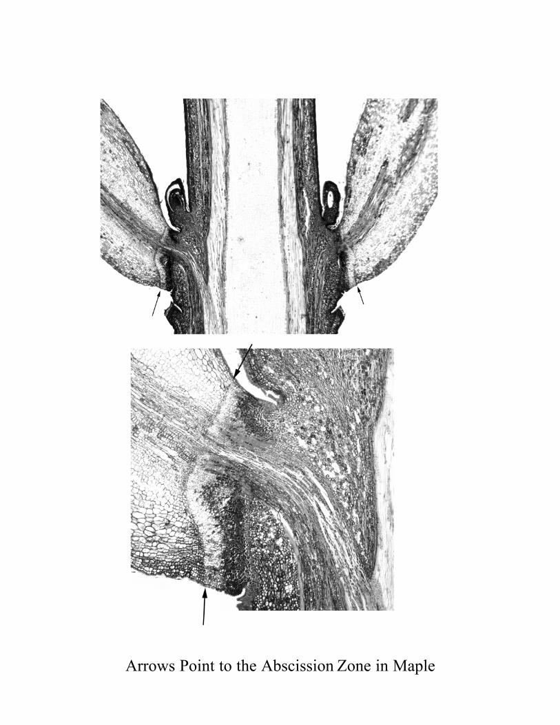
Arrows Point to the Abscission Zone in Maple


![[PPT]Shoot House Slideshow Presentation - Pennsylvaniaftig.png.pa.gov/Training/Documents/Shoot House/Shoot... · Web viewCAPABILITIES two story enclosed shoot house constructed of](https://img.pdfslide.us/doc/110x75/5ae5190a7f8b9a495c8f743e/pptshoot-house-slideshow-presentation-houseshootweb-viewcapabilities-two.jpg)