Embed Size (px)
Citation preview

April 2016 cliniciansbrief.com 89
Top 5 Tips on Radiographic Diagnosis of Obstructive Foreign BodiesNathalie Rademacher, Dr. med. vet., DECVDI, DACVRLouisiana State University
Ileus can be functional or mechanical. In functional ileus, peristaltic contrac-tions of the bowel cease as a result of vascular or neuromuscular abnormali-ties within the bowel wall, but the bowel remains patent. In mechanical ileus, physical obstruction occludes the lumen and can be caused by foreign bodies (FBs), intussusception, mural mass, or extraluminal lesions. GI FBs are a com-mon cause of mechanical obstruction, which may be complete or partial. The jejunum is reported to be the most com-mon location.1
The majority of obstructive GI FBs com-promise the blood supply by luminal distention, leading to intestinal wall edema and progressive necrosis.1 Com-plete obstruction is associated with more dramatic clinical and radiographic signs, whereas partial obstruction may be associated with more chronic signs.2
Differentiation between mechanical and functional ileus may be impossible because of overlapping radiographic findings.
The following focuses on the detection of obstructive FBs and provides a few tips to improve detection of FBs on radiographs to make a diagnosis of mechanical ileus easier and support the decision for surgical exploration.
1 Use Radiographic FindingsRadiographic features of small intestinal mechanical obstruc-tion depend on its completeness,
location, and duration.2 Whereas metallic FBs or those with mineral con-tent are easily recognized within the GI lumen (Figure 1), nonmineralized, nonmetallic objects (eg, cloth) within the GI tract are more difficult to iden-tify (Figure 2, next page). Some may be
TOP 5 h RADIOGRAPHY h PEER REVIEWED
1 VD abdominal radiograph of a 1-year-old cat with a 2-day history of vomiting and a nonobstructing metallic foreign body. A coin with a radiolucent center caused by erosion is present within the small intestinal loops in the right midabdomen. No dilated loops of small intestine are seen to indicate mechani-cal obstruction.
g
FB = foreign body
TOP 5 TIPS ON RADIOGRAPHIC DIAGNOSIS OF OBSTRUCTIVE FOREIGN BODIES1. Use Radiographic Findings
2. Repeat Radiography 24 to 36 Hours After Fasting
3. Use Positional Radiography
4. Perform Pneumoncolonography
5. Conduct Compression Radiography

90 cliniciansbrief.com April 2016
TOP 5 h RADIOGRAPHY h PEER REVIEWED
recognized by their geometric shape. The most consistent sign of mechanical obstruc-tion is variable dilation of intestinal loops proximal (orad) to the obstruction (described as a mixed population of small intestine; Figure 2). Determination of small intestinal diameter can be made by compar-ing it to the L5 vertebral body height in dogs.3 Values greater than 1.6 are suggestive of obstruction (Figure 2A). In cats, a ratio of the maximum small intestinal diameter to vertebral end plate height of L2 greater than 4 indicates a high likelihood of intestinal obstruction.4
Qualitative assessment of intestinal size by experienced clinicians may be as accurate in determining the presence of mechanical obstruction as calculation of ratios. Obstructed intestine usually contains fluid and gas; how-ever, if the obstruction is orad, reflux into the stomach can occur and limited intestinal dis-tension may be apparent. More distal (aborad) or more complete obstruction leads to greater dilation. Stacking of intestinal loops occurs with more severe dilation as segments become increasingly crowded in a relatively smaller space (Figure 2B).
2Repeat Abdominal Radiography 24 to 36 Hours After FastingFood or ingesta can look similar to foreign material and can be difficult
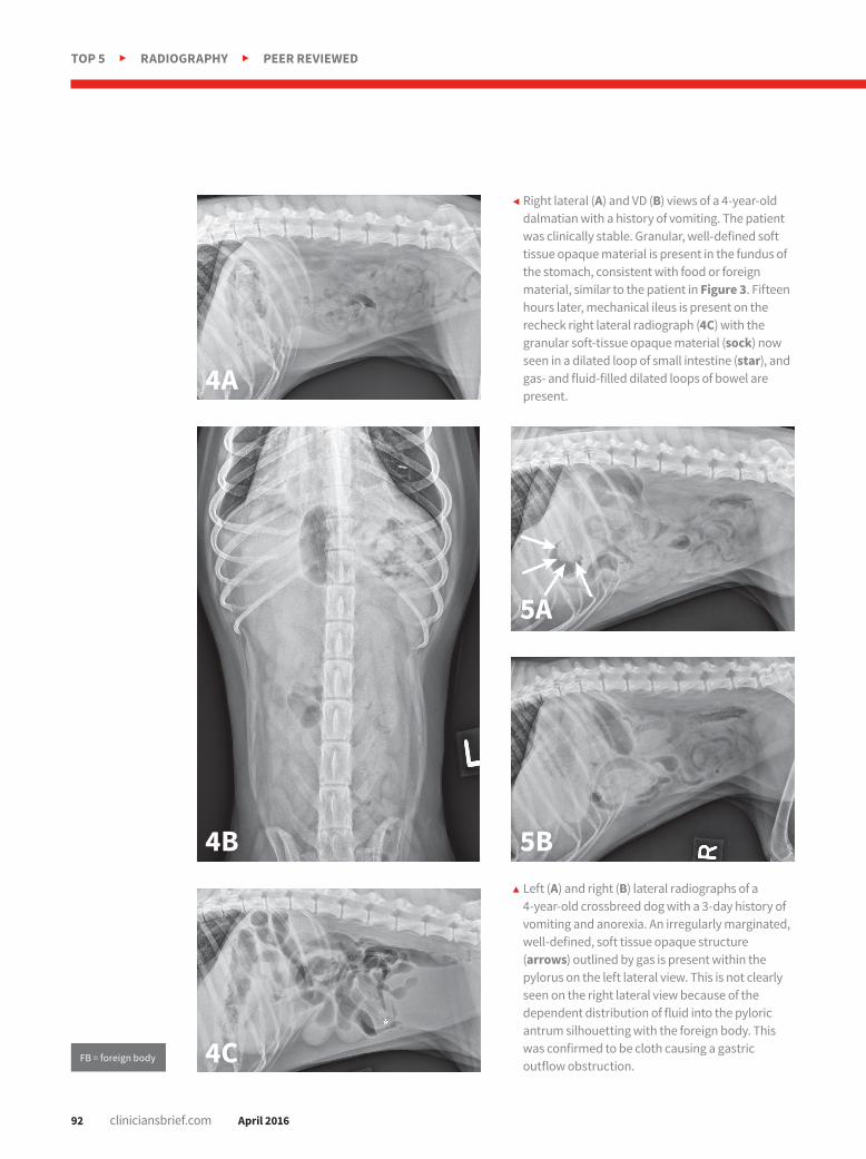
to differentiate. Depending on the presenta-tion, radiography can be repeated after fast-ing period to document passage (or lack thereof) of GI content (Figures 3, next page and 4, page 92). Persistence of opaque for-eign material in the same location over a 24- to 48-hour period should increase the suspicion of partial obstruction. In cases of complete mechanical obstruction, radio-graphic features of obstruction can develop during this time. This makes it easier to rec-ognize the obstruction radiographically (Figure 4C, page 92).
2A
2Bd Right lateral (A) and VD (B) abdominal radiographs
of a Labrador retriever with an obstructive ileus. A FB (sock) is present within a dilated small intestinal loop in the right caudoventral abdomen (arrows). The dashed lines outline a fluid-filled loop measuring 2.7 cm and a gas-filled loop measuring 2.4 cm, compared to the height of L5 (A; solid line, 1.1 cm); the resulting ratios of 2.4 and 2.2, respectively, are much greater than the upper limit of 1.6 for normal small intestine to L5 height ratio. Several stacked gas-filled loops of small intestine are present in the left midabdomen (B).
FB = foreign body

April 2016 cliniciansbrief.com 91
d Left lateral (A) and VD (B) radiographs of a 4-year-old crossbreed dog with a history of vomiting. The patient was clinically stable. Granular well-defined soft tissue opaque material is present within the fundus of the stomach, consistent with food or foreign material. The same dog after fasting for 24 hours (C & D). The previously seen soft tissue opaque material within the stomach is no longer present, and the GI tract is empty.
3A 3C
3B 3D
3Use Positional RadiographyRight and left lateral abdominal radio-graphs are easy to obtain and are often helpful in cases of suspected GI FBs
and obstruction because this positional
change shifts the fluid and gas present into different areas, which might highlight poten-tial intraluminal FBs (Figure 5, next page). The main benefit of this simple technique is in the identification of pyloric FBs in cases of sus-
92 cliniciansbrief.com April 2016
TOP 5 h RADIOGRAPHY h PEER REVIEWED
4A
5A
4C
4B 5Bd Left (A) and right (B) lateral radiographs of a
4-year-old crossbreed dog with a 3-day history of vomiting and anorexia. An irregularly marginated, well-defined, soft tissue opaque structure (arrows) outlined by gas is present within the pylorus on the left lateral view. This is not clearly seen on the right lateral view because of the dependent distribution of fluid into the pyloric antrum silhouetting with the foreign body. This was confirmed to be cloth causing a gastric outflow obstruction.
g Right lateral (A) and VD (B) views of a 4-year-old dalmatian with a history of vomiting. The patient was clinically stable. Granular, well-defined soft tissue opaque material is present in the fundus of the stomach, consistent with food or foreign material, similar to the patient in Figure 3. Fifteen hours later, mechanical ileus is present on the recheck right lateral radiograph (4C) with the granular soft-tissue opaque material (sock) now seen in a dilated loop of small intestine (star), and gas- and fluid-filled dilated loops of bowel are present.
FB = foreign body

April 2016 cliniciansbrief.com 93
6A 6C
6B 6Dd Left lateral (A) and VD (B) survey radiographs of a 4-year-old pit bull terrier with a 3-day history of vomiting,
diarrhea, and anorexia. A dilated loop of bowel is present within the midabdomen (star) caudal to the stomach, which could represent large or small bowel. Caudal to the ascending colon on the VD view, an irregularly marginated radiolucent rectangular foreign body is present (C & D; arrows). After pneumocolonography was performed, the colon can be easily traced, and the questionable loop was identified as small bowel (star). A mechanical ileus secondary to a foreign body was diagnosed. A corncob was identified as the cause for the mechanical obstruction in surgery.

94 cliniciansbrief.com April 2016
TOP 5 h RADIOGRAPHY h PEER REVIEWED
graphs are taken (Figures 6A and 6B, previous page), the animal is placed in lateral recumbency. Air is instilled into the rectum and colon using a large lubri-cated syringe or rubber catheter; approximately 10 to 12 mL/kg of air is needed for moderate distention of the colon.6,7 Standard lateral and ventrodor-sal radiographs are then taken (Figures 6C and 6D, previous page). In most ani-mals, it is desirable to fill the colon to the cecum. Partial filling of the distal jejunum and ileum may occur and is normal.
5Conduct Compression RadiographyCompression radiography is a simple technique that may help
clarify the presence of an obstruction.8
With the patient in lateral recumbency and using a wooden or plastic spoon or paddle, the abdomen is mildly com-
pected pyloric outflow obstruction, but it is also useful in the small intestine, espe-cially if duodenal FBs are suspected.
4Perform PneumocolonographyPneumocolonography is a rapid, safe, and relatively easy proce-
dure that can be performed to identify the position of the colon when it cannot be determined on survey radiographs. Usually the colon is filled with feces and gas, which makes its position apparent. If, however, the colon is poorly visual-ized or confusing fluid or gas opacities are present, the colon can be easily localized by creation of pneumocolon.5
In cases of suspected FBs, this technique can be helpful to differentiate dilated small intestinal loops from colon or determine whether mottled mineralized material is in the colon or small intesti-nal tract. After initial survey radio-
7A 7Bh Left lateral radiograph of a 9-year-old Jack Russell terrier with a 4-day history of vomiting and anorexia (A). Moderate dilation with
small gas bubbles (stars) of the small intestinal tract is noted (B). Left lateral compression radiograph of the same dog. A wooden spoon was used to apply compression to the caudal abdomen. Ventral to the colon, a well-defined ovoid mineral opacity (arrows) is visible within a dilated small intestinal loop. This was a piece of rubber confirmed at surgery.
FB = foreign body

April 2016 cliniciansbrief.com 95
Welcome our NEWEST addition to the familyIntroducing the new Ibex® EVO™
--the next generation of veterinary
portable ultrasound systems in the
E.I. Medical Imaging family of
products. The Ibex® EVO™ is as
durable and rugged as our other
ultrasound systems featuring our
innovative Durascan® technologinnovative Durascan® technology,
but it also features four times the
power which translates to better
image quality required of the most
advanced systems. Technology has
progressed in the world of portable
ultrasound systems and we
continue to lead the way in our continue to lead the way in our
advanced innovation in the industry.
We have taken the Ibex® EVO™ to
the next level and we are now
offering it to you. Wherever you
travel and wherever you use the
Ibex® EVO™ it will withstand the
tough conditions you have come to tough conditions you have come to
expect or require from a portable
ultrasound system. We have
engineered the Ibex® EVO™ to use
in nearly every imaginable location.
CALL US TOLL FREE: 1.866.365.6596
www.eimedical.com
April 2016 cliniciansbrief.com 95
pressed in an area of concern identified on survey films (Figure 7A). Using the paddle and compression, adjacent bowel or masses are displaced to increase radiographic con-spicuity. This results in better
visualization of potential for-eign material (Figure 7B), plication of small intestinal loops in cases of linear FBs, or intestinal masses other-wise obscured by superim-posed bowel. n
References1. Hayes G. Gastrointestinal foreign bodies
in dogs and cats: A retrospective study of 208 cases. J Small Anim Pract. 2009;50(11):576-583.
2. Riedesel EA. The small bowel. In: Thrall DE, ed. Textbook of Veterinary Diagnostic Radiology. 6th ed. St. Louis, MO: Saunders Elsevier; 2013:789-811.
3. Graham JP, Lord PF, Harrison JM. Quantitative estimation of intestinal dilation as a predictor of obstruction in the dog. J Small Anim Pract. 1998;39(11):521-524.
4. Adams WM, Sisterman LA, Klauer JM, Kirby BM, Lin TL. Association of intestinal disorders in cats with findings of abdominal radiography. JAVMA. 2010;236(8):880-886.
5. Nyland T, Ackerman N. Pneumocolon: A diagnostic aid in abdominal radiography. Vet Radiol Ultrasound. 1978;19(6):203-209.
6. Finck C, D’Anjou MA, Alexander K, Specchi S, Beauchamp G. Radiographic diagnosis of mechanical obstruction in dogs based on relative small intestinal external diameters. Vet Radiol Ultrasound. 2014;55(5):472-479.
7. Bradley K. Practical contrast radiography 2 Gastrointestinal studies. In Practice. 2005;27(8):412-417.
8. Schwarz T. The large bowel. In: Thrall DE, ed. Textbook of Veterinary Diagnostic Radiology. 6th ed. St. Louis, MO: Saunders Elsevier; 2013:812-824.