Embed Size (px)
Citation preview

Pulse of MR
Autumn 2017
RSNA Edition
Volume Twenty-Two
T O M O R R O W T O D A Y

gehealthcare.com/mr 2 Tomorrow Today
Outside the Bore
Publications Team:
Stephanie BroyhillEditor-in-ChiefAdvertising & Promotions Leader, Global MR
Anna BrownAssociate EditorAdvertising & Promotions Director, Global MR
Kerry AdapathyaAssociate EditorAdvertising & Promotions Leader, Global MR
Mary Beth MassatAssociate Editor
Steve LawsonClinical EditorGlobal Clinical Marketing Specialist, MR
Sylvain AdamClinical EditorClinical Marketing Specialist, MR
Brandon PascualClinical EditorApplications Specialist, MR
RocketLawnchairDesign & Production
GE Contributors:
Ibrahim AlabdulaalyClinical Leader, MR
Holly BlahnikClinical Development Specialist, MR
Raphael DoustalyZone Clinical Leader, MR
Almos Elekes, PhDGlobal Product Marketing Manager, PET/MR, Oncology and Molecular MR
Luciana Ferrari Fracchia Product Marketing Specialist, MR
Stephen GibbsGlobal Product Marketing Operations Director, MR
Kentaro GotoModality Sales Specialist, MR
Moonjung HwangScientist, MR
Brian KingRegion Modality Leader, MR
Dave LambRegion Product Marketing Manager, MR
Joonsung LeeAdvanced Applications Leader, MR
Greg LochmannRegion Modality Leader, MR
Jun Nagata Product Marketing Manager, MR
Barbara PirgousisRegion Clinical Applications Leader, MR
Richard ProrokStrategic Marketing Manager, MR
James SedorovichClinical Product Marketing Leader, MR
Janice SichRegulatory Affairs Manager, MR
Matt SuminskiBusiness Development Manager, PET/MR
Fotis Vlachos, PhDGlobal Product Manager, Premium MR
Guillermo ZannoliClinical Marketing Manager, MR
In Practice
Issue Spotlight
5 Break through to Tomorrow’s MR… Today with SIGNA™ Premier and SuperG
6 Weill Cornell Medical Center invests in ultra high-field 7T MR research
6 GE MR welcomes Michael Brandt
7 GE Healthcare welcomes new President/CEO
7 Whole-body staging of solid tumors in pediatrics without gadolinium
8 Synthetic diffusion: a robust sequence for prostate cancer diagnosis and patient management
13 Increased PET sensitivity with TOF & Q.Static elevates clinical confidence
16 Pursuing an MR-only approach to radiation therapy planning
20 Making a difference in MR oncology imaging
26 Optimized clinical pathway propels high utilization of PET/MR at Pitié-Salpêtrière Hospital
32 “Child first and always” philosophy drives investment in advanced MR technology at Intermountain Healthcare
38 Using an efficient, quantitative sequence to aid in patient treatment decisions
42 SIGNA™ Explorer Lift revives our MR
47 Going digital with SIGNA™ Artist helps prepare Dunedin Hospital for the future
52 A boost in productivity

gesignapulse.com 3 Autumn 2017
© 2017 General Electric Company, doing business as GE Healthcare. All rights reserved. The copyright, trademarks, trade names and other intellectual property rights subsisting in or used in connection with and related to this publication are, the property of GE Healthcare unless otherwise specified. Reproduction in any form is forbidden without prior written permission from GE Healthcare.
LIMITATION OF LIABILITY: The information in this magazine is intended as a general presentation of the content included herein. While every effort is made by the publishers and editorial board to see that no inaccurate or misleading data, opinion or statements occur, GE cannot accept responsibility for the completeness, currency or accuracy of the information supplied or for any opinion expressed. Nothing in this magazine should be used to diagnose or treat any disease or condition. Readers are advised to consult a healthcare professional with any questions. Products mentioned in the magazine may be subject to government regulation and may not be available in all locations. Nothing in this magazine constitutes an offer to sell any product or service.
Case Studies
Tech Trends68 Total freedom in coil
positioning and handling with the AIR Technology Suite
55 Two-minute MR ultra-fast neuro protocol
58 Pediatric brain tumor assessment using MAGiC
62 Pediatric imaging with MAGiC
65 Pediatric MR enterography

gehealthcare.com/mr 4 Tomorrow Today
Welcome
to 8 times faster imaging efficiency and
streamlined workflow. For example,
HyperSense technology delivers
increased speed for 3D exams by
incorporating compressed sensing
reconstruction for research and 88%
of all clinical procedures.
We’ve explored and invented, now it’s
time to transform. You’ll also see our
industry-first AIR Technology created
with people in mind. This unique RF coil
technology benefits patients with an
ultra-lightweight, flexible design that
conforms comfortably to all patient
shapes and sizes. Also included in
this suite is the 48-channel TDI Head
Coil, designed to fit 99.9% of patients
with high SNR and acceleration factors
for superior brain imaging. Available
initially with SIGNA Premier, the suite
allows technologists to experience new
freedom in coil handling and positioning.
Of course, there will be more to see
at RSNA, but I’d rather you hear about
those innovations from the passionate
people that made them possible. Stop
by the GE booth and see the strides
we’ve made in partnership with you as
we Explore, Invent and Transform to
deliver SIGNA: Tomorrow Today.
Earlier in October, I had the privilege of
hearing John Flannery, GE’s Chairman
and CEO, speak at the 2017 Minds +
Machines event in San Francisco. As he
succinctly retold GE’s 30+ year history
in MR—yes, it marks 35 years next
year!—I was struck by how closely his
narrative reflected the theme for RSNA.
This year’s theme of course revolves
around three core tenets: Explore,
Invent and Transform. Thirty-five years
ago, in GE’s labs, our engineers and
scientists were exploring magnetic
resonance imaging, leveraging
technology 30,000 times stronger
than the earth’s magnetic field. That
led to the invention of the industry’s
first high-field 1.5T MR scanner, our
first SIGNA™ system, which enabled
clinicians to see inside the human body
and obtain images and information
like never before. That was a quantum
leap that helped radiology transform
healthcare.
Now our challenge is to make the next
leap, and this time we are doing it in
the context of a more digitally-enabled
world. Along with our research and
clinical partners, we are focused on
applying deep learning-based Artificial
Intelligence (AI) and analytics to our
portfolio of SIGNA™ MR scanners and
connected devices. Our aim is to reduce
exam times and exam time variability,
improve consistency of image quality
and eliminate rescans and recalls. You’ll
benefit from real productivity gains and
your patients will benefit from a better
MR experience and maybe an improved
clinical outcome… our ultimate scorecard.
So, what will you see at RSNA this
year? We are excited to take you on
an interactive journey of innovation
that brings to the forefront technology
that embodies SIGNA™: Tomorrow
Today. Great products begin with
exploration and that exemplifies the
roots of the SIGNA™ Premier, a powerful
ultra-premium scanner you’ll have
the opportunity to view in our booth.
Developed in partnership with the NFL
and academic institutions, this scanner
is designed for both high-end research
and leadership clinical utility. SIGNA
Premier combines the exceptional
capabilities of 3.0T imaging with a
146-channel Total Digital Imaging
(TDI) RF chain and SuperG gradient
technology that delivers a 70 cm wide
bore with the cutting-edge stability and
Human Connectome performance only
available on 60 cm systems. And the
intelligence factor? SIGNA Premier’s
sophisticated platform uses advanced
diffusion applications to automatically
correct distortion and was developed
with machine learning to precisely
optimize SAR limits for each patient.
And cardiac imaging? Our newly
expanded ViosWorks post processing
platform leverages cloud computing
and deep learning algorithms to
automate cardiac MR analysis, delivering
accurate, quantifiable results.
Through exploration comes invention,
which brings us to SIGNA™Works.
Developed through partnerships
with our academic collaborators,
SIGNA™Works is a fully customizable
platform designed to drive productivity
and clinical excellence by optimizing
operational throughput with
greater imaging speed, accuracy
and consistency. This extensive
portfolio includes GE’s HyperWorks
applications: HyperSense, HyperBand
and HyperCube, designed to deliver up
Eric Stahre
President and CEO
Global MR, GE Healthcare
gesignapulse.com 5 Autumn 2017
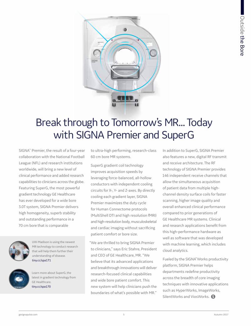
SIGNA™ Premier, the result of a four-year
collaboration with the National Football
League (NFL) and research institutions
worldwide, will bring a new level of
clinical performance and added research
capabilities to clinicians across the globe.
Featuring SuperG, the most powerful
gradient technology GE Healthcare
has ever developed for a wide bore
3.0T system, SIGNA Premier delivers
high homogeneity, superb stability
and outstanding performance in a
70 cm bore that is comparable
to ultra-high performing, research-class
60 cm bore MR systems.
SuperG gradient coil technology
improves acquisition speeds by
leveraging force-balanced, all-hollow
conductors with independent cooling
circuits for X-, Y- and Z-axes. By directly
cooling each gradient layer, SIGNA
Premier maximizes the duty cycle
for Human Connectome protocols
(MultiShell DTI and high resolution fMRI)
and high-resolution body, musculoskeletal
and cardiac imaging without sacrificing
patient comfort or bore size.
“We are thrilled to bring SIGNA Premier
to clinicians,” says Eric Stahre, President
and CEO of GE Healthcare, MR. “We
believe that its advanced applications
and breakthrough innovations will deliver
research-focused clinical capabilities
and wide bore patient comfort. This
new system will help clinicians push the
boundaries of what’s possible with MR.”
In addition to SuperG, SIGNA Premier
also features a new, digital RF transmit
and receive architecture. The RF
technology of SIGNA Premier provides
146 independent receive channels that
allow the simultaneous acquisition
of patient data from multiple high-
channel density surface coils for faster
scanning, higher image quality and
overall enhanced clinical performance
compared to prior generations of
GE Healthcare MR systems. Clinical
and research applications benefit from
this high-performance hardware as
well as software that was developed
with machine learning, which includes
cloud analytics.
Fueled by the SIGNA™Works productivity
platform, SIGNA Premier helps
departments redefine productivity
across the breadth of core imaging
techniques with innovative applications
such as HyperWorks, ImageWorks,
SilentWorks and ViosWorks.
Break through to Tomorrow’s MR… Today with SIGNA Premier and SuperG
UW-Madison is using the newest MR technology to conduct research that will help them further their understanding of disease.tiny.cc/spa171
Learn more about SuperG, the latest in gradient technology from GE Healthcare.tiny.cc/spa170
Outside the Bore

gehealthcare.com/mr 6 Tomorrow Today
of the brain while steadily expanding
to all anatomies. Researchers using
ultra high-field scanners hope that
their research will contribute to earlier
disease detection, more accurate
diagnosis and increased effectiveness
of investigational therapies for
disorders and diseases.
‡ The SIGNA 7T MR system is technology in development. It is not cleared or approved by the US FDA or other global regulatory body for commercial availability for clinical use. This system is for research only.
In early 2019, Weill Cornell Medical
Center will install GE Healthcare’s
SIGNA™ 7T‡ platform for use in research
applications and a training program
for ultra high-field MR clinical staff
and scientists. Weill Cornell Imaging
received an educational grant to serve
as a training center for ultra high-field
MR for clinical staff and scientists
from around the world. The goal of this
training is to equip the next generation
of MR users to better understand
and utilize ultra high-field MR in a
research setting.
GE Healthcare is pleased to announce
Michael Brandt has joined the team
as Chief Marketing Officer, Global MR.
Brandt has over 20 years of strategic
leadership experience in the medical
imaging industry as well as global
expertise in business development
for multi-national medical device
companies (specializing in MR-guided
radiotherapy, robotic radiosurgery,
CT and MR).
Prior to joining GE, Brandt served as
Vice President Sales, Marketing and
Business Development at Anatomage,
Inc., Senior Vice President of Sales and
Marketing at ViewRay, Inc. and General
Manager: Americas at Accuray. While
at ViewRay, Brandt oversaw the
commercial introduction of the world’s
first MR-guided radiation therapy system.
Brandt also distinguished himself at
Philips Medical Systems where he
“Our vision at GE Healthcare is to bring
MR technology to more patients around
the world and to push the boundaries
of what’s possible with MR; this
collaboration with Weill Cornell will
help achieve that vision through
novel research and training of the
next generation of MR users,” says
Eric Stahre, President and CEO of
GE Healthcare, MR.
7T MR systems are used for scientific
and medical research, primarily for
morphological and functional imaging
held a variety of international MR
management positions in the UK, the
Netherlands, South Africa and the U.S.
Brandt remains as passionate about
MR now as he was as a young engineer
installing and servicing MR equipment
in his hometown of Johannesburg,
South Africa. From understanding
the system mechanics to his vision
for advancing MR in radiation therapy,
Brandt believes the power of MR
imaging can continue to have a
significant impact on the diagnostic
advancements for cancer treatments,
neurodegenerative diseases and
cardiovascular disease.
“At GE Healthcare, we have a magnificent
product line and a strong engineering
team committed to patient care,”
Brandt says. “There are many marketing
channels available to us in the 21st
century, but our primary focus is the
patient.”
Keeping his sights set on what’s
important—elevating patient care
through innovation—he sees more
similarities than differences in how
MR is utilized worldwide.
“Our customers, regardless of where
they are located, are essentially reading
the same literature and examining the
same patient on the same equipment
for the same disease,” he says.
Brandt is excited to be part of the
GE Healthcare team and hopes
to continue the vision that has
consistently held the company at the
forefront of medical imaging technology.
Brandt is a graduate of Vaal University
of Technology in Vanderbijlpark, South
Africa with a degree in electrical
engineering.
Weill Cornell Medical Center invests in ultra high-field 7T MR research
GE MR welcomes Michael Brandt

gesignapulse.com 7 Autumn 2017
Outside the Bore
provider Vericore, Novartis, Adprotech,
ML Laboratories and Innovata plc.
Having earned his bachelor’s degree
in 1984 from University College, Dublin,
he subsequently graduated from the
University of Manchester Institute of
Science and Technology with a master’s
degree in marketing.
Kieran Murphy was appointed President
and CEO of GE Healthcare, succeeding
John Flannery who will replace GE
Chairman and CEO Jeff Immelt. Prior to
his appointment, Murphy was President
and CEO of GE Healthcare Life Sciences,
where he oversaw significant revenue
growth and geographic expansion of
the Molecular Imaging business.
In a released statement, Immelt
said, “Having led the strategic
combination of GE’s Life Sciences
and Medical Diagnostics units, Kieran
is universally respected across GE
Stanford researchers have achieved
whole-body staging of solid tumors
in pediatric patients without the use
of gadolinium. A study published in
Molecular Imaging and Biology in July
2017, reports that the use of ferumoxytol-
enhanced PET/MR scans provided
images showing the location and size of
the tumor and whether the cancer had
spread. Researchers were able to stage
the tumor in less than 1 hour with equal
or superior results compared to clinical
and has distinguished himself as a
strong customer advocate with great
commercial instincts. Alongside the
outstanding team at GE Healthcare,
we anticipate that the business will
experience continued global growth
under Kieran’s leadership.”
Murphy has over 20 years of experience
in the global life sciences and
biotechnology industry beginning his
career with Janssen Pharmaceutical,
a division of Johnson and Johnson,
followed by leadership roles with
Mallinckrodt, veterinary medicines
standard tests in 17 out of 20 patients.
The study also reported significantly
reduced radiation exposure with a
mean of 3.4 mSv compared to a mean
of 13.1 mSv with PET/CT. While PET/MR
had comparable detection rates to
PET/CT for pulmonary nodules with
diameters of 5 mm or greater, it
detected fewer nodules with diameters
less than 5 mm.
GE Healthcare welcomes new President/CEO
Whole-body staging of solid tumors in pediatrics without gadolinium
Read the full study at:tiny.cc/spa172

gehealthcare.com/mr 8 Tomorrow Today
prostate cancer diagnosis and patient management
Synthetic diffusion: a robust sequence for
Isabelle Boulay-Coletta, MD Groupe hospitalier Paris Saint-Joseph (St. Joseph Hospital) in Paris, France
Located in the heart of Paris, the Groupe hospitalier Paris Saint-Joseph (St. Joseph Hospital) has a strong reputation for the diagnosis and treatment of urologic cancers, including prostate, kidney and bladder. For the last 15 years, Isabelle Boulay-Coletta, MD, radiologist, has been using MR for prostate imaging—first at 1.5T and in the last eight years at 3.0T with the Discovery™ MR750. An average of 20 MR prostate exams are conducted each week.
When Dr. Boulay-Coletta began to use
MR for prostate imaging, the exam
was most often used to depict the
extent of the disease—patients were
already diagnosed with the cancer
based on prostate biopsy. Today, this
has dramatically changed. MR is being
increasingly used prior to prostate
biopsy to avoid artifacts due to
hemorrhage and to aid in the diagnosis
of prostate cancer in men with elevated
prostate-specific antigen (PSA) levels.
Although there is not yet a clear
consensus in the literature about when
to perform prostate MR, the role of MR
imaging is rapidly extending to a variety
of clinical situations, both before and
after prostate biopsy. For example,
when performed after a positive
prostate biopsy, MR is an important
tool for treatment decisions. In cases
of a negative prostate biopsy with
elevated PSA, a clinician will want to
be sure that no significant prostate
cancer is undetected by the biopsy
before reassuring the patient on the
status of their disease.
In the same setting, patients potentially
eligible for watchful waiting, or active
surveillance, are referred to MR to
rule out significant cancer before
being included in the program. During
active surveillance, if PSA increases
with no prostatitis then another MR is
performed to detect any modification
and/or appearance of a new target for
MR transrectal ultrasound (TRUS)
fusion biopsy.
Finally, more patients are being referred
to MR prior to prostate biopsy as a means
of aiding the clinician with a diagnosis
and to image the suspected cancer.
Diffusion MR sequences are of
increasing importance to delineate
the target and guide the TRUS biopsy.
Based on MR, Dr. Boulay-Coletta will
decide whether to add targeted biopsy,
especially to lateral and anterior locations.

gesignapulse.com 9 Autumn 2017
Issue Spotlight
A
D
B
E
C
Figure 2. FOCUS DWI: (A) b50; (B) b500; (C) b1000. Using MAGiC DWI, (D) b2000 and (E) b2500 are synthetic.
A
CB
ED
Figure 1. A 67-year-old patient with elevated PSA (9 ng/ml) referred to MR prior to biopsy. (A) Axial T2 FSE; (B) acquired FOCUS DWI b2000; (C) synthetic FOCUS DWI b2000; (D) ADC map based on FOCUS DWI. It is very challenging to detect the prostate cancer based on Axial T2 FSE. With diffusion imaging (acquired or synthetic) and ADC map, it is possible to delineate a significant cancer in the right apex of the prostate. In addition to randomized standard biopsies, apical biopsies could be performed in this case.
Value of MR imaging
In cases of known cancer confirmed by
biopsy, the hospital’s protocol initially
included three T2 sequences acquired
in each orthogonal plane, followed by
FOCUS DWI and contrast-enhanced
DISCO. In cases referred for diagnosis
of prostate cancer, Dr. Boulay-Coletta
performs a Sagittal 3D HyperCube T2
followed by an Axial T2, FOCUS DWI
and contrast-enhanced DISCO. She
explains that HyperCube provides the
reference MR images that, when fused
with ultrasound, can guide a transrectal
targeted biopsy if needed.
While DWI has always been a useful
sequence for prostate cancer, the
sequence has become essential in the
PI-RADS™ Version 2 reporting scheme.
gehealthcare.com/mr 10 Tomorrow Today
Dr. Boulay-Coletta explains, “So, you
must be able to succeed with this
sequence all the time. Utilizing FOCUS
for diffusion-weighted images with a
small FOV has improved overall image
quality at 3.0T. To maximize the quality
and minimize artifacts inherent to DWI,
patient preparation before the exam
is important. In our institution, we ask
patients to administer an enema 2 hours
before the MR to eliminate rectal gas.”
To fulfill the PI-RADS™ technical
requirement concerning diffusion,
Dr. Boulay-Coletta relies on an ADC map
acquired with b-values of less than 1,000
as well as diffusion-weighted images
with a high b-value greater than 2,000.1
Therefore, it is necessary to acquire
two different sets of diffusion images.
However, acquiring the b-value of 2,000
comes with a time penalty. So, when
MAGiC DWI, also known as synthetic
diffusion, became available, it was
added to St. Joseph’s 3.0T scanner via
an upgrade to SIGNA™Works. For the
first three months, Dr. Boulay-Coletta
and colleagues compared the image
quality of acquired diffusion with a
b-value of 2,000 with MAGiC DWI at
the same b-value. Most importantly,
Dr. Boulay-Coletta wanted to ensure
that MAGiC DWI provided the same
information as the high b-value acquired
images. Also, the images generated
by MAGiC DWI are slightly different in
terms of noise and appearance, and
she wanted to verify there would be
no change in diagnostic confidence.
After comparing the high b-value MAGiC
DWI images with those acquired using
FOCUS, Dr. Boulay-Coletta determined
that all the same necessary information
was provided by MAGiC DWI. Radiologists
at St. Joseph Hospital also became
accustomed to reading and reporting
from the MAGiC DWI images. After
three months of image evaluation,
the department confidently replaced
conventional diffusion acquisitions of
high b-values with MAGiC DWI. This
has also led to time savings.
The multiple b-value DWI sequence
would take approximately 4:50 minutes
with b-values of 50, 500, 1,000 and
2,000. However, with MAGiC DWI and
comparable image quality, 50, 500
and 1,000 FOCUS b-value images
can be acquired in 2:30 minutes and
synthetically processed to generate the
b2000 images. This helped save more
than 2 minutes off the acquisition.
HyperSense with HyperCube is another
sequence enabling higher image quality
without a time penalty. Dr. Boulay-
Coletta acquires HyperCube T2 in the
Sagittal plane and then reconstructs
in the Coronal plane. She continues
to acquire a 2D Axial T2 to obtain the
highest quality Axial plane. PROPELLER
Multi-Blade can be added if the patient
cannot control movement or has
contractions.
“We decided to use HyperSense with
3D HyperCube T2 to improve the image
quality rather than reduce scan time,”
Dr. Boulay-Coletta explains.
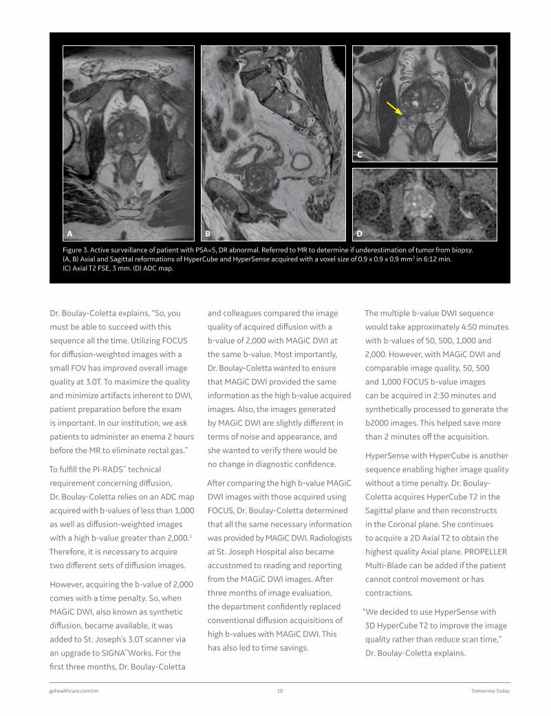
Figure 3. Active surveillance of patient with PSA=5, DR abnormal. Referred to MR to determine if underestimation of tumor from biopsy. (A, B) Axial and Sagittal reformations of HyperCube and HyperSense acquired with a voxel size of 0.9 x 0.9 x 0.9 mm3 in 6:12 min. (C) Axial T2 FSE, 3 mm. (D) ADC map.
A B
C
D

gesignapulse.com 11 Autumn 2017
Issue SpotlightIssue Spotlight
Technical inputs
“Now, we have very high confidence
when using MR to detect prostate
cancer,” she adds. “All these new
sequences also give us the information
we need to report into PI-RADS™
Version 2. So, when we tell the patient
that his imaging results are normal, we
can be very reassuring that if the MR
exam is negative, the patient has no
significant cancer.”
MR for treatment planning
In cases of a positive diagnosis of
prostate cancer, MR is an important
tool for determining the extent of
disease and the appropriate treatment.
For example, today many men with
non-significant, localized cancer will
undergo a period of active surveillance.
The idea behind active surveillance is
that often, prostate cancer will grow
slowly and if localized to only this gland,
the cancer is unlikely to harm the patient
or decrease his life expectancy. In fact, a
recent study on active surveillance has
found that less than 1% of men with
local, slow-growing and non-metastatic
disease had meaningful progression of
their disease after 15 years.2
“The key is, of course, to monitor
these patients regularly and begin
treatment if the disease becomes more
aggressive,” Dr. Boulay-Coletta says.
MR is considered as a baseline exam
at St. Joseph Hospital and it is used to
determine if the patient will be placed
on active surveillance. The information
from MR can also impact the planning
for radical prostatectomy in patients
undergoing surgical treatment.
“Surgeons and radiologists are using
MR imaging data to determine the type
of surgery for the patient,” Dr. Boulay-
Coletta explains. “They review the
images to detect any irregularity of the
While multi-parametric MR of the
prostate is increasingly easy to
perform, the ability to report per
medical association guidelines has
been challenging. PROView is a new
solution from GE Healthcare that
simplifies reading and reporting and
applies PI-RADS™ Version 2 report
guidelines to help standardize the
MR evaluation of prostate cancer.
PROView delivers simple, guided
workflow including prostate volume
calculation, PSA density, lesion
mapping and measurement. It
intelligently displays MR images to
guide workflow and enables MR-
to-MR rigid registration to reduce
patient motion effects. Clinicians
can score T2-weighted, diffusion-
weighted and dynamic contrast
enhanced acquisitions as well as
localize lesions per guidelines and
add new lesions with flexible options
to create a comprehensive report.
Tool-tip reminders of PI-RADS™ scoring
definition assists with reporting
compliance along with standardized
report creation and export. Additional
clinical information through relative
region-of-interest measurements,
curves and color parametric images
can be created to provide the referring
physician with clinically relevant
information that may aid in patient
management.
PROView, accessible from a PC,
laptop or PACS/RIS workstation, is
fully integrated with GE Healthcare’s
Volume Viewer for access to standard
measurement options.
Simplified review, assessment and reporting of multi-parametric
MR prostate exams
capsule and evaluate the extension to
the neurovascular bundle. For capsule
extension, high-resolution Axial T2
is mandatory and sometimes high
spatial resolution DISCO can help to
distinguish normal vessels from
capsule extension.”
All prostate MR imaging examinations
are performed on the Discovery
MR750 3.0T scanner, mainly because
the department does not use an
endorectal coil and therefore, they need
an intrinsically high SNR. Dr. Boulay-
Coletta continues to use DISCO each
time DWI and T2 show an abnormality

gehealthcare.com/mr 12 Tomorrow Today
BA
C ED
Figure 4. A 65-year-old patient with elevated PSA (14.5 ng/ml); biopsy indicated a tumor of Gleason 6 and the patient was referred to MR due to the PSA level and discrepant pathology finding. (A) Axial T2 FSE 3 mm; (B) HyperCube and HyperSense with voxel size 0.9 x 0.9 x 0.9 mm3 acquired in 6:12 min; (C) MAGiC DWI (synthetic DWI) b2000; (D) ADC map; (E) DISCO, 0.9 x 1.3 x 1.6 mm3, 15 sec/phase. MR demonstrated an anterior apical target of 22 mm. MR fusion TRUS biopsy was performed and confirmed 88 mm cancer extension on four targeted biopsies. The volume of cancer found on the MR-guided biopsy was in line with the high PSA level. A radical prostatectomy was recommended.
because it increases her confidence
for diagnosis and helps determine
the extent of disease. Yet, it is the
inclusion of MAGiC DWI and ADC
maps that she believes has made
the greatest impact.
“I would advise other institutions to use MAGiC DWI. It is a robust sequence that considerably reduces the exploration time in every prostate case.”
Dr. Isabelle Boulay-Coletta
References
1. PI-RADS™ v2 guidelines suggest the use of diffusion b-values greater than 1,400.
2. Tosoian JJ, Mamawala M, Epstein JI, et al. Intermediate and Longer-Term Outcomes from a Prospective Active- Surveillance Program for Favorable-Risk Prostate Cancer. J Clin Oncol, 2015; 33:3379-3385.

gesignapulse.com 13 Autumn 2017
Issue Spotlight
each coincident pair of photons are
more precisely detected, and the time
difference between them is used to
localize the PET signal accurately. TOF
leads to improved PET image quality
with higher structural detail and
improved signal-to-noise ratio.
Results
Nuclear physicians were asked to
provide their assessment regarding
the overall PET image quality (image
contrast and resolution) and the ease
of interpretation. They compared the
In October 2015, Michael Soussan, MD, began evaluating a newly installed SIGNA™ PET/MR system at CEA Service Hospitalier Frédéric Joliot (SHFJ) in Orsay, France. Dr. Soussan compared sensitivity, specificity and clinical confidence between the new PET/MR and an existing prior generation PET/CT in order to better understand the role that PET/MR should play in patient management.
the physician comes to a conclusion
based on the PET and MR images
would certainly contribute to a large
acceptance of the new system for
patient management.
The PET component on the SIGNA
PET/MR features a digital Silicon
PhotoMultiplier (SiPM) detector that
is up to three times more sensitive
than conventional PET technology. It
also features ultra-fast coincidence
timing resolution enabling Time-of-
Flight (TOF) reconstruction. With TOF
reconstruction, the arrival times of
Oncology staging is currently
the primary utilization of PET/CT,
accounting for approximately 80%
of patient scans at CEA-SHFJ. PET/CT
is also used for brain exams.
According to Dr. Soussan, a major
challenge is to demonstrate the ability
of PET/MR to enhance the confidence
associated with the interpretation
of PET and MR imaging data. The
hospital’s clinicians currently rely on
the interpretation of PET/CT images. He
believes demonstrating that PET/MR
increases the certainty with which
TOF & Q.Static elevates clinical confidence
Increased PET sensitivity with

gehealthcare.com/mr 14 Tomorrow Today
PET/MR scans to the PET/CT scans.
For this assessment, 150 patients
underwent both a PET/CT and PET/MR.
“Many of the differences we observed in
the images are due to the different PET
technology between our PET/CT and
PET/MR scanners. This demonstrates
the huge progress made in PET
detector technology over the years, by
taking advantage of TOF, enhanced
reconstruction including point spread
function modeling and scatter recovery,”
Dr. Soussan says. “The PET/MR
clearly demonstrated better contrast,
resolution and image quality over the
prior generation PET/CT scanner.”
The PET/MR is a big advancement in
technology, he adds, with this new
combination of modalities providing
improved lesion detectability and more
accurate interpretation of the signal.
Motion correction using Q.Static
was also evaluated on every patient
undergoing a PET/MR exam at CEA-SHFJ.
“We could already see the difference in
quantitation and lesion appearance but
the most important point is that we can
use it routinely, which will help us make
a comprehensive assessment of the
clinical benefit of motion compensation.
Using PET/MR, motion correction can
now enter the clinical practice, and it
is possible to use it for every patient.
With the high PET sensitivity, we can
perform a shorter acquisition with
Q.Static retaining about half the signal
and still have images perfectly suitable
for interpretation,” he says.
“Improving the spatial resolution and
image quality of PET is very important—
it helps make the technique more
precise and efficient than with prior
generation technology,” Dr. Soussan
adds. He is hopeful that a higher PET
sensitivity can help clinicians address
the issue of false positives and false
negative lesions when these situations
occur in clinical practice.
Case 1
A 75-year-old patient with colorectal
cancer, treated six months earlier with
adjuvant chemotherapy, referred to
PET/CT (non-TOF PET/CT system: 371
MBq, 75 min post IV, 4 min/bed position,
no PSF modeling in reconstruction)
followed by PET/MR (100 min post
IV, 4 min/bed position, TOF and PSF
modeling reconstruction).
The improvement in sensitivity leads to
homogenous liver and better contrast
recovery of small lesions. This case
shows that PET/MR enables a precise
staging of liver metastasis, improving
the therapeutic strategy.
Figure 1. (B) The PET image from the PET/MR shows better conspicuity of one liver subcapsular lesion, only slightly visible in the (A) PET image from the PET/CT.
Figure 2. Illustrates the higher diagnostic confidence with multi-modality imaging. Additional liver metastases are slightly visible on the PET from the (B, arrows) PET/MR, not visible on the PET from the (A) PET/CT, and clearly seen on the MR diffusion-weighted images.
A
AA
C
B
B

gesignapulse.com 15 Autumn 2017
Issue SpotlightIssue Spotlight
Figure 3. The impact of respiratory gating with (B) Q.Static can be clearly seen. In the (A) static images, two right liver lesions are blurred (black and red arrows) and the upper lesion almost appears as two lesions (black arrow, SUVpeak 1.9, metabolic volume [42% threshold]: 3 cm3). With (B) Q.Static reconstruction, the images are clearer with an increase in lesion conspicuity for a diagnosis of two lesions (B, black arrows). The quantitation of the upper lesion is improved (SUVpeak 2.2, metabolic volume [42% threshold] 0.8 cm3). Furthermore, in the Q.Static image, a third lesion previously poorly visible was also identified (blue arrow).
A B
Case 2
A 76-year-old patient with initial
diagnosis of well-differentiated,
midgut neuroendocrine tumor with
lymph nodes and liver metastasis. The
patient was scanned with F18-DOPA
PET/MR (245 MBq, 94 min post IV, 6
min/bed, 4 beds, TOF and PSF modeling
reconstruction). Q.Static was employed
for respiratory gating.

gehealthcare.com/mr 16 Tomorrow Today
For over a decade, the University Hospital of Umeå has been using MR for radiation therapy (RT) planning. MR imaging is a useful adjunct to CT because of its ability to depict soft tissue anatomy. The clinical impact is most established for cervical cancer. In fact, the European Society for Radiotherapy & Oncology (ESTRO) released new guidelines in 2012 for the use of MR in volumetric treatment planning in cervical cancer brachytherapy.1
Pursuing an MR-only approach to radiation therapy planning

gesignapulse.com 17 Autumn 2017
Issue Spotlight
According to Professor Tufve Nyholm,
PhD, Department Head, Radiation
Physics, Oncology at Umeå University,
MR is used at the institution in treatment
planning for nearly all cancers across
different therapy regimens—external
beam RT (EBRT), brachytherapy and
proton therapy. A standalone CT is still
acquired for density information on
each patient for use in dose calculations.
“The target is defined by MR imaging,”
Professor Nyholm says. Umeå was
one of the first institutions worldwide
to install the SIGNA™ PET/MR in
January 2015.
Because of the value of MR for lesion
visualization and delineation, Umeå has
transitioned a majority of treatment
planning cases to SIGNA PET/MR. The
exception are suspected lung tumors
and head and neck cases where PET/CT
is used in the diagnostic workup and
therefore, also utilized for RT planning.
Depending on the body part, either
T1-weighted or T2-weighted images
provide a good depiction of the
patient’s anatomy, Professor Nyholm
explains. Then, to define the volume
of the pathology, T2-weighted images
are used for cancers of the pelvic region
and contrast-enhanced T1-weighted
images are preferred for brain cancers.
“The main difference between diagnostic and therapy planning with PET/MR is we tend to use more 3D imaging in RT. We also use much longer acquisition times in the bed position where the tumor is located.”
Professor Tufve Nyholm
Typically, the patient is scanned for
25-30 minutes in one bed position on
the PET/MR. The goal is to acquire as
much information on the target area as
possible. Patients referred to RT have
already received a diagnostic workup
and are identified with localized, not
metastatic, disease.
“We know the area to treat so there
is no need to image the rest of the
body with another bed position,” he
adds. “We want to acquire as much
information as possible to define the
volume for treatment. Even a millimeter
in one direction is important for the
patient’s plan—in these instances
more is better.”
Professor Nyholm also points out
that it is well known that most
Treatment Planning Systems (TPS)
are not optimized for MR imaging—
they are simply not designed to
handle the volume of data that MR
Umeå has modified its MR protocols
with respect to contrast, minimal
slice thickness and adequately high
bandwidth to enhance visualization and
reliability of where the tumor starts and
stops—critical information in treatment
planning. Umeå strives to utilize a
maximum slice thickness of 2.5 mm
without slice gap and 3D isotropic
sequences such as Cube are used when
possible. Diffusion is another common
sequence Umeå uses. In many cases,
there is a need for both PET and MR
imaging data.
“It is a big advantage to acquire PET and MR data simultaneously for clinical decision making. Then we have the same coordinates in the data and avoid registration problems when fusing the data.”
Professor Tufve Nyholm

gehealthcare.com/mr 18 Tomorrow Today
Head and neck cancer is another area
where MR has made a significant
impact. Images from patients with
implants or fillings in their teeth result
in significant artifacts in CT.
“With MR, we can suppress these
artifacts and obtain good image quality.
Obviously, in patients with brain tumors
MR is preferred. Even in an ideal case
we often can’t get the information we
need with CT.”
Addressing prior challenges
to using MR in RT
Acquiring images for planning with the
patient in the treatment position is
important for the accuracy of the plan.
Historically, this has been an issue
with the use of MR in RT. However,
the development of wider bores and
smaller, more flexible coils has helped
to diminish this challenge.
imaging generates. While this can be
problematic, Umeå has addressed this
issue by providing only the key images
in the TPS while making the remaining
MR imaging data available for review
in PACS.
“The underlying problem is that the MR
vendors see the need to adapt to RT,”
Professor Nyholm says. “But the way I
see it, the right thing is for RT to adapt
to MR.”
Impact of MR images in RT
Radiation therapy is radiotoxic to
normal, healthy tissue. Gains in
curative and survival rates along with
enhanced quality of life for cancer
patients have largely been attributed
to advancements in treatment delivery
systems, such as multi-leaf collimators,
Intensity Modulated RT (IMRT) and
Volumetric Modulated Arc Therapy
(VMAT). The accuracy of treatment
delivery is enabled by the clarity and
precision of modern medical imaging
such as CT and MR. These imaging
modalities provide the detailed
information physicists and oncologists
need to treat only the cancerous lesion
and avoid healthy tissue.
At Umeå, Professor Nyholm and his
colleagues see the impact of MR
imaging in treatment planning because
they can clearly visualize the lesion and
surrounding healthy tissue.
“We have not yet reduced margins
but we have seen a change in the
volumes we are treating,” he explains.
“In prostate cancer, we have reduced
target volumes by using MR imaging.”
Figure 1. Patient MR images in a treatment planning system, demonstrating an overlay of diffusion b800 on a T2w MR image. The patient was imaged with PET/MR and PSMA-tracer and the delineated subvolume within the prostate represents the PSMA uptake. Umeå is in the validation phase for planning radiotherapy based on PSMA.

gesignapulse.com 19 Autumn 2017
Issue Spotlight
Umeå has been working with
GE Healthcare to develop the
infrastructure needed to acquire
patient images in the right position.
However, Professor Nyholm believes
other options exist, as well.
“One idea is to explore the possibility to treat the patient in the imaging position rather than image them in the treatment position. If this is possible, then we won’t have this problem. It seems to be the obvious thing to do.”
Professor Tufve Nyholm
Geometric inaccuracies are another
area that have historically been a
concern when using MR for RT. MR
distortions can be caused by gradient
field nonlinearity, which can impact
PET attenuation correction.
Professor Nyholm believes that if
the physicist has a knowledge of MR
imaging and potential issues, they
can avoid this issue. He is a co-author
of a published paper that concluded
when using a well-adjusted sequence
bandwidth, the distortions caused
by patient susceptibility effects can
be kept at an acceptable level.2 It
has already been verified in several
publications that when using 3D
distortion correction the small
remaining distortions have a minimal
effect on the quality of the treatment.
Umeå is also involved in a collaboration
between academic institutions in
Sweden and industry leaders, including
GE Healthcare. The focus of the
consortium, Gentle Radiotherapy, is to
develop MR-only RT. Professor Nyholm
shares that Umeå is responsible for
developing the methodology for MR-
based RT in the pelvis, e.g., bladder,
cervix and anal cancer, excluding
prostate. The consortium already
has a joint publication on MR-only
treatment planning for prostate.3
“The biggest challenge is education,”
Professor Nyholm says. “MR is complex
and it requires both the physicist and
the radiation oncologist to have this
knowledge before it is possible to take
full advantage of MR in RT.” Hence
close collaboration with radiologists
is essential.
In general, Professor Nyholm believes
the benefits of MR in RT outweigh
any remaining educational gaps. “The
quality of the imaging data is really
phenomenal,” he says.
“RT is almost entirely an image- driven specialty. The importance of MR imaging will increase and start to impact RT in other ways, such as how we shape the treatment beams and position and treat the patient.”
Professor Tufve Nyholm
References
1. Dimopoulos J, Petrow P, Tanderup K, et al. Recommendations from Gynaecological (GYN) GEC-ESTRO Working Group (IV): basic principles and parameters for MR imaging within the frame of image based adaptive cervix cancer brachytherapy. Radiother oncol. 2012;103(1):113-22.
2. Lundman JA, Bylund M, Garpebring A, Karlsson CT, Nyholm T. Patient-induced susceptibility effects simulation in magnetic resonance imaging. Phys Imag Radiat Oncol, 2017; 41-47. http://dx.doi.org/10.1016/j.phro.2017.02.004.
3. Persson E, Gustafsson C, Norström F, et al. MR-OPERA–A Multi-center/multi-vendor validation of MRI-only prostate treatment planning using synthetic CT images. Int J Radiat Oncol Biol Phys. 2017 Jun 16. pii: S0360-3016(17)31055-6. doi: 10.1016/j.ijrobp.2017.06.006. [Epub ahead of print]

gehealthcare.com/mr 20 Tomorrow Today
MAGiC DWI, is very helpful in rectal,
prostate and gynecologic cancers.
MAGiC DWI helps overcome the issue
of poor signal-to-noise ratios (SNR)
when using high b-values in diffusion-
weighted imaging (DWI).
“The beauty of synthetic diffusion is
we can achieve excellent SNR and
increase our sensitivity from the high
b-value,” Dr. Atkins explains. “That
gain in SNR and sensitivity is very
useful when looking for subtle lesions
in the prostate and evaluating tumor
extension for staging in the rectum or
cervix. Using traditional diffusion, we
are limited by spatial resolution as the
b-value increases.”
Oncologic imaging represents a steady volume of the MR imaging performed at Fairfax Radiology Consultants (FRC), the largest private radiology practice in the Washington D.C. metropolitan area. Referring physicians rely on the expertise of 70 subspecialized radiologists and skilled technologists to deliver the information they need for patient management. Two of the practice’s three 3.0T MR systems, SIGNA™ Architect and Discovery™ MR750, are increasingly utilized for body, pelvic, rectal and gynecologic cancers.
So, it comes as no surprise that Tom
Schrack, ARMRIT, CS, Manager of MR
Education and Technical Development
for MR Services at FRC, has seen
an increase in volumes as referring
physicians gain confidence in FRC’s
diagnostic quality. Most of the growth
has been in rectal and prostate cancer
imaging and, to a lesser extent, other
pelvic cancer imaging, he notes.
“Our prostate MR imaging growth has
been substantial, in the area of 60%,”
Schrack says. “A year ago, we averaged
five to six prostate MR cases each week.
Now we are imaging two to three
each day.”
Guidelines now recommend the use of
high-resolution MR as the first choice
for primary staging and postoperative
chemoradiation restaging of rectal
cancer.1 As a result, FRC developed a
colorectal imaging program utilizing
standardized reporting, well-defined
protocols and subspecialty radiologists
for rectal MR imaging.
According to Melany Atkins, MD,
Radiologist at FRC, GE Healthcare’s
SIGNA™Works productivity software
platform, which features BodyWorks,
OncoWorks and innovative
HyperWorks sequences, is making a
difference in MR oncology imaging. The
synthetic diffusion sequence, called
differenceMR oncology
Making a
in
imaging
Tom Schrack, ARMRIT, CS Fairfax Radiology Consultants (FRC) in Fairfax, VA
Melany Atkins, MD Fairfax Radiology Consultants (FRC) in Fairfax, VA

gesignapulse.com 21 Autumn 2017
Issue Spotlight
Nearly all prostate MR imaging is
performed with an endorectal coil
utilizing MAGiC DWI, FOCUS, and to
a limited extent DISCO. With FOCUS,
users can obtain high resolution DWI
images by reducing the phase field-
of-view (FOV) without the concern
of wrap artifacts. With DISCO, very
high temporal resolution T1-weighted
images can be obtained for evaluating
contrast enhanced characteristices of
lesions. Dr. Atkins sees image quality
improvement when using FOCUS,
especially in patients with hip prosthesis.
“We love FOCUS compared to regular diffusion and find it is superior with less geometric distortion in a small field of view. Plus, it is easy to use and doesn’t take a lot of time.”
Tom Schrack
Figure 1. Prostate with MAGiC DWI, ADC and Axial T2. (A) b400; (B) b400 synthetic; (C) b1000; (D) b1000 synthetic; (E) b1400 synthetic; (F) b2000 synthetic; (G) ADC map; (H) Axial T2.
A
C
E
G
B
D
F
H
gehealthcare.com/mr 22 Tomorrow Today
to reduce scan times, something
that Schrack is closely watching.
“These time savings allow us to add
sequences like HyperCube without
increasing our time slots,” he adds.
FRC uses HyperCube often as the first
sequence on pelvic malignancies to
help select the imaging plane.
While DISCO hasn’t yet been integrated
into many of the oncology imaging
protocols due to the practice’s
continued use of multi-phasic LAVA
imaging, Dr. Atkins anticipates it will
be adopted more in the near future as
they continue to evaluate its impact.
In cases where patient motion is an
issue, PROPELLER Multi-Blade, an
advanced motion correction technique,
is available to use at a moment’s notice
to eliminate the associated artifacts.
There is also HyperSense, a sequence
that Schrack says the practice absolutely
loves. “We use it on every imaging study
that we possibly can.” HyperSense
enables the use of compressed sensing
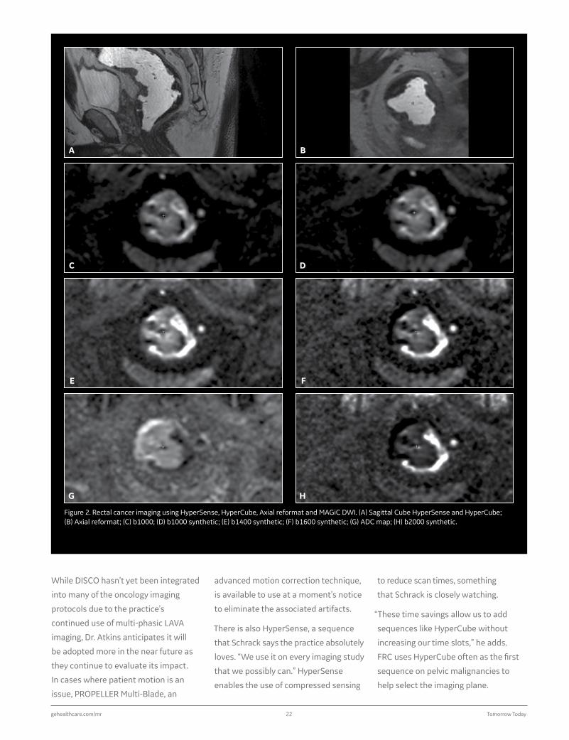
Figure 2. Rectal cancer imaging using HyperSense, HyperCube, Axial reformat and MAGiC DWI. (A) Sagittal Cube HyperSense and HyperCube; (B) Axial reformat; (C) b1000; (D) b1000 synthetic; (E) b1400 synthetic; (F) b1600 synthetic; (G) ADC map; (H) b2000 synthetic.
A
C
E
G
B
D
F
H

gesignapulse.com 23 Autumn 2017
Issue Spotlight
HyperCube enables aliasing-free
isotropic imaging with a reduced FOV
and increased matrix. The combination
of HyperCube and HyperSense, in
conjunction with conventional parallel
imaging, helps decrease imaging time
yet delivers the very thin, isotropic
heavy-weighted T2 imaging that aids
a radiologist in their diagnosis.
“First we acquire HyperCube—it is our ‘lay of the land’ sequence— and then we can reformat in any plane, which is very useful especially if a patient returns for a post-treatment exam.”
Tom Schrack
Figure 3. Rectal cancer with MAGiC DWI. (A) Axial T2; (B) ADC map; (C) b1000; (D) b1000 synthetic; (E) b1200 synthetic; (F) b1400 synthetic; (G) b1600 synthetic; (H) b2000 synthetic.
A
C
E
G
B
D
F
H

gehealthcare.com/mr 24 Tomorrow Today
Although MR imaging for gynecological
cancers—cervix, vaginal and
endometrial—hasn’t grown as much
as prostate, FRC has solid volume from
referrers in the area.
“The integration of these new sequences
and capabilities has been helpful for
our throughput and image quality,”
Dr. Atkins adds. “Across the board,
SIGNA™Works has improved SNR and
our diagnostic confidence.”
Reference
1. The National Accreditation Program for Rectal Cancer Standards Manual, 2017 Edition. American College of Surgeons, Chicago, IL.
For patients with prior exams, having
the ability to reformat to match the
plane used in a prior exam can be
invaluable for the radiologist.
Aside from accelerating with HyperCube,
Schrack has also worked on reducing
respiratory and abdominal wall motion
and has seen a significant improvement in
the image quality of the interior abdomen.
Dr. Atkins adds that they still acquire
the high resolution T2 in addition to
the T2 3D HyperCube images. The T2
images are of such high quality on their
MR systems that Dr. Atkins finds it
difficult to remove the sequence.
Growing use of MR in oncology
Dr. Atkins believes the adoption of
PI-RADS™, the American College of
Radiology’s Prostate Imaging Reporting
and Data System, has also spurred
growth in the utilization of MR imaging—
particularly to help guide biopsy.
Active surveillance for prostate
cancer has become well adopted by
providers—radiologists, oncologists
and urologists—with an increased
recognition that many prostate cancers
are slow-growing and localized. For
that subset of patients, receiving
radiation therapy for their prostate
cancer may do more harm due to the
side effects of treatment. However, the
location where the biopsy is performed
can significantly improve accuracy in
determining the exact type of cancer a
patient has. And that is where MR plays
a crucial role.
“MR images provide us with information to improve the accuracy of our biopsies, which, in turn, provides us with valid results across the entire gamut of prostate cancers. While we believe that accuracy has something to do with the quality of our MR scanners, it also is a reflection of the excellent product we put out by following PI-RADS™ and systematic reading and reporting.”
Dr. Melany Atkins
Figure 4. Cube with HyperSense and HyperCube.(A) Sagittal Cube HyperSense HyperCube; (B) acquired Axial T2; (C) Coronal Cube reformat; (D) Axial Cube reformat.
A
C
B
D

T O M O R R O W T O D AY
T H E N E W S T A N D A R D I S E X T R A O R D I N A R Y
JB46574US(1)
Elevate your clinical proficiency with SIGNA™Works, GE Healthcare’s latest productivity platform. Improve image quality, increase efficiency and streamline
workflow. Performing a clinical exam has never been easier. SIGNA™Works. Fueling the future of MR.
Visit www.signaworks.gehealthcare.com to learn more.

gehealthcare.com/mr 26 Tomorrow Today
The clinical PET/MR service is a
collaboration between the nuclear
medicine and radiology departments.
Professor Kas explains that in
addition to radiology, the PET/MR
implementation has further promoted
the development of new collaborations
with neurology, neuro-oncology,
urology, gynecology and surgery.
At Pitié-Salpêtrière Hospital, patients
with brain tumors, rare cancers and
rare neurodegenerative disorders, such
as atypical presentation of Alzheimer’s
According to Aurelie Kas, MD, PhD,
Head of the Department of Nuclear
Medicine at Pitié-Salpêtrière Hospital
Group, the utilization of the SIGNA™
PET/MR is equally divided between
clinical and research use, with 2.5 days a
week dedicated to each. The institution
is currently participating in 45 clinical
trials in oncology and neurology.
“In our experience, PET/MR imaging
is feasible in a clinical setting with a
throughput of 11 to 15 patients each
day,” Professor Kas says.
The hospital conducts a staggering
high volume of clinical PET/MR exams.
SIGNA PET/MR complements a robust
imaging environment in the nuclear
medicine department that includes
two PET/CT systems, two SPECT/CT
systems and one ultrasound system.
The department also provides
radioactive iodine therapy for thyroid
cancer patients with six beds that
accommodate 12 patients each
week. Six MR systems are also
located in the radiology department
primarily for neurology, oncology and
cardiac imaging.
As one of Europe’s largest teaching hospitals, Pitié-Salpêtrière Hospital is renowned for its innovative research and delivery of high-quality care to patients. In 2015, the Alzheimer Research Foundation in France, a partnership between the public hospital system and the University Hospital Brain and Spine Institute (IHU-A-ICM), provided funding for a PET/MR project at the hospital.
Optimized clinical pathway
propels high utilization of PET/MR at Pitié-Salpêtrière Hospital

gesignapulse.com 27 Autumn 2017
In Practice
disease (AD), are now preferably
referred to PET/MR rather than PET/CT
or MR alone to obtain detailed images
to aid in the detection, localization and
diagnosis of diseases and disorders.
Clinically, 75% of PET/MR imaging
studies are for neurodegenerative
disorders such as Alzheimer’s and
Parkinsonism, 23% for oncology
(approximately 33% neuro, 34%
maxillofacial and 33% abdominal/pelvic)
and 2% for drug-resistant partial epilepsy.
“We believe the diagnostic accuracy may be improved by combining metabolic information provided by PET with the structural and multi-parametric imaging of MR. This is especially helpful in the areas of neurodegenerative disorders with atypical presentation or vascular comorbidities, for the diagnosis of cancer recurrence and for tumor delineation before surgery in regions with complex anatomy.”
Professor Aurelie Kas
A
C
B
D
Figure 1. Pre-surgical assessment of a mandibular gingiva squamous cell carcinoma with FDG PET/MR imaging. MR findings: mandibular gingiva lesion with trabecular bone invasion, left necrotic lymph node. PET findings: hypermetabolism of the lesion and the left lymph node. Pathology confirms PET/MR findings.
Neuro Case 1
gehealthcare.com/mr 28 Tomorrow Today
“Our patients benefit with less examination time and an increase in comfort, especially the older patients with comorbidities.”
Professor Aurelie Kas
She adds, “Clinicians gain the
advantage of a reliable imaging exam
with increased diagnostic confidence
as the combination of these imaging
tools aid in accumulating evidence for
a specific diagnosis. Additional gains
in diagnostic accuracy can be achieved
since PET/MR studies are interpreted by
experts in both radiology and nuclear
medicine, who together provide a
single report.”
Since October 2015, more than 2,000
patients have undergone clinical
PET/MR examinations in the facility.
The department averages 11-15 clinical
patients each day from 8 am to 5:30 pm,
of which three to five exams are whole
body and eight to 10 are brain exams.
“In our facility, our research has found
that PET imaging provides early
biomarkers of many neurodegenerative
disorders, but MR provides unique
imaging information on age-related
or pathological cerebral atrophy,
vascular lesions such as stroke
sequelae, microbleeds and leukopathy,
and inflammatory abnormalities that
cannot be reliably assessed with PET
alone. In addition, using PET/MR image
interpretation and comparing imaging
biomarkers is optimal since the imaging
studies are acquired simultaneously.”
Diagnostic power of PET/MR
In complex neurology and oncology
cases requiring PET imaging, PET/MR
is now preferred rather than PET/CT
at Pitié-Salpêtrière Hospital. This is
especially important if MR imaging
is also indicated.
Professor Kas explains, “We utilize
PET/MR for visualization of advanced
head and neck cancers to assist with
the assessment of disease status and,
more specifically, to help with tumor
delineation before surgery in this
region with such complex anatomy. In
follow-up examinations, hybrid PET/MR
has advantages in the visualization of
tumor differentiation, scars and edema
after radiation therapy in head and
neck cancer. It is also of great interest
in brain tumors when MR images alone
may not enable us to differentiate the
viable tumor from necrosis with post-
therapeutic changes, especially after
radiation therapy.
“Hybrid PET/MR is also a helpful imaging
tool in cases of atypical presentation
or early-onset AD, Parkinson’s-plus
syndromes and suspected mixed
dementia when neurodegenerative
and vascular processes coexist,”
Professor Kas adds.
Prior to implementation of the
SIGNA PET/MR, patients with complex
indications in which PET and MR were
both recommended often underwent
two imaging exams. Now, a single
imaging exam—PET/MR—can
assist us in directly answering
the clinical question.
A B C
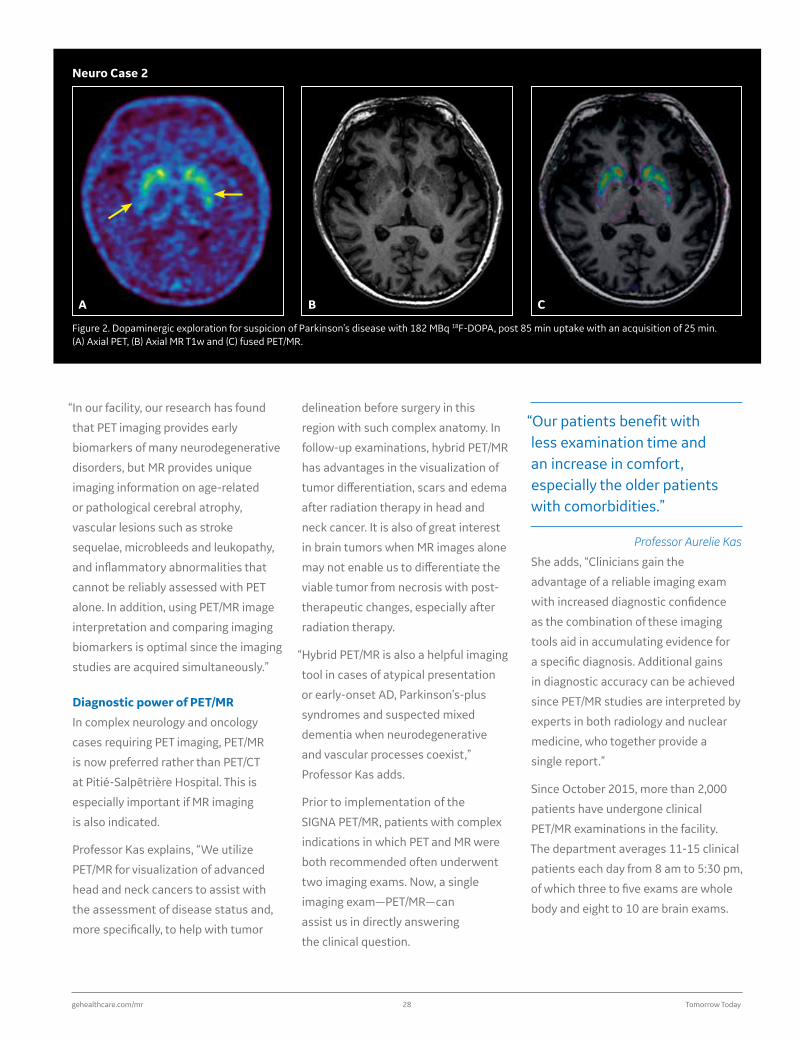
Figure 2. Dopaminergic exploration for suspicion of Parkinson’s disease with 182 MBq 18F-DOPA, post 85 min uptake with an acquisition of 25 min. (A) Axial PET, (B) Axial MR T1w and (C) fused PET/MR.
Neuro Case 2

gesignapulse.com 29 Autumn 2017
In Practice
B
E F G
C
D
Figure 3. Pelvic MR exam on a patient with endometrial adenocarcinoma. (A) Whole-body PET, 4 bed positions at 3 min/bed; (B) pelvic PET acquired in 16 min; (C) fused PET with Axial T2w PROPELLER; (D) Axial T1w LAVA post-contrast; (E) fused PET with Sagittal T2w PROPELLER; (F) Sagittal FOCUS DWI, b1000; (G) ADC map.
A
gehealthcare.com/mr 30 Tomorrow Today
At Pitié-Salpêtrière Hospital, the
typical protocol for neurodegenerative
disorders averages 25 minutes. For
oncology, it is slightly longer at a
median of 45 minutes which also
includes a regional multi-modal
exam—for example one bed position
on the neck or pancreatic area—and
a whole-body PET. Often, 18F-DOPA
PET/MR exams for brain tumors or
atypical Parkinsonism syndrome
indications last 45 minutes.
Optimizing the clinical pathway
To maximize clinical utilization and
benefit of the PET/MR system in as
many patients as possible, Professor
Kas and her colleagues in nuclear
medicine and radiology focused on
optimizing the clinical pathway. One
of the first steps was to start using the
system in well-defined areas of MR
imaging expertise, such as head and
neck, digestive and pelvis, with very
specific indications for each organ or
anatomic area being examined.
The PET/MR imaging service was
organized with a dedicated staff that
includes four trained technologists with
prior experience in MR, six physicians,
two physicists and one administrative
support position. The unit was
designed with the PET/MR exam room,
reading room and patient preparation
room in close proximity to facilitate
communication between technologists
and clinicians in order to help reduce
time between exams and enhance
patient safety and satisfaction.
All acquisition protocols were evaluated
and optimized by experts in nuclear
medicine and radiology to avoid the
collection of redundant information
by the two imaging modalities and
to ensure that protocols obtained all
the required information for diagnosis.
They also collaborated to determine
the minimal scanner occupancy times
to maximize clinical utilization since
the scanner would be used only 50%
for clinical applications. Regularly
scheduled meetings were held with all
PET/MR medical staff to review patient
flow, optimize MR sequences and
discuss any potential issues. Today, the
PET/MR team regularly holds multi-
disciplinary meetings to discuss the
contribution of PET/MR imaging to help
clinicians diagnose complex cases.
A
D
B
E
C
F
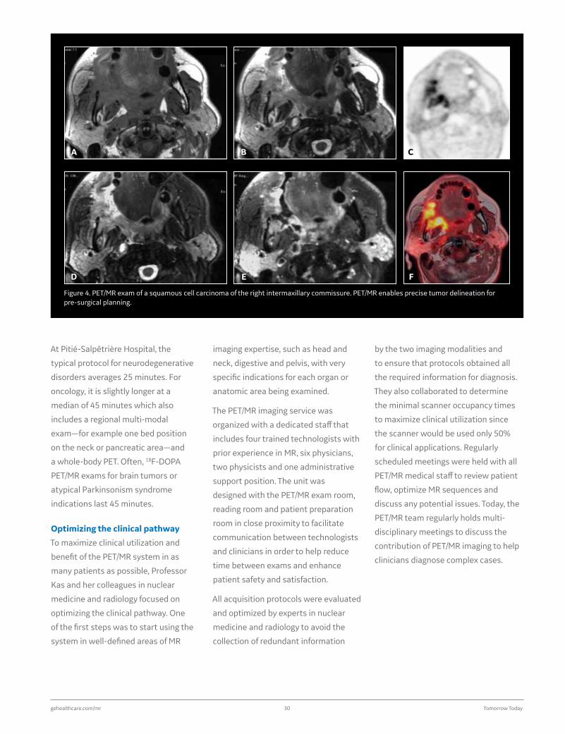
Figure 4. PET/MR exam of a squamous cell carcinoma of the right intermaxillary commissure. PET/MR enables precise tumor delineation for pre-surgical planning.

gesignapulse.com 31 Autumn 2017
In Practice
Overall, the utilization of PET/MR in a dual
clinical and research environment has
been successful. Professor Kas says, “It is
important to optimize imaging protocols
to ensure complementary imaging
data and avoid redundant information
between these modalities.”
La Pitié looking forward
One area of research that Professor
Kas and her colleagues plan to
investigate is to study the added
value of combined PET/MR biomarker
imaging. Specifically, Professor Kas
and her colleagues will examine the
diagnosis of neurodegenerative disease
and brain tumors and the prognosis and
evaluation of therapeutic response in
cancer patients.
The group is also examining the
reliability of Zero Echo Time (ZTE)
based attenuation correction (AC) for
brain, head and neck PET/MR images.
Currently, AC techniques in these
anatomic areas are based on a CT atlas
and the Dixon sequence to identify air,
water and soft tissue. This approach,
explains Professor Kas, is not always
satisfactory for the AC of bony regions,
especially in patients with a cancer of
the jaw or a tumor extending to the
base of the skull. The first results are
encouraging, she adds, and suggests
that ZTE sequences enable bone
visualization and are appropriate for
AC of the oral cavity.
“Efforts have been made to optimize the imaging protocol to maintain short PET/MR acquisition times to ensure that we can acquire more than 11 clinical imaging studies in one day. On the other hand, it was obvious to us that the protocol had to achieve the highest diagnostic capability to provide at least as much information as sequential PET/CT and MR examinations.”
Professor Aurelie Kas
Therefore scan times are balanced
between the desire for shorter
acquisition times and high-quality
imaging. While the hospital’s current
acquisition protocols typically last 25
minutes for a neuro protocol and up
to 50 minutes for exams that include
whole-body PET for oncology, Professor
Kas is currently working on new ways
to further reduce scan times.

gehealthcare.com/mr 32 Tomorrow Today
Named one of the top-ranked pediatric hospitals in the nation by U.S. News & World Report’s 2017-18 Best Children’s Hospitals1, Primary Children’s Hospital (Salt Lake City, UT) delivers exceptional care across 10 pediatric specialties, from cancer care to neurology. In fact, the Intermountain Healthcare facility is one of 24 hospitals in the nation to be ranked in all 10 specialties rated by the publication.
Embracing that commitment to
pediatric care, Primary Children’s was
one of the first hospitals in Utah to
implement a 3.0T MR system in 2005.
A decade later, the hospital replaced
its 60 cm 3.0T MR with the Discovery™
MR750w, a 70 cm wide bore system.
“We wanted to stay on the cutting edge
of technology with the more robust
sequences that became available, such
as the T1 PROPELLER and PROPELLER
Multi-Blade,” says Derek Maxfield,
MR Supervisor. When GE Healthcare
launched its SIGNA™ Lift program,
enabling existing Discovery MR750w
sites to upgrade to SIGNA™ Architect,
Primary Children’s embraced the
opportunity.
SIGNA Lift allows facilities to reset
the life of an existing GE MR scanner
with new applications and the latest
generation hardware. The new system
includes the SIGNA™Works productivity
platform and new electronics, software
and hardware that are built around the
site’s current magnet.
“ Child first and always”philosophy drives investment in advanced MR technology at Intermountain Healthcare
Jayson Argyle Intermountain Healthcare in Salt Lake City, UT
“As a dedicated pediatric hospital, we
strive to have an environment where
we have the full gamut of pediatric
imaging, whether it is for a routine
study to rule something out or the
most complex scenario working with
neurosurgeons or oncologists,” says
Jayson Argyle, Director of Imaging for
Intermountain Healthcare. “What really
differentiates us is our ability to care for
these children. Our philosophy is child
first and always.”
gesignapulse.com 33 Autumn 2017
In Practice
“Often, there is a financial concern
when the question of a new scanner is
brought up,” Maxfield adds. “However,
we worked with GE through the SIGNA
Lift program to navigate that concern
and make it affordable so we could
acquire the best technology.”
Adds Argyle, “We did our due diligence
and looked at the pros and cons of a
new system versus keeping our magnet
intact and upgrading the software
and hardware. It made more sense
economically and clinically to go with
the upgrade.” The hospital also avoided
the added expense of construction to
remove and replace the existing magnet.
Not only was the upgrade cost-effective,
it was streamlined. The GE engineering
and applications team arrived on a
Thursday, worked through the weekend
and had the upgraded scanner back up
and running by the following Tuesday.
“We were able to move the patients
requiring an MR study to other areas
where we had a scanner available,
such as scheduling outpatients in our
satellite clinics,” says Maxfield. “The
entire process was rapid and minimized
the negative impact on our patients.”
Embracing advanced apps
“When we first turned it on and started
scanning with it, my first impression
was that this is what 3.0T scanning
should be,” says Maxfield. “You get
more of everything and less of what
you don’t want.” Having scanned with
first generations of 3.0T MR scanners,
he has seen first-hand the leap in
technological advancements.
For example, dielectric effects that
can exist in 3.0T imaging have been
resolved with SIGNA Architect thanks
to the new RF receive chain. SAR is
also greatly reduced.
“It opens the door to shift exams from one scanner to another based on clinical and patient needs.”
Derek Maxfield
Technologists at Primary Children’s
are always trying to achieve the
best possible image quality. With
Total Digital Imaging (TDI) on SIGNA
Architect, they can achieve increases in
image quality thanks to the reduction of
noise with Direct Digital Interface (DDI),
intelligent Micro Electro-Mechanical
Switches (MEMS) in the RF coil design
and exceptional SNR and sensitivity
from surface coils along with superior
homogeneity and deeper signal
penetration with Digital Surround
Technology (DST).
The imaging team was also interested
in the 3D imaging options, particularly
SIGNA™Works’ Auto Navigated Turbo LAVA
free-breathing Magnetic Resonance
CholangioPancreatography (MRCP).
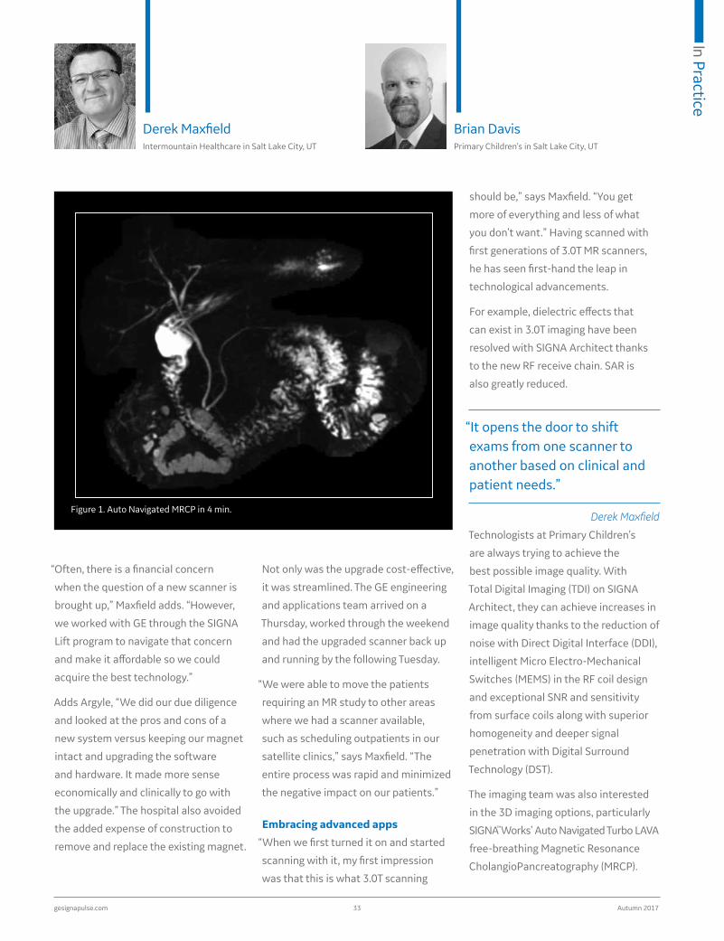
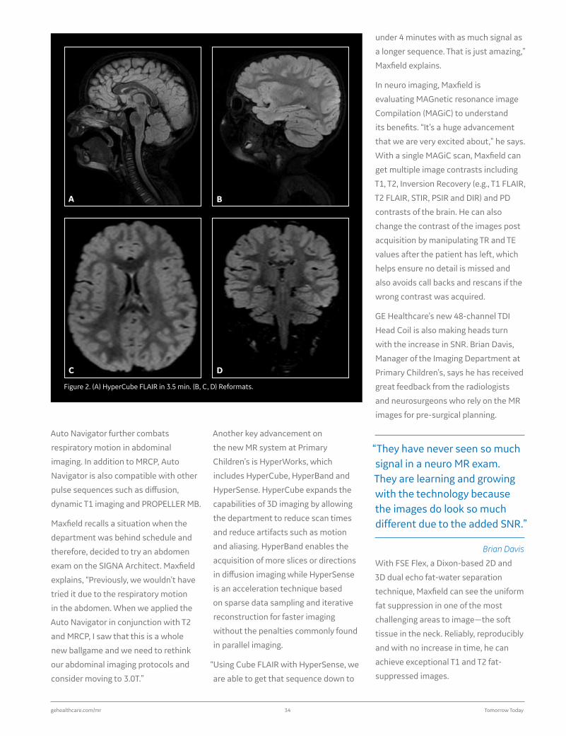
Figure 1. Auto Navigated MRCP in 4 min.
Derek Maxfield Intermountain Healthcare in Salt Lake City, UT
Brian Davis Primary Children’s in Salt Lake City, UT
gehealthcare.com/mr 34 Tomorrow Today
Auto Navigator further combats
respiratory motion in abdominal
imaging. In addition to MRCP, Auto
Navigator is also compatible with other
pulse sequences such as diffusion,
dynamic T1 imaging and PROPELLER MB.
Maxfield recalls a situation when the
department was behind schedule and
therefore, decided to try an abdomen
exam on the SIGNA Architect. Maxfield
explains, “Previously, we wouldn’t have
tried it due to the respiratory motion
in the abdomen. When we applied the
Auto Navigator in conjunction with T2
and MRCP, I saw that this is a whole
new ballgame and we need to rethink
our abdominal imaging protocols and
consider moving to 3.0T.”
Another key advancement on
the new MR system at Primary
Children’s is HyperWorks, which
includes HyperCube, HyperBand and
HyperSense. HyperCube expands the
capabilities of 3D imaging by allowing
the department to reduce scan times
and reduce artifacts such as motion
and aliasing. HyperBand enables the
acquisition of more slices or directions
in diffusion imaging while HyperSense
is an acceleration technique based
on sparse data sampling and iterative
reconstruction for faster imaging
without the penalties commonly found
in parallel imaging.
“Using Cube FLAIR with HyperSense, we
are able to get that sequence down to
under 4 minutes with as much signal as
a longer sequence. That is just amazing,”
Maxfield explains.
In neuro imaging, Maxfield is
evaluating MAGnetic resonance image
Compilation (MAGiC) to understand
its benefits. “It’s a huge advancement
that we are very excited about,” he says.
With a single MAGiC scan, Maxfield can
get multiple image contrasts including
T1, T2, Inversion Recovery (e.g., T1 FLAIR,
T2 FLAIR, STIR, PSIR and DIR) and PD
contrasts of the brain. He can also
change the contrast of the images post
acquisition by manipulating TR and TE
values after the patient has left, which
helps ensure no detail is missed and
also avoids call backs and rescans if the
wrong contrast was acquired.
GE Healthcare’s new 48-channel TDI
Head Coil is also making heads turn
with the increase in SNR. Brian Davis,
Manager of the Imaging Department at
Primary Children’s, says he has received
great feedback from the radiologists
and neurosurgeons who rely on the MR
images for pre-surgical planning.
“They have never seen so much signal in a neuro MR exam. They are learning and growing with the technology because the images do look so much different due to the added SNR.”
Brian Davis
With FSE Flex, a Dixon-based 2D and
3D dual echo fat-water separation
technique, Maxfield can see the uniform
fat suppression in one of the most
challenging areas to image—the soft
tissue in the neck. Reliably, reproducibly
and with no increase in time, he can
achieve exceptional T1 and T2 fat-
suppressed images.
Figure 2. (A) HyperCube FLAIR in 3.5 min. (B, C, D) Reformats.
A
C
B
D

gesignapulse.com 35 Autumn 2017
In Practice
In orthopedics, which represents a
large portion of the MR imaging service
at Primary Children’s, PROPELLER, T2
and the increased SNR are making a
big difference. In moderately compliant
teenaged children, technologists are
now adding PROPELLER to account for
any slight motion. The new generation
high-density coils—the 18-channel
T/R Knee, 16-channel T/R Wrist and
the 16-channel Shoulder—are helping
to propel faster imaging speeds with
increased image quality and resolution.
GE Healthcare’s Flex Coils are also
making an impact in patient care. While
the imaging team tries to use dedicated
coils whenever possible, Maxfield
recalls a situation where a Flex Coil was
used for spine imaging on a 4.5 lb. infant.
“The Flex Coils let us fill that gap for comprehensive body imaging and are our second line of defense when we don’t have a coil to fit on a particular body area,” he says.
Derek Maxfield
Figure 3. Images acquired with GE Healthcare’s 48ch TDI Head Coil. (A) Axial DTI HyperBand colored orientation; (B) Sagittal BRAVO; (C) Sagittal T2 FLAIR HyperCube PROMO in 4 min; (D) Axial TDI SWAN MinIP; (E) Axial SWAN phase image; (F) Axial TOF MRA MIP.
A
D
B
E
C
F
“This is really what neuro images should look like… we are just raising the bar on imaging with this system and the 48-channel TDI Head Coil.”
Derek Maxfield

gehealthcare.com/mr 36 Tomorrow Today
A Caring Suite
Calming anxious pediatric patients
and their parents is a challenge in
medical imaging and especially in MR.
To help alleviate anxiety and fear of
the MR scanner, Intermountain also
implemented GE Healthcare’s Caring
Suite, an environment designed to
help improve and humanize the patient
experience through the reassuring
senses of sight, sound and touch.
Sometimes, the most difficult aspect of
a pediatric MR exam is that initial break
from the parent—the child may cling
to their parent and may be unwilling to
enter the scanner.
“The Caring Suite really engages the child as they enter the room by providing a calming atmosphere and ambience. They can change the colors in the room, interact with pictures and watch a video on the TV, so they really feel in control. It really helps break that psychological barrier and also helps us to gain their trust and respect.”
Brian Davis
Figure 4. FSE Flex: (A) Sagittal PD FSE Flex in-phase and (B) water knee images; (C) Sagittal T1 FSE Flex in-phase and (D) water ankle images; and (E) Sagittal T1 FSE Flex finger image.
A
C
B
D
E

gesignapulse.com 37 Autumn 2017
In Practice
From the advanced SIGNA™Works
applications to the powerful new TDI
and the Caring Suite, SIGNA Lift has
enabled Intermountain to tap into all
that the SIGNA Architect has to offer.
According to Maxfield, in addition to
benefiting patients, the system is also
a great recruitment tool for attracting
the best and brightest technologists.
Argyle points out that financial
pressures will continue in healthcare
and the upgrade path through SIGNA
Lift is an excellent option that allows
facilities to spend more of their budget
on acquiring the latest technology while
spending less on construction.
Reference
1. U.S. News & World Report. Best Hospitals 2017-18. Available at: https://health.usnews.com/best-hospitals/area/ut/primary- childrens-medical-center-6870280.
Figure 5. PROPELLER MB images: (A) Axial and (B) Coronal T1 FatSat of the shoulder and arm; (C) Sagittal T2, (D) Sagittal T1 FLAIR, (E) Sagittal T2 STIR, (F) Axial T1 and (G) Axial T2 of the L-Spine.
A
C
F G
D E
B
“The ability to upgrade also offers a site that is growing or starting out with less advanced procedures a pathway to progress into new capabilities and more complex studies in an efficient and cost-effective timeframe,” says Argyle. “Most important for us, this has been an excellent tool that helps us focus on our philosophy of the child first and always.”
Jayson Argyle
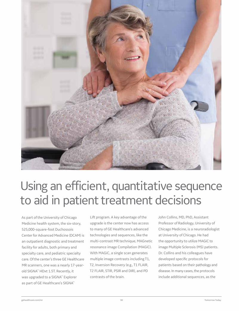
gehealthcare.com/mr 38 Tomorrow Today
John Collins, MD, PhD, Assistant
Professor of Radiology, University of
Chicago Medicine, is a neuroradiologist
at University of Chicago. He had
the opportunity to utilize MAGiC to
image Multiple Sclerosis (MS) patients.
Dr. Collins and his colleagues have
developed specific protocols for
patients based on their pathology and
disease. In many cases, the protocols
include additional sequences, as the
As part of the University of Chicago
Medicine health system, the six-story,
525,000-square-foot Duchossois
Center for Advanced Medicine (DCAM) is
an outpatient diagnostic and treatment
facility for adults, both primary and
specialty care, and pediatric specialty
care. Of the center’s three GE Healthcare
MR scanners, one was a nearly 17-year-
old SIGNA™ HDxt 1.5T. Recently, it
was upgraded to a SIGNA™ Explorer
as part of GE Healthcare’s SIGNA™
Lift program. A key advantage of the
upgrade is the center now has access
to many of GE Healthcare’s advanced
technologies and sequences, like the
multi-contrast MR technique, MAGnetic
resonance image Compilation (MAGiC).
With MAGiC, a single scan generates
multiple image contrasts including T1,
T2, Inversion Recovery (e.g., T1 FLAIR,
T2 FLAIR, STIR, PSIR and DIR), and PD
contrasts of the brain.
Using an efficient, quantitative sequence to aid in patient treatment decisions

In Practice
gesignapulse.com 39 Autumn 2017
goal is to obtain as much information
on the patient as possible: time on the
scanner is not a key concern.
For the MS patients who undergo
a neuro MR exam, the primary
benefit of MAGiC for these patients
is the sequence’s T1 and T2 mapping
capabilities. Dr. Collins finds the T1
and T2 mapping capabilities to be a
valuable addendum to the patient’s
neuroimaging evaluation.
“We’re using the T1 and T2 mapping capabilities to look for changes in the MS lesions resulting from therapy that we can’t otherwise image with conventional sequences.”
Dr. John Collins
Figure 1. A patient case conducted on a 3.0T scanner and then rescanned on the SIGNA Explorer 1.5T with MAGiC, demonstrating comparable imaging capabilities. (A) MAGiC T1; (B) Axial T1 SE on 3.0T; (C) MAGiC PSIR; (D) Axial T1 3D on 3.0T; (E) MAGiC T2 FLAIR; (F) Axial FLAIR reformat on 3.0T. With MAGiC, parameters can be changed to increase conspicuity of lesions without rescans or the patient returning for additional imaging. MAGiC can help improve productivity without sacrificing sensitivity or diagnostic confidence.
A B
C D
E F

gehealthcare.com/mr 40 Tomorrow Today
“This provides the potential to rethink
how we scan patients,” Dr. Collins adds.
“That’s not a new idea, we do this with
CT imaging right now with different
settings for soft tissue such as lungs, or
different window leveling for the liver,
kidney or bowels. It is intuitive that
we can benefit in MR from this same
approach. We could repeat sequences,
but clinically that is not efficient.
However, with MAGiC we can generate
different contrasts, such as T2 or Proton
Density, after the scan.”
Once the settings within MAGiC are
established, Dr. Collins says that
generating different contrasts does not
take a lot of additional time. There is
a learning curve, but the benefits are
clear to him.
“If we can see more lesions or get a truer
sense of the lesion size, then I think this
will catch on because it could change
patient management,” he adds.
While scanner time is not an issue at
DCAM, there is the potential for sites
to achieve scan time savings by using
Dr. Collins finds the T1 and T2 maps can
be more sensitive than conventional T2
or FLAIR sequences.
The process involves marking the
regions of interest in the lesions,
tabulating the T1 and T2 values and
then following the patient over time
to see if the values correlate with the
progress of the disease—and if the
patient is responding to therapy.
“Are there imaging characteristics that
we can use to predict whether a person
will respond to therapy?” Dr. Collins
asks. That’s the key question he is
hoping MAGiC with T1 and T2 mapping
capabilities can help him answer.
While still too early to share any
definitive results, Dr. Collins does see
the value of MAGiC for his study.
“Lesion conspicuity is definitely
enhanced with parametric T1, T2, R1, R2
and PD maps for our further analysis of
MR acquisition data,” he adds.
With MAGiC, Dr. Collins can set specific
parameters for T2 and FLAIR imaging,
including additional image contrasts.
For each disease process—whether it
be for an MS or oncology patient—he
can optimize these settings.
For example, the parameters for T2
of the brain are the same for most
conditions—infection, MS, suspected
stroke and tumors. However, there may
be a situation when he wants a more
sensitive evaluation, such that even the
subtlest lesions might be visualized in
an MS patient.
“Being able to see one or two tiny lesions
can mean the start or change of therapy
in an MS patient,” he explains. “There
are other situations where I don’t want
that level of detail.”
While with conventional sequences
only one contrast is acquired at
the scanner, with MAGiC a range of
contrasts can be potentially processed.
The optimal contrast for an MS patient
may not be the same for a head and
neck cancer patient.
Figure 2. This patient exam shows a large area of restricted diffusion posteriorly in both lobes of the brain. The normal T2 FLAIR isn’t sensitive enough to confirm this but MAGiC provided a higher sensitivity that enabled the clinician to confirm this was a real finding.
A B C
In Practice
gesignapulse.com 41 Autumn 2017
MAGiC. This could be particularly useful
in pediatric patients. In this patient
group, Dr. Collins sees the potential
to perform a diffusion-weighted
sequence and then use MAGiC to
generate additional contrasts. By
reducing sequences, scan time could
be dramatically shortened which
could lead to fewer pediatric patients
being sedated for an MR exam.
“We could almost obtain a complete brain MR in 10 minutes and that is not possible with conventional sequences. This is a new technology that we really need to step back and re-examine how we image with MR. We should not limit it to just a few pathologies. With the customized contrast levels, MAGiC could be helpful for a variety of conditions.”
Dr. John Collins
Head and neck tumors are another
condition where MAGiC may be helpful.
Daniel Ginat, MD, Assistant Professor
of Radiology, University of Chicago
Medicine, is interested in using MAGiC
to optimize MR protocols for these
types of patients.
“The literature suggests that lesions
are more conspicuous on the T1
sequences,” Dr. Ginat explains.1-4
“But these sequences also tend to be
noisier, so it would be an advantage
to optimize our T1 protocol for head
and neck applications.”
Currently at DCAM, additional sequences
beyond T1 are acquired for these patients.
However, shortening the scan time by
post processing additional contrasts
with MAGiC could help reduce motion
artifacts. Dr. Ginat explains that by
the end of a long scan, patients may
become irritable and move.
“If we can run a 5-minute scan to get
15 minutes worth of MR imaging data,
then it might lead to fewer motion
artifacts and incomplete exams.
Ultimately, shorter scan times are
better for the patients and increase
throughput for radiology departments,”
he says.
While his experience with MAGiC
is currently limited, Dr. Ginat did
have one case where a lesion was
more conspicuous on the MAGiC
post-processed FLAIR than on the
conventionally acquired FLAIR scan.
“T1 mapping may be useful to provide
an assessment of lesions,” Dr. Ginat
explains.
“I believe there are some important advantages of using MAGiC to characterize abnormalities in the head and neck, but further research is needed.”
Dr. Daniel Ginat
Both Dr. Collins and Dr. Ginat hope
to continue exploring MAGiC and the
impact it may have on managing MS
and head and neck cancer patients.
References
1. Blystad I, Warntjes JB, Smedby O, Landtblom AM, Lundberg P, Larsson EM. Synthetic MRI of the brain in a clinical setting. Acta Radiol. 2012 Dec 1;53(10):1158-63.
2. Granberg T, Uppman M, Hashim F, et al. Clinical Feasibility of Synthetic MRI in Multiple Sclerosis: A Diagnostic and Volumetric Validation Study. AJNR Am J Neuroradiol. 2016 Jun;37(6):1023-9.
3. Tanenbaum LN, Tsiouris AJ, Johnson AN, et al. Synthetic MRI for Clinical Neuroimaging: Results of the MAGnetic Resonance Image Compilation (MAGiC) Prospective, Multicenter, Multireader Trial. AJNR Am J Neuroradiol. 2017 Jun;38(6):1103-1110.
4. Betts AM, Leach JL, Jones BV, Zhang B, Serai S. Brain imaging with synthetic MR in children: clinical quality assessment. Neuroradiology. 2016 Oct;58(10):1017-1026.
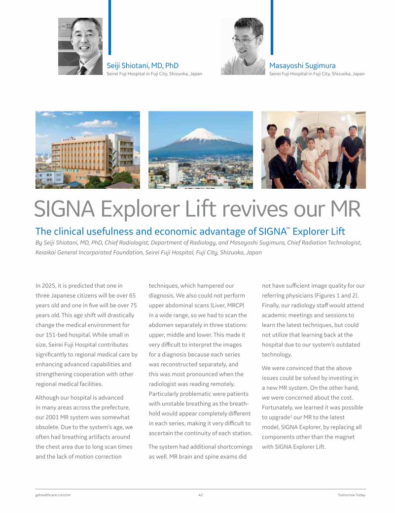
gehealthcare.com/mr 42 Tomorrow Today
not have sufficient image quality for our
referring physicians (Figures 1 and 2).
Finally, our radiology staff would attend
academic meetings and sessions to
learn the latest techniques, but could
not utilize that learning back at the
hospital due to our system’s outdated
technology.
We were convinced that the above
issues could be solved by investing in
a new MR system. On the other hand,
we were concerned about the cost.
Fortunately, we learned it was possible
to upgrade1 our MR to the latest
model, SIGNA Explorer, by replacing all
components other than the magnet
with SIGNA Explorer Lift.
In 2025, it is predicted that one in
three Japanese citizens will be over 65
years old and one in five will be over 75
years old. This age shift will drastically
change the medical environment for
our 151-bed hospital. While small in
size, Seirei Fuji Hospital contributes
significantly to regional medical care by
enhancing advanced capabilities and
strengthening cooperation with other
regional medical facilities.
Although our hospital is advanced
in many areas across the prefecture,
our 2001 MR system was somewhat
obsolete. Due to the system’s age, we
often had breathing artifacts around
the chest area due to long scan times
and the lack of motion correction
techniques, which hampered our
diagnosis. We also could not perform
upper abdominal scans (Liver, MRCP)
in a wide range, so we had to scan the
abdomen separately in three stations:
upper, middle and lower. This made it
very difficult to interpret the images
for a diagnosis because each series
was reconstructed separately, and
this was most pronounced when the
radiologist was reading remotely.
Particularly problematic were patients
with unstable breathing as the breath-
hold would appear completely different
in each series, making it very difficult to
ascertain the continuity of each station.
The system had additional shortcomings
as well. MR brain and spine exams did
SIGNA Explorer Lift revives our MRThe clinical usefulness and economic advantage of SIGNA™ Explorer Lift
By Seiji Shiotani, MD, PhD, Chief Radiologist, Department of Radiology, and Masayoshi Sugimura, Chief Radiation Technologist,
Keiaikai General Incorporated Foundation, Seirei Fuji Hospital, Fuji City, Shizuoka, Japan
Seiji Shiotani, MD, PhD Seirei Fuji Hospital in Fuji City, Shizuoka, Japan
Masayoshi Sugimura Seirei Fuji Hospital in Fuji City, Shizuoka, Japan

gesignapulse.com 43 Autumn 2017
In Practice
The main objective of upgrading our
MR was to improve image quality. We
visited another site that had recently
upgraded and compared images to
another recently installed (new)
SIGNA Explorer and found them to be
comparable. We therefore chose the
SIGNA Explorer Lift with confidence.
The cost was also lower since the
magnet did not need to be replaced,
making the downtime for the upgrade
significantly less.
After the upgrade
SIGNA Explorer Lift significantly
improved the performance and
image quality of our MR exams and
shortened exam times. We could now
provide a patient-friendly MR scan
and immediately accept emergency
patients. The scan time reduction
enabled an increase in the number of
patients in a day, providing an economic
benefit to our hospital.
Before the upgrade, we performed an average of 240 exams/month operating five days each week; now we increased that number to approximately 300 exams/month in the same operating time. A key reason for the increase in the number of examinations is the system’s image quality and clinical capability, further increasing referrals from local physicians.
Figure 1. A 70-year-old suffering from transient ischemic attack. Comparison of brain DWI images (A) before and (B) after SIGNA Explorer Lift. In Figure 1B, the SNR is significantly improved and brain structures are more clearly visualized than Figure 1A.
Figure 2. An 80-year-old with back pain. Comparing the lumber spine T2w images (A) before SIGNA Explorer Lift and (B) after SIGNA Explorer Lift, it is clear the vertebral body structures are distinctly visualized. The upgrade contributed to image quality improvement and motion artifact reduction as a result of shortened scan times and higher SNR through the Posterior Array Coil improvement.
A
A
B
B

gehealthcare.com/mr 44 Tomorrow Today
Our evaluation reported 27 cases
as "very good," 10 cases were "rescan
not necessary," and only three cases
required rescans. For the cases
rescanned, 3D frFSE was again used
under breath-hold but with increased
slice thickness. Since 3D frFSE collects
in-phase data with single shot, it is
necessary to decrease the number
of slices or shorten TR to achieve
scan time reduction. Increasing slice
thickness will lower the resolution but
improve SNR. Shortening TR causes
lower SNR even if fast recovery is used.
Considering these points, our hospital
optimizes the protocols based on slice
thickness, the number of slices and
TR to keep breath-hold times less
than 25 seconds (Figure 3).
In our case, we expect to realize a
return on our investment within three
years, including human resources
and maintenance expenses. With the
broader range of clinical capabilities,
our staff is now motivated to improve
their knowledge and skills.
Advanced clinical capabilities
With the prior system, we scanned
MRCP using 2D Fast Spin Echo (FSE).
The newly upgraded SIGNA Explorer
allows us to perform 3D Fast Recovery
FSE (frFSE) combined with ASSET
parallel imaging.
We reviewed 40 MRCP cases: 20
gallstone cholecystitis, 11 pancreatic
neoplastic lesion and 9 other cases.
The 18 male and 22 female patients
were an average age of 65.8 years
(maximum 85 and minimum 36 years).
All cases were scanned with respiratory
synchronization, although MRCP
can be acquired with diaphragm
synchronization. The use of Auto
Navigator was reported to improve
the image quality.
Figure 3. Comparison of MRCP images between respiratory synchronization scan and breath-hold scan. Depending on the patient, we can choose either (A) respiratory synchronization scan or (B) breath-hold scan.
A B
Using the 3D frFSE acquisition, it is possible to acquire higher signal with shorter TRs due to Fast Recovery. By using ASSET, it is possible to reduce the echo train of FSE, which helps to reduce blurring due to T2 attenuation. In addition, with 3D imaging we can view the data in various planes and angles. Furthermore, both respiratory synchronization and diaphragm synchronization are available.

gesignapulse.com 45 Autumn 2017
In Practice
LAVA Flex
With the upgrade, we can now use
LAVA Flex for T1-weighted (T1w)
imaging. LAVA Flex, a 3D acquisition,
uses a 2-point Dixon method. After
collecting data in-phase and out-of-
phase, the sequence estimates whether
the signals are water dominant or fat
dominant with reference to certain
pixel data. It then calculates magnetic
field inhomogeneity of all pixels from
neighboring pixels. By identifying the
signal intensity of each pixel from the
results, it is possible to calculate a uniform
water image (fat separation image) and
fat image (water separation image).
There are several advantages to using
LAVA Flex, including thinner slices
with 3D imaging, robust fat separation
and acquiring both with/without fat
separation images in one acquisition.
We can scan the same area on the
patient with gapless 3.2 mm slice
thickness in one breath-hold using LAVA
Flex and obtain both with/without fat
separation images (Figure 4).
Most importantly, these sequences
help us address the problem of a
patient who cannot hold their breath
to acquire the imaging data. In these
cases with the prior scanner, we often
sacrificed image resolution to shorten
breath-hold time by optimizing slice
thickness or using phase-encoding to
shorten breath-hold time. However,
SIGNA Explorer is compatible with
Auto Navigator and includes Auto
Navigator-gating, which enables the
acquisition of 3D T1-weighted images
with free breathing. By performing
LAVA Flex in conjunction with Auto
Navigator-gating, it is possible to scan
3D T1-weighted images both with and
without fat separation on patients who
cannot hold their breath (Figure 5).
Figure 4. Image comparison of T1w (A) before and (B, C) after upgrade. (A) Prior to upgrade, breath-hold T1w 2D FSPGR FatSat. After the upgrade, we are using a (B) breath-hold T1w LAVA Flex FatSat. (Water) and (C) breath-hold T1w LAVA Flex (in-phase). We can make the slices thinner using LAVA Flex, so the vessels in the liver can be observed without partial volume effect.
A B
C

gehealthcare.com/mr 46 Tomorrow Today
Conclusion
Just one month after the upgrade, our
hospital recorded its highest patient
volume for MR imaging. With SIGNA
Explorer Lift, we have the latest imaging
techniques at a fraction of the cost. A
key clinical benefit for our patients and
economic advantage for our hospital
is that we can improve image quality
without extending scan time. SIGNA
Explorer Lift is a good solution for facilities
seeking to improve clinical image
quality in an economic way. Footnotes
1. In the upgrade process, the existing magnet remains and all other components are replaced. After upgrading, the regulatory name and the number of the product will be changed.
Parallel imaging
Conventionally, the ASSET parallel
imaging method has been used to
shorten scan time. It can also be
used to reduce aliasing artifacts.
Theoretically, it is a parallel imaging
method that reduces the acquisition
time by decimating the amount of data
sampling. Typically, if the data sampling
amount is decimated then aliasing
artifacts appear. But with ASSET we
can obtain images without aliasing
artifacts due to the signal strength of
the wrapped-around part by calculation.
If we utilize this calculation to the
general aliasing artifact images (smaller
FOV scan than subject), we can obtain
non-aliasing artifact images by setting
the ASSET reduction factor to one.
Generally we utilize No-phase Wrap
(NPW) to eliminate aliasing artifacts.
NPW is a technique used to solve the
artifact by oversampling so the scan
time gets longer. Even in such cases,
it is possible to acquire non-aliasing
artifact images without extending scan
time by using ASSET. Using LAVA Flex,
it is possible to acquire a higher quality
image without non-aliasing artifact
and without a penalty in scan time.
Figure 5. The effect of Auto Navigator-gating in a LAVA Flex acquisition. (A, C) Breath-holding failure occurred at
hepatocyte phase scan with Primovist™ EOB and we rescanned it with (B, D) Auto Navigator-gating, which can be used in any plane. Blur conspicuous in (A) and (C) was improved with the use of Auto Navigator-gating.
A C
B D

gesignapulse.com 47 Autumn 2017
In Practice
“If we have up-to-date technology, then we can tackle clinical questions that we previously couldn’t answer, and that is clearly beneficial for our patients.”
Dr. David Smit
After witnessing installations in nearby
Australia and assessing image quality
and upgrade potential for hardware
and software, Dr. Smit, Jill and
their colleagues at Dunedin Hospital
chose the SIGNA™ Artist through
GE Healthcare’s SIGNA™ Lift program.
This economical upgrade path started
with the Optima™ MR450w with plans
to transition to the SIGNA Artist
In many hospitals, MR is a problem-solving tool that can help save the lives of critically ill or injured patients. This is especially true at Dunedin Hospital, a 388-bed tertiary and teaching facility that covers a large geographic area spanning the South Island of New Zealand’s second-largest city and surrounding areas.
“We have a fairly complex mix of
pathologies and patients, both referred
and brought into the ER by ambulance
or helicopter,” says David Smit, MB.BCh,
Director of MRI at Dunedin Hospital.
“So, we need very robust equipment to
handle the vast array of cases that we
see each day.”
It is not uncommon for inpatients at
Dunedin Hospital to have immobility
issues and complex disease processes
with multiple comorbidities. So, when
it came time to replace the hospital’s
11- to 12-year-old SIGNA™ HDxt MR
scanner, a key consideration was what
their patients would require.
“It was not what we thought our
patients would need, or what we
could find available. Rather we looked
at the complexity of our patients
and what would truly benefit them,”
explains Jill Oliver, DCR, MSc (MRI),
Lead MR Technologist. One of the key
considerations for any new system
would be patient comfort—and not just
the scanner hardware but also the coils.
Dr. Smit and Jill required a scanner
that could grow with the hospital—a
scalable platform that would
enable them to stay on pace with
advancements in technology, novel
sequences and coil development.
digitalSIGNA Artist
Going
helps prepare Dunedin Hospital for the future
with
David Smit, MB.BCh Dunedin Hospital in Dunedin, New Zealand
Jill Oliver, DCR, MSc Dunedin Hospital in Dunedin, New Zealand
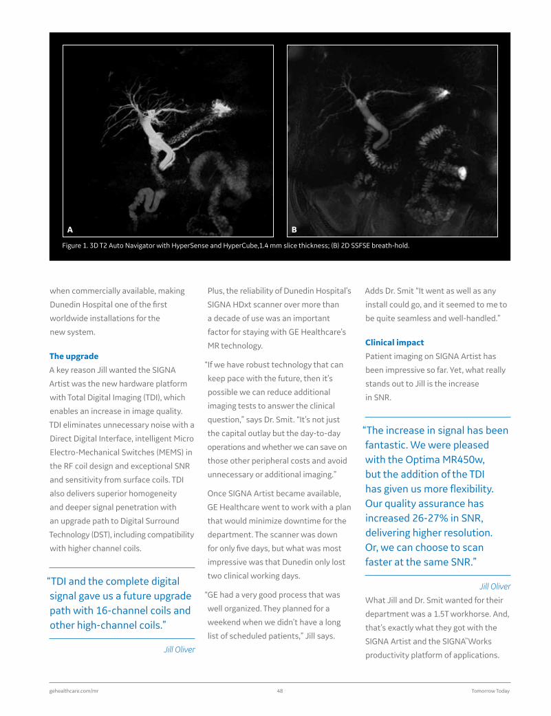
gehealthcare.com/mr 48 Tomorrow Today
when commercially available, making
Dunedin Hospital one of the first
worldwide installations for the
new system.
The upgrade
A key reason Jill wanted the SIGNA
Artist was the new hardware platform
with Total Digital Imaging (TDI), which
enables an increase in image quality.
TDI eliminates unnecessary noise with a
Direct Digital Interface, intelligent Micro
Electro-Mechanical Switches (MEMS) in
the RF coil design and exceptional SNR
and sensitivity from surface coils. TDI
also delivers superior homogeneity
and deeper signal penetration with
an upgrade path to Digital Surround
Technology (DST), including compatibility
with higher channel coils.
“TDI and the complete digital signal gave us a future upgrade path with 16-channel coils and other high-channel coils.”
Jill Oliver
Figure 1. 3D T2 Auto Navigator with HyperSense and HyperCube,1.4 mm slice thickness; (B) 2D SSFSE breath-hold.
A B
Plus, the reliability of Dunedin Hospital’s
SIGNA HDxt scanner over more than
a decade of use was an important
factor for staying with GE Healthcare’s
MR technology.
“If we have robust technology that can
keep pace with the future, then it’s
possible we can reduce additional
imaging tests to answer the clinical
question,” says Dr. Smit. “It’s not just
the capital outlay but the day-to-day
operations and whether we can save on
those other peripheral costs and avoid
unnecessary or additional imaging.”
Once SIGNA Artist became available,
GE Healthcare went to work with a plan
that would minimize downtime for the
department. The scanner was down
for only five days, but what was most
impressive was that Dunedin only lost
two clinical working days.
“GE had a very good process that was
well organized. They planned for a
weekend when we didn’t have a long
list of scheduled patients,” Jill says.
Adds Dr. Smit “It went as well as any
install could go, and it seemed to me to
be quite seamless and well-handled.”
Clinical impact
Patient imaging on SIGNA Artist has
been impressive so far. Yet, what really
stands out to Jill is the increase
in SNR.
“The increase in signal has been fantastic. We were pleased with the Optima MR450w, but the addition of the TDI has given us more flexibility. Our quality assurance has increased 26-27% in SNR, delivering higher resolution. Or, we can choose to scan faster at the same SNR.”
Jill Oliver
What Jill and Dr. Smit wanted for their
department was a 1.5T workhorse. And,
that’s exactly what they got with the
SIGNA Artist and the SIGNA™Works
productivity platform of applications.

gesignapulse.com 49 Autumn 2017
In Practice
two sequences are a large part of that
from my perspective,” says Dr. Smit.
Adds Jill, “The image quality for
prostate is shockingly good at 1.5T with
the new DWI sequences.” There is a
large demand for prostate MR in the
area, and Jill anticipates Dunedin can
help alleviate existing backlogs with
the speed and quality of the new MR
system. She believes they are delivering
the highest quality 1.5T MR prostate
exams in their region.
DISCO is also regularly used in both
prostate and liver imaging for its high
temporal resolution. In the liver, DISCO
has helped with the timing of breath-
hold acquisitions.
For orthopedic imaging, FSE Flex in the
spine and large field-of-views (FOVs)
have also resulted in excellent image
Dr. Smit has found that HyperSense
delivers clearer abdominal images
with less patient movement due to
the reduction in scan time. The
department has shaved 4 seconds
off Coronal FIESTA sequences of the
liver with excellent image quality and
signal. Dr. Smit says the department
also performs a lot of liver imaging
for HCC, metastatic disease and
post-RF ablation. It’s these types of
cases where the fast scans for contrast
enhancement really make a difference.
HyperSense can also be utilized to gain
spatial resolution while maintaining
scantimes. This is especially
important in Magnetic Resonance
CholangioPancreatography (MRCP)
exams in cases of suspected sclerosing
cholangitis.
“I can see the narrowing and dilation of the intrahepatic ducts more clearly and have more confidence in the findings.”
Dr. David Smit
HyperSense and HyperCube are
regularly used in pelvic imaging.
According to Jill, the ability to reformat
an Axial 3D HyperCube into Coronal
and Sagittal planes along with TDI are
making a big difference in the quality
of fast scanning techniques.
Dunedin Hospital also does a large
volume of prostate cancer imaging,
where the new synthetic diffusion,
MAGiC DWI, and FOCUS diffusion
sequences are regularly used. “I’m
more confident with a prostate cancer
diagnosis than I was before, and these
Figure 2. HyperCube + HyperSense in a patient with rectal cancer. Scan time 4 min, FOV 26 x 23.4 cm, slice thickness 1.2/0.6 mm.
Figure 3. 16-channel Shoulder Coil + PROPELLER MB. (A) Coronal T2 FSE PROPELLER MB 3/0.6 mm, 16 cm FOV, 3:26 min acquisition. (B) Coronal T1 FSE 3/0.6 mm, 16 cm FOV, 3:30 min acquisition.
A
A
B
C B

gehealthcare.com/mr 50 Tomorrow Today
Figure 4. High-resolution imaging of patient with “Turf Toe,” using the 16-channel Small Flex Coil. Sagittal FSE Flex PD and PD FatSat, 3/0.3 mm, 320 x 224, scan time of 2:24 min. (A) Water, (B) in-phase and (C) Coronal T1.
Figure 5. A 10-year-old patient with suspected Gaucher’s Disease. Protocol: Sagittal lumbar T1 & T2 with/without FatSat + Coronal femoral T1 + T2 without FatSat. Total exam time was 15 min versus 1 hour on prior system. (A, B) T2 FSE FLEX 3:30 min; (C, D) T1 FSE FLEX 3:20 min; (E, F) T1 FSE FLEX 3:03 min, in-phase and water only; and (G, H) T2 FSE FLEX 3:55 min, in-phase and water only.
C
A
E
G
C
B
F
H
D
A B

gesignapulse.com 51 Autumn 2017
In Practice
Jill also feels rather “spoiled” by
GE Healthcare and its support teams
given the geography of New Zealand
being separate from other countries.
Anytime Jill has a question she can pick
up the phone and get an answer right
away—and that filters right down to a
higher quality of patient care.
“The SIGNA Artist allows us to help more patients that we might otherwise not be able to help. It really lowers the barrier for patients who have difficulty tolerating MR so we can do what we need to get done on a daily basis, and that has been great for our patients and our clinicians.”
Dr. David Smit
quality. Based on this success, Jill
intends to utilize it in smaller FOVs such
as the hand, foot and wrist. The Proton
Density (PD) sequence—both with and
without FatSat—has produced very
good, high-resolution images and is
useful for differentiating bone infarction
from osteomyelitis while also helping
to shorten scan times.
Whole-body MR in rheumatology
patients is another area where the
SIGNA Artist shines. Although the exam
includes the use of multiple integrated
coils, patients have tolerated it well
and Jill says they are getting really good
images using whole-body STIR and
whole-body T1 sequences.
“The key improvement is the uniformity
over large FOVs, and that is really why
the whole-body imaging on this system
is so much better—it is predictable
and that enhances clinical confidence,”
Jill says.
Adds Dr. Smit, “Rheumatologists rely
on MR for determining the best area to
biopsy. As a radiologist, we don’t want
to review MR images with a lot of flare
as we are looking for brightness in the
muscle. With this system and using
many of these new imaging techniques,
we are now much more confident
that the signal in the muscle is truly
abnormal. In the past, we didn’t always
have that clarity of identification.”
Thanks to the speed and precision
of SIGNA Artist and SIGNA™Works,
patients are more comfortable and
there has been a noticeable reduction
in the use of sedation and general
anesthesia. The ability to image a
patient feet first has also enhanced
satisfaction.
“Feet-first imaging is a real plus for our
patients,” says Jill. “They have tolerated
it so much better.”
Figure 6. MAGiC DWI and eDWI for prostate imaging. (A) MAGiC DWI, b1400; (B) T2 Axial; (C) T2 Coronal; (D) eDWI, b800; (E) fused image. Protocols: Axial and Coronal T2, frFSE ETL 18, TR 5160/TE 100, 3/0.5 mm, 384 x 288, 4 NEX, 28 slices.
A
D
B
E
C

gehealthcare.com/mr 52 Tomorrow Today
At the end of March 2017, our SIGNA™ Pioneer 3.0T MR
system at PICTORU Izumo Imaging Center was upgraded
to the SIGNA™Works productivity platform. After
upgrading, this platform has helped dramatically increase
patient throughput without impacting image quality. We
also have the option to increase spatial resolution and
maintain prior scan times. As a result, we are receiving
excellent feedback from radiologists, referring physicians,
patients and C-level-suite administrators regarding the
SIGNA™Works upgrade.
Prior to having SIGNA™Works on SIGNA Pioneer, we
were scanning 16 patients each day on average.
Figure 1 shows the number of patients before and
after implementation.
With the addition of new sequences and techniques, such as HyperSense and HyperCube, we have increased patient throughput by 2.4 patients each day.
boostin productivity
A
SIGNA™Works helps imaging center increase both produtivity by up to 15% and daily patient throughput
By Koji Uchida, RT, Assistant Director, PICTORU Izumo Imaging Center, Eda Clinic Orthopedics Surgery and Rehabilitation in Japan
Before After
Number of exams (per day) 16.0 18.4
Figure 1. The benefit of new scan protocol with SIGNA™Works as shown in the increase in daily volume.
Before After
Koji Uchida, RT PICTORU Izumo Imaging CenterEda Clinic Orthopedics Surgery and Rehabilitation in Japan

gesignapulse.com 53 Autumn 2017
In Practice
Neuro imaging
In a routine neuro exam, head and
neck MR angiography (MRA) with
HyperSense and head DWI with
HyperBand have made significant
contributions in reducing scan time.
As shown in Figure 2, the scan time
for a neck MRA has been reduced by 4
minutes, from 9 minutes to 5 minutes,
representing a 45% reduction in scan
time. Further, HyperBand can be
applied to diffusion sequences, such
as diffusion-weighted imaging (DWI)
and diffusion-tensor imaging (DTI),
and we’ve seen up to a 50% scan time
reduction. The result is now a head
exam that can be completed in 20
minutes, including two MRA (head and
neck) sequences. (Figure 2).
Spine imaging
We are most impressed by SIGNA™Works’
increase in image quality for spine
exams. The signal-to-noise ratio (SNR)
of spine imaging is enhanced without
increasing scan time. This is primarily
due to SIGNA Pioneer’s Digital Surround
Technology (DST) which is compatible
with ARC, a parallel imaging method.
Coronal STIR PROPELLER gives us
good fat-suppressed images and MR
myelography, which is robust even in
the presence of patient movement.
Patients in severe pain may often
inadvertently move, degrading image
quality. Now, for these patients, we
apply PROPELLER at the beginning
of the examination. Prior to the
SIGNA™Works upgrade, an Axial
T2 acquisition covering only the
intervertebral foramen would take
more than 4 minutes. Now, we can
achieve wider coverage with an Axial
T2 of the spine in thin slices using
HyperCube with HyperSense in a scan
time that is less than 4 minutes.
Cube is suitable for imaging
lateral cervical disk herniation and
hyperostosis of Luschka’s joints
because we can apply thin slices
compared to a 2D sequence. Flow void
often occurs on 2D FSE and impacts
the clinician’s diagnosis. However, it
does not occur on Cube because of
the optimized flow compensation
technique. For lumber spine imaging
Before
Sequence Scan time (min)
SSFSE 3P 0:06
1 DWI Axial 1:30
2 Head TOF-MRA 5:04
3 FLAIR Axial 2:44
4 T2*w Axial 3:25
5 T1w Axial 3:21
6 T2w Axial 2:30
7 (opt.) Neck TOF-MRA 9:01
Total 27:40
After
Sequence scan time (min)
Advantage
SSFSE 3P 0:09
HB DWI Axial 0:45 50% scan time reduction by HyperBand
Head HS TOF-MRA 3:13 40% scan time reduction by HyperSense
FLAIR Axial 2:44
T2*w Axial 3:25
T1w Axial 3:21
T2w Axial 2:30
Neck HS TOF-MRA 4:51 45% scan time reduction by HyperSense
20:58
Figure 2. Routine head protocol before and after implementation of SIGNA™Works.
Figure 3. Routine head/neck MRA with HyperSense. (A) 1 mm slice thickness, 1 slab, FOV 20 cm, 352 x 224, ARC 2.0, HyperSense factor of 1.3, scan time of 3:13 min. (B) 2 mm slice thickness, ZIP 2, 34 x 8 slab, FOV 24 cm, 320 x 192, ARC 2.0, HyperSense factor of 1.5, scan time of 4:51 min.A B

gehealthcare.com/mr 54 Tomorrow Today
information is obtained in a scan time
of only 3 minutes.
Summary
SIGNA™Works can make a significant
impact on routine examinations. From
technologists to radiologists to referring
clinicians, we all strongly believe in
the clinical benefits of SIGNA™Works,
especially with HyperSense and
HyperCube. By implementing this
productivity platform in our facility,
we have increased patient throughput
as well as enhanced image quality.
Cube is effective for scanning the entire
vertebrae and herniated disc, which
is difficult to perform when using a 2D
sequence. Total scan time reduction
for routine spine imaging is 21%.
Abdomen imaging
3D MRCP with HyperSense delivers
high image quality with 1.2 mm slice
thickness and a 384 x 260 matrix
in a shorter scan time for routine
imaging. Before the upgrade to
SIGNA™Works, a thin slice 3D MRCP
sequence would take approximately 7
minutes to acquire. Now 3D MRCP with
HyperSense takes less than 3.5 minutes,
so we have built it into our routine
abdomen protocols (Figure 5).
Orthopedic imaging
In some clinical scenarios, our clinicians
request additional sequences that
tap into the exceptional image quality
that SIGNA Pioneer with SIGNA™Works
together deliver. An example is in
our routine knee exam. We’ve added
Sagittal PD-weighted scans with
HyperCube and HyperSense because
it provides high-quality meniscal and
cartilage images and excellent reformat
images. These reformatted images
contribute detailed image data to
assist our clinicians in their diagnosis
and identification of anteromedial/
posterolateral bundle on the anterior
cruciate ligament. This valuable
Figure 4. Routine spine examination. (A) T2w Flex of the spine; (B) DST with ARC; (C) PROPELLER STIR; (D) T2w HyperCube with HyperSense.
Figure 5. 3D MRCP with HyperSense. 1.2 mm slice thickness, 384 x 260, ARC 2.0, HyperSense factor of 2.0, scan time of 3:27 min.
A B C D

gesignapulse.com 55 Autumn 2017
Case Studies
MR imaging can be utilized for
differentiating healthy versus damaged
brain tissue in suspected stroke
patients even though MR imaging is
still challenging due to long scan times.
MR has been demonstrated to be
more sensitive for lesion detection and
more specific for delineation of healthy
versus damaged brain tissue compared
with CT in patients with acute ischemic
stroke.1-4 However, there is a need for
further improvements in MR acquisition
speed and limited availability.
The phrase “time is brain” emphasizes
that brain tissue is rapidly destructed
as ischemic stroke progresses, so
rapid evaluation with prompt therapy
is very important.
Therefore, the practicalities of using MR
for imaging brain tissue as one tool in
the diagnostic work up for suspected
acute stroke patients, especially in
restless and uncooperative patients,
remains challenging even for major
academic institutions.
In this patient case, there is a strong
suspicion of acute ischemic stroke.
The patient also had end-stage kidney
disease and showed a severe degree
of irritability during the physical
examination. Therefore, there were
Two-minute MR ultra-fast neuro protocolBy Hye Jin Baek, MD, PhD, Associate Professor Brain, Head & Neck Imaging, Department of Radiology,
Gyeongsang National University School of Medicine, Gyeongsang National University Changwon Hospital, South Korea
SIGNA™ Architect
Image Parameter Ultra-fast T1 Ultra-fast T2 Ultra-fast FLAIR
Ultra-fast DWI
Ultra-fast T2* Ultra-fast MRA
Sequence: T1 SPGR T2 SSFSE T2 FLAIR EPI SE EPI T2* Gradient 3D TOF SPGR
FOV (cm): 24 24 24 24 24 20
Phase FOV (cm): 0.75(18) 1(21.6) 0.9(21.6) 0.9(21.6) 1 1.84(16.8)
Slice thickness (mm): 5 5 5 5 5 1.6
TR (ms): 167.7 392.7 min 10000 2511(auto) 1600 27
TI (ms): 2200
TE (ms): 2.6 102 100 74.9 22.2 2.6
ETL (ms):
Frequency matrix (mXn):
260 320 128 128 128 320
Phase matrix (mXn): 190 320 256 128 320 160
Bandwidth (kHz): 31.25 83.33 250 250 250 35.71
ASSET/ARC factor: ARC 2 ASSET 3 ARC 2 ASSET 3 ARC 2, HS 2
Imaging options: FC, EDR EDR, TRF EDR,HB FC FC, EDR, ZIP2. ZIP512, FS
Scan time (sec): 15 8 25 13 6 38

gehealthcare.com/mr 56 Tomorrow Today
A B C
D E F
Figure 1. Clinician evaluations of MR images state that there are multiple diffusion restrictions with T1 hypointensity, T2 and FLAIR hyperintensity in both cerebellar hemispheres, suggesting acute infarction. On the GRE image, clinicians state that there are faint hemorrhages in both posterior cerebellar hemispheres, suggesting hemorrhagic transformation. On the MRA image, clinicians state that there are a few apparent focal stenoses seen in the basilar arteries.5
physiological difficulties in performing a
neuroimaging study.
Using HyperSense and HyperBand from
the SIGNA™Works productivity platform,
we performed a 2-minute ultra-fast
neuro protocol MR exam.
Patient history
An 83-year-old patient, weighing
128 lbs (58 kg) and 5 feet 4 inches
(164 cm) tall, with underlying disease
(hypertension, end-stage kidney
disease and diabetes mellitus) with
sudden onset of dysarthria and vertigo.
MR findings
MR images showed multiple
hyperintense lesions on diffusion-
weighted images and FLAIR with
coexisting hemorrhages in both
cerebellar hemispheres. With this
imaging data, we were able to identify
acute embolic infarctions due to
cytotoxic edema and distribution
of the lesions. We also found a few
apparent stenoses in the basilar artery
with underlying hypoplasia due to
fetal variant of both posterior cerebral
arteries. On the initial neurologic
examination, NIHSS was 7 points
(dysarthria 2 points, ataxia 2 points,
facial palsy 2 points and sensory
change 1 point). Atrial fibrillation
was confirmed during hospitalization,
causing cerebellar infarctions.
Discussion
Using HyperSense and HyperBand
on the SIGNA™ Architect, we acquired
valuable images which aided in
determining patient diagnosis and
management without the use of
contrast media or concern for motion
artifacts and scanning time (Figure 1).
Imaging data helped to confirm our
initial neurologic diagnosis by depicting
a conspicuous delineation of the lesions
despite the patient’s uncooperative
state. We also performed an ultra-
fast MRA to image major intracranial
arteries in a very short scan time. In this
case, our evaluation of the MR images
enabled us to achieve an appropriate
course for patient management.

gesignapulse.com 57 Autumn 2017
Case Studies
benefit for the patient who cannot
tolerate a longer scan time by reducing
motion artifact and minimizing the
need for sedation. We believe that
these advantages of a 2-minute
ultra-fast neuro protocol may extend
indications of MR examination into
the pediatric, non-cooperative and
emergency patients.
References
1. U-King-Im JM, et al. Utility of an ultrafast magnetic resonance imaging protocol in recent and semi-recent strokes. J Neurol Neurosurg Psychiatry. 2005 Jul;76(7):1002-1005.
2. Fiebach JB, et al. CT and diffusion-weighted MR imaging in randomized order: diffusion-weighted imaging results in higher accuracy and lower interrater variability in the diagnosis of hyperacute ischemic stroke. Stroke 2002;33:2206–2210.
3. Lev MH, Koroshetz WJ, Schwamm LH, Gonzalez RG. CT or MRI for imaging patients with acute stroke: visualization of
“tissue at risk”? [letter] Stroke 2002;33:2736–27377.
4. Saur D, Kucinski T, Gryzyska U, et al. Sensitivity and interrater agreement of CT and diffusion-weighted MR imaging in hyperacute stroke. AJNR Am J Neuroradiol 2003;24:878–885Ab.
5. The figures in this article will be used in future journal publications, for which the manucript is in preparation.
neuroimaging protocol in our institution.
Therefore, it was not difficult to
implement a 2-minute ultra-fast neuro
protocol as other challenging fast MR
protocols exist in our clinical practice.
Now we have more than 30 clinical
cases utilizing our 2-minute ultra-fast
neuro protocol. Our clinicians are
satisfied with this protocol due to its
shorter scan time, acceptable image
quality and diagnostic capability.
Although the image quality is perceived
as slightly inferior to that of a routine
20-minute protocol with MAGiC, the
use of a 2-minute ultra-fast neuro
protocol is feasible to visualize brain
tissue, which aids in our evaluation of
time-critical diseases like stroke. In
the clinical setting, it has a particular
This case suggests that our 2-minute
ultra-fast neuro protocol using HyperBand
and HyperSense can be a useful
imaging tool in patients with presumed
acute ischemic stroke and provide
an added benefit in patients who are
unable to endure longer acquisition
times or are contraindicated for contrast.
In this case, using this protocol aided in
our rapid diagnosis that assisted with
patient management decisions.
Additionally, we performed a two-day
follow-up MR using MAGnetic resonance
imaging Compilation (MAGiC) in this
patient, providing an interesting
comparison of two different fast
imaging protocols (Figure 2). Our
clinicians were already familiar with
fast imaging using MAGiC as a routine
D E F
A B C
D E F
Figure 2. MR images generated using MAGiC on the two-day follow-up. Clinicians determined that multi-focal acute cerebellar infarctions do not indicate significant interval change and demonstrate good correlation with initial ultra-fast MR.5
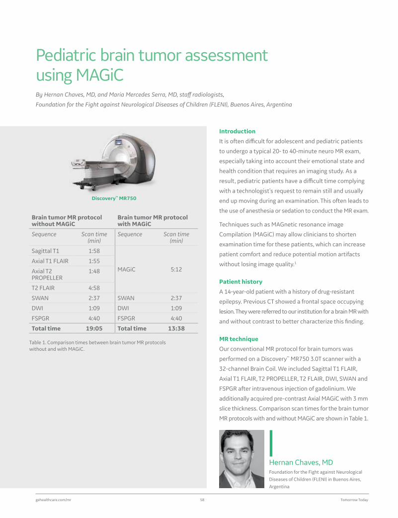
gehealthcare.com/mr 58 Tomorrow Today
Hernan Chaves, MDFoundation for the Fight against Neurological Diseases of Children (FLENI) in Buenos Aires, Argentina
Introduction
It is often difficult for adolescent and pediatric patients
to undergo a typical 20- to 40-minute neuro MR exam,
especially taking into account their emotional state and
health condition that requires an imaging study. As a
result, pediatric patients have a difficult time complying
with a technologist’s request to remain still and usually
end up moving during an examination. This often leads to
the use of anesthesia or sedation to conduct the MR exam.
Techniques such as MAGnetic resonance image
Compilation (MAGiC) may allow clinicians to shorten
examination time for these patients, which can increase
patient comfort and reduce potential motion artifacts
without losing image quality.1
Patient history
A 14-year-old patient with a history of drug-resistant
epilepsy. Previous CT showed a frontal space occupying
lesion. They were referred to our institution for a brain MR with
and without contrast to better characterize this finding.
MR technique
Our conventional MR protocol for brain tumors was
performed on a Discovery™ MR750 3.0T scanner with a
32-channel Brain Coil. We included Sagittal T1 FLAIR,
Axial T1 FLAIR, T2 PROPELLER, T2 FLAIR, DWI, SWAN and
FSPGR after intravenous injection of gadolinium. We
additionally acquired pre-contrast Axial MAGiC with 3 mm
slice thickness. Comparison scan times for the brain tumor
MR protocols with and without MAGiC are shown in Table 1.
Pediatric brain tumor assessment using MAGiCBy Hernan Chaves, MD, and Maria Mercedes Serra, MD, staff radiologists,
Foundation for the Fight against Neurological Diseases of Children (FLENI), Buenos Aires, Argentina
Discovery™ MR750
Brain tumor MR protocol without MAGiC
Brain tumor MR protocol with MAGiC
Sequence Scan time (min)
Sequence Scan time (min)
Sagittal T1 1:58
MAGiC 5:12Axial T1 FLAIR 1:55
Axial T2 PROPELLER
1:48
T2 FLAIR 4:58
SWAN 2:37 SWAN 2:37
DWI 1:09 DWI 1:09
FSPGR 4:40 FSPGR 4:40
Total time 19:05 Total time 13:38
Table 1. Comparison times between brain tumor MR protocols without and with MAGiC.
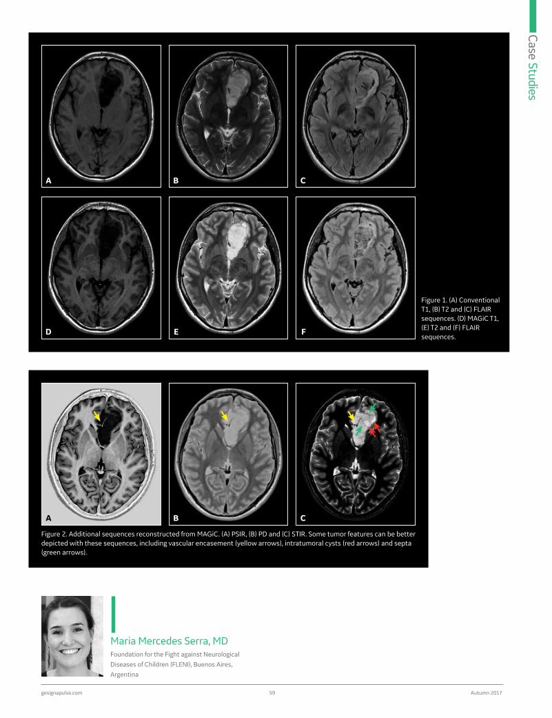
gesignapulse.com 59 Autumn 2017
Case Studies
Maria Mercedes Serra, MDFoundation for the Fight against Neurological Diseases of Children (FLENI), Buenos Aires, Argentina
A
A
B
B
C
C
D E F
Figure 1. (A) Conventional T1, (B) T2 and (C) FLAIR sequences. (D) MAGiC T1, (E) T2 and (F) FLAIR sequences.
Figure 2. Additional sequences reconstructed from MAGiC. (A) PSIR, (B) PD and (C) STIR. Some tumor features can be better depicted with these sequences, including vascular encasement (yellow arrows), intratumoral cysts (red arrows) and septa (green arrows).

gehealthcare.com/mr 60 Tomorrow Today
images, such as those processed with
MAGiC, we could reduce the total
exam time while maintaining image
quality with similar diagnostic utility.
Specifically, MAGiC could replace Axial
T1-weighted, T2-weighted and T2 FLAIR
acquisitions and can be acquired in any
plane if desired. Additional sequences
reconstructed from MAGiC, such as
Phase Sensitive Inversion Recovery
(PSIR), double IR, Proton Density (PD)
and Short Tau Inversion Recovery (STIR),
which are not typically acquired on
conventional brain tumor protocols,
could provide additional information for
further analysis of MR acquisition data.
PSIR is a phase sensitive reconstruction
contrast administration, isolated
nodules and rings of enhancement
were identified within the lesion. These
features suggested the diagnosis of
Dysembryoplastic Neuroepithelial
Tumor (DNET), which was pathologically
confirmed after surgery.
Discussion
MR scan duration in adolescents and
pediatrics is a primary concern due
to the difficulty in compliance with
these patients and the desire to avoid
or minimize sedation. Even when
optimized, simple brain protocols will
usually require at least 20 minutes
to complete the study. By replacing
conventional techniques with synthetic
MR findings
A cortical expansive lesion was
identified in the left mesial frontal
region. It presented with markedly
increased signal on T2-weighted and
decreased signal on T1-weighted
images with a bubbly appearance in
both the synthetic and conventional
images. On the synthetic FLAIR and
STIR images, internal septations and
multi-microcysts were observed with
better definition (Figures 1 and 2) than
on the conventional images. diffusion-
weighted imaging (DWI) was facilitated
within the lesion and no coarse
calcifications or blood products were
identified on SWAN. After intravenous
A
D
B
E
C
F
Figure 3. Additional conventional sequences. (A) Sagittal T1; (B) Axial post-contrast FSPGR; (C) DWI; (D) Sagittal reconstruction post-contrast FSPGR; (E) SWAN; and (F) ADC map. Areas of nodular and ring-like enhancement can be clearly depicted on post-contrast FSPGR (B, D, yellow arrows). (E) No coarse calcifications or blood products can be seen on SWAN. (C, F) Tumor diffusion was facilitated.

gesignapulse.com 61 Autumn 2017
Case Studies
• MAGiC is designed to fit in a routine
(320 x 224) 5-minute scan time with
3 mm slices and moderate resolution.
• MAGiC contrasts can be customized
to provide the best contrast, in our
opinion, especially PSIR and STIR (TI
set to the optimal WM/GM contrast).
• MAGiC can be post processed
to provide other contrasts
automatically depending on the
indication, a radiologist’s preference
or to add different contrasts that
were not initially acquired.
be used for further evaluation of tissues.
In conclusion, MAGiC may be acceptable
for clinical use in children; however,
users should be aware of its limitations.2
Noteworthy synthetic reconstructions
rely on the quality of a single scan,
so special care should be taken to
minimize motion artifacts during this
acquisition. References
1. Tanenbaum LN, Tsiouris AJ, Johnson AN, Naidich TP, DeLano MC, Melhem ER, et al. Synthetic MRI for Clinical Neuroimaging: Results of the Magnetic Resonance Image Compilation (MAGiC) Prospective, Multicenter, Multireader Trial. AJNR Am J Neuroradiol. 2017 Jun;38(6):1103-1110.
2. Betts AM, Leach JL, Jones BV, Zhang B, Serai S. Brain imaging with synthetic MR in children: clinical quality assessment. Neuroradiology. 2016 Oct;58(10):1017-1026.
that greatly improves T1 contrast as
it displays both negative and positive
longitudinal magnetization amplitudes
rather than normal magnitude
reconstruction. In our case, we also
used MAGiC T1 with a 100 ms TR and
a 5 ms TE, improving contrast of normal
T1 FSE.
The MR workstation enables immediate
and automatic reconstruction of all
MAGiC-derived series, which can then
be sent to PACS for reading. For further
inspection, radiologists have access to
raw data and can change the TR, TE and
TI of the derived. Quantitative maps are
also available for T1, T2 and PD and can
A B C
D E
Figure 4. MAGiC quantitative maps (T1, R1, T2, R2 and PD) allow direct visualization as well as information of T1, T2 and PD for enhanced evaluation of tissues inside the ROI.
Technical inputs

gehealthcare.com/mr 62 Tomorrow Today
Discovery™ MR750
MAGiC Parameters
Age: 1-6 years old
TR (ms): 5000
TE (ms): 22.8 (second TE is automatically decided)
TI (ms): 4 (saturation delay automatically calculated)
ETL (ms): 12
FOV (cm): 22
Slice thickness (mm):
4
Frequency (mXn): 320
Phase (mXn): 224
Scan time (min): 6:20
Age: less than 1 year old
TR (ms): 4000
TE (ms): 23.2 (second TE is automatically decided)
TI (ms): 4 (saturation delay automatically calculated)
ETL (ms): 12
FOV (cm): 20
Slice thickness (mm):
4
Frequency (mXn): 320
Phase (mXn): 224
Scan time (min): 6:08 So Mi Lee, MDKyungpook National University Medical Center, Daegu in South Korea
Pediatric imaging with MAGiCBy So Mi Lee, MD, Assistant Professor, Department of Radiology, Kyungpook National University Medical Center,
Daegu, South Korea
Introduction
Contrast-enhanced MR studies are a
sensitive imaging study for the
evaluation of brain pathology. Despite
the diagnostic utility of using an MR
contrast agent, there is concern regarding
the accumulation of gadolinium in
neuronal tissues.1-3 As a result, many
experts suggest if gadolinium is not
needed for a particular exam, especially
in pediatric patients, then it should not
be administered.
Our institution has evaluated MAGnetic
resonance image Compilation (MAGiC)
for use in non-contrast imaging of
pediatric patients with neurological
disorders. Many of these young patients
will require multiple follow-up MR scans
and, therefore, the avoidance of MR
contrast is desirable.
Sturge-Weber Syndrome (SWS) is a
rare neurological disorder present at
birth marked by a distinctive port-wine
stain (facial nevus flammeus) on the
forehead, scalp or around the eye. Many
people afflicted with SWS experience
seizures or convulsions. Other
complications may include eye
abnormalities, developmental delays
and weakness on one side of the body.
This disease usually worsens over
time with clinical symptoms such
as hemiparesis, mental retardation
and seizures. Specific pathologies
resulting from this disorder include
leptomeningeal angioma and
enlarged collateral vessels, such as
transmedullary and subependymal
veins. Demonstration of the extent of
the angioma is critical in determining
the patient’s prognosis and the
necessary extent of cortical resection
for seizure management.
In the following case, the patient
underwent conventional and synthetic
imaging (with MAGiC), to determine
the extent of disease and evaluate if
the use of MAGiC without contrast
may be utilized to replace conventional
contrast-enhanced scans for the
evaluation of SWS.

gesignapulse.com 63 Autumn 2017
Case Studies
Patient history
A nine-month-old patient with an
episode of focal seizure was referred for
evaluation. The patient also had facial
nevus flammeus (left side) and angioma
involving the choroid of the eye.
MR findings
Both conventional and synthetic
imaging with MAGiC were performed
on this patient.
Conventional: Axial T2-weighted
image (Figure 1A) shows minimal
hypointensity of the left hemispheric
white matter and dilated medullary and
subependymal veins. The ipsilateral
cerebral hemisphere shows mild
atrophic change. On the contrast-
enhanced image (Figure 1B) the
leptomeningeal angioma especially
involves the parieto-occipital region.
Enlarged collateral vessels, such as
subependymal and transmedullary
veins, enlarged choroid plexus and
marked enhancement of the choroid
of the ocular globe are shown in the
left side. These imaging features are
consistent with SWS.
Using synthetic imaging (Figure 2B),
the leptomeningeal angioma—the
pathognomic feature of this pathology
being most clearly depicted on contrast
exam—can be demonstrated without
the use of an MR contrast agent.
In addition, using MAGiC PSIR (Figure 1D),
the dilated and enlarged collateral
vessels as well as parenchymal atrophic
changes (Figure 2C) can be well visualized
without use of an MR contrast agent.
In this case, the white matter underlying
the angioma typically shows prominent
hypointensity on T2-weighted images
compared to the remainder of the
brain. This is most likely secondary
to abnormal hypermyelination that
presumably results from abnormal
venous congestion or from repeated
seizures. The synthetic imaging that
includes myelin partial volume map
as well as quantitative imaging such
as T1, T2 and PD maps are useful
for evaluation of early white matter
changes in an infant with SWS.
Discussion
The use of synthetic imaging facilitated
diagnosis of the patient’s condition as
well as the evaluation of the extent of
the lesion without the use of a contrast
agent. Concerns regarding intracranial
C D
A B
Figure 1.
A. Conventional T2 B. Conventional CE T1 C. Synthetic T2 D. PSIR
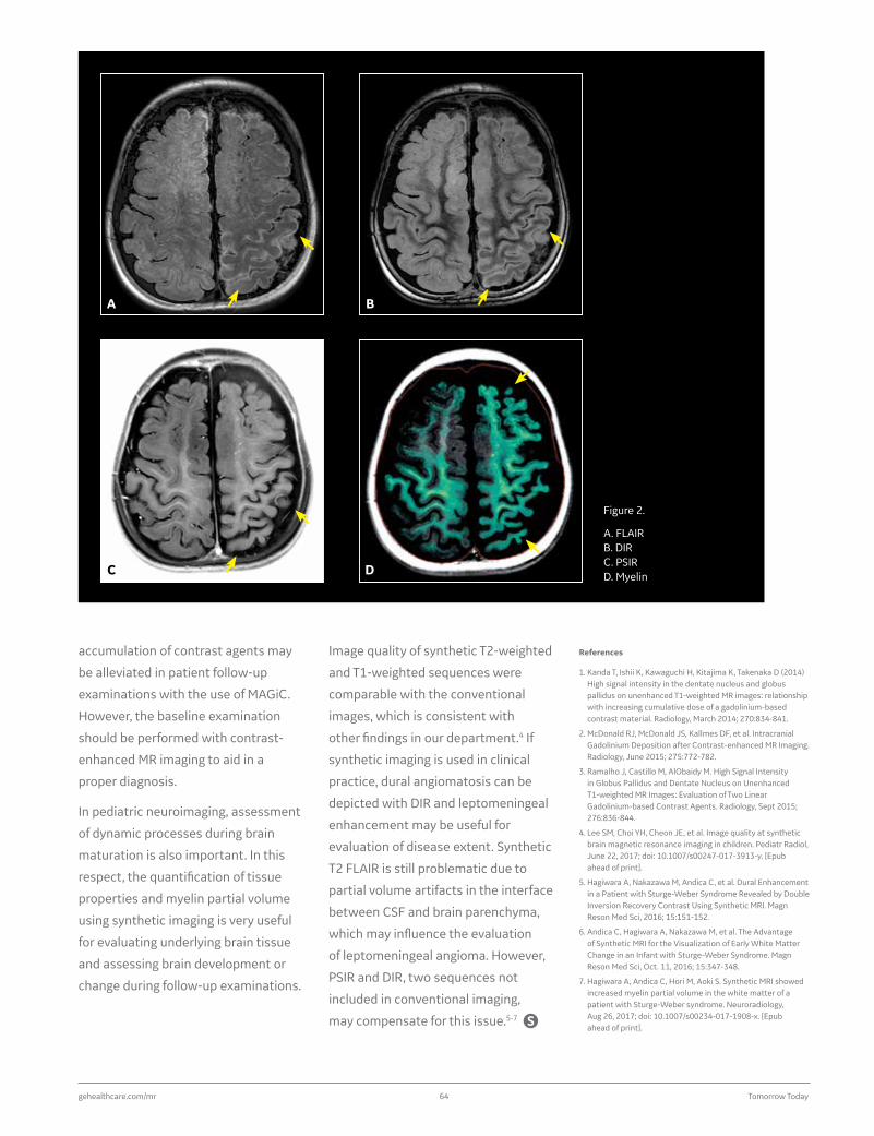
gehealthcare.com/mr 64 Tomorrow Today
References
1. Kanda T, Ishii K, Kawaguchi H, Kitajima K, Takenaka D (2014) High signal intensity in the dentate nucleus and globus pallidus on unenhanced T1-weighted MR images: relationship with increasing cumulative dose of a gadolinium-based contrast material. Radiology, March 2014; 270:834-841.
2. McDonald RJ, McDonald JS, Kallmes DF, et al. Intracranial Gadolinium Deposition after Contrast-enhanced MR Imaging. Radiology, June 2015; 275:772-782.
3. Ramalho J, Castillo M, AlObaidy M. High Signal Intensity in Globus Pallidus and Dentate Nucleus on Unenhanced T1-weighted MR Images: Evaluation of Two Linear Gadolinium-based Contrast Agents. Radiology, Sept 2015; 276:836-844.
4. Lee SM, Choi YH, Cheon JE, et al. Image quality at synthetic brain magnetic resonance imaging in children. Pediatr Radiol, June 22, 2017; doi: 10.1007/s00247-017-3913-y. [Epub ahead of print].
5. Hagiwara A, Nakazawa M, Andica C, et al. Dural Enhancement in a Patient with Sturge-Weber Syndrome Revealed by Double Inversion Recovery Contrast Using Synthetic MRI. Magn Reson Med Sci, 2016; 15:151-152.
6. Andica C, Hagiwara A, Nakazawa M, et al. The Advantage of Synthetic MRI for the Visualization of Early White Matter Change in an Infant with Sturge-Weber Syndrome. Magn Reson Med Sci, Oct. 11, 2016; 15:347-348.
7. Hagiwara A, Andica C, Hori M, Aoki S. Synthetic MRI showed increased myelin partial volume in the white matter of a patient with Sturge-Weber syndrome. Neuroradiology, Aug 26, 2017; doi: 10.1007/s00234-017-1908-x. [Epub ahead of print].
Image quality of synthetic T2-weighted
and T1-weighted sequences were
comparable with the conventional
images, which is consistent with
other findings in our department.4 If
synthetic imaging is used in clinical
practice, dural angiomatosis can be
depicted with DIR and leptomeningeal
enhancement may be useful for
evaluation of disease extent. Synthetic
T2 FLAIR is still problematic due to
partial volume artifacts in the interface
between CSF and brain parenchyma,
which may influence the evaluation
of leptomeningeal angioma. However,
PSIR and DIR, two sequences not
included in conventional imaging,
may compensate for this issue.5-7
accumulation of contrast agents may
be alleviated in patient follow-up
examinations with the use of MAGiC.
However, the baseline examination
should be performed with contrast-
enhanced MR imaging to aid in a
proper diagnosis.
In pediatric neuroimaging, assessment
of dynamic processes during brain
maturation is also important. In this
respect, the quantification of tissue
properties and myelin partial volume
using synthetic imaging is very useful
for evaluating underlying brain tissue
and assessing brain development or
change during follow-up examinations.
C D
A B
Figure 2.
A. FLAIR B. DIR C. PSIR D. Myelin

gesignapulse.com 65 Autumn 2017
Case StudiesPediatric MR enterographyBy Hesham Alshaalan, MBBS, FRCPC, dABR, Ped CAQ, Deputy Chairman Medical Imaging Department and Consultant Pediatric
Radiology & Pediatric Neuroradiology, King Abdullah Specialist Children’s Hospital Ministry of National Guard, Riyadh,
Kingdom of Saudi Arabia
Optima™ MR450w GEM
Hesham Alshaalan, MBBS, FRCPC, dABR, Ped CAQKing Abdullah Specialist Children’s Hospital Ministry of National Guard, Riyadh, Kingdom of Saudi Arabia
Pediatric MR enterography (MRE) presents additional
challenges due to the small body size and small field of
view that may come at the expense of SNR. There is often
suboptimal oral contrast injection as many children find it
tastes unpleasant. Rapid breathing can also create image
artifacts from respiratory motion.
The introduction of fast scanning techniques may overcome
some of these pediatric MRE obstacles and enable the
diagnosis of these patients without the use of ionizing
radiation.
Patient history
Three-year-old with suspicion of Crohn’s disease. The patient
was prepared with oral contrast for bowel distension and the
procedure was done under general anesthesia.
Fast imaging was employed to minimize scanning as well
as sedation timing for the patient with a motion correction
series. MR was chosen over other modalities to avoid a
radiation hazard.
Parameters
Sequence Slice thickness (mm)
Scan time (min)
Resolution
Axial FIESTA FatSat – respiratory triggered
4 3:06 224 x 224
Axial PROPELLER T2 FatSat motion correction
4 6:13 256 x 256
Axial DWI with multiple b values 0, 50, 150 & 800 with corresponding ADC; respiratory triggered
4 5:07 80 x 192
Axial LAVA Flex dynamic series, arterial-venous & delayed, with Auto Navigator
3 1:39 256 x 192
Coronal LAVA Flex post gadolinium injection (delayed)
3 1:22 256 x 224

gehealthcare.com/mr 66 Tomorrow Today
A
A
C
B
D
B
Figure 1. (A) Axial FIESTA FatSat, 4 mm, Respiratory Trigger, acquisition time of 3:06 min; (B) Axial PROPELLER T2 FatSat, 4 mm, Respiratory Trigger, acquisition time of 6:13 min.
Figure 2. (A) Axial DWI, 4 mm, Respiratory Trigger, acquisition time of 5:07 min, b600; (B) pre-contrast LAVA Flex Axial; (C) PROPELLER T2 FatSat; (D) Fusion of the DWI to the LAVA Flex post contrast.
MR findings
Short segment terminal ileum bowel
wall thickening with enhancement,
restricted diffusion and prominent
mesenteric lymph nodes. The diagnosis
was confirmed by colonoscopy biopsy.

gesignapulse.com 67 Autumn 2017
Case Studies
patients. Image quality was excellent.
These techniques provided a high level
of confidence in my diagnosis and led
to appropriate initiation of therapy.
Continued development of appropriate
respiratory-triggered sequences for
pediatrics will help to further overcome
inherent challenges in MR imaging of
these patients. Our referring pediatric
gastroenterologists now rely on our
ability to diagnose inflammatory bowel
disease utilizing MRE.
Discussion
The use of fast scanning and motion
correction sequences with Auto
Navigator and respiratory-triggered
solutions helped to overcome inherent
challenges in imaging pediatric
A
A
B
B
C
C
Figure 3. Axial LAVA (A) pre-contrast, (B) arterial and (C) venous phases.
Figure 4. Coronal LAVA dynamic (delayed), 3 mm, Auto Navigator acquisition time of 1:22 min; (B) post-contrast LAVA Flex; (C) post-contrast LAVA Flex fused with the Axial DWI.
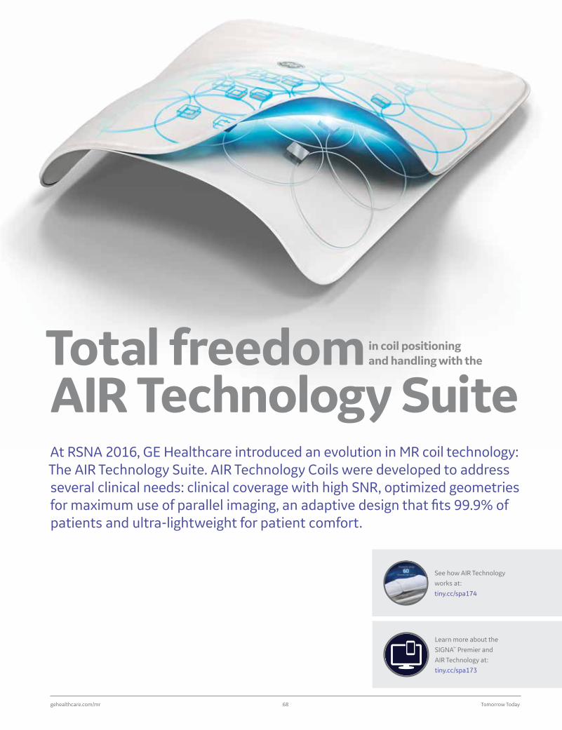
gehealthcare.com/mr 68 Tomorrow Today
At RSNA 2016, GE Healthcare introduced an evolution in MR coil technology: The AIR Technology Suite. AIR Technology Coils were developed to address several clinical needs: clinical coverage with high SNR, optimized geometries for maximum use of parallel imaging, an adaptive design that fits 99.9% of patients and ultra-lightweight for patient comfort.
Total freedomAIR Technology Suite
in coil positioning and handling with the
Learn more about the SIGNA™ Premier and AIR Technology at:tiny.cc/spa173
See how AIR Technology works at:tiny.cc/spa174

gesignapulse.com 69 Autumn 2017
Tech Trends
GE Healthcare scientists developed
several new underlying technologies
in the new coil design. A multiple
resonator conductor arrangement
replaces lumped components and
circuit boards with an exceptionally
durable and flexible loop, named the
INCA conductor. AIR Technology also
incorporates proprietary E-Mode
electronics designed to reduce
component volume by more than
60%, decrease coupling flex conductor
geometries by 30% and deliver 95%
more transparency for MR-based
attenuation correction. The E-Mode
electronics also help lessen current
noise, boost linearity and improve
tolerance to varying coil loading
conditions. The combination of these
technologies makes AIR Technology
Coils well suited to work in low- to high-
density coil applications.
The goal is total freedom in coil positioning and handling with a ≥60% lighter weight and 80% more flexible design opportunities.
The University of Wisconsin-Madison
recently evaluated the AIR Technology
Suite on the SIGNA™ Premier 3.0T
system using volunteers in a clinical
study which was reviewed and
monitored by an Institutional Review
Board (IRB). Tammy Heydle, RT(R)
(MR), Senior MR Technologist, and
Shawn Pulver, RT(R)(MR), Objective
MR Technologist, performed a series
of scans on the prostate, female pelvis,
musculoskeletal, abdomen, ENT (ear,
nose and throat), neck, spine and
vasculature. They used the Posterior
AIR Technology Coil and Anterior AIR
Technology Coil for all applications.
In addition, Heydle and Pulver also
used the AIR Technology Coils
simultaneously with conventional coil
technology—Head Neck Unit (HNU).
When the HNU was combined with the
AIR Technology Coil it enabled whole-
body imaging without repositioning
or re-landmarking the patient. All
scanning was performed using existing
UW-Madison MR protocols.
“The signal on the images acquired with the AIR Technology Coil appeared greater than the images obtained with the conventional Abdominal Body Array coil. The AIR Technology Coils are also lighter and more flexible so they conform to the body habitus.”
Tammy Heydle
She explains that with traditional coils,
the Anterior Abdominal Array does
not conform to the patient and needs
to be fastened to the table to prevent
coil movement. However, with the
AIR Technology Coil conforming and
molding to a patient’s unique body
and size, the issue of coil movement
was minimized.
Pulver adds that the material of the
coil also makes a difference—it is
smoother and more sleek so it is easier
to slide the patient into the bore of the
magnet. He was also impressed that
he could obtain a signal beyond where
the AIR Technology Coil is positioned
A B
Figure 1. The AIR Technology Coil allows for extended coverage in z-direction as demonstrated in this Coronal T2 SSFSE breath-hold exam.

gehealthcare.com/mr 70 Tomorrow Today
“The AIR Technology Coil is a vast
improvement over the conventional,
hard-shell coils that cover the patient’s
face,” Heydle says. “It also conforms to
the neck providing better comfort.”
Pulver adds, “Since the AIR Technology
Coil is lighter, there is less weight on
the arms, so the patient doesn’t feel all
that pressure on their shoulders when
we image them with their arms above
their head.”
Another advantage of the AIR Technology
Suite in brachial plexus exams is the
ability to overlap elements. Pulver used
the HNU and the AIR Technology Coil
simultaneously.
“We had great signal penetration all the way through with no artifacts or any issues due to the overlap.”
Shawn Pulver
The AIR Technology Coil also performed
well with parallel imaging. Pulver
and Heydle used HyperSense, an
acceleration technique that enables
faster imaging without the penalties
commonly found with conventional
parallel imaging. HyperCube for 3D
isotropic imaging and MAGiC DWI
(synthetic diffusion) were also utilized.
“Using HyperSense with parallel imaging
enables a decrease in scan times
without affecting image quality,” Heydle
says. “With HyperSense, we can obtain
higher resolution imaging or faster scan
times for better patient throughput.”
MAGiC DWI allows the tech to acquire
additional b-values without an addition to
exam time when compared to acquiring
separate scans for each b-value.”
Using the AIR Technology Coil in a
brachial plexus study, Heydle and
Pulver acquired Sagittal T1 BRAVO and
Sagittal T1 Cube images that were then
reformatted into other imaging planes.
on the patient. An indentation and
gray colored area helps indicate coil
coverage, enabling a streamlined coil
set up. A thinner coil, thanks to the
reduction in component volume, also
translates to more available room in the
magnet bore to scan very large patients.
The design of the coils may also lead to
higher SNR. The Abdominal Array Coil
provides superb SNR and acceleration
performance while the Posterior Array
Coil improves SNR in the spine and
deeper anatomical structures in the body.
“The way the coil elements are combined
and situated as many smaller coils
appears to enhance SNR,” Heydle
says. She didn’t conduct comparison
QA testing, but when compared to a
traditional Abdominal Body Array
Coil she could qualitatively see a boost
in SNR.
Figure 2. Coronal brachial plexus with Cube STIR and HyperCube acquired with 1.2 mm isotropic resolution.

gesignapulse.com 71 Autumn 2017
Tech Trends
“You don’t want too much pressure on
the patient if they are sedated, so the
AIR Technology Coil could be very nice
for pediatric imaging.”
In addition to pediatrics, Pulver
also sees an opportunity to use the
lightweight, flexible AIR Technology
Coils for extremity imaging. Both Pulver
and Heydle are excited to use the AIR
Technology Suite once they are fully
implemented at UW-Madison.
Heydle adds, “The AIR Technology Coils
are very versatile, which opens up
almost endless possibilities for
using them.”
With some coils, an overlap can lead
to signal loss or image degradation.
He also notes that one nice feature of
the SIGNA Premier is the system will
alert the technologist if there is too
much overlap so they can adjust the
coil placement.
Heydle believes the AIR Technology
Coils will also help with claustrophobic
patients. “The lighter coil will feel more
like a blanket, so the patient won’t
feel as weighed down,” she says. Plus,
the ability to scan feet first on the
SIGNA Premier may further lessen
claustrophobia.
While the IRB at UW-Madison was only
for adult volunteer scanning, Pulver
sees an opportunity to use the AIR
Technology Coils on pediatric patients.
Figure 3. Axial LAVA Flex. Figure 4. High resolution Sagittal T2 PROPELLER Multi-Blade of the female pelvis.

T O M O R R O W T O D AY
© 2017 General Electric Company – All rights reserved.
MR-0507-11.17-EN-US JB53475XX
Experience freedom.Designed for freedom in coil positioning, GE Healthcare’s revolutionary AIR Technology is reinventing the way imaging should be. Created with people in mind, the ultra-lightweight design conforms comfortably to all patients, leaving them more at ease during a scan, allowing the technologist to focus more on the patient and not on the technology.
#imagingwithAIR
Highly flexible to fit all sizes, shapes and ages
Highest channel count in the industry
≥60% lighter design without compromising image quality