Embed Size (px)
Citation preview

1
Title: Recessive inactivating mutations in TBCK, encoding a Rab GTPase-activating protein that modulates mTOR signaling, cause severe infantile syndromic encephalopathy Authors: Jessica X. Chong1
Viviana Caputo2
Ian G. Phelps1
Lorenzo Stella3
Lisa Worgan4 Jennifer C. Dempsey1 Alina Nguyen1 Vincenzo Leuzzi5 Richard Webster6,7 Antonio Pizzuti2 Colby T. Marvin1 Gisele E. Ishak8 Simone Ardern–Holmes7 Zara Richmond9 University of Washington Center for Mendelian Genomics Michael J. Bamshad1,10,11 Xilma R. Ortiz-Gonzalez12 Marco Tartaglia13,17,** Maya Chopra14-16,17 Dan Doherty1,11,* 1. Department of Pediatrics, University of Washington, Seattle, WA 98195, USA 2. Dipartimento di Medicina Sperimentale, Università La Sapienza, 00161 Rome, Italy 3. Dipartimento di Scienze e Tecnologie Chimiche, Università di Roma Tor Vergata, 00133 Rome, Italy 4. Department of Clinical Genetics, Liverpool Hospital, Liverpool, NSW, Australia 5. Dipartimento di Pediatria e di Neuropsichiatria Infantile, Università La Sapienza, 00185 Rome, Italy 6. T.Y. Nelson Department of Neurology and Neurosurgery 7. Institute of Neuroscience and Muscle Research, the Children’s Hospital at Westmead 8. Department of Radiology, University of Washington, Seattle, WA 98195, USA 9. Department of Genomic Medicine, Royal Prince Alfred Hospital, Missenden Road, Camperdown, Sydney
NSW, Australia 10. Department of Genome Sciences, University of Washington, Seattle, WA 98195, USA 11. Division of Genetic Medicine, Seattle Children's Hospital, Seattle, WA 98105 12. Division of Neurology, Children’s Hospital of Philadelphia and Department of Neurology, Perelman School
of Medicine, University of Pennsylvania, Philadelphia, PA, 19104 13. Area di Ricerca " Genetica e Malattie Rare", Ospedale Pediatrico Bambino Gesù - IRCCS, 00146 Rome,
Italy 14. Department of Genomic Medicine, Royal Prince Alfred Hospital, Missenden Road, Camperdown, Sydney
NSW, Australia 15. School of Genomic Medicine, University of Sydney, Sydney, NSW Australia 16. Shanghai First Maternity and Infant Hospital, Tongji University School of Medicine, West Gaoke Road,
Pudong, Shanghai 17. These authors contributed equally to this work
Corresponding authors: *Dan Doherty, MD, PhD [email protected] **Marco Tartaglia, PhD [email protected]
.CC-BY-NC 4.0 International licensepeer-reviewed) is the author/funder. It is made available under aThe copyright holder for this preprint (which was not. http://dx.doi.org/10.1101/036111doi: bioRxiv preprint first posted online Jan. 6, 2016;

2
Abstract:
Infantile encephalopathies are a group of clinically and biologically heterogeneous disorders for which the
genetic basis remains largely unknown. Here, we report a previously unrecognized syndromic neonatal
encephalopathy characterized by profound developmental disability, severe hypotonia, seizures, diminished
respiratory drive requiring mechanical ventilation, brain atrophy, corpus callosum dysgenesis, cerebellar vermis
hypoplasia, and facial dysmorphism. Biallelic inactivating mutations in TBCK (TBC1 domain-containing kinase)
were independently identified by Whole-Exome Sequencing (WES) as the cause of this condition in four
unrelated families. Matching these families was facilitated by sharing phenotypic profiles and WES data in a
recently released web-based tool (Geno2MP) that links phenotypic information to rare variants in families with
Mendelian traits. TBCK is a putative GTPase-activating protein (GAP) for small GTPases of the Rab family and
has been shown to control cell growth and proliferation, actin cytoskeleton dynamics, and mTOR signaling.
Two of the three mutations are predicted to truncate the protein (c.376C>T [p.Arg126*] and c.1363A>T
[p.Lys455*]), and loss of the major TBCK isoform was confirmed in primary fibroblasts from one affected
individual. The third mutation, c.1532G>A [p.Arg511His], alters a conserved residue within the TBC1 domain.
Structural analysis implicates Arg511 as a required residue for Rab-GAP function, and in silico homology
modeling predicts impaired GAP function in the corresponding mutant. These results suggest loss of Rab-GAP
activity is the underlying mechanism of disease. In contrast to other disorders caused by dysregulated mTOR
signaling associated with focal or global brain overgrowth, impaired TBCK function results in progressive loss
of brain volume.
KEYWORDS
exome sequencing, Mendelian disease, infantile encephalopathy, mTOR signaling, GTPase activating protein
.CC-BY-NC 4.0 International licensepeer-reviewed) is the author/funder. It is made available under aThe copyright holder for this preprint (which was not. http://dx.doi.org/10.1101/036111doi: bioRxiv preprint first posted online Jan. 6, 2016;

3
Severe infantile encephalopathy is a non-specific clinical condition that can be caused by hypoxemia,
hemorrhage, toxins (e.g., hyperbilirubinemia or withdrawal from selective serotonin reuptake inhibitors or
narcotics), and mutations in multiple genes such as PURA [MIM 600473], UNC80 [MIM 612636] or NALCN
[MIM 611549].1-9 Over the past 5 years, substantial progress has been made toward defining the genetic basis
of infantile encephalopathies; however, the genetic cause remains unknown for a large proportion of affected
individuals, and these conditions have few distinguishing clinical features, making it challenging to stratify
affected individuals for gene discovery efforts. Whole-Exome Sequencing (WES) of multiple independent
families with non-specific infantile encephalopathies is a powerful way to discover shared candidate genes
among affected individuals, which upon further clinical evaluation are often found to share phenotypic features
delineating a distinctive condition within this otherwise non-specific category. Here, we report on a previously
unrecognized syndromic infantile encephalopathy characterized by severe developmental disability, brain
atrophy, focal seizures with early-onset, central respiratory failure, and facial dysmorphism. WES in four
families led to the discovery of mutations in TBCK, a gene encoding a putative Rab-specific GTPase-activating
protein (GAP), as the underlying molecular cause.
Clinical data and biological samples were obtained from three unrelated families after written informed
consent was provided, and studies for each family were approved by the respective institutional review boards
of the University of Washington (Seattle, USA), the Genetics Services Advisory Committee (New South Wales,
Australia), and Università La Sapienza (Rome, Italy). A single affected individual with neonatal onset
encephalopathy, brain atrophy with cerebellar malformation, and later-onset seizures (Figure 1, Table 1, and
Table S2 Family A) was referred to one of us with the tentative diagnosis of Joubert syndrome [MIM
PS213300] with atypical features. Twenty-seven genes previously implicated in Joubert syndrome were
screened,10 but no predicted-pathogenic mutations were found. Upon re-evaluation, the clinical and MRI and
features were inconsistent with the diagnosis of Joubert syndrome (i.e., early respiratory failure, epilepsy,
developmental regression, dysmorphic features, and absence of classic imaging features), which suggested
that this person instead had a condition involving hindbrain malformation that had not been previously
delineated. Subsequently WES was performed by the University of Washington Center for Mendelian
Genomics (UW-CMG) as described previously.11 Single nucleotide variants (SNVs) were annotated with the
.CC-BY-NC 4.0 International licensepeer-reviewed) is the author/funder. It is made available under aThe copyright holder for this preprint (which was not. http://dx.doi.org/10.1101/036111doi: bioRxiv preprint first posted online Jan. 6, 2016;
4
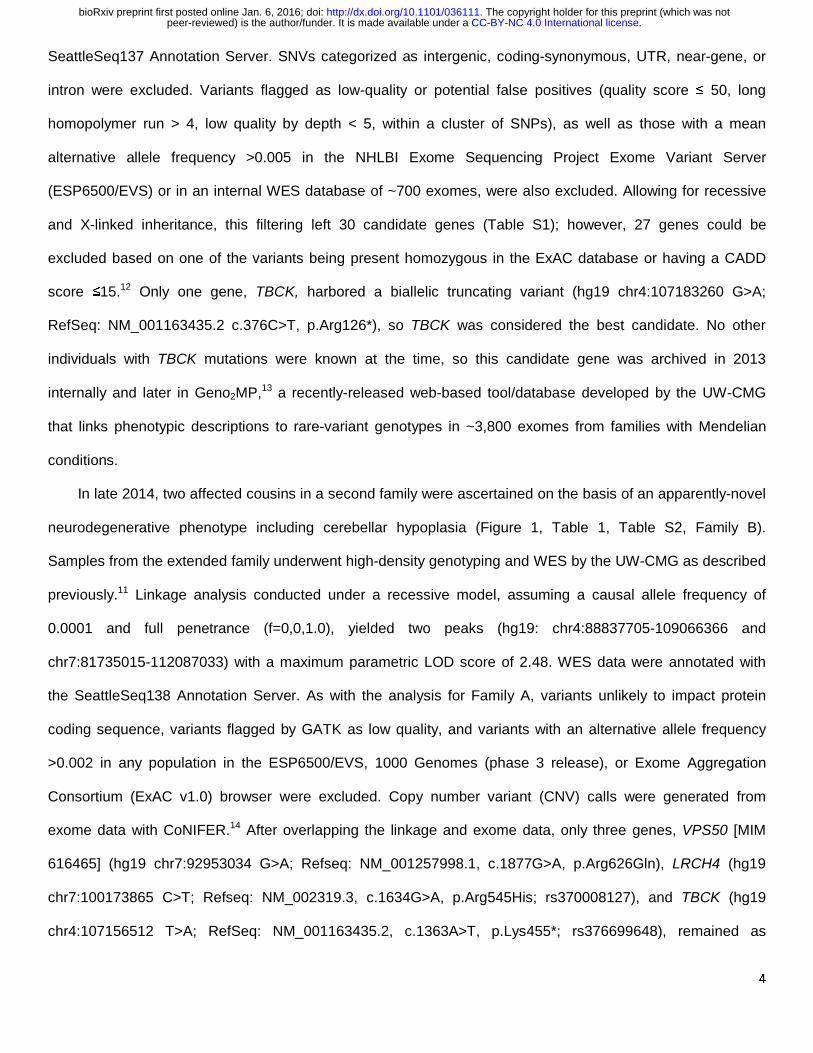
SeattleSeq137 Annotation Server. SNVs categorized as intergenic, coding-synonymous, UTR, near-gene, or
intron were excluded. Variants flagged as low-quality or potential false positives (quality score ≤ 50, long
homopolymer run > 4, low quality by depth < 5, within a cluster of SNPs), as well as those with a mean
alternative allele frequency >0.005 in the NHLBI Exome Sequencing Project Exome Variant Server
(ESP6500/EVS) or in an internal WES database of ~700 exomes, were also excluded. Allowing for recessive
and X-linked inheritance, this filtering left 30 candidate genes (Table S1); however, 27 genes could be
excluded based on one of the variants being present homozygous in the ExAC database or having a CADD
score ≤15.12 Only one gene, TBCK, harbored a biallelic truncating variant (hg19 chr4:107183260 G>A;
RefSeq: NM_001163435.2 c.376C>T, p.Arg126*), so TBCK was considered the best candidate. No other
individuals with TBCK mutations were known at the time, so this candidate gene was archived in 2013
internally and later in Geno2MP,13 a recently-released web-based tool/database developed by the UW-CMG
that links phenotypic descriptions to rare-variant genotypes in ~3,800 exomes from families with Mendelian
conditions.
In late 2014, two affected cousins in a second family were ascertained on the basis of an apparently-novel
neurodegenerative phenotype including cerebellar hypoplasia (Figure 1, Table 1, Table S2, Family B).
Samples from the extended family underwent high-density genotyping and WES by the UW-CMG as described
previously.11 Linkage analysis conducted under a recessive model, assuming a causal allele frequency of
0.0001 and full penetrance (f=0,0,1.0), yielded two peaks (hg19: chr4:88837705-109066366 and
chr7:81735015-112087033) with a maximum parametric LOD score of 2.48. WES data were annotated with
the SeattleSeq138 Annotation Server. As with the analysis for Family A, variants unlikely to impact protein
coding sequence, variants flagged by GATK as low quality, and variants with an alternative allele frequency
>0.002 in any population in the ESP6500/EVS, 1000 Genomes (phase 3 release), or Exome Aggregation
Consortium (ExAC v1.0) browser were excluded. Copy number variant (CNV) calls were generated from
exome data with CoNIFER.14 After overlapping the linkage and exome data, only three genes, VPS50 [MIM
616465] (hg19 chr7:92953034 G>A; Refseq: NM_001257998.1, c.1877G>A, p.Arg626Gln), LRCH4 (hg19
chr7:100173865 C>T; Refseq: NM_002319.3, c.1634G>A, p.Arg545His; rs370008127), and TBCK (hg19
chr4:107156512 T>A; RefSeq: NM_001163435.2, c.1363A>T, p.Lys455*; rs376699648), remained as
.CC-BY-NC 4.0 International licensepeer-reviewed) is the author/funder. It is made available under aThe copyright holder for this preprint (which was not. http://dx.doi.org/10.1101/036111doi: bioRxiv preprint first posted online Jan. 6, 2016;

5
candidates. All three genes were submitted to GeneMatcher and Geno2MP. No matches were found for VPS50
or LRCH4 in either database, but Geno2MP yielded a single individual (Figure 1 and Table 1, Family A) with
“cerebellar malformation” who was homozygous for a nonsense variant in TBCK. Sharing of the same
candidate gene and an overlapping phenotype suggested that the putative loss of function mutations in TBCK
were causal.
In parallel, a third family was ascertained due to profound developmental disability associated with brain
atrophy before two years of age in a son, and death of a 12 month old daughter exhibiting similar features
(Figure 1, Table 1, and Table S2, Family C). WES was performed on a sample from the affected son (Family
C-II-1). Variants were called using an in-house pipeline,15-17 and filtered against public and in-house databases,
retaining only clinically-associated variants and variants with MAF <0.001 (dbSNP142), <0.002 (ExAC), <0.01
(in-house database, ~500 exomes), or unknown MAF. The SnpEff toolbox (v4.1) was used to predict the
impact of variants, which were filtered to retain only functionally relevant variants (i.e., missense, nonsense,
coding indel variants, and intronic variants located from −5 to +5 with respect to an exon-intron junction).
Functional annotation of variants was performed using SnpEff v4.1 and dbNSFP2.8. Parental first cousin
consanguinity and the death of a female sibling with similar clinical features (i.e., suggestive facies, severe
developmental delay, generalized hypotonia and congenital heart malformation) strongly suggested autosomal
recessive inheritance, and based on the hypothesis of homozygosity by descent, 16 candidate genes were
identified. These candidates were stratified through a mixed filtering/prioritization strategy designed to retain
genes with predicted-damaging variants (CADD_phred > 15), ranked on the basis of their biological relevance
on the developmental processes altered in the disorder (GeneDistiller) (see Table S1).18 Among the 8 retained
genes, TBCK (hg19 chr4:107154202 C>T; RefSeq: NM_001163435.2 c.1532G>A, p.Arg511His) received the
highest score from GeneDistiller and was considered the best candidate. At this point, M.T. contacted M.J.B.
and inquired whether TBCK was a candidate gene for any phenotypes being studied at the UW-CMG.
Another individual of Puerto Rican descent (Figures 1 and 2, Family D II-1) with severe hypotonia, chronic
respiratory insufficiency requiring nighttime BiPAP (Bilevel Positive Airway Pressure), brain atrophy, and
similar facial features (Family D-II-1 in Figure 1) was also ascertained in parallel. Clinical exome sequencing
identified the same homozygous p.Arg126* variant present in Family A-II-1, supporting the existence of a
.CC-BY-NC 4.0 International licensepeer-reviewed) is the author/funder. It is made available under aThe copyright holder for this preprint (which was not. http://dx.doi.org/10.1101/036111doi: bioRxiv preprint first posted online Jan. 6, 2016;

6
founder mutation in the Puerto Rican population. The phenotypic features in Family D-II-1 are quite similar to
the other affected individuals (Tables 1, S2, and S3).
The variants in each family were either rare or not present in reference population databases. Specifically,
the maximum alternate allele frequencies in any population from EVS, 1000 Genomes phase 3, or ExAC v0.3
was 0.5208% for p.Arg126* (Families A and D) and 0.0107% for p.Lys455* (Family B) in ExAC Latinos, while
p.Arg511His (Family C) was not present in any database. Each variant had a high CADD score:12 37.0 for
p.Arg126*, 42.0 for p.Lys455*, and 35.0 for p.Arg511His.
All affected individuals displayed profound developmental disability, making little progress beyond infancy,
and a relatively homogeneous phenotype (Table 1, Figure 1). Additionally, Family A-II-1 and Family B-IV-4 and
IV-6 exhibited developmental regression (loss of visual fixation and following). All four individuals also required
gastrostomy tube feedings and had decreased respiratory drive, with the three older children requiring
tracheostomies for chronic mechanical ventilation. Family A-II-1, Family B-IV-4 and Family B-IV-6 also
developed focal seizures between 2 and 6 years of age, and Family C-II-1 had an abnormal
electroencephalogram before 2 years of age without clinical seizures. Family A-II-1, Family B-IV-4 and Family
C-II-1 had decreased to absent reflexes, while Family B-IV-6 developed hyperreflexia. Shared facial features
included bitemporal narrowing, arched eyebrows, deep set eyes, high nasal bridge, anteverted nares, and an
exaggerated “Cupid's bow” of the upper lip (Figure 1D-H). Family A-II-1 and Family B-IV-6 developed
osteoporosis, which could be either a non-specific effect of chronic, severe disability or a more specific feature
of TBCK-related disease. At three years of age, Family A-II-1 had an acute neurological decompensation with
restricted diffusion of the right parieto-occipital cortex, indicative of a metabolic stroke, from which he
recovered to his baseline neurological status. In all four individuals, brain imaging revealed increased
ventricular and extra-axial spaces and diffusely decreased white matter volume, with atrophy confirmed by
serial MRI in Family A-II-1 and Family B-IV-4 (Figure 2 and Table S3). Despite the loss of brain volume,
microcephaly was not observed. In addition, all individuals have corpus callosum dysgenesis (thinning, partial
agenesis or both), increased T2/FLAIR signal in the periventricular white matter, and cerebellar vermis
hypoplasia.
.CC-BY-NC 4.0 International licensepeer-reviewed) is the author/funder. It is made available under aThe copyright holder for this preprint (which was not. http://dx.doi.org/10.1101/036111doi: bioRxiv preprint first posted online Jan. 6, 2016;

7
TBCK encodes a protein with a TBC (Tre-2, Bub2, and Cdc16) domain flanked by an N-terminal kinase-
like domain and a rhodanese homology domain at the C-terminus, but its function has not been extensively
characterized. Due to the lack of key catalytic residues, the kinase domain is presumably inactive.19 Multiple
TBCK mRNAs have been reported with two major isoforms differing in the presence or absence of the N-
terminal portion that encodes the kinase-like domain (Figure 3A).20 To characterize the impact of the truncating
mutations, the TBCK protein level was evaluated by western blot using skin fibroblasts obtained from an
individual who was homozygous for p.Arg126*, Family A-II-1 (Figure 3B). As expected, two major bands at
~101 and 71 kD, representing respectively the long (full-length) protein, and the shorter isoform without the N-
terminal kinase-like domain,20 were observed in two control fibroblast lines, with the full-length isoform
representing the more abundant product. In contrast, the full-length protein was nearly absent in the fibroblasts
from Family A-II-1, and the 71 kD isoform was also reduced. While the transcript for the 71kD isoform is not
directly affected by the mutation, the reduced protein level indicates that the mutation perturbs the levels of
both major TBCK isoforms in fibroblasts.
The c.1532G>A mutation in Family C-II-1 is predicted to alter a highly conserved arginine residue located
in the TBC1 domain (Figure 3C). This structural unit of approximately 200 amino acids is characteristic of most
GAPs that regulate the Rab family of small GTPases.21,22 TBC domains negatively control Rab function by
promoting GTP hydrolysis via stabilization of the transition state of the reaction. Specifically, the TBC domain
interacts with the substrate, and catalyzes the reaction by using a so-called “dual finger” mechanism, in which
a key Arg residue projects into the Rab active site.23-25 Since no crystallographic structure of TBCK was
available, a homology model of the TBC1 domain of TBCK (residues 467-648) in complex with a Rab protein
was constructed to explore the structural impact of the disease-causing amino acid substitution. This model
was generated using DeepView and the SwissModel server,26 based on the crystallographic structure of the
Gyp1 TBC domain (36% sequence identity with the TBCK TBC domain) complexed with Rab33 bound to GDP
and AlF3 (PDB code 2G77).23 TBCK was originally incorrectly classified as an unconventional TBC protein
lacking the “arginine finger”;21 however, our modeling data support more recent bioinformatics analyses21
indicating that Arg511 does in fact represent an arginine finger essential for GAP function. The p.Arg511His
substitution was introduced in silico with UCSF Chimera, 27 and even though the Arg-to-His substitution
.CC-BY-NC 4.0 International licensepeer-reviewed) is the author/funder. It is made available under aThe copyright holder for this preprint (which was not. http://dx.doi.org/10.1101/036111doi: bioRxiv preprint first posted online Jan. 6, 2016;

8
introduces a side chain that could maintain the overall positive charge under appropriate conditions,28 the
introduced residue is significantly bulkier and shorter than Arg, and unable to project into the GTPase active
site. In the model, the minimum distance between Arg511 and the GDP phosphate is 3 Å, while this distance
increases to 7 Å in the mutant. These considerations strongly point to impaired GAP activity as the predicted
mechanism of disease associated with the p.Arg511His substitution.
TBCK was recently documented to play a role in the control of cell proliferation, cell size, and actin
cytoskeleton dynamics.19,20 While the lack of several residues with critical function in mediating the activity of
the kinase domain supports the view that TBCK is catalytically inactive,29,30 the conservation of the key
catalytic residues of the TBC1 domain, as well as the identification of a disease-causing mutation specifically
targeting one of the two key-residues of this domain (Arg511), point to the relevance of the Rab-GAP activity for
TBCK function, even though its physiological targets have not been identified.19 Indeed, the “Arg finger” is
conserved in virtually all TBC domains with GAP activity,21,22,25 and it is largely accepted that unconventional
TBC domains lacking this residue do not stimulate GTP hydrolysis in Rab proteins but have different
functions.21,22,25 Of note, one of the human unconventional TBC proteins (TBC1D26) and its Chlamydomonas
reinhardtii orthologue have a histidine residue in place of the “catalytic” arginine, and no GAP activity was
detected in the algal protein.31 Mutation of the “catalytic” Arg in TBC domain-containing proteins is commonly
used as a tool to investigate their function and identify their substrate Rabs,22,32-35 since substitution of this
amino acid greatly reduces the catalytic efficiency of these GAPs.14
TBCK’s role in controlling cell proliferation/growth and actin cytoskeleton organization has been reported
to be mediated by modulation of the mTOR signaling network and transcriptional regulation of components of
the mTOR complex.19 The mTOR pathway is a growth-regulating network, which in an activated state
promotes angiogenesis, cell growth, and cell proliferation. A number of developmental brain disorders,
collectively termed “TORopathies”, have been shown to result from dysregulated mTOR signaling.36,37 These
disorders are characterized by disorganized cortical lamination, seizures and cytomegaly.38 Tuberous sclerosis
complex (TSC) [MIM PS191100] is caused by heterozygous loss of function mutations in TSC1 [MIM 605284]
or TSC2 [MIM 191092], encoding proteins that negatively regulate the mTOR pathway. Similarly, germline or
postzygotic activating de novo mutations in components of the PI3K-AKT3-mTOR pathway cause the
.CC-BY-NC 4.0 International licensepeer-reviewed) is the author/funder. It is made available under aThe copyright holder for this preprint (which was not. http://dx.doi.org/10.1101/036111doi: bioRxiv preprint first posted online Jan. 6, 2016;

9
overlapping hemimegalencephaly phenotypes megalencephaly-capillary malformation-polymicrogyria [MIM
602501] (usually caused by somatic mutations in PIK3CA [MIM 171834]) and megalencephaly-polymicrogyria-
polydactyly-hydrocephalus [MIM PS603387] (usually caused by germline mutations in PIK3R2 [MIM 603157],
CCND2 [MIM 123833, and AKT3 [MIM 611223]), in addition to their well-documented role in cancer. More
recently, germline gain-of-function mutations in MTOR [MIM 601231] have been described in two families with
features overlapping the megalencephaly and RASopathy spectrum of disorders.39,40 These TORopathies are
all caused by activation of the mTOR pathway and generally characterized by brain overgrowth at a global
(hemimegalencephaly spectrum) or focal level (TSC). In contrast, loss of TBCK function is associated with loss
of brain volume. In combination with available biochemical and functional data supporting a positive
modulatory role of TBCK on mTOR signaling,19 our findings suggest that loss of TBCK function has an
opposite effect on this signaling network compared to what is observed in other mTOR-related disorders, and
importantly, that over-inhibition of the mTOR pathway may lead to this distinct and severe phenotype. Some of
the affected individuals were found to have moderate mitochondrial dysfunction which did not meet Walker
diagnostic criteria41 for a primary respiratory chain disorder and was not associated with increased brain lactate
in the three individuals evaluated by magnetic resonance spectroscopy (Table S3). Given that the mTOR
pathway is known to positively affect mitochondrial biogenesis42 and negatively regulate autophagy/mitophagy
(reviewed in Bockaert and Marin 201543) and that several individuals have decreased mitochondrial enzyme
levels, we hypothesize that the progression of the disease may be due, in part, to decreased mitochondrial
biogenesis and/or increased mitophagy.
In summary, we establish that biallelic mutations in TBCK cause a severe neurodevelopmental disorder
whose major features include profound developmental delay/cognitive deficit, brain atrophy without
microcephaly, dysgenesis of the corpus callosum, white matter signal abnormality, cerebellar vermis
hypoplasia, seizures, diminished respiratory function, and distinctive facies. The recent report of a similar
individual exhibiting severe developmental disability, poor feeding, abnormal eye movements, epilepsy, facial
dysmorphism, severe hypotonia, and diffuse brain atrophy associated with a homozygous canonical splice-site
mutation (NM_001163435.1 c.1897+1G>A) in TBCK44 further strengthens this view. Structural and molecular
modeling analyses predict a key role for the substituted residue in mediating the Rab-GAP activity of the
.CC-BY-NC 4.0 International licensepeer-reviewed) is the author/funder. It is made available under aThe copyright holder for this preprint (which was not. http://dx.doi.org/10.1101/036111doi: bioRxiv preprint first posted online Jan. 6, 2016;

10
protein, suggesting that loss of TBCK GAP function is sufficient to cause disease. Of note, many
encephalopathies are caused by de novo dominant mutations and therefore have low risk of recurrence, so
distinguishing TBCK-related encephalopathy is essential for accurate recurrence risk counseling and
reproductive planning. Recognition of this disorder will also make it possible to delineate the natural history of
TBCK-related disease, and provide more precise prognostic information that is essential to guide decisions
about invasive treatments such as tracheostomy and gastrostomy in neonates and young children.
More than three years passed between the identification of TBCK as the strongest candidate gene in
Family A and the identification of TBCK mutations in Families B, C, and D. Importantly, even though two
families were sequenced through the same center (University of Washington Center for Mendelian Genomics),
their shared phenotype was not initially recognized, in part because the clinicians rightfully focused on different
aspects of each affected individual’s condition. The identification of a shared candidate gene by searching
Geno2MP prompted comparison of the clinical findings and led to delineation of an overlapping phenotype. It is
anticipated that matchmaking platforms in development, such as Matchmaker Exchange,45 will help to speed
up this process, but only if investigators and clinicians worldwide all participate in such data sharing efforts.
Historically, ascertaining persons with a highly similar phenotype and then conducting subsequent gene
discovery within that group has been a successful method for understanding new Mendelian phenotypes;
however, this approach requires the Mendelian phenotype to be common enough that a single investigator
could be expected to encounter multiple affected individuals. The small number of individuals with biallelic
mutations in TBCK identified thus far, and the international assemblage of investigators who identified these
individuals, underscores the fact that many, if not most, Mendelian phenotypes now being studied will be more
rapidly and successfully tackled by widespread sharing of phenotypes, genotypes, and candidate genes.13,45,46
Supplemental Data
Supplemental data consists of 3 tables.
Acknowledgments
We thank the families for their participation and support. Our work was supported in part by grants from the
National Institutes of Health: National Human Genome Research Institute and the National Heart, Lung and
.CC-BY-NC 4.0 International licensepeer-reviewed) is the author/funder. It is made available under aThe copyright holder for this preprint (which was not. http://dx.doi.org/10.1101/036111doi: bioRxiv preprint first posted online Jan. 6, 2016;
11
Blood Institute (1U54HG006493 to M.B., D.N., and J.S.; 1RC2HG005608 to M.B., D.N., and J.S.), the National
Institute of Neurological Diseases and Stroke (K12NS049453-09 to X.O.G.), the Eunice Kennedy Shriver
National Institute of Child Health and Human Development (U54HD083091, Genetics Core and Sub-project
6849 to D.D.), Telethon-Italy (GGP13107 to M.T.) and the Ospedale Pediatrico Bambino Gesù (GeneRare to
M.T.). The authors would like to thank the University of Washington Center for Mendelian Genomics and all
contributors to Geno2MP for use of data included in Geno2MP. The authors would also like to thank the Exome
Aggregation Consortium and the groups that provided exome variant data for comparison. A full list of
contributing groups can be found at http://exac.broadinstitute.org/about.
Web Resources The URLs for data presented herein are as follows: Exome Variant Server (NHLBI Exome Sequencing Project ESP6500): http://evs.gs.washington.edu/EVS/. Exome Aggregation Consortium (ExAC), Cambridge, MA: http://exac.broadinstitute.org (accessed April 2015). Geno2MP, NHGRI/NHLBI University of Washington-Center for Mendelian Genomics (UW-CMG), Seattle, WA: http://geno2mp.gs.washington.edu (accessed April 2015) GeneDistiller 2014: http://www.genedistiller.org/ Online Mendelian Inheritance in Man: http://www.omim.org References
1. Lalani, S.R., Zhang, J., Schaaf, C.P., Brown, C.W., Magoulas, P., Tsai, A.C.-H., El-Gharbawy, A., Wierenga, K.J., Bartholomew, D., Fong, C.-T., et al. (2014). Mutations in PURA cause profound neonatal hypotonia, seizures, and encephalopathy in 5q31.3 microdeletion syndrome. Am J Hum Genet 95, 579–583.
2. Hunt, D., Leventer, R.J., Simons, C., Taft, R., Swoboda, K.J., Gawne-Cain, M., DDD Study, Magee, A.C., Turnpenny, P.D., and Baralle, D. (2014). Whole exome sequencing in family trios reveals de novo mutations in PURA as a cause of severe neurodevelopmental delay and learning disability. J Med Genet 51, 806–813.
3. Perez, Y., Kadir, R., Volodarsky, M., Noyman, I., Flusser, H., Shorer, Z., Gradstein, L., Birnbaum, R.Y., and Birk, O.S. (2015). UNC80 mutation causes a syndrome of hypotonia, severe intellectual disability, dyskinesia and dysmorphism, similar to that caused by mutations in its interacting cation channel NALCN. J Med Genet jmedgenet–2015–103352.
4. Stray-Pedersen, A., Cobben, J.-M., Prescott, T.E., Lee, S., Cang, C., Aranda, K., Ahmed, S., Alders, M., Gerstner, T., Aslaksen, K., et al. (2015). Biallelic Mutations in UNC80 Cause Persistent Hypotonia, Encephalopathy, Growth Retardation, and Severe Intellectual Disability. Am J Hum Genet 1–8.
5. Shamseldin, H.E., Faqeih, E., Alasmari, A., Zaki, M.S., Gleeson, J.G., and Alkuraya, F.S. (2015). Mutations in UNC80, Encoding Part of the UNC79-UNC80-NALCN Channel Complex, Cause Autosomal-Recessive
.CC-BY-NC 4.0 International licensepeer-reviewed) is the author/funder. It is made available under aThe copyright holder for this preprint (which was not. http://dx.doi.org/10.1101/036111doi: bioRxiv preprint first posted online Jan. 6, 2016;

12
Severe Infantile Encephalopathy. Am J Hum Genet 1–6.
6. Chong, J.X., Mcmillin, M.J., Shively, K.M., Beck, A.E., Marvin, C.T., Armenteros, J.R., Buckingham, K.J., Nkinsi, N.T., Boyle, E.A., Berry, M.N., et al. (2015). De Novo Mutations in NALCN Cause a Syndrome Characterized by Congenital Contractures of the Limbs and Face, Hypotonia, and Developmental Delay. Am J Hum Genet 96, 462–473.
7. Koroglu, C., Seven, M., and Tolun, A. (2013). Recessive truncating NALCN mutation in infantile neuroaxonal dystrophy with facial dysmorphism. J Med Genet 50, 515–520.
8. Al-Sayed, M.D., Al-Zaidan, H., Albakheet, A., Hakami, H., Kenana, R., Al-Yafee, Y., Al-Dosary, M., Qari, A., Al-Sheddi, T., Al-Muheiza, M., et al. (2013). Mutations in NALCN Cause an Autosomal-Recessive Syndrome with Severe Hypotonia, Speech Impairment, and Cognitive Delay. Am J Hum Genet 93, 721–726.
9. Tan, S., and Wu, Y. (2015). Etiology and pathogenesis of neonatal encephalopathy. In UpToDate, T.W. Post, ed. (Waltham, MA).
10. Bachmann-Gagescu, R., Dempsey, J.C., Phelps, I.G., O'Roak, B.J., Knutzen, D.M., Rue, T.C., Ishak, G.E., Isabella, C.R., Gorden, N., Adkins, J., et al. (2015). Joubert syndrome: a model for untangling recessive disorders with extreme genetic heterogeneity. J Med Genet 52, 514–522.
11. Chong, J.X., Burrage, L.C., Beck, A.E., Marvin, C.T., Bacino, C.A., Jain, M., Alanay, Y., Berry, S.A., Carey, J.C., Gibbs, R.A., et al. (2015). Autosomal-Dominant Multiple Pterygium Syndrome Is Caused by Mutations in MYH3. Am J Hum Genet 96, 841–849.
12. Kircher, M., Witten, D.M., Jain, P., O'Roak, B.J., Cooper, G.M., and Shendure, J. (2014). A general framework for estimating the relative pathogenicity of human genetic variants. Nat Genet 46, 310–315.
13. Chong, J.X., Buckingham, K.J., Jhangiani, S.N., Boehm, C., Sobreira, N., Smith, J.D., Harrell, T.M., Mcmillin, M.J., Wiszniewski, W., Gambin, T., et al. (2015). The Genetic Basis of Mendelian Phenotypes: Discoveries, Challenges, and Opportunities. Am J Hum Genet 97, 199–215.
14. Krumm, N., Sudmant, P.H., Ko, A., O'Roak, B.J., Malig, M., Coe, B.P., NHLBI Exome Sequencing Project, Quinlan, A.R., Nickerson, D.A., and Eichler, E.E. (2012). Copy number variation detection and genotyping from exome sequence data. Genome Res 22, 1525–1532.
15. Niceta, M., Stellacci, E., Gripp, K.W., Zampino, G., Kousi, M., Anselmi, M., Traversa, A., Ciolfi, A., Stabley, D., Bruselles, A., et al. (2015). Mutations Impairing GSK3-Mediated MAF Phosphorylation Cause Cataract, Deafness, Intellectual Disability, Seizures, and a Down Syndrome-like Facies. Am J Hum Genet.
16. Cordeddu, V., Redeker, B., Stellacci, E., Jongejan, A., Fragale, A., Bradley, T.E.J., Anselmi, M., Ciolfi, A., Cecchetti, S., Muto, V., et al. (2014). Mutations in ZBTB20 cause Primrose syndrome. Nat Genet 46, 815–817.
17. Kortüm, F., Caputo, V., Bauer, C.K., Stella, L., Ciolfi, A., Alawi, M., Bocchinfuso, G., Flex, E., Paolacci, S., Dentici, M.L., et al. (2015). Mutations in KCNH1 and ATP6V1B2 cause Zimmermann-Laband syndrome. Nat Genet 47, 661–667.
18. Seelow, D., Schwarz, J.M., and Schuelke, M. (2008). GeneDistiller--distilling candidate genes from linkage intervals. PLoS ONE 3, e3874.
19. Liu, Y., Yan, X., and Zhou, T. (2013). TBCK influences cell proliferation, cell size and mTOR signaling pathway. PLoS ONE 8, e71349.
20. Wu, J., Li, Q., Li, Y., Lin, J., Yang, D., Zhu, G., Wang, L., He, D., Lu, G., and Zeng, C. (2014). A Long Type of TBCK Is a Novel Cytoplasmic and Mitotic Apparatus-Associated Protein Likely Suppressing Cell
.CC-BY-NC 4.0 International licensepeer-reviewed) is the author/funder. It is made available under aThe copyright holder for this preprint (which was not. http://dx.doi.org/10.1101/036111doi: bioRxiv preprint first posted online Jan. 6, 2016;

13
Proliferation. Journal of Genetics and Genomics 41, 69–72.
21. Frasa, M.A.M., Koessmeier, K.T., Ahmadian, M.R., and Braga, V.M.M. (2012). Illuminating the functional and structural repertoire of human TBC/RABGAPs. Nat Rev Mol Cell Biol 13, 67–73.
22. Fukuda, M. (2011). TBC proteins: GAPs for mammalian small GTPase Rab? Biosci. Rep. 31, 159–168.
23. Pan, X., Eathiraj, S., Munson, M., and Lambright, D.G. (2006). TBC-domain GAPs for Rab GTPases accelerate GTP hydrolysis by a dual-finger mechanism. Nature 442, 303–306.
24. Gavriljuk, K., Gazdag, E.-M., Itzen, A., Kötting, C., Goody, R.S., and Gerwert, K. (2012). Catalytic mechanism of a mammalian Rab·RabGAP complex in atomic detail. Proceedings of the National Academy of Sciences 109, 21348–21353.
25. Cherfils, J., and Zeghouf, M. (2013). Regulation of small GTPases by GEFs, GAPs, and GDIs. Physiol. Rev. 93, 269–309.
26. Biasini, M., Bienert, S., Waterhouse, A., Arnold, K., Studer, G., Schmidt, T., Kiefer, F., Cassarino, T.G., Bertoni, M., Bordoli, L., et al. (2014). SWISS-MODEL: modelling protein tertiary and quaternary structure using evolutionary information. Nucleic Acids Res. 42, W252–W258.
27. Pettersen, E.F., Goddard, T.D., Huang, C.C., Couch, G.S., Greenblatt, D.M., Meng, E.C., and Ferrin, T.E. (2004). UCSF Chimera--a visualization system for exploratory research and analysis. J Comput Chem 25, 1605–1612.
28. Creighton, T.E. (1993). Proteins (W.H. Freeman & Company).
29. Boudeau, J., Miranda-Saavedra, D., Barton, G.J., and Alessi, D.R. (2006). Emerging roles of pseudokinases. Trends in Cell Biology 16, 443–452.
30. Scheeff, E.D., Eswaran, J., Bunkoczi, G., Knapp, S., and Manning, G. (2009). Structure of the pseudokinase VRK3 reveals a degraded catalytic site, a highly conserved kinase fold, and a putative regulatory binding site. Structure 17, 128–138.
31. Bhogaraju, S., and Lorentzen, E. (2014). Crystal structure of a Chlamydomonas reinhardtii flagellar RabGAP TBC-domain at 1.8 Å resolution. Proteins 82, 2282–2287.
32. Liegel, R.P., Handley, M.T., Ronchetti, A., Brown, S., Langemeyer, L., Linford, A., Chang, B., Morris-Rosendahl, D.J., Carpanini, S., Posmyk, R., et al. (2013). Loss-of-function mutations in TBC1D20 cause cataracts and male infertility in blind sterile mice and Warburg micro syndrome in humans. Am J Hum Genet 93, 1001–1014.
33. Chotard, L., Mishra, A.K., Sylvain, M.-A., Tuck, S., Lambright, D.G., and Rocheleau, C.E. (2010). TBC-2 regulates RAB-5/RAB-7-mediated endosomal trafficking in Caenorhabditis elegans. Mol. Biol. Cell 21, 2285–2296.
34. Chadt, A., Leicht, K., Deshmukh, A., Jiang, L.Q., Scherneck, S., Bernhardt, U., Dreja, T., Vogel, H., Schmolz, K., Kluge, R., et al. (2008). Tbc1d1 mutation in lean mouse strain confers leanness and protects from diet-induced obesity. Nat Genet 40, 1354–1359.
35. Haas, A.K., Fuchs, E., Kopajtich, R., and Barr, F.A. (2005). A GTPase-activating protein controls Rab5 function in endocytic trafficking. Nature Cell Biology 7, 887–893.
36. Smith, L.D., Saunders, C.J., Dinwiddie, D.L., Atherton, A.M., Miller, N.A., Soden, S.E., Farrow, E.G., Abdelmoity, A.T.G., and Kingsmore, S.F. (2013). Exome Sequencing Reveals De Novo Germline Mutation of
.CC-BY-NC 4.0 International licensepeer-reviewed) is the author/funder. It is made available under aThe copyright holder for this preprint (which was not. http://dx.doi.org/10.1101/036111doi: bioRxiv preprint first posted online Jan. 6, 2016;
14
the Mammalian Target of Rapamycin (MTOR) in a Patient with Megalencephaly and Intractable Seizures. Jge 2, 63–72.
37. Crino, P.B. (2011). mTOR: A pathogenic signaling pathway in developmental brain malformations. Trends in Molecular Medicine 17, 734–742.
38. Crino, P.B. (2009). Focal brain malformations: seizures, signaling, sequencing. Epilepsia 50 Suppl 9, 3–8.
39. Baynam, G., Overkov, A., Davis, M., Mina, K., Schofield, L., Allcock, R., Laing, N., Cook, M., Dawkins, H., and Goldblatt, J. (2015). A germline MTOR mutation in Aboriginal Australian siblings with intellectual disability, dysmorphism, macrocephaly, and small thoraces. Am J Med Genet A 167, 1659–1667.
40. Smith, L., Saunders, C., Dinwiddie, D., Atherton, A., Soden, S., Farrow, E., Abdelmoity, A., and Kingsmore, S. (2013). Exome Sequencing Reveals De Novo Germline Mutation of the Mammalian Target of Rapamycin (MTOR) in a Patient with Megalencephaly and Intractable Seizures. Jge 2, 63–72.
41. Walker, U.A., Collins, S., and Byrne, E. (1996). Respiratory chain encephalomyopathies: a diagnostic classification. Eur. Neurol. 36, 260–267.
42. Cunningham, J.T., Rodgers, J.T., Arlow, D.H., Vazquez, F., Mootha, V.K., and Puigserver, P. (2007). mTOR controls mitochondrial oxidative function through a YY1-PGC-1 alpha transcriptional complex. Nature 450, 736–740.
43. Bockaert, J., and Marin, P. (2015). mTOR in Brain Physiology and Pathologies. Physiol. Rev. 95, 1157–1187.
44. Alazami, A.M., Patel, N., Shamseldin, H.E., Anazi, S., Al-Dosari, M.S., Alzahrani, F., Hijazi, H., Alshammari, M., Aldahmesh, M.A., Salih, M.A., et al. (2015). Accelerating Novel Candidate Gene Discovery in Neurogenetic Disorders via Whole-Exome Sequencing of Prescreened Multiplex Consanguineous Families. Cell Rep 10, 148–161.
45. Philippakis, A.A., Azzariti, D.R., Beltran, S., Brookes, A.J., Brownstein, C.A., Brudno, M., Brunner, H.G., Buske, O.J., Carey, K., Doll, C., et al. (2015). The Matchmaker Exchange: A Platform for Rare Disease Gene Discovery. Hum Mutat 36, 915–921.
46. Krawitz, P., Buske, O., Zhu, N., Brudno, M., and Robinson, P.N. (2015). The Genomic Birthday Paradox: How Much Is Enough? Hum Mutat 36, 989–997.
.CC-BY-NC 4.0 International licensepeer-reviewed) is the author/funder. It is made available under aThe copyright holder for this preprint (which was not. http://dx.doi.org/10.1101/036111doi: bioRxiv preprint first posted online Jan. 6, 2016;

15
Figure Legends
Figure 1. Pedigrees and pictures of individuals with TBCK-related encephalopathy (photographs
removed at request of bioRxiv).
(A-D) Pedigrees for four families segregating loss of function mutations in TBCK. Solid black fill indicates
individuals affected with TBCK-related encephalopathy. Light blue fill in Family B indicates individuals affected
by a blood-related phenotype without neurodevelopmental features; neither was homozygous for a mutation in
TBCK. WES was performed on individuals marked with *’s. (E-J) Similar facial features and hypotonia in
Family A-II-1 at 25 months (E) and 13 years (F), Family B-IV-4 at 17 months (G) and 4 years 3 months (H),
Family B-IV-6 at 18 months (I), Family C-II-1 at 21 months (J), and Family D-II-1 at 14 years. See Table 1 for
detailed clinical information for each affected individual and Figure 2 and Table S3 for imaging information.
Figure 2. Brain imaging features in individuals with TBCK-related encephalopathy
(A-D) Progressive gray and white matter volume loss, most severe in the frontal lobes, demonstrated by
increasing ventriculomegaly and extraxial spaces (cortical and cerebellar) in Family A-II-1 between 22 days of
age (A-B) and 29 months of age (C-D). Swelling of the right parieto-occipital lobe, presumably due to a
metabolic stroke (bracket in C), diffusely thin corpus callosum with absent rostrum (arrowhead in B and D), and
mild cerebellar vermis hypoplasia with relatively spared brainstem are also present. (E-F) Ventriculomegaly,
prominent extraxial spaces, diffusely thin but complete corpus callosum, and mild cerebellar vermis hypoplasia
in Family B-IV-4 at 18 months of age. Right plagiocephaly is also present. (G-H) Marked ventriculomegaly,
prominent extraxial spaces, diffusely thin but complete corpus callosum, and mild cerebellar vermis hypoplasia
in Family B-IV-6 at 6 years of age. Synechiae are also present in the right frontal horn (asterisks in G). (I-J)
Ventriculomegaly, prominent extraxial spaces, diffusely thin corpus callosum with absent rostrum and anterior
body (arrowhead in J), and mild cerebellar vermis hypoplasia in Family C-II-1 at 21 months of age. (K-L) Mild
ventriculomegaly, prominent extraxial spaces, diffusely thin corpus callosum, mild cerebellar vermis hypoplasia
with relatively preserved brainstem, and thick frontal bone (arrow in K) in Family D-II-1 at 14 years of age. (A,
.CC-BY-NC 4.0 International licensepeer-reviewed) is the author/funder. It is made available under aThe copyright holder for this preprint (which was not. http://dx.doi.org/10.1101/036111doi: bioRxiv preprint first posted online Jan. 6, 2016;

16
C, E, G, K) axial T2-weighted images, (I) axial T1-weighted image, (B, D, F, H, J, L) sagittal T1-weighted
images.
Figure 3. Biochemical and structural characterization of TBCK mutations. (A) TBCK encodes two
isoforms containing a TBC1 domain (TBC) flanked by a rhodanese domain (RHOD) at the C-terminus. The
long isoform also contains a pseudokinase domain (STYKc) at the N-terminus. Location of the identified
mutations is reported. (B) Western blot showing dramatically reduced levels of TBCK in cell line from Family A-
II-1 versus two control cell lines. The TBCK monoclonal antibody (1/250, Sigma HPA039951) recognizes a C-
terminal fragment of the protein depicted as a red line in (A). Following stripping, β-actin (1/5,000, Sigma
A5441) was used as a loading control. Predicted sizes of TBCK long and short isoforms are 101kD and 71kD
respectively. (C) Conservation of the catalytic arginine “finger” (Arg511 in TBCK) in TBCK orthologs. (D)
Homology model of the TBCK TBC1 domain complexed with the Rab33 GTPase. In the overall structure of the
complex (left), both proteins are shown in a surface representation, with Rab colored in pink, GDP in gray, the
TBC1 domain in light blue and Arg511 in blue. In the enlarged view of the active site of the GTPase (right), GDP
is reported in gray sticks, with the phosphates colored in orange. AlF3, which in the crystal structure mimics the
transition state for GTP hydrolysis, is shown in yellow and the Mg atom as a purple sphere. The TBC1 domain
is reported in ribbon representation, with the side-chains of key residues shown as sticks. The two catalytic
“fingers” Arg511 (blue) and Gln546 (green) are shown together with the disease-associated His511 reported in
semitransparent red.
.CC-BY-NC 4.0 International licensepeer-reviewed) is the author/funder. It is made available under aThe copyright holder for this preprint (which was not. http://dx.doi.org/10.1101/036111doi: bioRxiv preprint first posted online Jan. 6, 2016;

A. Family A
*
I
II
1 2
1
**** * *
* ** *4
431 2
1 2
31 2 5 6 7
6 831 2
8
54 7
I
II
III
IV
Family BB. Family C
*
I
II
1 2
1 2
C.
+
Figure 1. Pedigrees and pictures of patients with TBCK-related encephalopathy
D. Family D
*
I1 2
1
II
.CC-BY-NC 4.0 International licensepeer-reviewed) is the author/funder. It is made available under aThe copyright holder for this preprint (which was not. http://dx.doi.org/10.1101/036111doi: bioRxiv preprint first posted online Jan. 6, 2016;

A B G
FE
DC
H
I J
Figure 2. Brain imaging features in patients with TBCK-related encephalopathy
**
K L
.CC-BY-NC 4.0 International licensepeer-reviewed) is the author/funder. It is made available under aThe copyright holder for this preprint (which was not. http://dx.doi.org/10.1101/036111doi: bioRxiv preprint first posted online Jan. 6, 2016;

STYKc TBC RHODN C
N C
Immunogen
TBC RHOD
Long Isoform
Short Isoform 626aa
893aa
A
B
100kD75kD TBCK Ab
50kD37kD
β-Actin Ab
Control1
5 2.552.552.5 µg
p.R12
6*
p.L45
5*p.R
511H
C
D
Figure 3. Biochemical and structural characterization of TBCK mutations.
Control2 FamA-II-1
.CC-BY-NC 4.0 International licensepeer-reviewed) is the author/funder. It is made available under aThe copyright holder for this preprint (which was not. http://dx.doi.org/10.1101/036111doi: bioRxiv preprint first posted online Jan. 6, 2016;
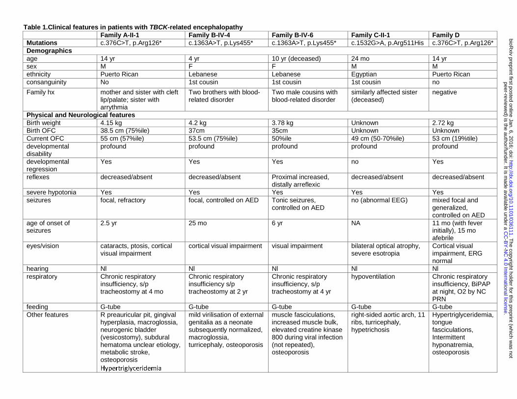
Table 1.Clinical features in patients with TBCK-related encephalopathy Family A-II-1 Family B-IV-4 Family B-IV-6 Family C-II-1 Family D Mutations c.376C>T, p.Arg126* c.1363A>T, p.Lys455* c.1363A>T, p.Lys455* c.1532G>A, p.Arg511His c.376C>T, p.Arg126* Demographics age 14 yr 4 yr 10 yr (deceased) 24 mo 14 yr sex M F F M M ethnicity Puerto Rican Lebanese Lebanese Egyptian Puerto Rican consanguinity No 1st cousin 1st cousin 1st cousin no
Family hx mother and sister with cleft lip/palate; sister with arrythmia
Two brothers with blood-related disorder
Two male cousins with blood-related disorder
similarly affected sister (deceased)
negative
Physical and Neurological features Birth weight 4.15 kg 4.2 kg 3.78 kg Unknown 2.72 kg Birth OFC 38.5 cm (75%ile) 37cm 35cm Unknown Unknown Current OFC 55 cm (57%ile) 53.5 cm (75%ile) 50%ile 49 cm (50-70%ile) 53 cm (19%tile) developmental disability
profound profound profound profound profound
developmental regression
Yes Yes Yes no Yes
reflexes decreased/absent decreased/absent Proximal increased, distally arreflexic
decreased/absent decreased/absent
severe hypotonia Yes Yes Yes Yes Yes seizures focal, refractory focal, controlled on AED Tonic seizures,
controlled on AED no (abnormal EEG) mixed focal and
generalized, controlled on AED
age of onset of seizures
2.5 yr 25 mo 6 yr NA 11 mo (with fever initially), 15 mo afebrile
eyes/vision cataracts, ptosis, cortical visual impairment
cortical visual impairment visual impairment bilateral optical atrophy, severe esotropia
Cortical visual impairment, ERG normal
hearing Nl Nl Nl Nl Nl respiratory Chronic respiratory
insufficiency, s/p tracheostomy at 4 mo
Chronic respiratory insufficiency s/p tracheostomy at 2 yr
Chronic respiratory insufficiency, s/p tracheostomy at 4 yr
hypoventilation Chronic respiratory insufficiency, BiPAP at night, O2 by NC PRN
feeding G-tube G-tube G-tube G-tube G-tube Other features R preauricular pit, gingival
hyperplasia, macroglossia, neurogenic bladder (vesicostomy), subdural hematoma unclear etiology, metabolic stroke, osteoporosis Hypertriglyceridemia
mild virilisation of external genitalia as a neonate subsequently normalized, macroglossia, turricephaly, osteoporosis
muscle fasciculations, increased muscle bulk, elevated creatine kinase 800 during viral infection (not repeated), osteoporosis
right-sided aortic arch, 11 ribs, turricephaly, hypetrichosis
Hypertriglyceridemia, tongue fasciculations, Intermittent hyponatremia, osteoporosis
.C
C-B
Y-N
C 4.0 International license
peer-reviewed) is the author/funder. It is m
ade available under aT
he copyright holder for this preprint (which w
as not.
http://dx.doi.org/10.1101/036111doi:
bioRxiv preprint first posted online Jan. 6, 2016;

Facial features bitemporal narrowing Yes Mild Mild Mild Yes arched eyebrows Yes Yes Yes No Yes deep set eyes No Yes Yes No Yes high nasal bridge Yes Yes Yes Yes Yes anteverted nares Yes Yes Yes Mild No exaggerated Cupid's bow
Yes Yes Yes Mild Yes
coarse features Yes No No Yes Yes macroglossia Yes No No No Yes gingival hyperplasia Yes Yes Unknown Unknown No
AED: anti-epileptic drugs, BiPAP: Bilevel Positive Airway Pressure, EEG: electroencephalogram, L: left, NA: not applicable, Nl: Normal, OFC: occipital frontal circumference, R: right, yr: years.
.C
C-B
Y-N
C 4.0 International license
peer-reviewed) is the author/funder. It is m
ade available under aT
he copyright holder for this preprint (which w
as not.
http://dx.doi.org/10.1101/036111doi:
bioRxiv preprint first posted online Jan. 6, 2016;