Embed Size (px)
Citation preview
Title ABCs of Skin Lesions: Benign, Pre-cancerous, & Cancerous
Authored By Wanda Lockwood RN, BA, MA
Course No SL081707
Contact Hours 3
Purpose
The purpose of this course is to differentiate between benign, pre-cancerous and cancerous skin lesions and to
explain the appearance, symptoms, and treatment for those and common lesions.
Objectives
1. Identify at least 5 benign lesions that pose little danger of malignancy. Identify 2 common pre-cancerous
lesions.
2. Explain the difference between common and dysplastic nevi. Identify 4 types of malignant skin lesions.
3. Explain the difference between basal cell carcinoma and squamous cell carcinoma.
4. Identify at least 8 different treatment options for skin lesions. Outline the symptoms and treatment for
malignant melanoma.
5. Identify 3 different variants of Kaposi’s sarcoma.
6. Explain treatment options for Kaposi’s sarcoma.
Introduction
Almost everyone will develop some type of skin lesion over the course of a lifetime, and differentiating among those
that are benign, pre-cancerous, or cancerous is an important part of assessment. Many lesions are related to sun
exposure, and incidence is increasing, despite warnings to avoid overexposure to ultraviolet light. Benign lesions,
such as acrochordons (skin tags) and seborrheic keratoses, are those that are not clinically significant but may be a
cosmetic concern. Generally, they do not spread and do not recur after they are removed. Precancerous lesions,
such as dysplastic nevi and actinic keratoses, have the potential of evolving into malignancies and may be difficult
to diagnose accurately without biopsy. Other lesions, such as basal cell carcinoma, squamous cell carcinoma,
melanoma, and Kaposi’s sarcoma are malignant, and early diagnosis and treatment can prevent metastasis, which
can be life threatening.
Benign and pre-cancerous lesions
Acrochordons (skin tags)
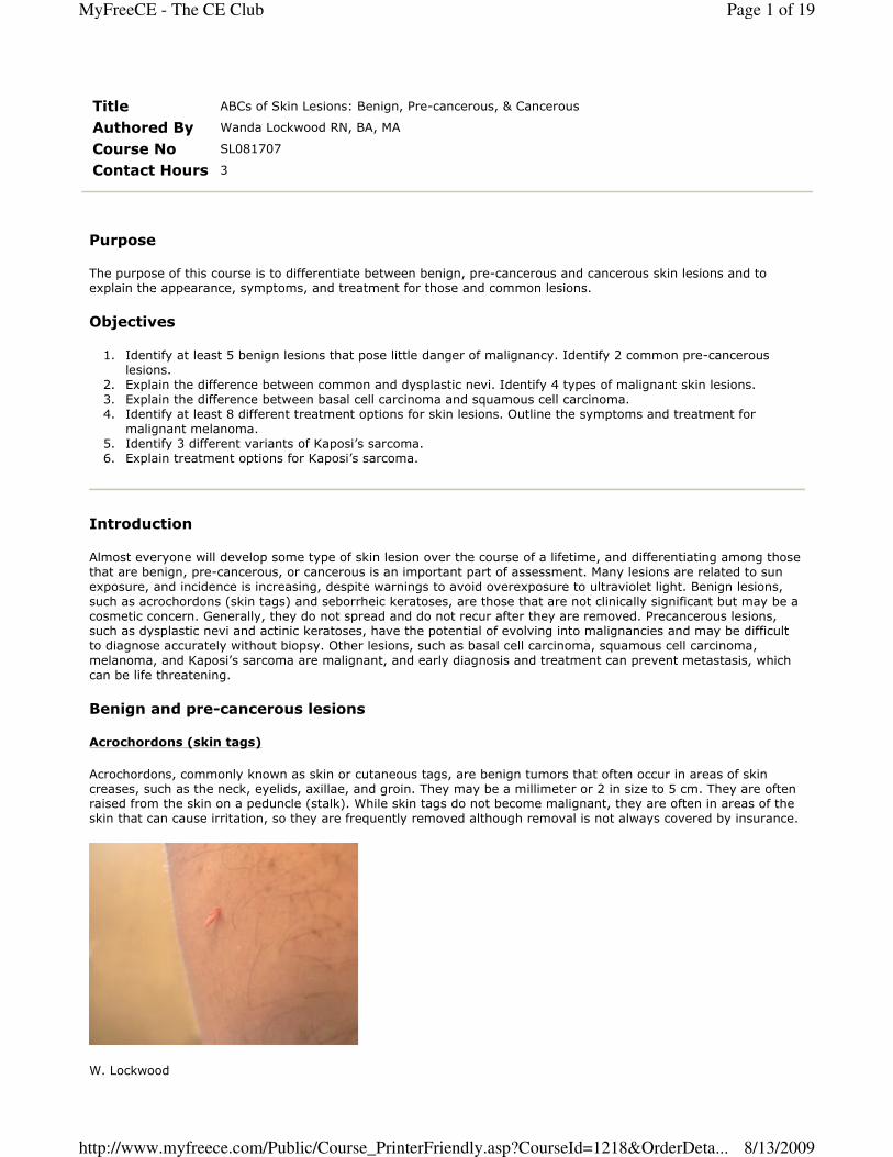
Acrochordons, commonly known as skin or cutaneous tags, are benign tumors that often occur in areas of skin
creases, such as the neck, eyelids, axillae, and groin. They may be a millimeter or 2 in size to 5 cm. They are often
raised from the skin on a peduncle (stalk). While skin tags do not become malignant, they are often in areas of the
skin that can cause irritation, so they are frequently removed although removal is not always covered by insurance.
W. Lockwood
Page 1 of 19MyFreeCE - The CE Club
8/13/2009http://www.myfreece.com/Public/Course_PrinterFriendly.asp?CourseId=1218&OrderDeta...
Acrochordons are associated with age, pregnancy, and obesity and are common in those with diabetes mellitus,
occurring in both males and females. Studies of those with acrochordons indicate that about 40% are type II
diabetics or are insulin-resistant so those with skin tags should be tested for diabetes. Acrochordons are also a
symptom of Birt-Hogg-Dube (BHD) syndrome, an autosomal dominant disorder of the skin involving a number of
different types of lesions. Studies have indicated that human papillomavirus (HPV) types 6 and 11 are found in a
large percentage of skin tab biopsies, suggesting that viral infections are a cause. There are 3 types of
acrochordons:
� Small 1-2mm papules, usually on the neck and axillae
� Single or multiple filiform lesion 2mm x 5 mm occurring in other areas of the body.
� Large soft pedunculated fibromas, usually on lower trunk (1, 2).
Actinic keratosis
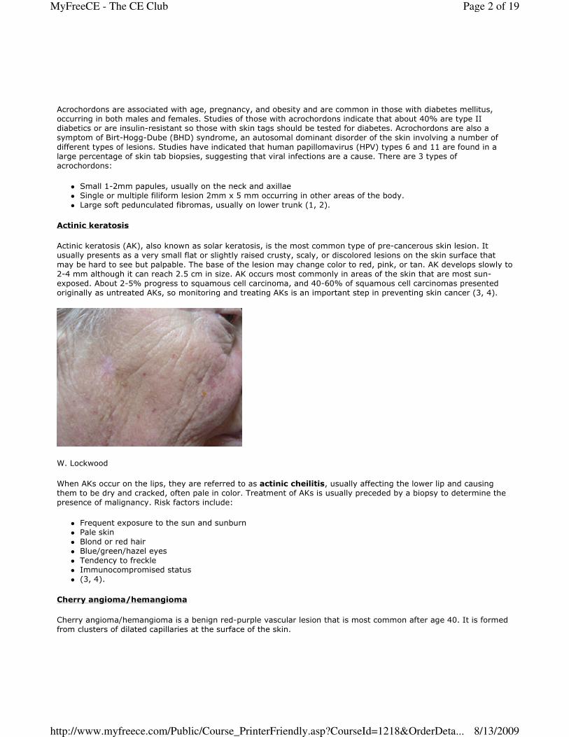
Actinic keratosis (AK), also known as solar keratosis, is the most common type of pre-cancerous skin lesion. It
usually presents as a very small flat or slightly raised crusty, scaly, or discolored lesions on the skin surface that
may be hard to see but palpable. The base of the lesion may change color to red, pink, or tan. AK develops slowly to
2-4 mm although it can reach 2.5 cm in size. AK occurs most commonly in areas of the skin that are most sun-
exposed. About 2-5% progress to squamous cell carcinoma, and 40-60% of squamous cell carcinomas presented
originally as untreated AKs, so monitoring and treating AKs is an important step in preventing skin cancer (3, 4).
W. Lockwood
When AKs occur on the lips, they are referred to as actinic cheilitis, usually affecting the lower lip and causing
them to be dry and cracked, often pale in color. Treatment of AKs is usually preceded by a biopsy to determine the
presence of malignancy. Risk factors include:
� Frequent exposure to the sun and sunburn
� Pale skin
� Blond or red hair
� Blue/green/hazel eyes
� Tendency to freckle
� Immunocompromised status
� (3, 4).
Cherry angioma/hemangioma
Cherry angioma/hemangioma is a benign red-purple vascular lesion that is most common after age 40. It is formed
from clusters of dilated capillaries at the surface of the skin.
Page 2 of 19MyFreeCE - The CE Club
8/13/2009http://www.myfreece.com/Public/Course_PrinterFriendly.asp?CourseId=1218&OrderDeta...
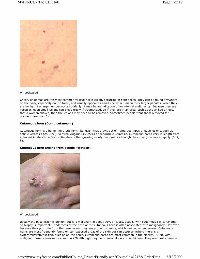
W. Lockwood
Cherry angiomas are the most common vascular skin lesion, occurring in both sexes. They can be found anywhere
on the body, especially on the torso, and usually appear as small cherry-red macules or larger papules. While they
are benign, if a large number occur suddenly, it may be an indication of an internal malignancy. Because they are
vascular, even small lesions can bleed freely if traumatized, so if they are in an area, such as the axillae or legs,
that a woman shaves, then the lesions may need to be removed. Sometimes people want them removed for
cosmetic reasons (5).
Cutaneous horn (Cornu cutaneum)
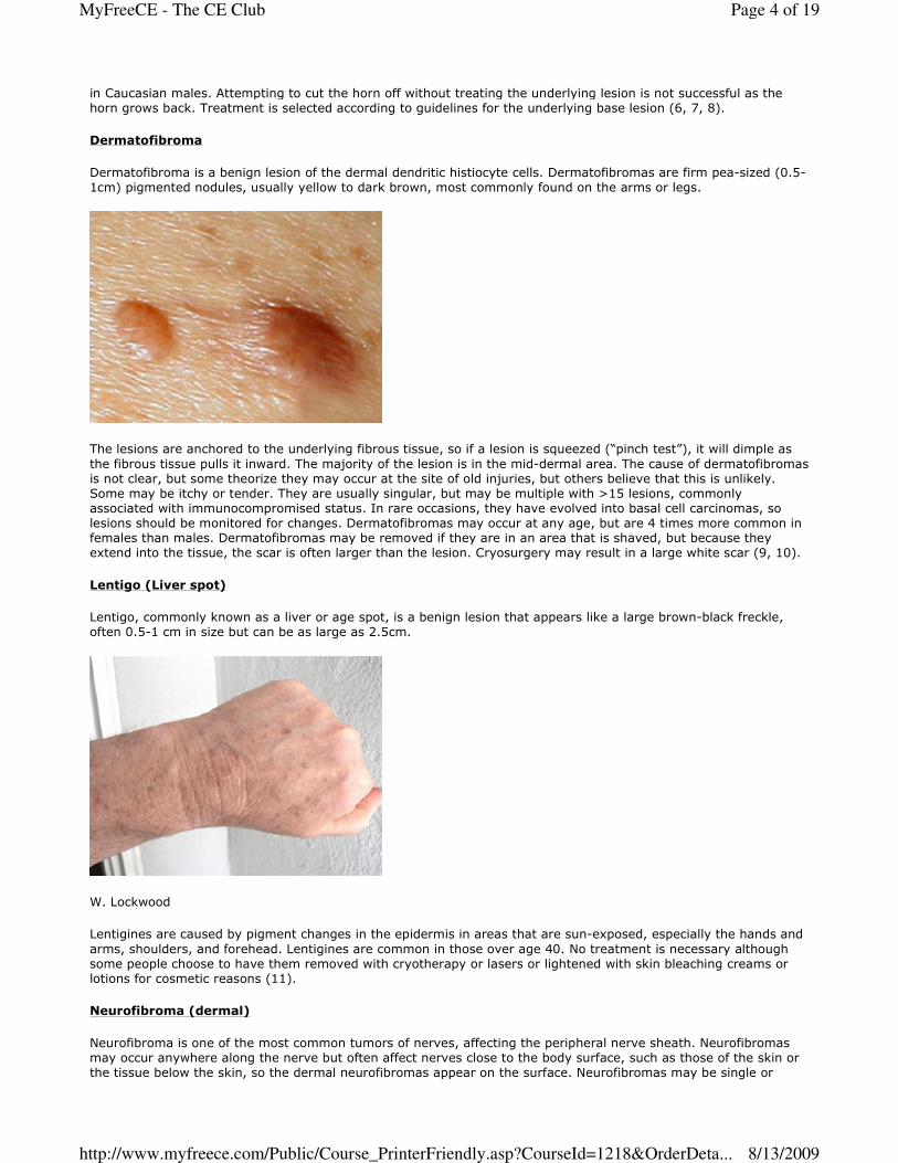
Cutaneous horn is a benign keratotic horn-like lesion that grows out of numerous types of base lesions, such as
actinic keratosis (25-35%), verruca vulgaris (15-25%) or seborrheic keratosis. Cutaneous horns vary in length from
a few millimeters to a few centimeters, often growing slowly over years although they may grow more rapidly (6, 7,
8).
Cutaneous horn arising from actinic keratosis:
W. Lockwood
Usually the base lesion is benign, but it is malignant in about 20% of cases, usually with squamous cell carcinoma,
so biopsy is important. Tenderness at the base of the cutaneous horn is often associated with malignancy. However,
because they protrude from the base lesion, they are prone to trauma, which can cause tenderness. Cutaneous
horns are most frequently found on sun-exposed areas of the skin but can occur anywhere there is a
hyperproliferative lesion, such as on the penis. Cutaneous horns are most common in the elderly, 60-70, with
malignant base lesions more common ?70 although they do occasionally occur in children. They are most common
Page 3 of 19MyFreeCE - The CE Club
8/13/2009http://www.myfreece.com/Public/Course_PrinterFriendly.asp?CourseId=1218&OrderDeta...
in Caucasian males. Attempting to cut the horn off without treating the underlying lesion is not successful as the
horn grows back. Treatment is selected according to guidelines for the underlying base lesion (6, 7, 8).
Dermatofibroma
Dermatofibroma is a benign lesion of the dermal dendritic histiocyte cells. Dermatofibromas are firm pea-sized (0.5-
1cm) pigmented nodules, usually yellow to dark brown, most commonly found on the arms or legs.
The lesions are anchored to the underlying fibrous tissue, so if a lesion is squeezed (“pinch test”), it will dimple as
the fibrous tissue pulls it inward. The majority of the lesion is in the mid-dermal area. The cause of dermatofibromas
is not clear, but some theorize they may occur at the site of old injuries, but others believe that this is unlikely.
Some may be itchy or tender. They are usually singular, but may be multiple with >15 lesions, commonly
associated with immunocompromised status. In rare occasions, they have evolved into basal cell carcinomas, so
lesions should be monitored for changes. Dermatofibromas may occur at any age, but are 4 times more common in
females than males. Dermatofibromas may be removed if they are in an area that is shaved, but because they
extend into the tissue, the scar is often larger than the lesion. Cryosurgery may result in a large white scar (9, 10).
Lentigo (Liver spot)
Lentigo, commonly known as a liver or age spot, is a benign lesion that appears like a large brown-black freckle,
often 0.5-1 cm in size but can be as large as 2.5cm.
W. Lockwood
Lentigines are caused by pigment changes in the epidermis in areas that are sun-exposed, especially the hands and
arms, shoulders, and forehead. Lentigines are common in those over age 40. No treatment is necessary although
some people choose to have them removed with cryotherapy or lasers or lightened with skin bleaching creams or
lotions for cosmetic reasons (11).
Neurofibroma (dermal)
Neurofibroma is one of the most common tumors of nerves, affecting the peripheral nerve sheath. Neurofibromas
may occur anywhere along the nerve but often affect nerves close to the body surface, such as those of the skin or
the tissue below the skin, so the dermal neurofibromas appear on the surface. Neurofibromas may be single or
Page 4 of 19MyFreeCE - The CE Club
8/13/2009http://www.myfreece.com/Public/Course_PrinterFriendly.asp?CourseId=1218&OrderDeta...
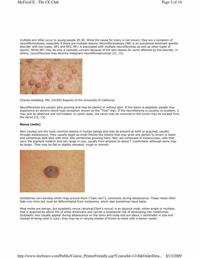
multiple and often occur in young people 20-30. While the cause for many is not known, they are a symptom of
neurofibromatosis, especially if there are multiple lesions. Neurofibromatosis (NF) is an autosomal dominant genetic
disorder with two types: NF1 and NF2. NF1 is associated with multiple neurofibromas as well as other types of
lesions. While NF1 may be only a cosmetic concern because of the skin lesions for some affected by the disorder, in
others, neurofibromas may become malignant neurofibrosarcomas (12, 13).
Charles Goldberg, MD, (UCSD) Regents of the University of California
Neurofibromas are usually slow growing and may be painful or without pain. If the lesion is palpated, people may
experience an electric-shock type sensation, known as the “Tinel” sign. If the neurofibroma is causing no problem, it
may just be observed and not treated. In some cases, the nerve may be removed or the tumor may be excised from
the nerve (12, 13).
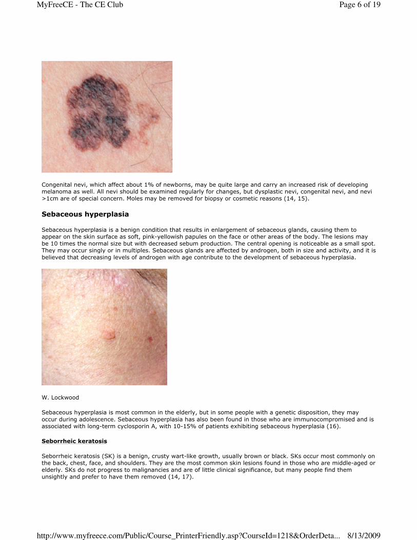
Nevus (mole)
Nevi (moles) are the most common lesions in human beings and may be present at birth or acquired, usually
through adolescence. They usually begin as small freckle-like lesions that may grow and darken to brown or black
and sometimes dark blue with time, also sometimes growing hairs. Nevi are composed of melanocytes, cells that
carry the pigment melanin and can range in size, usually from pinpoint to about ? 1centimeter although some may
be larger. They may be flat or slightly elevated, rough or smooth.
Sometimes nevi develop white rings around them (“halo nevi”), commonly during adolescence. These moles often
fade over time but must be differentiated from melanoma, which also sometimes have halos.
Most moles are benign, but dysplastic nevus (atypical/Clark’s nevus) is an atypical mole, either single or multiple,
that is acquired by about 4% of white Americans and carries a substantial risk of developing into melanomas.
Dysplastic nevi usually appear during adolescence on the torso and scalp and are about 1 centimeter in size and
instead of being solid in color; they may be in varying shades of brown to black with a darker center.
Page 5 of 19MyFreeCE - The CE Club
8/13/2009http://www.myfreece.com/Public/Course_PrinterFriendly.asp?CourseId=1218&OrderDeta...
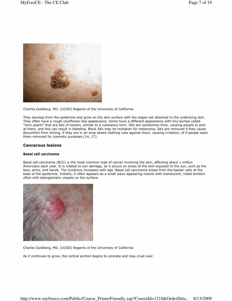
Congenital nevi, which affect about 1% of newborns, may be quite large and carry an increased risk of developing
melanoma as well. All nevi should be examined regularly for changes, but dysplastic nevi, congenital nevi, and nevi
>1cm are of special concern. Moles may be removed for biopsy or cosmetic reasons (14, 15).
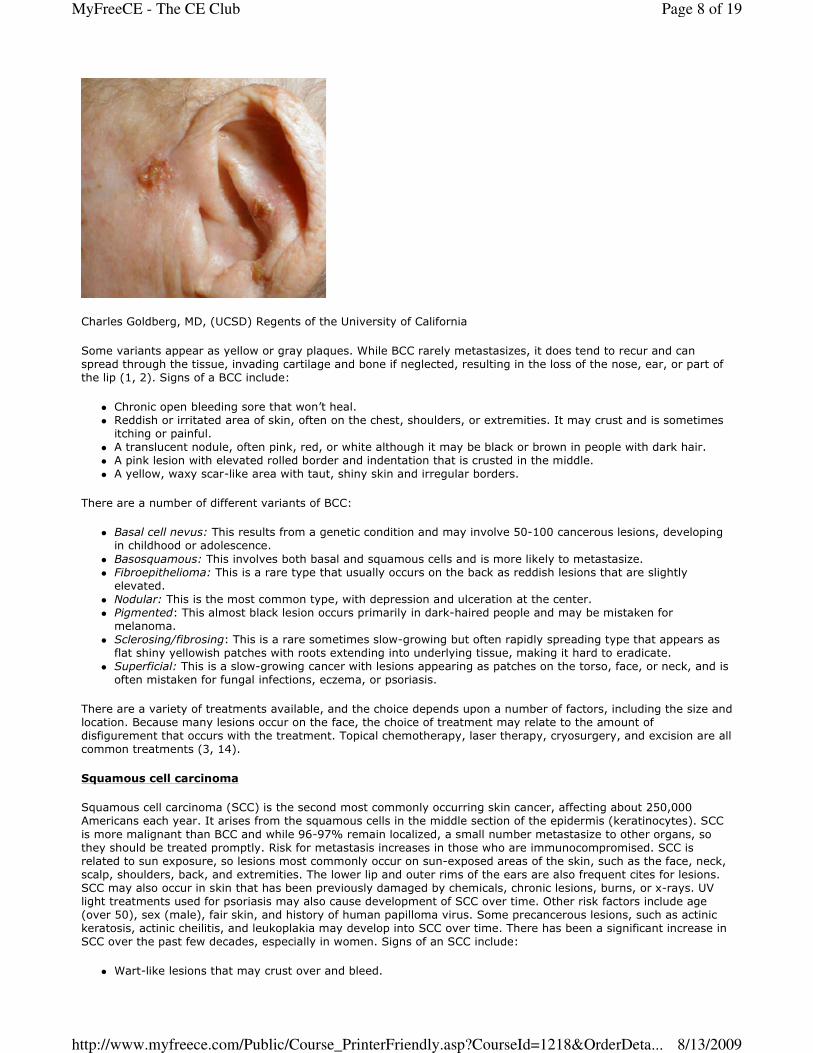
Sebaceous hyperplasia
Sebaceous hyperplasia is a benign condition that results in enlargement of sebaceous glands, causing them to
appear on the skin surface as soft, pink-yellowish papules on the face or other areas of the body. The lesions may
be 10 times the normal size but with decreased sebum production. The central opening is noticeable as a small spot.
They may occur singly or in multiples. Sebaceous glands are affected by androgen, both in size and activity, and it is
believed that decreasing levels of androgen with age contribute to the development of sebaceous hyperplasia.
W. Lockwood
Sebaceous hyperplasia is most common in the elderly, but in some people with a genetic disposition, they may
occur during adolescence. Sebaceous hyperplasia has also been found in those who are immunocompromised and is
associated with long-term cyclosporin A, with 10-15% of patients exhibiting sebaceous hyperplasia (16).
Seborrheic keratosis
Seborrheic keratosis (SK) is a benign, crusty wart-like growth, usually brown or black. SKs occur most commonly on
the back, chest, face, and shoulders. They are the most common skin lesions found in those who are middle-aged or
elderly. SKs do not progress to malignancies and are of little clinical significance, but many people find them
unsightly and prefer to have them removed (14, 17).
Page 6 of 19MyFreeCE - The CE Club
8/13/2009http://www.myfreece.com/Public/Course_PrinterFriendly.asp?CourseId=1218&OrderDeta...
Charles Goldberg, MD, (UCSD) Regents of the University of California
They develop from the epidermis and grow on the skin surface with the edges not attached to the underlying skin.
They often have a rough cauliflower-like appearance. Some have a different appearance with tiny bumps called
“horn pearls” that are bits of keratin, similar to a cutaneous horn. SKs are sometimes itchy, causing people to pick
at them, and this can result in bleeding. Black SKs may be mistaken for melanoma. SKs are removed if they cause
discomfort from itching, if they are in an area where clothing rubs against them, causing irritation, of if people want
them removed for cosmetic purposes (14, 17).
Cancerous lesions
Basal cell carcinoma
Basal cell carcinoma (BCC) is the most common type of cancer involving the skin, affecting about 1 million
Americans each year. It is related to sun damage, so it occurs on areas of the skin exposed to the sun, such as the
face, arms, and hands. The incidence increases with age. Basal cell carcinoma arises from the basilar cells at the
base of the epidermis. Initially, it often appears as a small waxy-appearing nodule with translucent, rolled borders
often with telangiectatic vessels on the surface.
Charles Goldberg, MD, (UCSD) Regents of the University of California
As it continues to grow, the central portion begins to ulcerate and may crust over.
Page 7 of 19MyFreeCE - The CE Club
8/13/2009http://www.myfreece.com/Public/Course_PrinterFriendly.asp?CourseId=1218&OrderDeta...
Charles Goldberg, MD, (UCSD) Regents of the University of California
Some variants appear as yellow or gray plaques. While BCC rarely metastasizes, it does tend to recur and can
spread through the tissue, invading cartilage and bone if neglected, resulting in the loss of the nose, ear, or part of
the lip (1, 2). Signs of a BCC include:
� Chronic open bleeding sore that won’t heal.
� Reddish or irritated area of skin, often on the chest, shoulders, or extremities. It may crust and is sometimes
itching or painful.
� A translucent nodule, often pink, red, or white although it may be black or brown in people with dark hair.
� A pink lesion with elevated rolled border and indentation that is crusted in the middle.
� A yellow, waxy scar-like area with taut, shiny skin and irregular borders.
There are a number of different variants of BCC:
� Basal cell nevus: This results from a genetic condition and may involve 50-100 cancerous lesions, developing
in childhood or adolescence.
� Basosquamous: This involves both basal and squamous cells and is more likely to metastasize.
� Fibroepithelioma: This is a rare type that usually occurs on the back as reddish lesions that are slightly
elevated.
� Nodular: This is the most common type, with depression and ulceration at the center.
� Pigmented: This almost black lesion occurs primarily in dark-haired people and may be mistaken for
melanoma.
� Sclerosing/fibrosing: This is a rare sometimes slow-growing but often rapidly spreading type that appears as
flat shiny yellowish patches with roots extending into underlying tissue, making it hard to eradicate.
� Superficial: This is a slow-growing cancer with lesions appearing as patches on the torso, face, or neck, and is
often mistaken for fungal infections, eczema, or psoriasis.
There are a variety of treatments available, and the choice depends upon a number of factors, including the size and
location. Because many lesions occur on the face, the choice of treatment may relate to the amount of
disfigurement that occurs with the treatment. Topical chemotherapy, laser therapy, cryosurgery, and excision are all
common treatments (3, 14).
Squamous cell carcinoma
Squamous cell carcinoma (SCC) is the second most commonly occurring skin cancer, affecting about 250,000
Americans each year. It arises from the squamous cells in the middle section of the epidermis (keratinocytes). SCC
is more malignant than BCC and while 96-97% remain localized, a small number metastasize to other organs, so
they should be treated promptly. Risk for metastasis increases in those who are immunocompromised. SCC is
related to sun exposure, so lesions most commonly occur on sun-exposed areas of the skin, such as the face, neck,
scalp, shoulders, back, and extremities. The lower lip and outer rims of the ears are also frequent cites for lesions.
SCC may also occur in skin that has been previously damaged by chemicals, chronic lesions, burns, or x-rays. UV
light treatments used for psoriasis may also cause development of SCC over time. Other risk factors include age
(over 50), sex (male), fair skin, and history of human papilloma virus. Some precancerous lesions, such as actinic
keratosis, actinic cheilitis, and leukoplakia may develop into SCC over time. There has been a significant increase in
SCC over the past few decades, especially in women. Signs of an SCC include:
� Wart-like lesions that may crust over and bleed.
Page 8 of 19MyFreeCE - The CE Club
8/13/2009http://www.myfreece.com/Public/Course_PrinterFriendly.asp?CourseId=1218&OrderDeta...
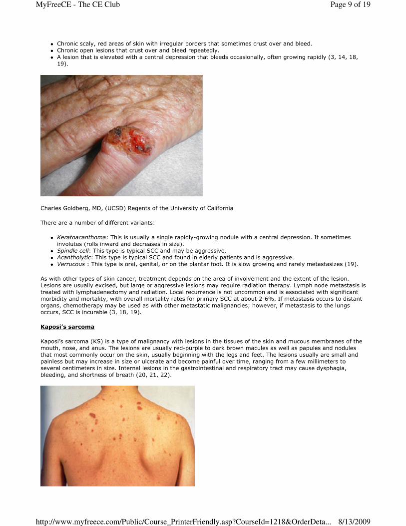
� Chronic scaly, red areas of skin with irregular borders that sometimes crust over and bleed.
� Chronic open lesions that crust over and bleed repeatedly.
� A lesion that is elevated with a central depression that bleeds occasionally, often growing rapidly (3, 14, 18,
19).
Charles Goldberg, MD, (UCSD) Regents of the University of California
There are a number of different variants:
� Keratoacanthoma: This is usually a single rapidly-growing nodule with a central depression. It sometimes
involutes (rolls inward and decreases in size).
� Spindle cell: This type is typical SCC and may be aggressive.
� Acantholytic: This type is typical SCC and found in elderly patients and is aggressive.
� Verrucous : This type is oral, genital, or on the plantar foot. It is slow growing and rarely metastasizes (19).
As with other types of skin cancer, treatment depends on the area of involvement and the extent of the lesion.
Lesions are usually excised, but large or aggressive lesions may require radiation therapy. Lymph node metastasis is
treated with lymphadenectomy and radiation. Local recurrence is not uncommon and is associated with significant
morbidity and mortality, with overall mortality rates for primary SCC at about 2-6%. If metastasis occurs to distant
organs, chemotherapy may be used as with other metastatic malignancies; however, if metastasis to the lungs
occurs, SCC is incurable (3, 18, 19).
Kaposi’s sarcoma
Kaposi’s sarcoma (KS) is a type of malignancy with lesions in the tissues of the skin and mucous membranes of the
mouth, nose, and anus. The lesions are usually red-purple to dark brown macules as well as papules and nodules
that most commonly occur on the skin, usually beginning with the legs and feet. The lesions usually are small and
painless but may increase in size or ulcerate and become painful over time, ranging from a few millimeters to
several centimeters in size. Internal lesions in the gastrointestinal and respiratory tract may cause dysphagia,
bleeding, and shortness of breath (20, 21, 22).
Page 9 of 19MyFreeCE - The CE Club
8/13/2009http://www.myfreece.com/Public/Course_PrinterFriendly.asp?CourseId=1218&OrderDeta...
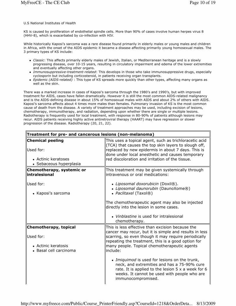
U.S National Institutes of Health
KS is caused by proliferation of endothelial spindle cells. More than 90% of cases involve human herpes virus 8
(HHV-8), which is exacerbated by co-infection with HIV.
While historically Kaposi’s sarcoma was a rare disease found primarily in elderly males or young males and children
in Africa, with the onset of the AIDS epidemic it became a disease affecting primarily young homosexual males. The
3 primary types of KS include:
� Classic: This affects primarily elderly males of Jewish, Italian, or Mediterranean heritage and is a slowly
progressing disease, over 10-15 years, resulting in circulatory impairment and edema of the lower extremities
and eventually affecting other organs.
� Immunosuppressive-treatment-related: This develops in those who take immunosuppressive drugs, especially
cyclosporin but including corticosteroid, in patients receiving organ transplants.
� Epidemic (AIDS-related) : This type of KS spreads more quickly than other types, affecting many organs as
well as the skin.
There was a marked increase in cases of Kaposi’s sarcoma through the 1980’s and 1990’s, but with improved
treatment for AIDS, cases have fallen dramatically. However it is still the most common AIDS-related malignancy
and is the AIDS-defining disease in about 15% of homosexual males with AIDS and about 2% of others with AIDS.
Kaposi’s sarcoma affects about 4 times more males than females. Pulmonary invasion of KS is the most common
cause of death from the disease. A variety of treatment approaches may be used, including excision of lesions,
chemotherapy, immunotherapy, and radiation, depending upon whether there are single or multiple lesions.
Radiotherapy is frequently used for local treatment, with response in 80-90% of patients although lesions may
recur. AIDS patients receiving highly active antiretroviral therapy (HAART) may have regression or slower
progression of the disease. Radiotherapy (20, 21, 22).
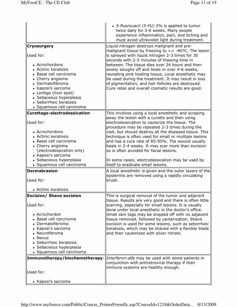
Treatment for pre- and cancerous lesions (non-melanoma)
Chemical peeling
Used for:
� Actinic keratoses
� Sebaceous hyperplasia
This uses a topical agent, such as trichloracetic acid
(TCA) that causes the top skin layers to slough off,
replaced by new epidermis in about 7 days. This is
done under local anesthetic and causes temporary red discoloration and irritation of the tissue.
Chemotherapy, systemic or
intralesional
Used for:
� Kaposi’s sarcoma
This treatment may be given systemically through
intravenous or oral medications:
� Liposomal doxorubicin (Doxil®).
� Liposomal daunorubin (DaunoXome®)
� Paclitaxel (Taxol®)
The chemotherapeutic agent may also be injected directly into the lesion in some cases.
� Vinblastine is used for intralesional
chemotherapy.
Chemotherapy, topical
Used for:
� Actinic keratosis
� Basal cell carcinoma
This is less effective than excision because the
cancer may recur, but it is simple and results in less
scarring, so even though it may require periodically repeating the treatment, this is a good option for
many people. Topical chemotherapeutic agents
include:
� Imiquimod is used for lesions on the trunk,
neck, and extremities and has a 75-90% cure rate. It is applied to the lesion 5 x a week for 6
weeks. It cannot be used with people who are
immunocompromised.
Page 10 of 19MyFreeCE - The CE Club
8/13/2009http://www.myfreece.com/Public/Course_PrinterFriendly.asp?CourseId=1218&OrderDeta...
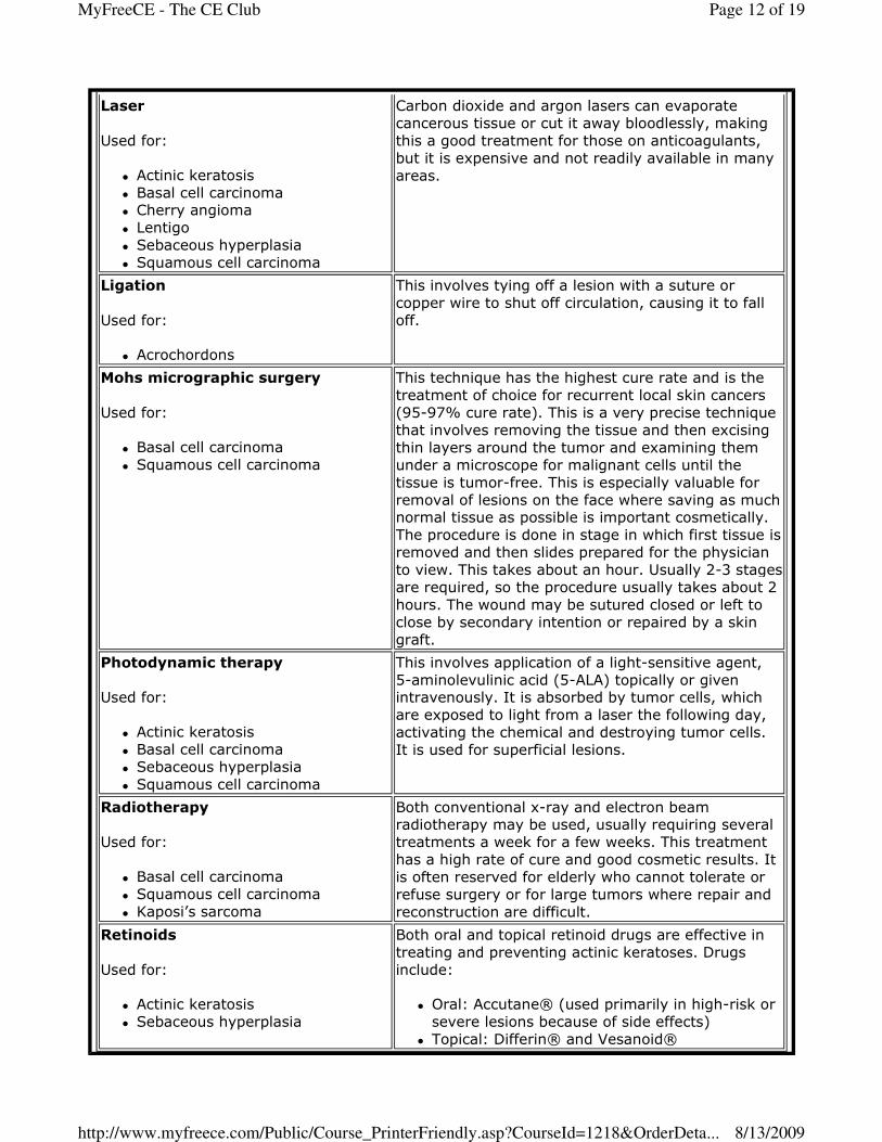
� 5-fluorouacil (5-FU) 5% is applied to tumor
twice daily for 3-6 weeks. Many people
experience inflammation, pain, and itching and
must avoid ultraviolet light during treatment.
Cryosurgery
Used for:
� Acrochordons
� Actinic keratosis
� Basal cell carcinoma
� Cherry angioma
� Dermatofibroma
� Kaposi’s sarcoma
� Lentigo (liver spot) � Sebaceous hyperplasia
� Seborrheic keratosis
� Squamous cell carcinoma
Liquid nitrogen destroys malignant and pre-
malignant tissue by freezing to >= -40?C. The lesion
is sprayed with liquid nitrogen 2-3 times for 30
seconds with 2-5 minutes of thawing time in
between. The tissue dies over 24 hours and then slowly sloughs off and heals in over 4-6 weeks,
revealing pink healing tissue. Local anesthetic may
be used during the treatment. It may result in loss
of pigmentation, and hair follicles are destroyed.
Cure rates and overall cosmetic results are good.
Curettage-electrodessication
Used for:
� Acrochordons
� Actinic keratosis
� Basal cell carcinoma
� Cherry angioma
(electrodessication only)
� Kaposi’s sarcoma
� Sebaceous hyperplasia
� Squamous cell carcinoma
This involves using a local anesthetic and scraping
away the lesion with a curette and then using
electrodessication to cauterize the tissue. The
procedure may be repeated 2-3 times during the
visit, but should destroy all the diseased tissue. This
technique is often used for small or multiple lesions
and has a cure rate of 85-95%. The wound usually heals in 2-4 weeks. It may scar more than excision
so is often avoided for facial lesions.
In some cases, electrodessication may be used by
itself to eradicate small lesions.
Dermabrasion
Used for:
� Actinic keratosis
A local anesthetic is given and the outer layers of the
epidermis are removed using a rapidly circulating
brush.
Excision/ Shave excision
Used for:
� Acrochordon
� Basal cell carcinoma
� Dermatofibroma
� Kaposi’s sarcoma
� Neurofibroma
� Nevus
� Seborrheic keratosis.
� Sebaceous hyperplasia
� Squamous cell carcinoma
This is surgical removal of the tumor and adjacent
tissue. Results are very good and there is often little
scarring, especially for small lesions. It is usually
done under local anesthetic in the doctor’s office.
Small skin tags may be snipped off with no adjacent
tissue removed, followed by cauterization. Shave
excision is used for some lesions, such as seborrheic keratosis, which may be shaved with a flexible blade
and then cauterized with silver nitrate.
Immunotherapy/biochemotherapy:
Used for:
� Kaposi’s sarcoma
Interferon-alfa may be used with some patients in conjunction with antiretroviral therapy if their
immune systems are healthy enough.
Page 11 of 19MyFreeCE - The CE Club
8/13/2009http://www.myfreece.com/Public/Course_PrinterFriendly.asp?CourseId=1218&OrderDeta...
Laser
Used for:
� Actinic keratosis
� Basal cell carcinoma � Cherry angioma
� Lentigo
� Sebaceous hyperplasia
� Squamous cell carcinoma
Carbon dioxide and argon lasers can evaporate
cancerous tissue or cut it away bloodlessly, making
this a good treatment for those on anticoagulants,
but it is expensive and not readily available in many
areas.
Ligation
Used for:
� Acrochordons
This involves tying off a lesion with a suture or
copper wire to shut off circulation, causing it to fall
off.
Mohs micrographic surgery
Used for:
� Basal cell carcinoma
� Squamous cell carcinoma
This technique has the highest cure rate and is the
treatment of choice for recurrent local skin cancers
(95-97% cure rate). This is a very precise technique
that involves removing the tissue and then excising
thin layers around the tumor and examining them
under a microscope for malignant cells until the
tissue is tumor-free. This is especially valuable for
removal of lesions on the face where saving as much normal tissue as possible is important cosmetically.
The procedure is done in stage in which first tissue is
removed and then slides prepared for the physician
to view. This takes about an hour. Usually 2-3 stages
are required, so the procedure usually takes about 2
hours. The wound may be sutured closed or left to
close by secondary intention or repaired by a skin
graft.
Photodynamic therapy
Used for:
� Actinic keratosis
� Basal cell carcinoma
� Sebaceous hyperplasia
� Squamous cell carcinoma
This involves application of a light-sensitive agent,
5-aminolevulinic acid (5-ALA) topically or given intravenously. It is absorbed by tumor cells, which
are exposed to light from a laser the following day,
activating the chemical and destroying tumor cells.
It is used for superficial lesions.
Radiotherapy
Used for:
� Basal cell carcinoma
� Squamous cell carcinoma
� Kaposi’s sarcoma
Both conventional x-ray and electron beam radiotherapy may be used, usually requiring several
treatments a week for a few weeks. This treatment
has a high rate of cure and good cosmetic results. It
is often reserved for elderly who cannot tolerate or
refuse surgery or for large tumors where repair and
reconstruction are difficult.
Retinoids
Used for:
� Actinic keratosis
� Sebaceous hyperplasia
Both oral and topical retinoid drugs are effective in
treating and preventing actinic keratoses. Drugs
include:
� Oral: Accutane® (used primarily in high-risk or
severe lesions because of side effects)
� Topical: Differin® and Vesanoid®
Page 12 of 19MyFreeCE - The CE Club
8/13/2009http://www.myfreece.com/Public/Course_PrinterFriendly.asp?CourseId=1218&OrderDeta...
(1, 2, 3, 4, 11, 19, 20, 21).
Melanoma
Malignant melanoma is one of the most serious forms of skin cancer, resulting in 2% of the deaths from cancer each
year. Melanomas originate in melanocytes within the skin. In rare cases melanomas may also arise in the uvea of
the eyes or the meninges, digestive tract, or lymph nodes where melanocytes are found. Melanocytes produce
melanin, which is a pigment that gives color to the skin. With exposure to the sun, the skin produces more melanin,
causing the skin to tan. Sometimes melanocytes group together and form moles, but in some cases they become
malignant. The incidence of melanoma has increased markedly worldwide, doubling every 10 years. This increase is
believed related to increased sun exposure in recreational activities, the use of tanning beds, and better detection.
Early diagnosis is important because lesions >1.5 mm in thickness or with regional lymph node involvement have a
poor prognosis with only a 20-50% chance of surviving for 5 years compared to 95% chance for those with thinner
lesions and no lymph node involvement. The American Cancer Society estimates >= 8000 deaths a year in the
United States, with almost twice as many males as females. Melanomas can occur at any age, most after
adolescence. Recent research has identified mutated genes, including BRAF and p53, which are associated with
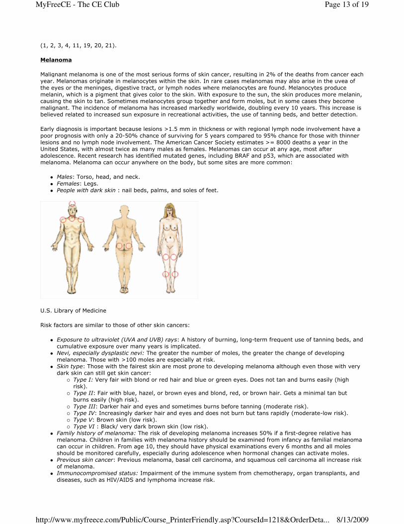
melanoma. Melanoma can occur anywhere on the body, but some sites are more common:
� Males: Torso, head, and neck.
� Females: Legs.
� People with dark skin : nail beds, palms, and soles of feet.
U.S. Library of Medicine
Risk factors are similar to those of other skin cancers:
� Exposure to ultraviolet (UVA and UVB) rays: A history of burning, long-term frequent use of tanning beds, and
cumulative exposure over many years is implicated.
� Nevi, especially dysplastic nevi: The greater the number of moles, the greater the change of developing
melanoma. Those with >100 moles are especially at risk.
� Skin type: Those with the fairest skin are most prone to developing melanoma although even those with very
dark skin can still get skin cancer:
� Type I: Very fair with blond or red hair and blue or green eyes. Does not tan and burns easily (high
risk).
� Type II: Fair with blue, hazel, or brown eyes and blond, red, or brown hair. Gets a minimal tan but
burns easily (high risk).
� Type III: Darker hair and eyes and sometimes burns before tanning (moderate risk).
� Type IV: Increasingly darker hair and eyes and does not burn but tans rapidly (moderate-low risk).
� Type V: Brown skin (low risk).
� Type VI : Black/ very dark brown skin (low risk).
� Family history of melanoma: The risk of developing melanoma increases 50% if a first-degree relative has
melanoma. Children in families with melanoma history should be examined from infancy as familial melanoma
can occur in children. From age 10, they should have physical examinations every 6 months and all moles
should be monitored carefully, especially during adolescence when hormonal changes can activate moles.
� Previous skin cancer: Previous melanoma, basal cell carcinoma, and squamous cell carcinoma all increase risk
of melanoma.
� Immunocompromised status: Impairment of the immune system from chemotherapy, organ transplants, and
diseases, such as HIV/AIDS and lymphoma increase risk.
Page 13 of 19MyFreeCE - The CE Club
8/13/2009http://www.myfreece.com/Public/Course_PrinterFriendly.asp?CourseId=1218&OrderDeta...
It is especially important that those with nevi inspect their skin on a regular basis and observe for the ABCDEs of
melanomas.
The ABCDEs of Melanomas
� Asymmetry.
� Borders uneven, scalloped or notched.
� Color variations within a mole.
� Diameter is usually larger than 1/4 inch (6 mm).
� E volving changes in size, shape, color, or appearance.
There are 4 primary types of melanoma, and determining the type is an important part of diagnosis and follow-up
treatment:
� Superficial spreading: These affect about 70%, especially those who are young. This spreads along the top
layers of the skin for long periods before invading underlying tissue. This occurs most often on the trunk and
upper back in males and the legs and upper back in females. Lesions are usually flat or slightly raised with
irregular borders and may occur in a previous mole.
� Lentigo-maligna: These are slowly evolving lesions that progress with changes in size and color over time,
often many years. They spread along the surface of the skin before invading. They are most common in the
elderly on the hands, head, and neck.
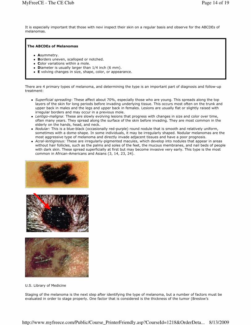
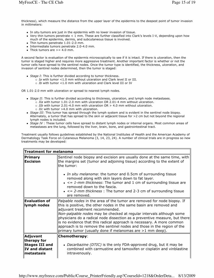
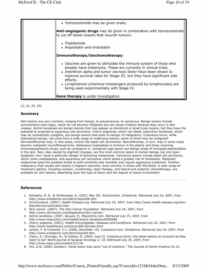
� Nodular: This is a blue-black (occasionally red-purple) round nodule that is smooth and relatively uniform,
sometimes with a dome-shape. In some individuals, it may be irregularly shaped. Nodular melanomas are the
most aggressive type of melanoma and directly invade adjacent tissues and have a poor prognosis.
� Acral-lentiginous: These are irregularly-pigmented macules, which develop into nodules that appear in areas
without hair follicles, such as the palms and soles of the feet, the mucous membranes, and nail beds of people
with dark skin. These spread superficially at first but may become invasive very early. This type is the most
common in African-Americans and Asians (3, 14, 23, 24).
U.S. Library of Medicine
Staging of the melanoma is the next step after identifying the type of melanoma, but a number of factors must be
evaluated in order to stage properly. One factor that is considered is the thickness of the tumor (Breslow’s
Page 14 of 19MyFreeCE - The CE Club
8/13/2009http://www.myfreece.com/Public/Course_PrinterFriendly.asp?CourseId=1218&OrderDeta...
thickness), which measure the distance from the upper layer of the epidermis to the deepest point of tumor invasion
in millimeters:
� In situ tumors are just in the epidermis with no lower invasion of tissue.
� Very thin tumors penetrate < 1 mm. These are further classified into Clark’s levels I-V, depending upon how
much of the epidermis, dermis, and subcutaneous tissue is involved.
� Thin tumors penetrate 1.01-2.0 mm.
� Intermediate tumors penetrate 2.0-4.0 mm.
� Thick tumors are >= 4.0 mm.
A second factor is evaluation of the epidermis microscopically to see if it is intact. If there is ulceration, then the
tumor is staged higher and requires more aggressive treatment. Another important factor is whether or not the
tumor cells have spread to the sentinel nodes. Once the tumor type is identified, the thickness, ulceration, and
invasion of sentinel nodes determined, then the tumor is staged:
� Stage I: This is further divided according to tumor thickness.
� Ia with tumor <1.0 mm without ulceration and Clark level II or III.
� Ib with tumor <1.0 mm with ulceration and Clark level III or IV
OR 1.01-2.0 mm with ulceration or spread to nearest lymph nodes.
� Stage II: This is further divided according to thickness, ulceration, and lymph node metastases.
� IIa with tumor 1.01-2.0 mm with ulceration OR 2.01-4 mm without ulceration.
� IIb with tumor 2.01-4.0 mm with ulceration OR > 4.0 mm without ulceration.
� IIc with tumor >4.0 mm with ulceration.
� Stage III: This tumor has spread through the lymph system and is evident in the sentinel node biopsy.
Alternately, a tumor that has spread to the skin or adjacent tissue for >2 cm but not beyond the regional
lymph nodes is included.
� Stage IV : These tumor cells have spread to distant lymph nodes or internal organs. Most common areas of
metastases are the lung, followed by the liver, brain, bone, and gastrointestinal tract.
Treatment usually follows guidelines established by the National Institutes of Health and the American Academy of
Dermatology Task Force on Cutaneous Melanoma (3, 14, 23, 24). A number of clinical trials are in progress so new
treatments may be developed:
Treatment for melanoma
Primary
Excision
Sentinel node biopsy and excision are usually done at the same time, with
the margins set (tumor and adjoining tissue) according to the extent of
the tumor:
� In situ melanoma: the tumor and 0.5cm of surrounding tissue
removed along with skin layers down to fat layer. � <= 1-mm thickness: The tumor and 1 cm of surrounding tissue are
removed down to the fascia.
� <= 2-mm thickness : The tumor and 2-3 cm of surrounding tissue
are removed.
Evaluation of
lymph nodes
Palpable nodes in the area of the tumor are removed for node biopsy. If
this is positive, the other nodes in the same basin are removed and
adjuvant treatment recommended.
Non-palpable nodes may be checked at regular intervals although some
physicians do a radical node dissection as a preventive measure, but there
is no evidence that this radical approach is necessary. A more common approach is to remove the sentinel nodes and those in the region of the
primary tumor (usually done if melanomas are >1 mm deep).
Adjuvant
therapy for
Stages III and
IV and distant
metastasis
Chemotherapy:
� Dacarbazine (DTIC) is the only FDA-approved drug, but it may be
combined with carmustine and tamoxifen or cisplatin and vinblastine
intravenously.
Page 15 of 19MyFreeCE - The CE Club
8/13/2009http://www.myfreece.com/Public/Course_PrinterFriendly.asp?CourseId=1218&OrderDeta...
(3, 14, 23, 24)
Summary
Skin lesions are very common, ranging from benign, to precancerous, to cancerous. Benign lesions include
acrochordons (skin tags), which do not become malignant but can cause irritation because they occur in skin
creases. Actinic keratoses are benign lesions that may appear as discolored or small scaly lesions, but they have the
potential to progress to squamous cell carcinoma. Cherry angiomas, which can bleed, seborrheic keratoses, which
may be cosmetically unsightly, are benign lesions that pose no danger of malignancy. Cutaneous horns, while
themselves benign, can arise from a wide range of underlying lesions, some of which may be malignant.
Dermatofibromas may, in rare cases, evolve into basal cell carcinomas. Neurofibromas, in turn, may in some cases
become malignant neurofibrosarcoma. Sebaceous hyperplasia is common in the elderly and those receiving
immunosuppressive drugs, such as cyclosporin A. Lentigines (age spots) are benign areas of increased pigmentation
in the skin. Nevi, also caused by pigment changes, are the most common lesion in human beings, but one type—
dysplastic nevi—pose a particular danger of becoming melanomas. Cancerous lesions include basal cell carcinoma,
which rarely metastasizes, and squamous cell carcinoma, which poses a greater risk of metastasis. Malignant
melanomas pose the greatest threat to both morbidity and mortality and require aggressive treatment. Another
malignancy that causes skin lesions is Kaposi’s sarcoma, most common in those with HIV/AIDS. A wide range of
treatment options, including excision, cryotherapy, laser therapy, and topical and systemic chemotherapy, are
available for skin lesions, depending upon the type of lesion and the degree of tissue involvement.
References
1. Schwartz, R. A., & Terlikowska, A. (2007, May 30). Acrochordon. Emedicine. Retrieved July 24, 2007, from
http://www.emedicine.com/derm/topic606.htm
2. Acrochordons. (2007). Health-Disease.org. Retrieved July 24, 2007, from http://www.health-disease.org/skin-
disorders/acrochordons.htm
3. Skin cancer. (2007). The Skin Cancer Foundation. Retrieved July 24, 2007, from
http://www.skincancer.org/blogsection/skin-cancer/
4. Actinic keratosis. (2007, January 4). MayoClinic.com. Retrieved July 25, 2007, from
http://www.mayoclinic.com/health/actinic-keratosis/DS00568
5. Cherry angioma. (2001). Health Encyclopedia—Diseases and Conditions. Retrieved July 24, 2007, from
http://www.healthscout.com/ency/68/18/main.html
6. Larson, F. & Cockerell, C.J. (2006, December 15). Cutaneous horn. Emedicine. Retrieved July 24, 2007, from
http://www.emedicine.com/derm/topic90.htm
7. Copcu, E.; Sivrioglu, N.; & Culhaci, N. (2004, June 3). Cutaneous horns: Are these lesions as innocent as they
seem to be? World journal of Surgical Oncology 2: 18. Retrieved July 24, 2007, from
http://www.wjso.com/content/2/1/18
8. Fox, G.N. (2004, October). Facial lesion that came “out of nowhere.” The Journal of Family Practice 53.10.
� Temozozlomide may be given orally.
Anti-angiogenic drugs may be given in combination with temozozlomide
to cut off blood vessels that nourish tumors:
� Thalidomide � Angiostatin and endostatin
Immunotherapy/biochemotherapy:
� Vaccines are given to stimulate the immune system of those who
already have melanoma. These are currently in clinical trials.
� Interferon alpha and tumor necrosis factor have been shown to
improve survival rates for Stage III, but they have significant side
effects.
� Lymphokines (chemical messengers produced by lymphocytes) are
being used experimentally with Stage IV.
Gene therapy is under investigation.
Page 16 of 19MyFreeCE - The CE Club
8/13/2009http://www.myfreece.com/Public/Course_PrinterFriendly.asp?CourseId=1218&OrderDeta...

Retrieved July 25, 2007, from http://www.jfponline.com/Pages.asp?AID=1800&UID
9. Dermatofibroma. (2006, December 26). DermNet NZ. Retrieved July 25, 2007, from
http://dermnetnz.org/lesions/dermatofibroma.html
10. Pierson, J.C., & Pierson, D.M. (2007, January 24). Dermatofibroma. EMedicine. Retrieved July 25, 2007, from
http://www.emedicine.com/DERM/topic96.htm
11. Lehrer, M.S. (2006, October 26). Liver spots. Medline Plus. Retrieved July 25, 2007, from
http://www.nlm.nih.gov/medlineplus/ency/article/001141.htm
12. Neurofibroma. (2006). Children’s Hospital Boston. Retrieved July 26, 2007, from
http://www.childrenshospital.org/az/Site1085/mainpageS1085P0.html
13. Neurofibromatosis. (2005, September 6). The Doctor’s Doctor. Retrieved July 26, 2007, from
http://www.thedoctorsdoctor.com/diseases/neurofibromatosis.htm
14. Smeltzer, S.C., & Bare, B. (2004). Brunner and Suddarth’s Textbook of Medical-Surgical Nursing. 10th ed.
Philadelphia: Lippincott Willimans & Wilkins.
15. Moles. (2007). Dermatology Channel. Retrieved July 25, 2007, from
http://www.dermatologychannel.net/moles/
16. Hogan, D., & Jones, R. W. (2007, February 27). Sebaceous hyperplasia. Emedicine. Retrieved July 25, 2007,
from http://www.emedicine.com/derm/topic395.htm
17. Brannon, H. (2007). Seborrheic keratosis. About.com. Retrieved July 25, 2007, from
http://dermatology.about.com/cs/benignlesions/a/sebk.htm
18. Berman, K. (2007, February 12). Squamous cell carcinoma. Medline Plus. Retrieved July 24, 2007, from
http://www.nlm.nih.gov/medlineplus/ency/imagepages/9947.htm
19. Hess, S. D. (2006, June 1). Squamous cell carcinoma. Emedicine. Retrieved July 24, 2007, from
http://www.emedicine.com/derm/topic401.htm
20. What is Kaposi sarcoma? (2006, March 14). American Cancer Society. Retrieved July 26, 2007, from
http://www.cancer.org/docroot/CRI/content/CRI_2_4_1X_What_is_Kaposis_Sarcoma_21.asp
21. Kaposi’s sarcoma. (2007, May 8). UCSF Medical Center. Retrieved July 26, 2007, from
http://www.ucsfhealth.org/adult/medical_services/cancer/aids_related/conditions/kaposis_sarcoma/signs.html
22. Fishman, A.D., & Sparano, J.A. (2004, November 15). Kaposi’s sarcoma. Emedicine. Retrieved July 26, 2007,
from http://www.emedicine.com/med/topic1218.htm
23. What is Melanoma? (2007). Melanoma.com. Retrieved July 26, 2007, from
http://www.melanoma.com/whatis.html
24. U.S. National Institutes of Health. (2003, March 31). What you need to know about melanoma. National
Cancer Institute. Retrieved July 26, 2007, from http://www.cancer.gov/cancertopics/wyntk/melanoma
Course Exam
1. Benign lesions are those that are not clinically significant but may be of cosmetic concern.
True False
2. Precancerous lesions begin as benign lesions but always eventually become cancerous.
True False
3. Acrochordons (skin tags) are benign tumors that grow in skin creases, such as neck, eyelids, axillae, and groin.
True False
4. Actinic keratoses (solar keratoses) may be very small, flat or slightly raised crusty, scaly, or discolored lesions
on the skin surface, and about 2-5% progress to squamous cell carcinoma.
True False
5. Cherry angiomas are benign lesions that can indicate an internal malignancy.
True False
6. Cutaneous horns are pre-cancerous lesions that arise from benign base lesions.
True False
7. Dermatofibromas are unrelated to immunocompromised status.
True False
8. Lentigines are caused by pigment changes in the epidermic areas that are sun-exposed, especially in those
>40.
True False
Page 17 of 19MyFreeCE - The CE Club
8/13/2009http://www.myfreece.com/Public/Course_PrinterFriendly.asp?CourseId=1218&OrderDeta...
9. Neurofibromas, which may be single or multiple, are most common in those >60.
True False
10. While nevi are usually benign, dysplastic nevi may evolve into melanoma.
True False
11. Sebaceous hyperplasia may be caused by decreasing levels of androgen with age and the use of cyclosporin A.
True False
12. Seborrheic keratoses may develop into basal cell carcinomas.
True False
13. Basal cell carcinoma has a high occurrence of metastasis.
True False
14. Basal cell carcinoma often appear first as a waxy looking nodule that later ulcerates in the center and crusts
over.
True False
15. There are a number of different variants of basal cell carcinoma, including, basal cell nevus, basosquamous,
fibroepithelioma, nodular, pigmented, sclerosing/fibrosing, and superficial.
True False
16. Squamous cell carcinoma is less malignant than basal cell carcinoma.
True False
17. Squamous cell carcinoma may develop from some precancerous lesions, such as actinic keratosis, actinic
cheilitis, and leukoplakia.
True False
18. Squamous cell carcinoma lesions may vary from wart-like lesions to open or scaly lesions but all have a
tendency to bleed.
True False
19. Verrucous squamous cell carcinoma is a fast-growing type of lesion that frequently metastasizes.
True False
20. Kaposi's sarcoma is a type of malignancy with red-purple to brown lesions in the tissues of the skin and mucous
membranes of the mouth, nose, and anus.
True False
21. More than 90% of Kaposi's sarcoma involve human herpes virus 8 (HHV-8), which is exacerbated by co-
infection with HIV.
True False
22. Kaposi's sarcoma cases related to the AIDS epidemic continue to rise.
True False
23. Imiquimod and 5-flurouacil are two commonly used topical chemotherapeutic agents for treatment of squamous
cell carcinoma.
True False
24. Cryosurgery uses liquid nitrogen to destroy malignant and pre-malignant tissue by freezing to >= -40?C.
True False
25. Mohs micrographic surgery has the highest cure rate for basal cell and squamous cell carcinoma and is the
treatment of choice for recurrent local skin cancers.
Page 18 of 19MyFreeCE - The CE Club
8/13/2009http://www.myfreece.com/Public/Course_PrinterFriendly.asp?CourseId=1218&OrderDeta...

True False
26. Melanomas originate in the melanocytes, which produce melanin, within the skin.
True False
27. The incidence of melanoma has remained stable over the past 20 years.
True False
28. Melanoma lesions >1.5 mm in thickness with regional lymph node involvement have a poor prognosis with a
20-50% survival rate at 5 years.
True False
29. Malignant melanoma is more common in females than males.
True False
30. Common signs of a melanoma include asymmetry, uneven borders, color variations, diameter >6 mm, and
evolving change in size, shape, color, or appearance.
True False
Page 19 of 19MyFreeCE - The CE Club
8/13/2009http://www.myfreece.com/Public/Course_PrinterFriendly.asp?CourseId=1218&OrderDeta...