Embed Size (px)
Citation preview

TISSUES
UNIT 3

I. Types of Tissues
1. Epithelial tissuea. Covers body surfacesb. Lines hollow organs,
body cavities and ducts
c. Forms glands
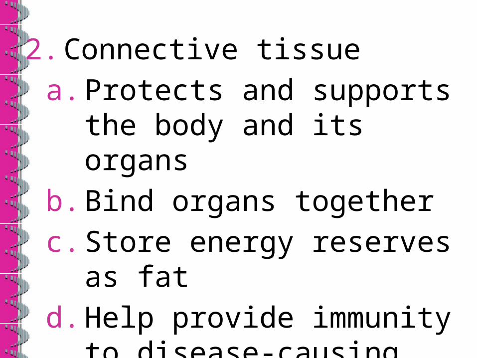
2. Connective tissuea. Protects and supports the
body and its organsb. Bind organs togetherc. Store energy reserves as
fatd. Help provide immunity to
disease-causing organisms

3. Muscular tissuea. Generates the physical
force needed to make body structures move

4. Nervous tissuea. Detects changes in a
variety of conditions inside and outside the body
b. Responds to changes by generating nerve impulses
c. Nerve tissue in the brain helps maintain homeostasis


1. EPITHELIAL TISSUE
• Consists of cells arranged in continuous sheets
• in either single or multiple layers
• There is little intercellular space between adjacent cells
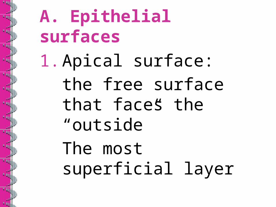
A. Epithelial surfaces
1. Apical surface:the free surface that faces the “outside”The most superficial layer

2. Lateral surface:faces the adjacent cells
3. Basal surface:opposite the apical surfaceadhered to extracellular materialsThe deepest layer


B. Types of Epithelium1. Covering and lining:
form the outer covering of the skin and some internal organsalso forms the inner lining of blood vessels, ducts and body cavitiesand the interior of many organ systems
a. Classification of covering and lining cells• C & L cells are classified according to two characteristics:
• Arrangement of cells into layers
• Cell shape
i. Arrangement in layers
• Arranged in one or more layers depending on the function performed

Simple Epithelium
• A single layer of cellsthat function in diffusion, osmosis, filtration, secretion (production and release) and absorption (intake)

Stratified Epithelium
• Two or more layers of cells that protect underlying tissues
• Found in locations of considerable wear and tear

ii. Cell shape
• Squamous cells: thin and arranged like floor tilesallowing for the rapid movement of substances through themoccur in cells in which diffusion or infiltration occurs

Cuboidal cells• Are as tall as they are wide
• Shaped like cubes…or hexagons
• May have microvilli on apical surface
• Function in secretion or absorption

Columnar cells
• Taller than they are wide• Protect underlying tissue
• Apical surface may have microvilli or cilia
• Function in secretion or absorption
Transitional cells
• Cells that change shape from cuboidal to flat or visa versa
• This occurs as organs stretch or collapse


2. Glandular Epithelium
constitutes the secreting portion of glands such as:thyroid, adrenal and sweat glands.

2. CONNECTIVE TISSUE
• One of the most abundant and widely distributed tissues
• Performs a variety of functions:–binds together, supports and strengthens other body tissues

–protects and insulates internal organs
–compartmentalizes structures such as skeletal muscles
–is the major site of immune responses
–is the major transport system (as blood is a fluid connective tissue)

• Consists of cells and an extracellular matrixwhich is located between the widely spaced cells and consists of protein fibers and ground substance (the stuff between the cells and the fibers)

• The ground substance:• supports cells, • binds cells together• stores water• provides a medium for exchange of materials between the blood and cells
• The fibers include collagen fibers, elastic fibers and reticular fibers. They strengthen and support ct.
• Collagen fibers are found in most types of ct and are made of the protein collagen. This is the most abundant protein in the body!
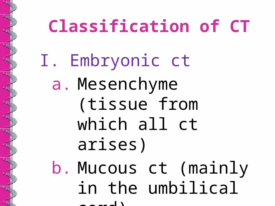
Classification of CT
I. Embryonic cta. Mesenchyme (tissue
from which all ct arises)
b. Mucous ct (mainly in the umbilical cord)
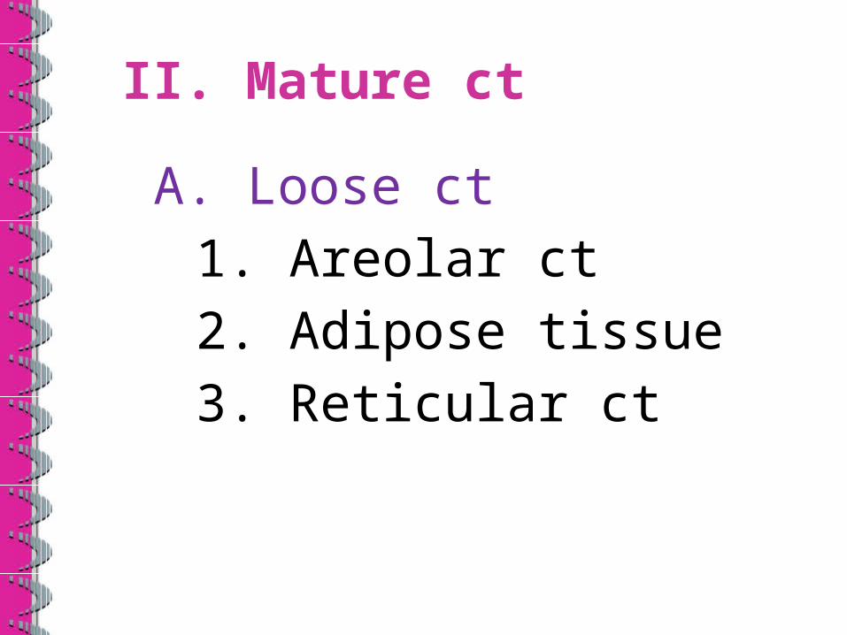
II. Mature ct
A. Loose ct1. Areolar ct2. Adipose tissue3. Reticular ct
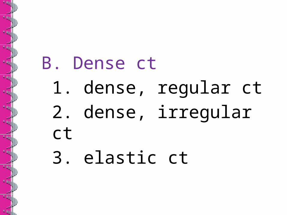
B. Dense ct1. dense, regular ct2. dense, irregular ct3. elastic ct

C. Cartilage1. Hyaline cartilage2. Fibrocartilage3. Elastic cartilage

D. Bone tissueE. Liquid ct1. Blood tissue2. Lymph

• Areolar connective tissue is the most widely distributed ct and is the only kind that occurs on an external surface and is the tissue that lines joint cavities
• C.T. is usually highly vascular therefore has a rich blood supply
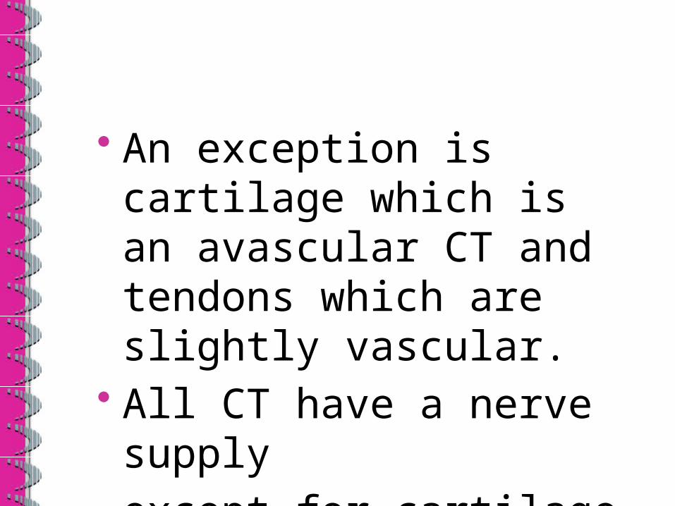
• An exception is cartilage which is an avascular CT and tendons which are slightly vascular.
• All CT have a nerve supplyexcept for cartilage

A. TISSUE CELLS• Mesenchymal cells give rise to the all tissue cells
• The immature form of tissue cells are–Fibroblasts (CT), chondroblasts (cartilage) and osteoblasts (bone)

• Immature “blast” cells are able to divide and secrete the matrix of its tissue.
• Mature “cyte” cells have a reduced capacity for cell division and are more involved in matrix maintenance
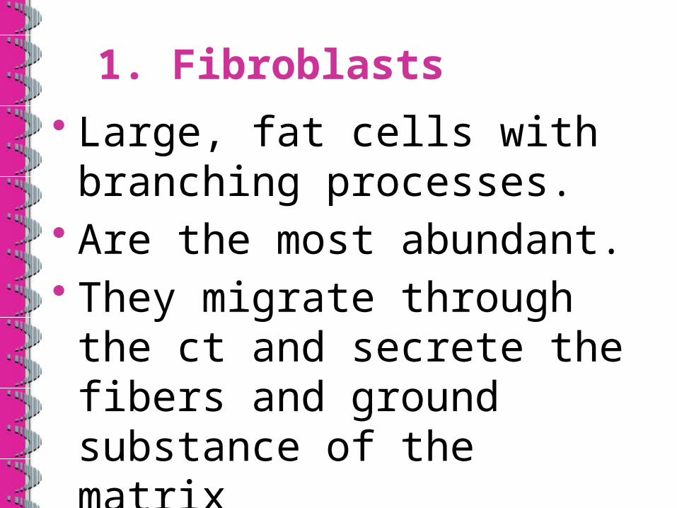
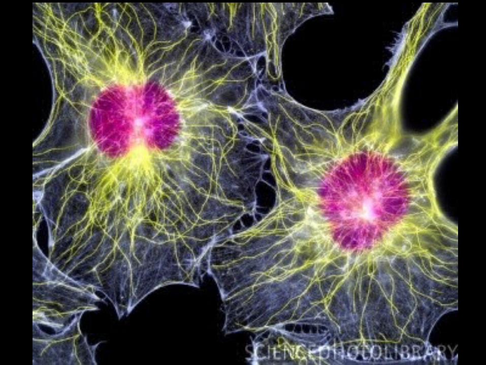
1. Fibroblasts
• Large, fat cells with branching processes.
• Are the most abundant.• They migrate through the ct and secrete the fibers and ground substance of the matrix

2. Macrophages• Have an irregular shape with short branching projections
• Capable of engulfing bacteria and cellular debris by phagocytosis
• Some are fixed (remain in one cell) and others wander and gather at the site of infection


3. Plasma cells
• Cells that develop from β lymphocytes.
• Secrete antibodies that attack or neutralize foreign bodies

4. Mast cells
• Abundant along the blood vessels that supply the ct
• Produce histamine: dilates blood vessels as an inflammatory response
• and heparin: an anticoagulant

5. Adipocytes
• Fat cells• Store triglycerides• Found deep beneath the skin…around organs such as the heart and kidneys

6. White Blood Cells
• Not normally abundant in ct
• Migrate from blood to ct in response to events such as infection, parasitic invasions and allergic reactions
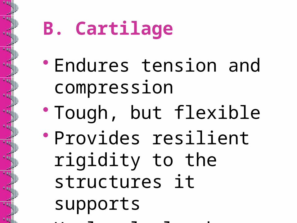
B. Cartilage
• Endures tension and compression
• Tough, but flexible• Provides resilient rigidity to the structures it supports
• Heals slowly when injured

Three types of cartilage
1. Hyaline cartilage: most abundant; provides firm support with some pliability
Found at: ends of long bones, tip of nose, between ribs and sternum, as a support to the respiratory system

2. Elastic cartilage• Found where strength
and stretchability are needed
• Form the external ear and the epiglottis

3. Fibrocartilage– Compressible and
resists tension– Found where strong
support and ability to withstand pressure are needed
– Intervertebral discs and the menisci of the knee

C. Bone (osseous tissue)
• Exceptional ability to support and protect body structures
• Well supplied by invading blood vessels
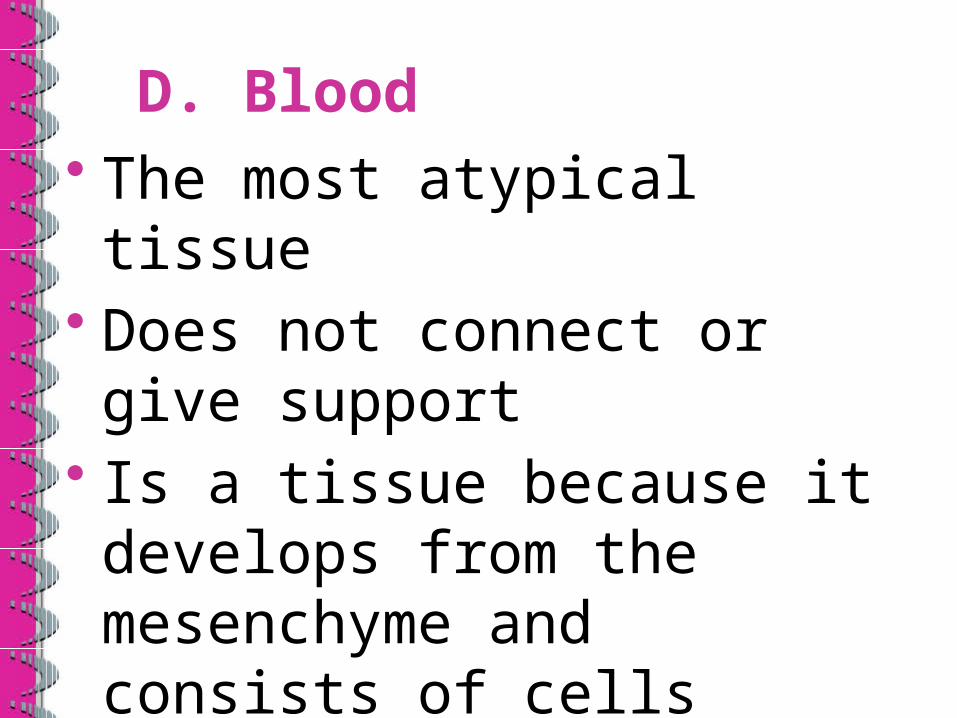
D. Blood• The most atypical tissue• Does not connect or give support
• Is a tissue because it develops from the mesenchyme and consists of cells surrounded by a fluid matrix

E. Muscle Tissue
• Highly cellular, well-vascularized tissues
• Responsible for most types of body movements
• Skeletal, cardiac and smooth are the three types of muscle tissue

F. Nervous Tissue
• Main component of the nervous system consists of various types of supporting cells, nonconducting cells that support, insulate and protect the delicate nerve cells (neurons)
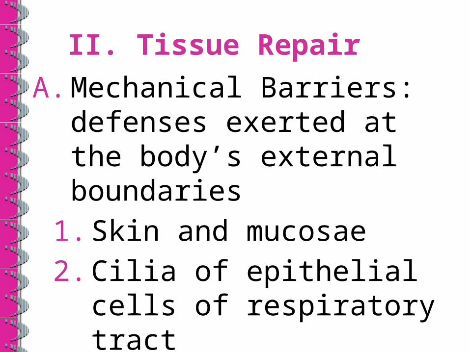
II. Tissue Repair
A. Mechanical Barriers: defenses exerted at the body’s external boundaries
1. Skin and mucosae2. Cilia of epithelial cells
of respiratory tract3. Acid produced by
stomach
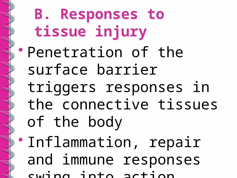
B. Responses to tissue injury
• Penetration of the surface barrier triggers responses in the connective tissues of the body
• Inflammation, repair and immune responses swing into action

1. Inflammation
• Tissue trauma triggers the release of inflammatory chemicals that cause capillaries to dilate and become more permeable

• WBCs and plasma fluid seep into the injured area
• The clotting process takes place stopping the loss of blood and holding the edges of the injured area together isolating the injury from further harm
2. Organization
• The blood clot is replaced by granulation tissuewhich contains capillaries that grow in from nearby areas giving the tissue a “nubby” or granular appearance

• Granulation tissue is resistant to infection because of the bacteria inhibiting substances it produces
• Fibroblasts produce growth factors and collagen fibers to bridge the gap created by the injury
• Granulation tissue eventually becomes scar tissue

• Once granulation tissue has accumulated enough to form a fibrous patch,the fibroblasts either revert to the resting stage or undergo apoptosis

3. Regeneration• Surface epithelial tissue grows under the forming scab
• Fibrous tissue matures and contracts resulting in fully regenerated epithelial tissue and underlying scar tissue (which may be visible depending upon the severity of the wound)
• The entire repair process is put into motion following a wound that goes through the epithelial barrier.
• An infection only requires the regeneration portion of the process

THE END!