Embed Size (px)
Citation preview
TISSUE LE
VEL OF
ORGANIZAT
ION
F AL L 2
01
2
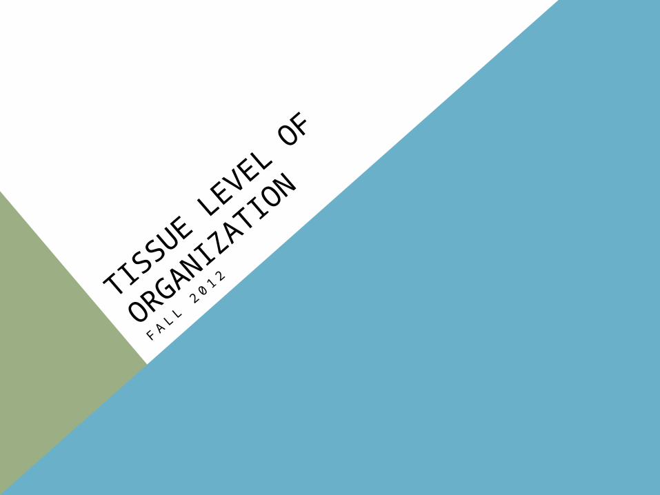
• a – without
• apo – front
• cardium – heart
• chondros – cartilage
• dendron – tree
• desmos – ligament
• glia – glue
• histos – tissue
• holos – entire
• hyalos – glass
• inter – between
• krinein – to separate
• lacus – lake
• meros – part
• neuro – nerve
• os – bone
VOCAB DEVELOPMENT
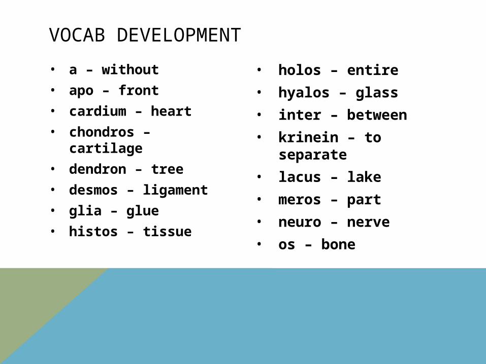
• peri – around
• phagein – to eat
• pleura – rib
• pseudes – false
• sistere – to set
• soma – body
• squama – plate or scale
• vas – vessel
VOCAB DEVELOPMENT
FOUR TISSUE TYPES
• cells combine to form tissues• tissues are groups of specialized cells and cell
products that perform a limited number of functions
• histology is the study of tissues
• 4 basic types of tissues• epithelial• connective• muscle• neural
EPITHELIAL TISSUE
• includes epithelia and glands• epithelia are layers of cells that cover internal or external surfaces •glands are composed of secreting cells derived from epithelia
EPITHELIAL TISSUE
• Important Characteristics•cells that are bound closely together •a surface exposed to the environment or to some internal chamber or passageway•attachment to underlying connective tissue by a basement membrane•absence of blood vessels•continual replacement or regeneration of epithelial cells that are damaged or lost at the exposed surface
EPITHELIAL TISSUE
• Functions of Epithelial Tissue• Provide physical protection•Control permeability• Provide sensation• Produce specialized secretions •Gland cells •Glandular epithelium• Exocrine- secretions are discharged to the surface of the epithelium• Endocrine- secretions are released into the surrounding tissue fluid and blood • These secretions are usually called hormones.
INTERCELLULAR CONNECTIONS
• Undamaged epithelia form effective barriers.• The plasma membranes are held together by CAMs
and by a thin layer of intercellular cement. The CAMs bind to:
Cytoskeletal filaments Each other Extracellular materials
The CAMs form specialized attachment sites called cell junctions 3 types of junctions
Tight junctions Gap junctions Desmosomes

BASEMENT MEMBRANE
• Lies between the epithelium and underlying connective tissues
• No cells, it is made up of a network of proteins
• Provides strength and resists distortion
EPITHELIAL RENEWAL & REPAIR
• Maintains its structures by the continuous division of stem cells • Stem cells are unspecialized cells • Found in the deepest layer of the epithelium, near
the basement membrane
CLASSIFICATION OF EPITHELIA
• Classified according to:• Number of cell layers• Shape of exposed cells
CLASSIFICATION OF EPITHELIA
• Cell layers• Simple epithelium• Single layer of cells covering the basement
membrane • Thin• Found in protected areas inside the body • Line internal compartments & passageways • Common where absorption & secretion takes
place
CLASSIFICATION OF EPITHELIUM
• Cell layers• Stratified Epithelium • Several layers thick above the basement membrane • Found in areas subject to mechanical or chemical
stresses
CLASSIFICATION OF EPITHELIUM
• Cell shapes• Squamous • Cells are thin & flat• Nucleus occupies the thickest portion of each cell
• Cuboidal • Appear square in 2 dimensions• Appear hexagonal in 3 dimensions • Nucleus is near the center of each cell
CLASSIFICATION OF EPITHELIUM
• Cell shapes• Columnar • Hexagonal, but taller & more slender than cuboidal • Nucleus is found near basement membrane

CLASSIFICATION OF EPITHELIA
• Simple Squamous Epithelia• Found in protected regions where absorption takes
place or a slippery surface • Ex: exchange surfaces of lungs, lining of ventral body
cavities, lining of blood vessels
• Simple Cuboidal Epithelia • Provides limited protection & occurs where absorption
& secretion take place • Secret enzymes & buffers in the pancreas & salivary
glands
CLASSIFICATION OF EPITHELIA
• Simple Columnar Epithelia • Provide some protection & may occur in areas of
absorption or secretion• Ex: lines the stomach, the intestinal tract, and many
excretory ducts
• Stratified Squamous Epithelia• Found where mechanical stresses are severe• Ex: surface of the skin, lining of the mouth, tongue,
esophagus
CLASSIFICATION OF EPITHELIA
• Stratified Cuboidal Epithelia • Relatively rare• Ex: along the ducts of sweat glands and the ducts of
the mammary glands
• Stratified Columnar Epithelia• Relatively rare• Ex: portions of the pharynx, epiglottis, urethra
CLASSIFICATION OF EPITHELIA
• Psuedostratified Epithelia • Called this because it looks stratified but it is not • Found in portions of the respiratory tract
• Transitional Epithelia• Stratified epithelium that tolerates repeated
stretching • Lines the urinary bladder
GLANDULAR EPITHELIA
• Exocrine secretions are produced by exocrine glands and are discharged through a duct to the outside.
• Endocrine secretions are produced by ductless glands and are released into blood or tissue fluids

GLANDULAR EPITHELIA
• Mechanisms of Secretion • Merocrine secretion• Most common form of secretion• Leaves the cell intact and able to function • Product is released from secretory vesicles by
exocytosis • Ex: mucus
GLANDULAR EPITHELIA
• Mechanisms of Secretion• Apocrine secretion• Involves the loss of cytoplasm and the secretory
product• Leaves the cell intact and able to function
GLANDULAR EPITHELIA
• Mechanisms of Secretion• Holocrine secretions • Entire cell becomes packed with secretions and
bursts and dies • Ex: sebaceous glands
GLANDULAR EPITHELIA
• Types of secretions • Serous • Watery solution contains enzymes (saliva)
• Mucous• Thick, slippery mucus (snot)
• Mixed • Contains more than one type of secretion
CONNECTIVE TISSUES
• 3 basic components• Specialized cells• Protein fibers• Ground substance• Fluid
• Matrix- extracellular protein fibers & ground substance that surround the cells of connective tissue
CONNECTIVE TISSUES
• Never exposed to the outside
• Highly vascularized
• Contain receptors that provide pain, pressure, temperature, & other sensations
CONNECTIVE TISSUES
• Functions • Support & Protection• Transportation of materials• Storage of energy reserves • Defense of the body
CONNECTIVE TISSUES
• Three major types • Connective tissue proper• Fluid connective tissues • Blood • lymph
• Supporting connective tissues • Cartilage • Bone
CONNECTIVE TISSUE PROPER
• Cells found in connective tissue proper• Fibroblasts • Fibrocytes• Macrophages• Fat cells (adipocytes) • Mast cells
CONNECTIVE TISSUE PROPER
• 3 types of connective tissue fibers • Collagen fibers• Most common fibers in connective tissue proper
• Elastic fibers • Return to their original shape after stretching
• Reticular fibers• Least common • Form the framework of various organs
CONNECTIVE TISSUE PROPER
• Types of connective tissue proper• Classified into types based on their relative
proportions or cells, fibers, & ground substance • Loose• Dense
CONNECTIVE TISSUE PROPER
• Loose Connective Tissues • Locations• Beneath the dermis of the skin • Digestive tract• Respiratory and urinary tracts • Between muscles• Around blood vessels, nerves, & joints
CONNECTIVE TISSUE PROPER
• Loose connective tissue functions. • Cushions organs• Provides support, but allows independent movement• Phagocytic cells defend against pathogens
CONNECTIVE TISSUE PROPER
• Loose Connective Tissue• Adipose tissue (composed mainly of triglycerides) • Locations:• Underneath the deep skin (sides, buttocks,
breasts)• Padding around eyes and kidneys
• Functions: • Provides padding & cushions shocks • Insulates• Stores energy reserves

CONNECTIVE TISSUE PROPER
• Dense Connective Tissue• Locations: • Between skeletal muscles & skeleton• Tendons- connect muscle to bone
• Between bones • Ligaments – connect bone to bone
• Covering skeletal muscles• Capsules of internal organs
CONNECTIVE TISSUE PROPER
• Dense connective tissue • Functions:• Provides firm attachment• Conducts pull of muscles• Reduces friction between muscles• Stabilizes relative position of bones• Helps prevent over expansion of organs (bladder)
FLUID CONNECTIVE TISSUES
• 2 types• Blood• Plasma- watery matrix• Red blood cells• Account for half the volume of blood• Transport oxygen through the blood
• White blood cells- immune response • Platelets- clotting
• Lymph • Forms as interstitial fluid enter lymphatic vessels • Circulated throughout the entire body• Supports your immune system
SUPPORTING CONNECTIVE TISSUES
• 2 types • Cartilage • Bone
SUPPORTING CONNECTIVE TISSUES
• Cartilage• Heals poorly because it is avascular• 3 types of cartilage• Hyaline cartilage• Elastic cartilage• Fibrous cartilage
SUPPORTING CONNECTIVE TISSUES
• Elastic Cartilage • Locations: • Auricle of external ear• Epiglottis • Auditory tube• Part of larynx
• Functions:• Provides support• tolerates distortion without damage and returns to
original shape
SUPPORTING CONNECTIVE TISSUES
• Hyaline Cartilage• Locations: • Between tips of ribs & bones of the sternum • Cover bone surfaces at synovial joints• Supporting larynx, trachea, & bronchii• Forms part of the nasal septum
• Functions:• Provides stiff but somewhat flexible support• Reduces friction between bony surfaces
SUPPORTING CONNECTIVE TISSUES
• Fibrous Cartilage• Locations:• Pads within knee joint• Between the pubic bones of the pelvis &
intervertebral discs• Functions:• Resists compression• Prevents bone on bone contact • Limits relative movement

SUPPORTING CONNECTIVE TISSUES
• Bone• Osteocytes- cells that make up bones (hard outer part
of the bone)• Periosteum- covering around the bone
MEMBRANES
• Membranes are physical barriers
• 4 types • Mucous• Serous• Cutaneous• Synovial
MEMBRANES
• Mucous Membranes • Line cavities• Communicate with the exterior
MEMBRANES
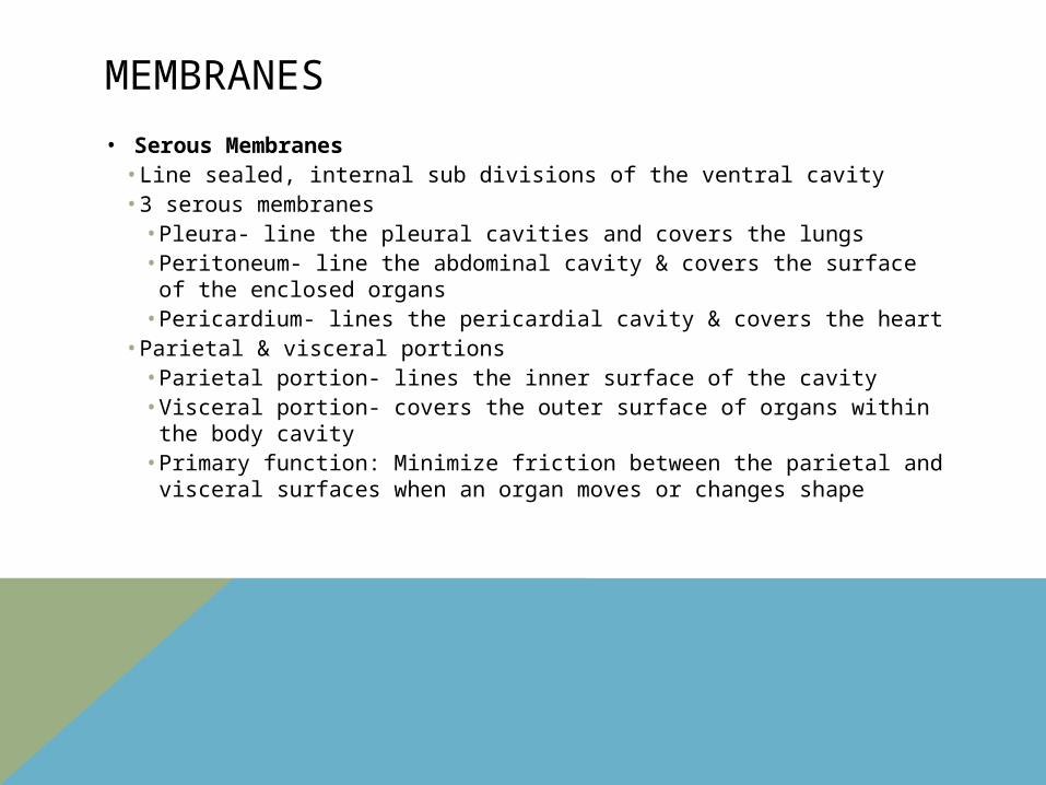
• Serous Membranes • Line sealed, internal sub divisions of the ventral cavity • 3 serous membranes • Pleura- line the pleural cavities and covers the lungs• Peritoneum- line the abdominal cavity & covers the surface of
the enclosed organs • Pericardium- lines the pericardial cavity & covers the heart
• Parietal & visceral portions • Parietal portion- lines the inner surface of the cavity• Visceral portion- covers the outer surface of organs within the
body cavity• Primary function: Minimize friction between the parietal and
visceral surfaces when an organ moves or changes shape
MEMBRANES
• Cutaneous Membrane• Covers the surface of the body (skin) • Thick • Waterproof• Usually dry • Consists of: stratified squamous epithelium &
underlying dense connective tissues
MEMBRANES
• Synovial Membranes• Consist primarily of loose connective tissue & an
incomplete layer of epithelial tissue• Found in joints that are allowed to move free • Lubricates joints
MUSCLE TISSUE
• Specialized from contraction
• 3 types:• Skeletal• Cardiac• Smooth
MUSCLE TISSUE
• Skeletal Muscle Tissue• Cells- long, cylindrical, striated, multinucleate • Locations: • Combined with connective tissues & neural tissue in
skeletal muscles• Functions:• Moves or stabilized the position of the skeleton• Guards entrances and exits to the digestive,
respiratory, and urinary tracts• Generates heat• Protects internal organs
MUSCLE TISSUE
• Cardiac Muscle Tissue• Cells: short, branched, and striated; usually have a
single nucleus• Location:• Heart
• Functions:• Circulates blood• Maintains blood pressure
MUSCLE TISSUE
• Smooth Muscle Tissue • Cells: short, spindle shaped, and non striated; single
central nucleus • Location: • Walls of blood vessels• Digestive, respiratory, urinary, & reproductive organs
• Functions:• Moves food, urine, and reproductive tract secretions• Controls diameter of respiratory passageways• Regulates diameter of blood vessels
NEURAL TISSUE
• Specialized for the conduction of electrical impulses from one region of the body to another.
• Most is found in the brain and spinal cord
• 2 basic types of cells• Neurons• 3 main parts • Cellbody• Dendrites• Axon
• neuroglia


![DEMENTIA CLINICAL SERVICE MAPPING SOUTH COAST ...nhs.joindementiaresearch.nihr.ac.uk/wp-content/uploads/...Dementia Clinical Service Map- South Coast DeNDRoN -Version 1 (SW SHA) [5]](https://img.pdfslide.us/doc/110x75/60b26a65f6c58544614b2c65/dementia-clinical-service-mapping-south-coast-nhs-dementia-clinical-service.jpg)