Embed Size (px)
Citation preview
861
Abstract. – OBJECTIVE: To investigate the im-pact of tissue glue on mast cells (MC) and epider-mal growth factor receptor (EGFR) in rat peptic ul-cer.
MATERIALS AND METHODS: SD rats were used to establish peptic ulcer model. Cimeti-dine gavage was adopted in the positive control, while cimetidine and endoscopic tissue glue therapy were applied in the experimental group. The ulcer inhibition rate and ulcer index were measured to evaluate healing quality. Real-time PCR was performed to test EGFR mRNA in the ulcer surrounded gastric mucosa. Immunohisto-chemistry was selected to determine MC quan-tity. HE staining and apoptosis detection were used to evaluate cell apoptosis.
RESULTS: Tissue glue significantly reduced MC number in the peptic ulcer rat compared with control with dose dependence (p < 0.05). Tissue glue significantly decreased ulcer area, and el-evated ulcer index and inhibition rate (p < 0.05). EGFR mRNA in the mucosa markedly declined after modeling. Tissue glue upregulated EGFR mRNA to a certain extent (p > 0.05). Tissue glue induced MC apoptosis with dose dependence.
CONCLUSIONS: Endoscopic application of tissue glue accelerated ulcer mucosa healing via up-regulating EGFR mRNA, enhancing gastroin-testinal mucous membrane regeneration and re-pair ability, and decreasing MC number.
Key Words:Peptic ulcer, Endoscopic tissue glue, Healing quali-
ty, Mast cell, EGFR.
Introduction
Peptic ulcer, a common disease in digestive system, refers to the mucosal ulcer caused by pep-sase and gastric acid auto-digestion. It exhibits
complex etiology and long-term recurrent attack1. The etiology and pathogenesis of peptic ulcer are still unclear. At present, the imbalance between damage factor and defending factor is the main cause of ulcer2,3. Damage factors include drugs, pepsase, gastric acid, prostaglandin, epidermal growth factor, Helicobacter pylori, spirit, and genetic factors. The healing of peptic ulcer is a conundrum of its treatment. Various histological and ultra-structural abnormalities affect the de-fense of mucous membrane and cell oxygenation capacity, becoming the physiological and patho-logical bases of ulcer healing and recurrence. Its treatment not only needs to repair mucosal dele-tion, but also rebuild the sub-mucosal tissue4. The mucosal healing process is affected by growth factor and its receptor, leading to stimulate the digestive tract mucosa cells proliferation, exert the regulatory function of the growth factors, promote the gastric mucosal blood flow, inhibit gastric acid secretion, facilitate mucosal healing, and exhibit strong mucosal cell protection. Cur-rent studies found that epidermal growth factor receptor (EGFR) showed good digestive tract mu-cosal protection. Its expression in normal gastric mucosa is few, while gastrointestinal damage can increase its level. It can stimulate epithelial cells and surrounding cells proliferation, with high af-finity of the digestive tract mucosa and healing effect to the ulcer surface5. Also, there are various mast cells (MC) with multiple biological activities and different anatomical parts that have different impact on inflammation. In the digestive tract, MC release mediator to promote muscle contrac-tion, increase vascular permeability and mucous secretion, and accelerate the inflammatory cells stimulating the pain nerve, cause the digestive
European Review for Medical and Pharmacological Sciences 2017; 21: 861-866
Y.-Y. SHI1, H.-F. LIU1, M. MIN2, W. WANG1, J. LI1, C.-Y. HE1, Q.-P. DU3
1Department of Gastroenterology, Beijing Armed Police General Hospital, Beijing, China2Departmentof Gastroenterology, People ‘s Liberation Army 307 Hospital, Beijing, China3Department of Gastroenterology, Beijing First Integrative Traditional Chinese and Western Medicine Hospital, Beijing, China
Yingying Shi and HaifengLiu contributed equally to this work
Corresponding Author: Qiuqeng Du, MD; e-mail: [email protected]
Correlation analysis of mast cells and EGFR with endoscopic application of tissue glue for treatment of peptic ulcer healing
Y.-Y. Shi, H.-F. Liu, M. Min, W. Wang, J. Li, C.-Y. He, Q.-P. Du
862
tract local edema, weaken uptake of nutrients, give rise to local blood loss, and generate pain6. It is considered that MC involved in the occur-rence, development, and healing of peptic ulcer7. In recent years, endoscopic application of tissue glue obtained good clinical effect, with the role of hemostasis and promoting local inflammation resolution. However, its treatment mechanism is still controversy. Therefore, we explored the role of endoscopic tissue glue on peptic ulcer healing and related mechanism through using the animal model, aiming to provide new thought for the treatment and prevention of peptic ulcer.
Materials and Methods
Experimental AnimalMale SD rats weighted 180-220 g were bought
from Beijing Animal Center (license key: SCXK 2016-0263). The rats were raised in SPF grade an-imal feeding room with temperature at 21-24°C, humidity at 40%-60%, free eating and drinking, and 12 h day-night cycle.
Rats were used for all experiments, and all procedures were approved by the Animal Eth-ics Committee of Beijing Armed Police General Hospital, Beijing, China.
Drugs and ReagentsEGFR and MC were got from Sigma-Aldrich
(St. Louis, MO, USA). Endoscopic tissue glue was obtained from Ankang Chia Tai Pharmaceutical co., Ltd (Beijing, China). Biochemistry kit was purchased from Merlin medical science co., Ltd (Shanghai, China). PCR primers were construct-ed by Sangon (Shanghai, China).
InstrumentsInjection needle, GIF-Q150 electronic gas-
troscope, histotome, Real-time PCR amplifier, microplate reader, high-speed centrifuge, em-bedding machine, and analytical balance were purchased from Olympus (Tokio, Japan). α-butyl-cyanoacrylate tissue glue at 0.5 ml was obtained from Suncon Medical Adhesive (Beijing, China).
Experimental Method
Animal Model EstablishmentA total of 80 male SD rats were randomly di-
vided into two groups, including 70 in the ex-perimental group and 10 in the negative control.
The rats in experimental group were anesthetized by 1% pentobarbital sodium (Haoran Bio. Ltd., Beijing, China) and given 0.002 ml 100% glacial acetic acid (Haoran Bio. Ltd., Beijing, China) in-jection at the junction of the antrum of the stom-ach and gastric body to construct the peptic ulcer animal model. When the white ulcer ring on the serosal layer reached 3 mm, the stomach was re-turned to the abdominal cavity. Tissue glue was endoscopically injected at 1.5 ml, 1 ml, or 0.5 ml for 2 days, respectively. The endoscopic normal saline injection was selected as normal control. The rats in sham group were opened abdominal cavity without injection.
Index ObservationAfter two days’ treatment, the rat was anesthe-
tized to extract blood from the abdominal aorta. The serum was separated, while the gastric mu-cosa was put into the liquid nitrogen and embed-ded after paraformaldehyde (Sigma-Aldrich, St. Louis, MO, USA) fixation.
Mast Cells HE Staining and Apoptosis Detection
Apoptosis index (AI) = apoptotic cell number/total cell number × 100%. Apoptotic MC was stained by TUNEL kit (Roche, Mannheim, Ger-many) and observed under the microscope (Olym-pus, Tokyo, Japan). The apoptotic cytoplasm was stained as yellow, whereas the cell nucleus was stained as hyacinthine. The apoptotic cells were accounted to calculate AI.
EGFR mRNA DetectionPCR reaction was performed at pre-degen-
eration at 95°C for 2 min, followed by 40 cy-cles of 95°C for 20 s, 62°C for 30 s, and 68°C for 60 s. Total RNA was extracted from gas-tric mucosal tissue using Trizol method. Re-al-time PCR was used to test EGFR mRNA expression. The primers sequences were as fol-lows. EGFR mRNA, forward, 5’-ACTCGCAG-GAAAGACTAGCA-3’, reverse, 5’-AGCAGT-GGAAGAATCGGACC-3’. GAPDH, forward, 5’-CATCAGCAATGCCTCCTGCAC-3’, reverse, 5’-TGAGTCCTTCCACGATACCAAAGTT -3’.
Statistical AnalysisAll data analyses were performed by SPSS
20.0 software (SPSS, Inc., Chicago, IL, USA). Enumeration data were compared by x2-test, while measurement data were analyzed by t-test. p < 0.05 was depicted as statistical significance.
Tissue glue promotes gastric ulcer healing
863
Results
The Impact of Tissue Glue on MC Number in Rat Serum
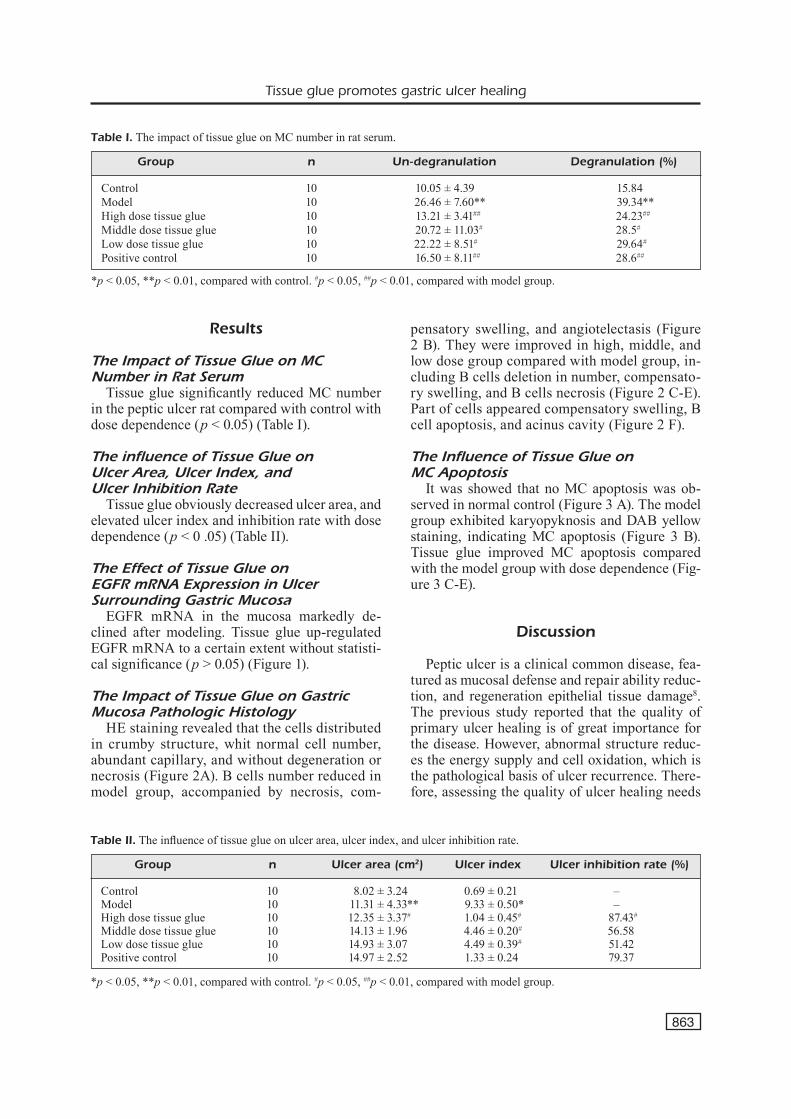
Tissue glue significantly reduced MC number in the peptic ulcer rat compared with control with dose dependence (p < 0.05) (Table I).
The influence of Tissue Glue on Ulcer Area, Ulcer Index, and Ulcer Inhibition Rate
Tissue glue obviously decreased ulcer area, and elevated ulcer index and inhibition rate with dose dependence (p < 0 .05) (Table II).
The Effect of Tissue Glue on EGFR mRNA Expression in Ulcer Surrounding Gastric Mucosa
EGFR mRNA in the mucosa markedly de-clined after modeling. Tissue glue up-regulated EGFR mRNA to a certain extent without statisti-cal significance (p > 0.05) (Figure 1).
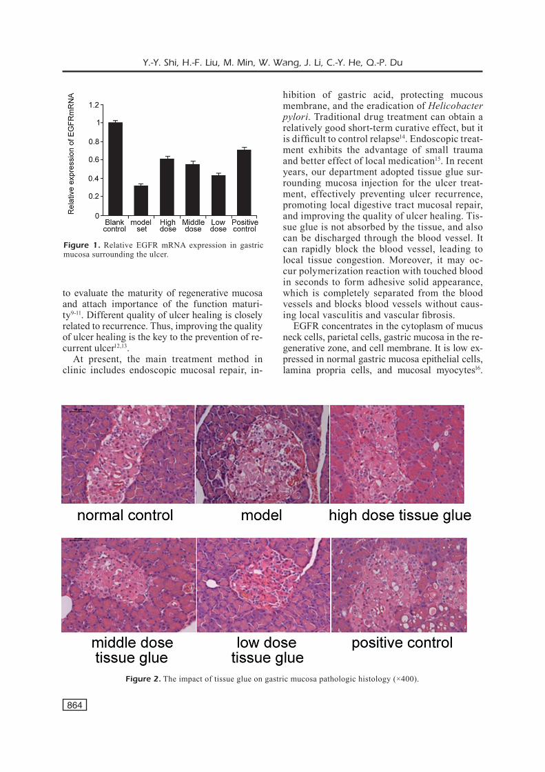
The Impact of Tissue Glue on Gastric Mucosa Pathologic Histology
HE staining revealed that the cells distributed in crumby structure, whit normal cell number, abundant capillary, and without degeneration or necrosis (Figure 2A). B cells number reduced in model group, accompanied by necrosis, com-
pensatory swelling, and angiotelectasis (Figure 2 B). They were improved in high, middle, and low dose group compared with model group, in-cluding B cells deletion in number, compensato-ry swelling, and B cells necrosis (Figure 2 C-E). Part of cells appeared compensatory swelling, B cell apoptosis, and acinus cavity (Figure 2 F).
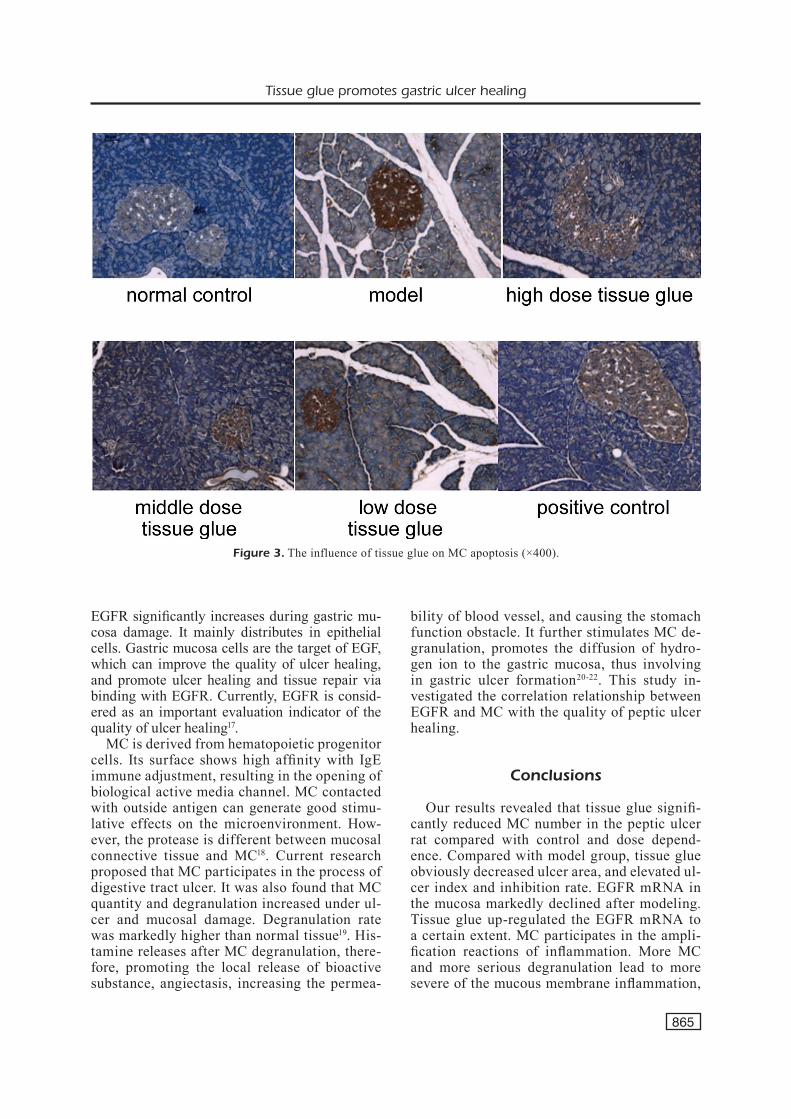
The Influence of Tissue Glue onMC Apoptosis
It was showed that no MC apoptosis was ob-served in normal control (Figure 3 A). The model group exhibited karyopyknosis and DAB yellow staining, indicating MC apoptosis (Figure 3 B). Tissue glue improved MC apoptosis compared with the model group with dose dependence (Fig-ure 3 C-E).
Discussion
Peptic ulcer is a clinical common disease, fea-tured as mucosal defense and repair ability reduc-tion, and regeneration epithelial tissue damage8. The previous study reported that the quality of primary ulcer healing is of great importance for the disease. However, abnormal structure reduc-es the energy supply and cell oxidation, which is the pathological basis of ulcer recurrence. There-fore, assessing the quality of ulcer healing needs
Table I. The impact of tissue glue on MC number in rat serum.
Group n Un-degranulation Degranulation (%)
Control 10 10.05 ± 4.39 15.84Model 10 26.46 ± 7.60** 39.34**High dose tissue glue 10 13.21 ± 3.41## 24.23##
Middle dose tissue glue 10 20.72 ± 11.03# 28.5#
Low dose tissue glue 10 22.22 ± 8.51# 29.64#
Positive control 10 16.50 ± 8.11## 28.6##
*p < 0.05, **p < 0.01, compared with control. #p < 0.05, ##p < 0.01, compared with model group.
*p < 0.05, **p < 0.01, compared with control. #p < 0.05, ##p < 0.01, compared with model group.
Table II. The influence of tissue glue on ulcer area, ulcer index, and ulcer inhibition rate.
Group n Ulcer area (cm2) Ulcer index Ulcer inhibition rate (%)
Control 10 8.02 ± 3.24 0.69 ± 0.21 –Model 10 11.31 ± 4.33** 9.33 ± 0.50* –High dose tissue glue 10 12.35 ± 3.37# 1.04 ± 0.45# 87.43#
Middle dose tissue glue 10 14.13 ± 1.96 4.46 ± 0.20# 56.58Low dose tissue glue 10 14.93 ± 3.07 4.49 ± 0.39# 51.42Positive control 10 14.97 ± 2.52 1.33 ± 0.24 79.37
Y.-Y. Shi, H.-F. Liu, M. Min, W. Wang, J. Li, C.-Y. He, Q.-P. Du
864
to evaluate the maturity of regenerative mucosa and attach importance of the function maturi-ty9-11. Different quality of ulcer healing is closely related to recurrence. Thus, improving the quality of ulcer healing is the key to the prevention of re-current ulcer12,13.
At present, the main treatment method in clinic includes endoscopic mucosal repair, in-
hibition of gastric acid, protecting mucous membrane, and the eradication of Helicobacter pylori. Traditional drug treatment can obtain a relatively good short-term curative effect, but it is difficult to control relapse14. Endoscopic treat-ment exhibits the advantage of small trauma and better effect of local medication15. In recent years, our department adopted tissue glue sur-rounding mucosa injection for the ulcer treat-ment, effectively preventing ulcer recurrence, promoting local digestive tract mucosal repair, and improving the quality of ulcer healing. Tis-sue glue is not absorbed by the tissue, and also can be discharged through the blood vessel. It can rapidly block the blood vessel, leading to local tissue congestion. Moreover, it may oc-cur polymerization reaction with touched blood in seconds to form adhesive solid appearance, which is completely separated from the blood vessels and blocks blood vessels without caus-ing local vasculitis and vascular fibrosis.
EGFR concentrates in the cytoplasm of mucus neck cells, parietal cells, gastric mucosa in the re-generative zone, and cell membrane. It is low ex-pressed in normal gastric mucosa epithelial cells, lamina propria cells, and mucosal myocytes16.
Figure 1. Relative EGFR mRNA expression in gastric mucosa surrounding the ulcer.
Figure 2. The impact of tissue glue on gastric mucosa pathologic histology (×400).
Tissue glue promotes gastric ulcer healing
865
EGFR significantly increases during gastric mu-cosa damage. It mainly distributes in epithelial cells. Gastric mucosa cells are the target of EGF, which can improve the quality of ulcer healing, and promote ulcer healing and tissue repair via binding with EGFR. Currently, EGFR is consid-ered as an important evaluation indicator of the quality of ulcer healing17.
MC is derived from hematopoietic progenitor cells. Its surface shows high affinity with IgE immune adjustment, resulting in the opening of biological active media channel. MC contacted with outside antigen can generate good stimu-lative effects on the microenvironment. How-ever, the protease is different between mucosal connective tissue and MC18. Current research proposed that MC participates in the process of digestive tract ulcer. It was also found that MC quantity and degranulation increased under ul-cer and mucosal damage. Degranulation rate was markedly higher than normal tissue19. His-tamine releases after MC degranulation, there-fore, promoting the local release of bioactive substance, angiectasis, increasing the permea-
bility of blood vessel, and causing the stomach function obstacle. It further stimulates MC de-granulation, promotes the diffusion of hydro-gen ion to the gastric mucosa, thus involving in gastric ulcer formation20-22. This study in-vestigated the correlation relationship between EGFR and MC with the quality of peptic ulcer healing.
Conclusions
Our results revealed that tissue glue signifi-cantly reduced MC number in the peptic ulcer rat compared with control and dose depend-ence. Compared with model group, tissue glue obviously decreased ulcer area, and elevated ul-cer index and inhibition rate. EGFR mRNA in the mucosa markedly declined after modeling. Tissue glue up-regulated the EGFR mRNA to a certain extent. MC participates in the ampli-fication reactions of inflammation. More MC and more serious degranulation lead to more severe of the mucous membrane inflammation,
Figure 3. The influence of tissue glue on MC apoptosis (×400).

Y.-Y. Shi, H.-F. Liu, M. Min, W. Wang, J. Li, C.-Y. He, Q.-P. Du
866
leading to body damage. After treatment, tissue glue induced MC apoptosis with dose depend-ence. Therefore, tissue glue effectively inhibits inflammation, suppresses EGFR reaction to in-flammatory cytokines, and reduces MC genera-tion, thus is advantageous to the quality of ulcer healing.
AcknowledgementsThis work was supported by Capital Medical Development and scientific research fund (NO. Z151100004015213).
Conflict of InterestThe Authors declare that they have no conflict of interests.
References
1) Takeuchi k, Ohishi M, OTa s, suzuMura k, NaraOka h, OhaTa T, seki J, MiyaMae y, hONMa M, sOga T. Meta-bolic profiling to identify potential serum biomark-ers for gastric ulceration induced by nonsteroid anti-inflammatory drugs. J Proteome Res 2013; 12: 1399-1407.
2) ryTer sW, clOONaN sM, chOi aM. Autophagy: a crit-ical regulator of cellular metabolism and homeo-stasis. Mol Cells 2013; 36: 7-16.
3) cheN hc, FONg Th, lee aW, chiu WT. Autophagy is activated in injured neurons and inhibited by methylprednisolone after experimental spinal cord injury. Spine (Phila Pa 1976) 2012; 37: 470-475.
4) kaTO M, ONO s, yOshida T, Mabe k, shiMizu y, asa-ka M. Significance of H. pylori eradication in treat-ment and prevention for low-dose aspirin induced gastric ulcer of elderly. Nihon Rinsho 2010; 68: 2089-2095.
5) NagaNO y, MaTsui h, TaMura M, shiMOkaWa O, Na-kaMura y, kaNekO T, hyOdO i. NSAIDs and acidic environment induce gastric mucosal cellular mi-tochondrial dysfunction. Digestion 2012; 85: 131-135.
6) he lQ, lu Jh, yue zy. Autophagy in ageing and ageing-associated diseases. Acta Pharmacol Sin 2013; 34: 605-611.
7) liu g, bi y, WaNg r, WaNg X. Self-eating and self-defense: autophagy controls innate immuni-ty and adaptive immunity. J Leukoc Biol 2013; 93: 511-519.
8) david s, krONer a. Repertoire of microglial and macrophage responses after spinal cord injury. Nat Rev Neurosci 2011; 12: 388-399.
9) kaNNO h, OzaWa h, sekiguchi a, yaMaya s, iTOi e. In-duction of autophagy and autophagic cell death
in damaged neural tissue after acute spinal cord injury in mice. Spine (Phila Pa 1976) 2011; 36: E1427-1434.
10) bOike Jr, kaO r, Meyer d, Markle b, rOseNberg J, Niebruegge J, sTeiN ac, berkes J, gOldsTeiN Jl. Does concomitant use of paracetamol potenti-ate the gastroduodenal mucosal injury associ-ated with aspirin? A prospective, randomised, pilot study. Aliment Pharmacol Ther 2012; 36: 391-397.
11) Oh Je, lee hk. Autophagy in innate recognition of pathogens and adaptive immunity. Yonsei Med J 2012; 53: 241-247.
12) seO PJ, kiM N, kiM Jh, lee bh, NaM rh, lee hs, Park Jh, lee Mk, chaNg h, JuNg hc, sONg is. Compar-ison of indomethacin, diclofenac and aspirin-in-duced gastric damage according to age in rats. Gut Liver 2012; 6: 210-217.
13) TaMura i, FuJiTa T, TsuMura h, MOriTa y, yOshida M, TOyONaga T, hiraNO s, iNOkuchi h, kuTsuMi h, azu-Ma T. Low-dose aspirin-induced gastroduodenal mucosal injury in Japanese patients with arteri-osclerotic disease. Intern Med 2010; 49: 2537-2545.
14) cheN y, yu l. Autophagic lysosome reformation. Exp Cell Res 2013; 319: 142-146.
15) WONg PM, PueNTe c, gaNley ig, JiaNg X. The ULK1 complex: sensing nutrient signals for autophagy activation. Autophagy 2013; 9: 124-137.
16) Jaber N, dOu z, cheN Js, caTaNzarO J, JiaNg yP, bal-lOu lM, seliNger e, OuyaNg X, liN rz, zhaNg J, zONg WX. Class III PI3K Vps34 plays an essential role in autophagy and in heart and liver function. Proc Natl Acad Sci USA 2012; 109: 2003-2008.
17) Faure M, laFONT F. Pathogen-induced autophagy signaling in innate immunity. J Innate Immun 2013; 5: 456-470.
18) cheN gy, yaNg hJ, lu ch, chaO yc, hWaNg sM, cheN cl, lO kW, suNg ly, luO Wy, TuaN hy, hu yc. Simultaneous induction of autophagy and toll-like receptor signaling pathways by graphene oxide. Biomaterials 2012; 33: 6559-6569.
19) Ng NM, JiaNg sP, zhaNg W. 2-Aminoethoxydiphe-nyl borate reduces degranulation and release of cytokines in a rat mast cell line. Eur Rev Med Pharmacol Sci 2012; 16: 1017-1021.
20) JOuNai N, kObiyaMa k, shiiNa M, OgaTa k, ishii kJ, TakeshiTa F. NLRP4 negatively regulates autophag-ic processes through an association with beclin1. J Immunol 2011; 186: 1646-1655.
21) lei y, WeN h, TiNg JP. The NLR protein, NLRX1, and its partner, TUFM, reduce type I interferon, and enhance autophagy. Autophagy 2013; 9: 432-433.
22) WaNg W, liu J, Wu Q. MiR-205 suppresses auto-phagy and enhances radiosensitivity of prostate cancer cells by targeting TP531NP1. Eur Rev Med Pharmacol Sci 2016; 20: 92-100.