Embed Size (px)
Citation preview
Tissue engineering andtissue repair
Dragica Maja SMRKE, MD,Ph.D., Professor

Tissue engineering
Biology, medicine and technology are today closely interleaved with each other
Tissue engineering – combining cells and biomaterials into functional tissues
Cells are seeded onto a biomaterial scaffold to be integrated into a specific tissue
Examples
• Artificial pancreas
• Artificial bladders
• Cartilage
• Artificial skin
• Artificial bone marrow
• Artificial bone
• Tissue engineered vessels, …
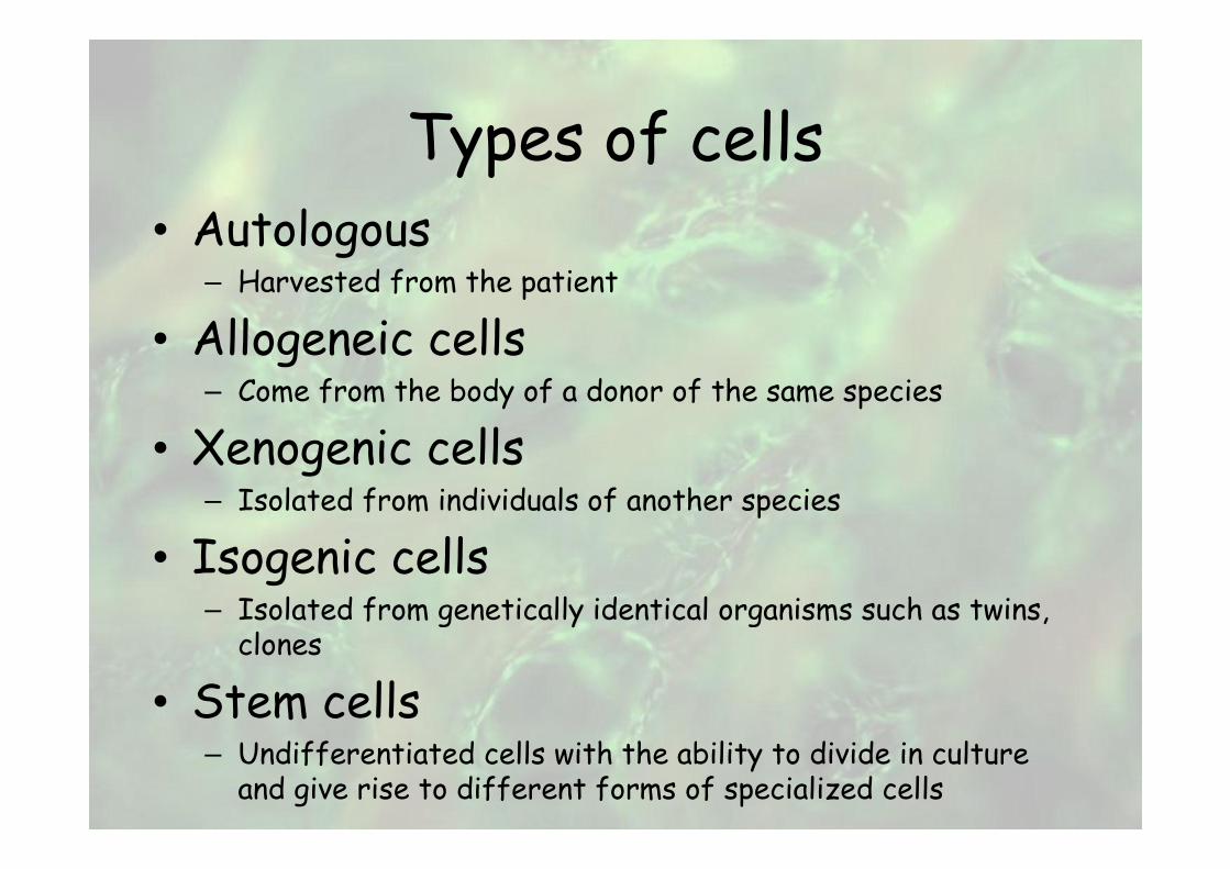
Types of cells• Autologous
– Harvested from the patient
• Allogeneic cells– Come from the body of a donor of the same species
• Xenogenic cells– Isolated from individuals of another species
• Isogenic cells– Isolated from genetically identical organisms such as twins, clones
• Stem cells– Undifferentiated cells with the ability to divide in culture and give rise to different forms of specialized cells
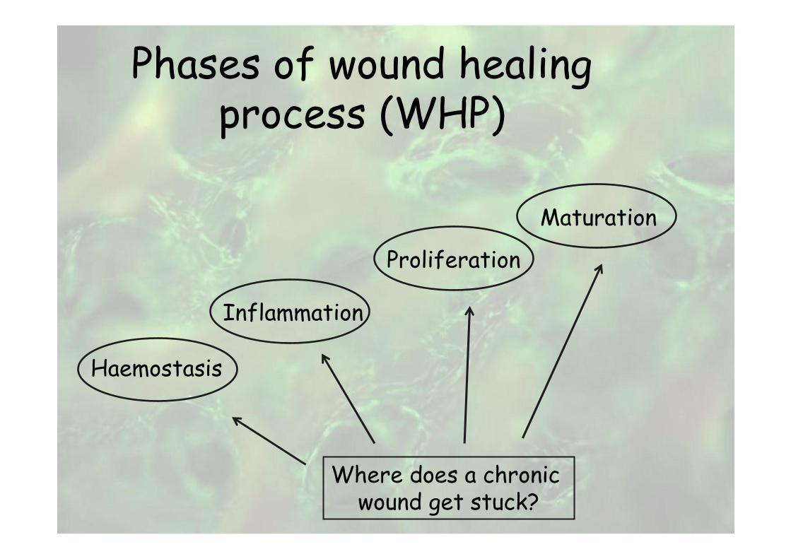
Phases of wound healing process (WHP)
Haemostasis
Inflammation
Proliferation
Maturation
Where does a chronic wound get stuck?
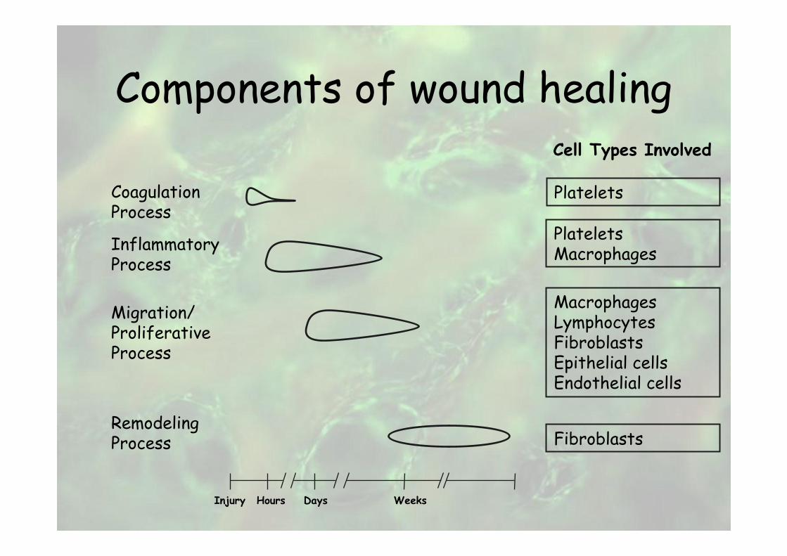
Components of wound healing
CoagulationProcess
InflammatoryProcess
Migration/ProliferativeProcess
RemodelingProcess
Platelets
Cell Types Involved
PlateletsMacrophages
MacrophagesLymphocytesFibroblastsEpithelial cellsEndothelial cells
Fibroblasts
Injury Hours Days Weeks
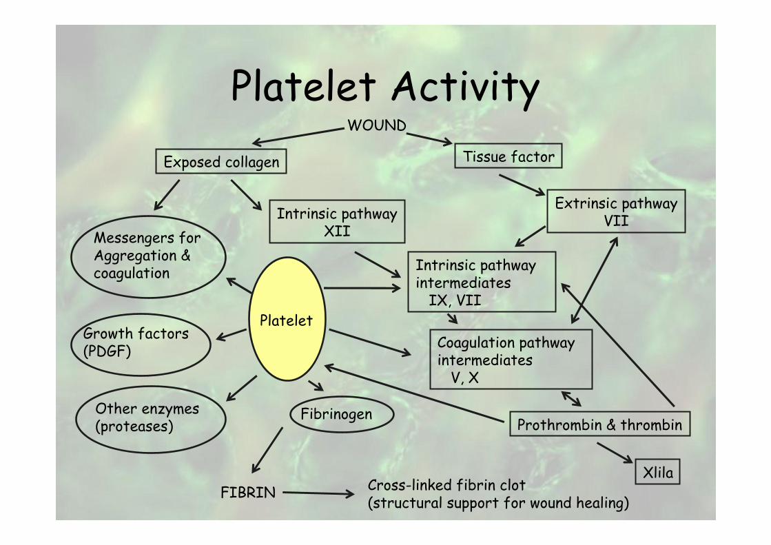
Platelet ActivityWOUND
Exposed collagen
Intrinsic pathwayXII
Intrinsic pathwayintermediatesIX, VII
Extrinsic pathwayVII
Coagulation pathwayintermediatesV, X
Tissue factor
Messengers forAggregation &coagulation
Growth factors(PDGF)
Other enzymes(proteases)
Platelet
Fibrinogen
FIBRIN
Prothrombin & thrombin
XlilaCross-linked fibrin clot(structural support for wound healing)
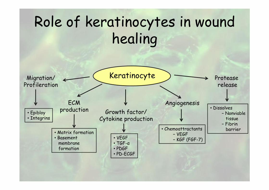
Role of keratinocytes in wound healing
Migration/Profileration
Protease release
AngiogenesisECMproduction Growth factor/
Cytokine production• Epibloy• Integrins
• Matrix formation• Basementmembraneformation
• VEGF• TGF-α• PDGF• PD-ECGF
• Chemoattractants− VEGF− KGF (FGF-7)
• Dissolves− Nonviabletissue
− Fibrinbarrier
Keratinocyte
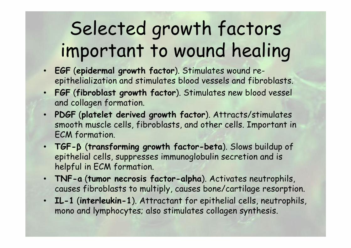
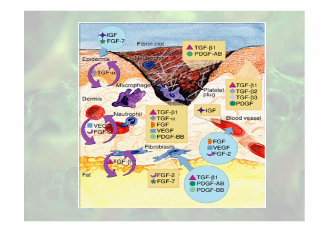
Selected growth factors important to wound healing
• EGF (epidermal growth factor). Stimulates wound re-epithelialization and stimulates blood vessels and fibroblasts.
• FGF (fibroblast growth factor). Stimulates new blood vessel and collagen formation.
• PDGF (platelet derived growth factor). Attracts/stimulates smooth muscle cells, fibroblasts, and other cells. Important in ECM formation.
• TGF-β (transforming growth factor-beta). Slows buildup of epithelial cells, suppresses immunoglobulin secretion and is helpful in ECM formation.
• TNF-a (tumor necrosis factor-alpha). Activates neutrophils, causes fibroblasts to multiply, causes bone/cartilage resorption.
• IL-1 (interleukin-1). Attractant for epithelial cells, neutrophils, mono and lymphocytes; also stimulates collagen synthesis.
Local factors
• Growth factors
• Edema and ischemia
• Low oxygen tension
• Infection
Regional factors
• Arterial insufficiency
• Venous insufficiency
• neuropathy
Systemic factors
• Inadequate perfusion
• Metabolic disease (diabetes)
Wound bed preparation
• Debridement
• Bacterial balance
• Dressing therapies (f.i. silver dressings prevent of infections, help reduce healing time)

Local wound care
debridement moisture balance
SurgicalAutolyticEnzymaticBiological
FoamsCalcium alginatesHydrogelsHydrocolloidsAdhesive filmsNegative pressure therapy
Tissue engineering
Advances– Biological wound dressings
– Material scaffolds and cell material interactions
– The use of stem cells for tissue engineering
– Combination of stem cells and material scaffolds into tissue engineered replacements of tissues and organs
Tissue engineering implants
Synthetic polymeric biomaterials• Nonbiodegradable
Is required to provide and maintain optimal cellular function -> e.g. alginate, liposome,…
• Biodegradable To restore the histological structure and replace the cellular function of recipients -> e.g. poly L-lactic acid, poly glycolic acid, …
Biomaterials in surgery
• Collagen based biomaterials and tissue engineering
Extracellular matrix of connective tissue is composed of proteins:– Collagens (30% of all)
– Proteoglycans
– Glycoproteins
– Elastin
• Collagen provides the principal source of mechanical strength in tissues such as bone, cartilage, skin, tendon, ligaments
• Collagen contributes a structural framework to other tissues (blood vessels, most organs)
Local delivery systems contain gentamicin
• Bone cement (PMMA beads) -> PolyMethyl-Meth-Acrylate
• Collagen (fleeces)
as drug carrier

Wound healing promoting anti-adhesive matrix
The collagen grafting is also applied to produce a healing / promoting antiadhesive membrane
• Particularly necessary in peritoneal surgery to prevent postoperative adhesion


Methods of tissue bioengineering
•Photobiomodulation (modulate cellular activity in red to near infrared light)
•Hyperbaric oxygen therapy: as therapeutic benefit in WT
•Growth factors (from blood)
Methods of tissue bioengineering
Skin replacement
– Cultured epidermal graft
– Cultured human autologous and allogeneic keratinocytes
– Semi synthetic materials
Allogeneic cultivated human skin keratinocytes
• Make rapid healing of the ulcers particularly those that are difficult to heal
• No clinical or laboratory evidence of rejection
• No evidence of preexisting cytotoxic antibodies specific fort the HLA class I antigens expressed on HSE cells
• A fibrin-based skin substitute produced in the defined keratinocyte medium could be safely used to threat a number of skin defect
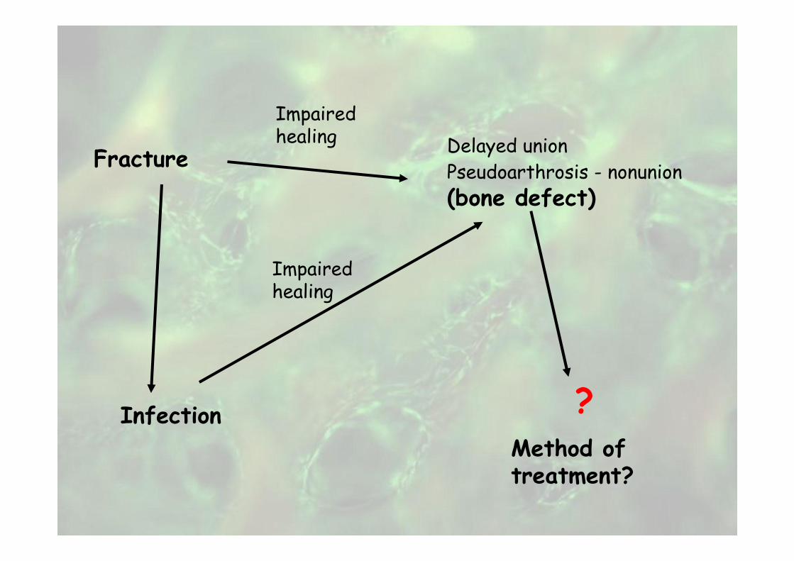
FractureDelayed union
Pseudoarthrosis - nonunion
(bone defect)
Infection ?Method of treatment?
Impaired healing
Impaired healing
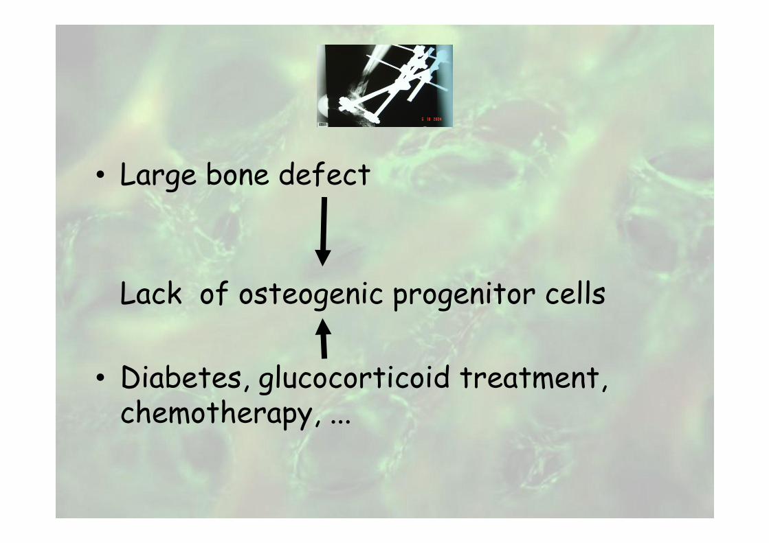
• Large bone defect
Lack of osteogenic progenitor cells
• Diabetes, glucocorticoid treatment, chemotherapy, ...
Reconstruction of bone defect
• Stability
• No infections
• Soft tissue defect reconstruction
Accepted methods of treatment
• Autologous bone transplants– Cancellous bone graft (contains all necessary characteristics of bone substitutes)
– Corticocancellous graft (possibly vascularized�limited amount)
• Homologous (allogeneic) graftBone banks, treated (no rejection), contains only osteoconductive properties
• Ilizarow intercallary bone transport (traction method)
Alternatives
Bone substitute (biomaterials for scaffold):
– Demineralized bone matrix
– Biocompatible ceramics
– Synthetic Calcium phosphate
– Mineral bone
– Collagen
– Composite grafts
– Osteoinductive collagen
Alternatives
Role of GROWTH FACTORS
Role of STEM CELLS
Properties of bone grafts
• Osteogenesis (bone marrow, cancellous bone)
• Osteoinduction– Demineralized bone matrix– Growth factors (platelet rich plasma, bone morphogenic proteins – BMPs)
• Osteoconduction– Ceramics– Collagen
Growth factors (GF)
• Bone and cartilage GF are selected proteins
• Generally are produced locally in the skeleton microenvironment
• Regulate growth and repair
Fracture healing promoting molecules
Growth factors– The transforming growth factor-β (TGF-β) superfamily
• Bone morphogenetic proteins(osteoprogenitors, mesenchymal cells, osteoblasts and chondrocytes within the extracellular matrix produce BMPs.)BMP-2, BMP-4BMP-5, BMP-6, BMP-7GDF-5 (BMP-14), GDF-6 (BMP-13), GDF-7 (BMP-12)BMP-3 (Osteogenin), GDF-10 (BMP-3b)
– Platelet-derived growth factor (PDGF)
– Fibroblast growth factor (FGFs)
– Insulin-like growth factor (IGFs)

Current knowledge of clinical applications of bone
morphogenetic proteins• Marshal R. Wrist 40y ago discovered a substance in bone matrix that has inductive properties for the development of bone and cartilage
• Now are 20 individual human bone morphogenetic proteins (BMPs) with varying degrees of inductive activities
• BMP-2 and BMP-7 have the main role for the restoration and treatment of skeletal conditions
• Efficiency equivalent to autologous bone graft (clinical studies)
• Enhance bone healing
Reconstruction of bone defect
• BMPs
• Autologous cancellous bone with stem cells and allogeneic platelet gel graft
Methods of tissue bioengineering
• Autologous platelet rich plasma product (platelet gel)
• Allogeneic platelet gel
The effect is attributed to the growth factors

Platelet rich plasma (contains high concentrations of growth factors) especially TGF-B and PDGF
Autologous
AllogeneicPLATELETS
PDGFPDGF
TGFTGF--ββ
MONOCITE
MACROPHAGE
FIBROBLAST
ENDOTHELIUMOSTEOBLASTS
NEUTROPHILS
SMOOTH MUSCLE
PDGF
TGF-β
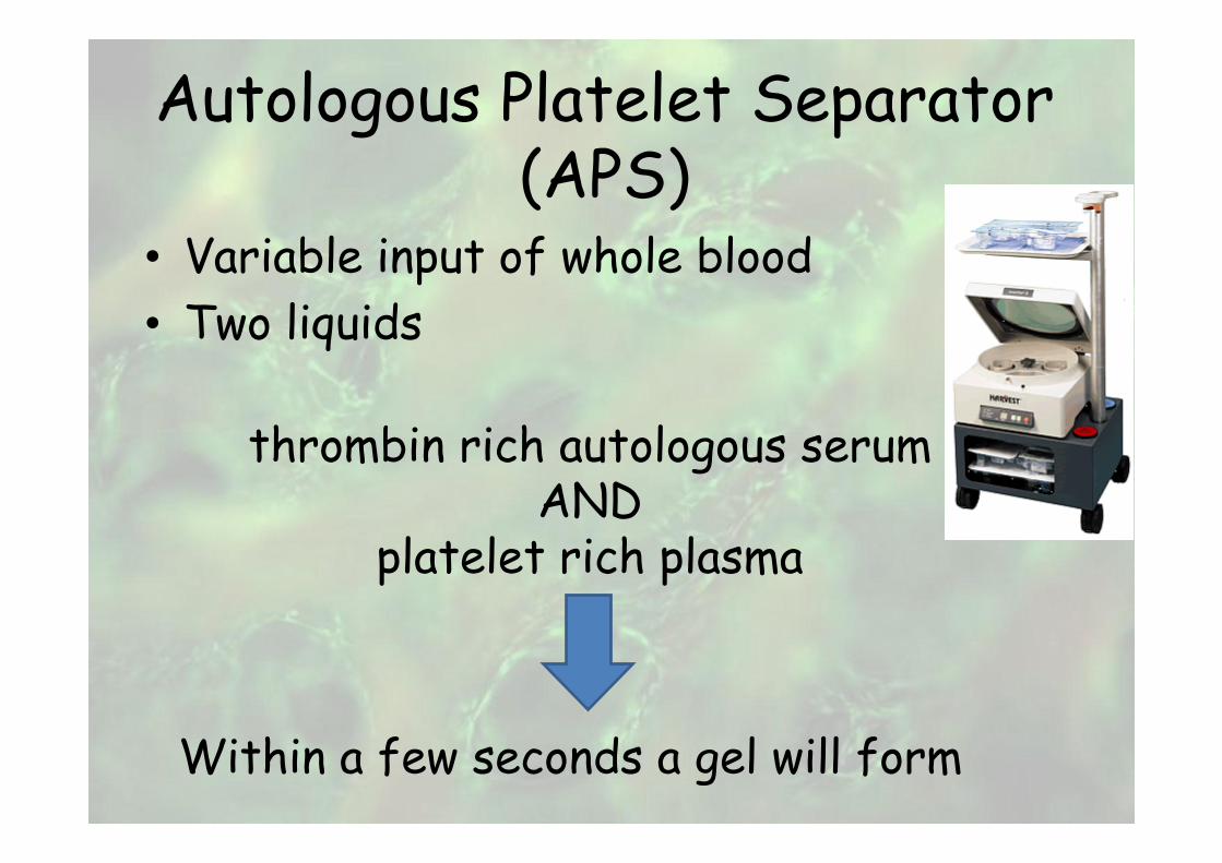
Autologous Platelet Separator (APS)
• Variable input of whole blood
• Two liquids
thrombin rich autologous serumAND
platelet rich plasma
Within a few seconds a gel will form
Advantages of APS
• Use intraoperatively
• Safe and rapid preparation of platelet poor plasma and platelet concentrate of whole blood
• Totally automated system for efficient time utilization
• Requires a small amount of blood

Stem cells
• May be useful for repair of diseased or damaged tissues
• May be used to grow new organs
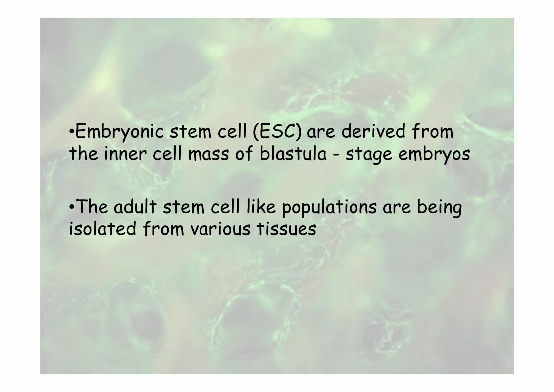
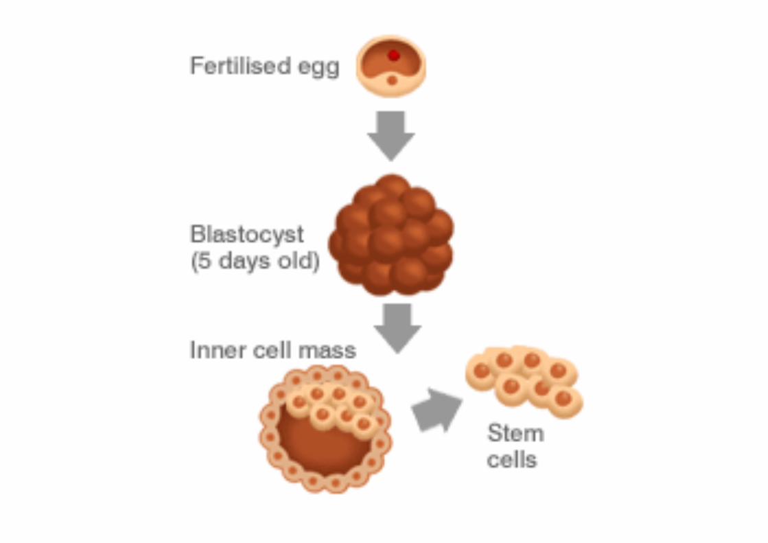
•Embryonic stem cell (ESC) are derived from the inner cell mass of blastula - stage embryos
•The adult stem cell like populations are being isolated from various tissues

Stem cells
Some cells are totipotent in the earliest stages of embryo
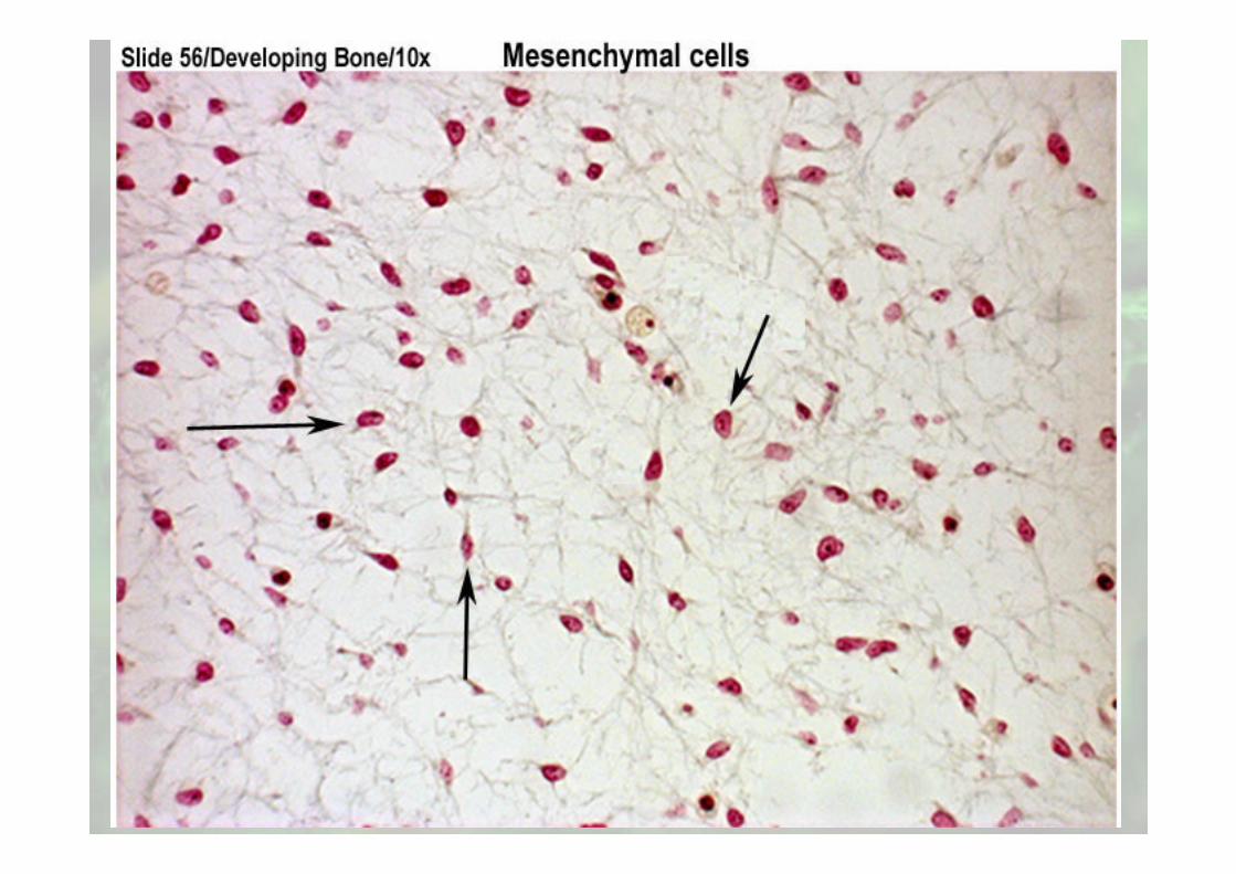
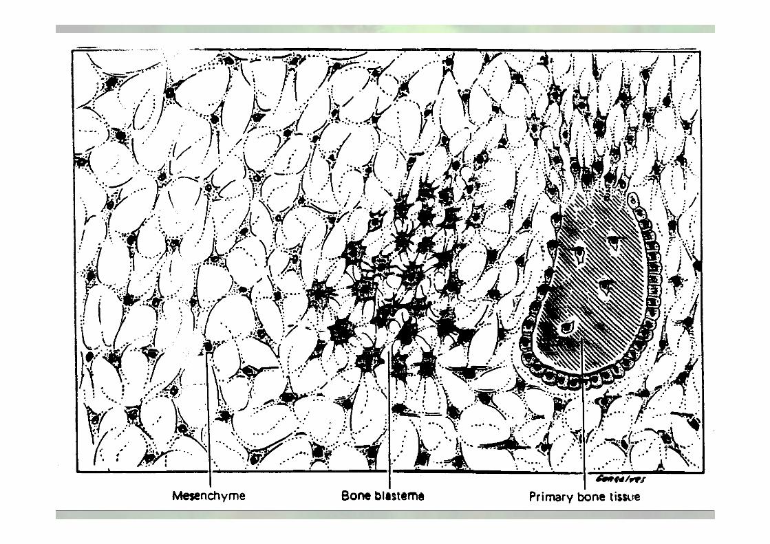
Mesenchymal stem cell: the promise for treating skeletal disorders
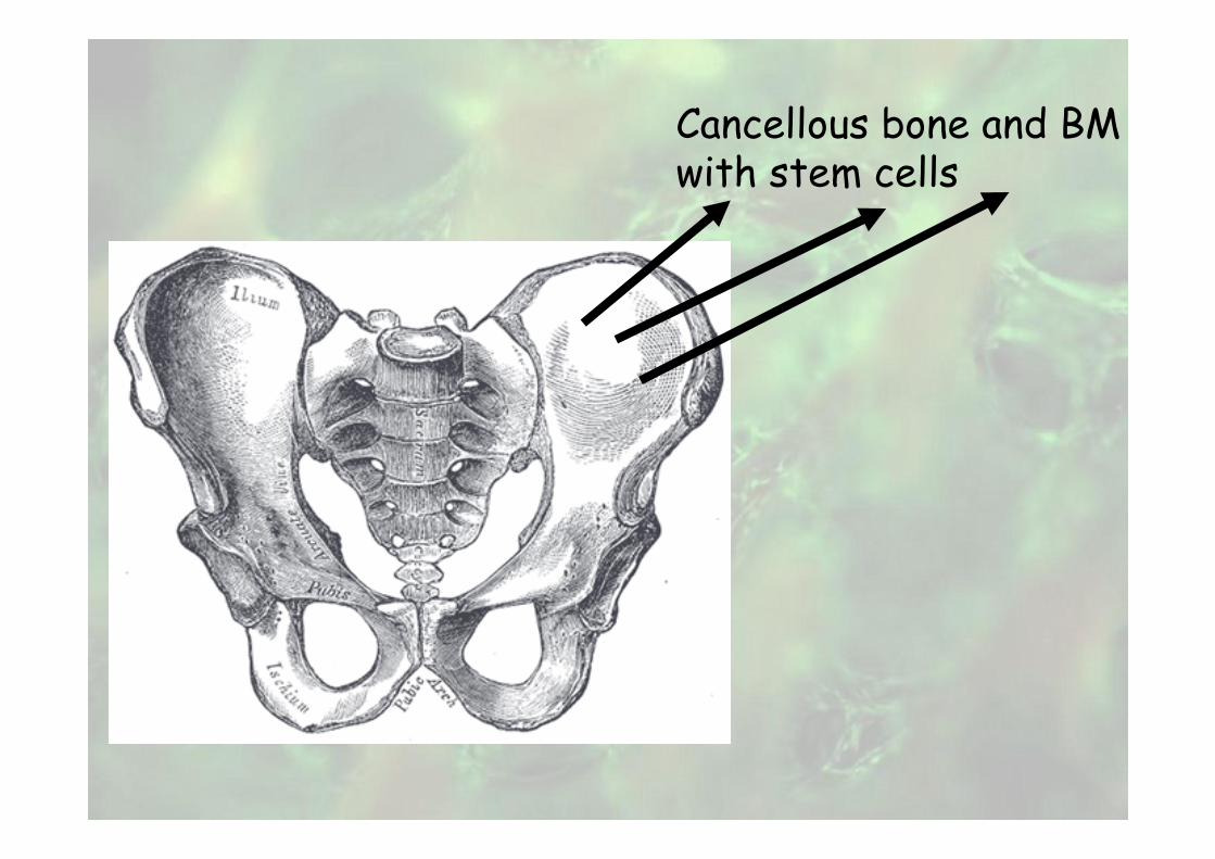
Cancellous bone and BMwith stem cells

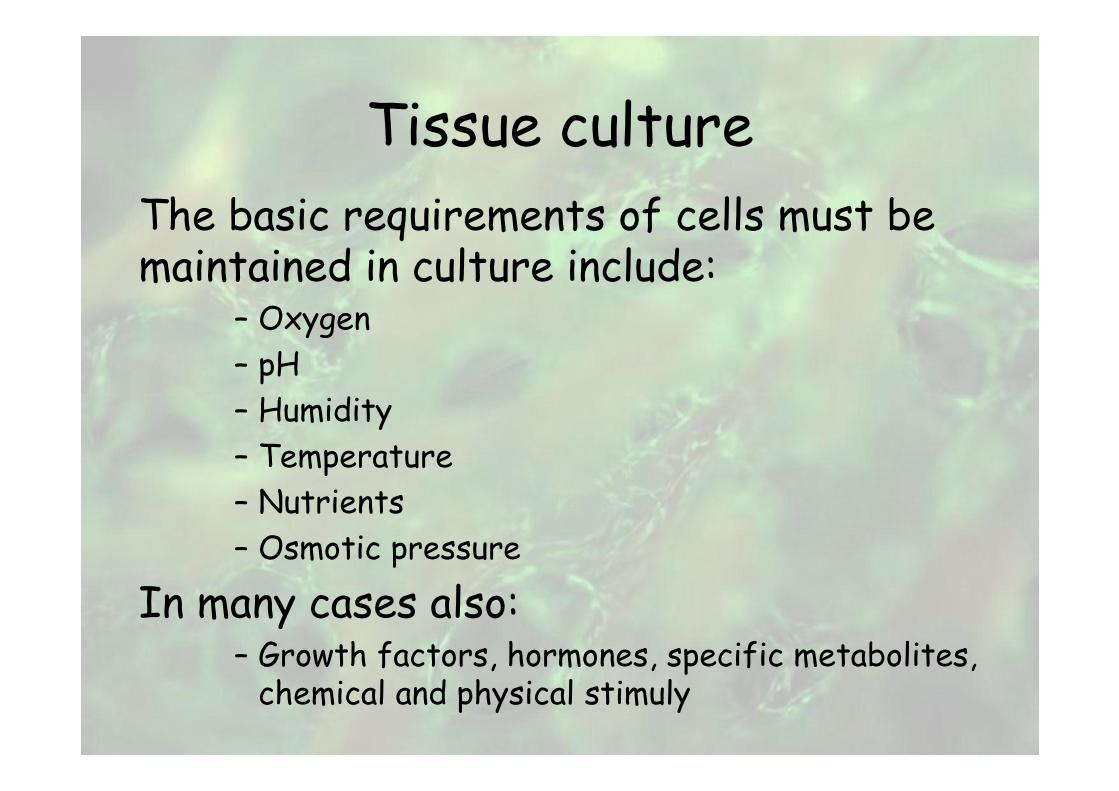
Tissue culture
The basic requirements of cells must be maintained in culture include:
– Oxygen
– pH
– Humidity
– Temperature
– Nutrients
– Osmotic pressure
In many cases also:– Growth factors, hormones, specific metabolites, chemical and physical stimuly
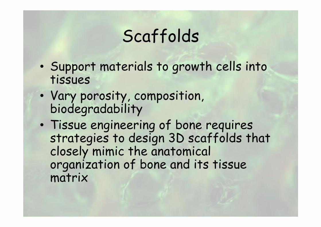
Scaffolds
• Support materials to growth cells into tissues
• Vary porosity, composition, biodegradability
• Tissue engineering of bone requires strategies to design 3D scaffolds that closely mimic the anatomical organization of bone and its tissue matrix
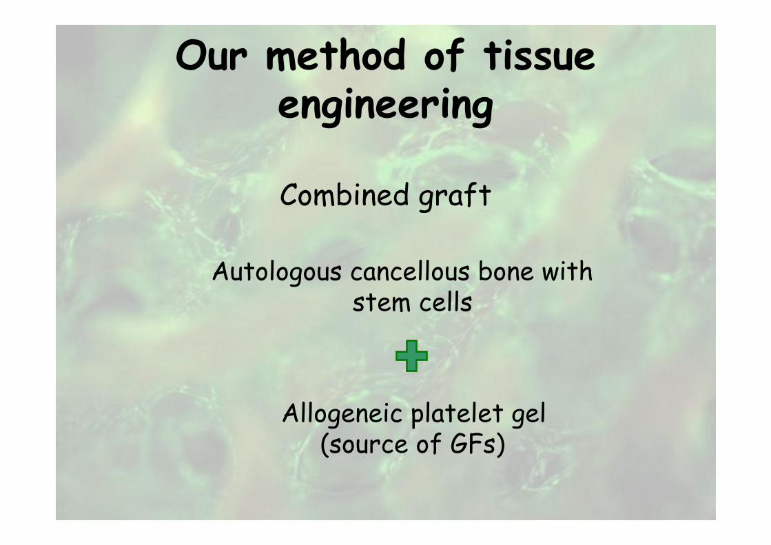
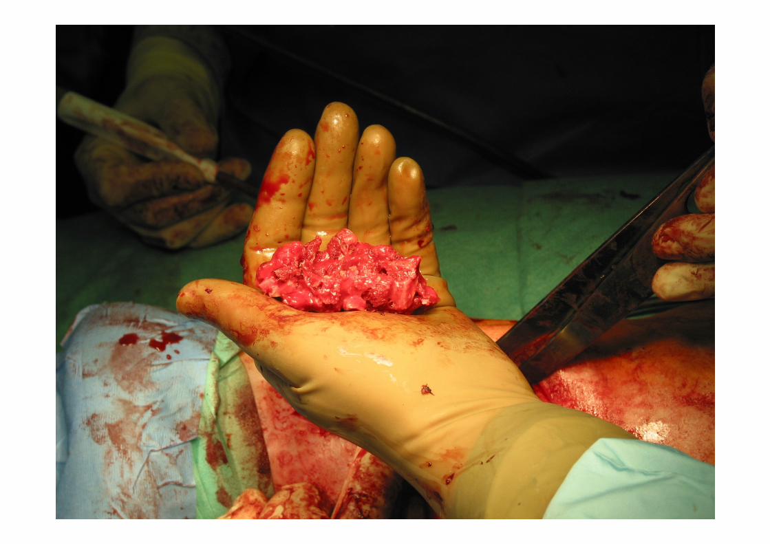
Our method of tissue engineering
Combined graft
Autologous cancellous bone withstem cells
Allogeneic platelet gel(source of GFs)
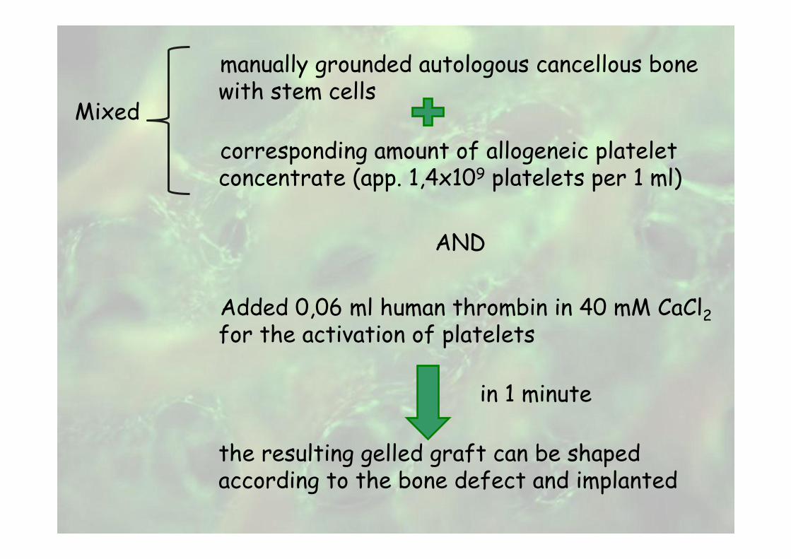
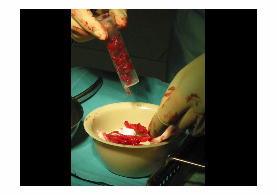
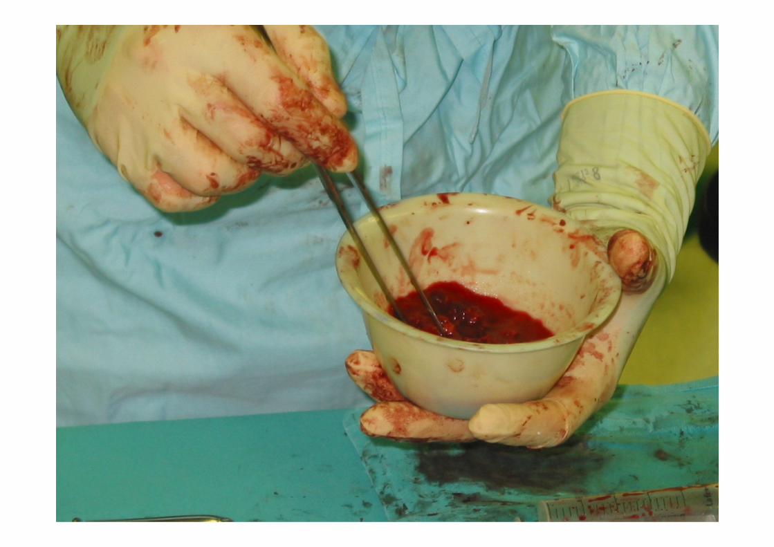
manually grounded autologous cancellous bone with stem cells
corresponding amount of allogeneic platelet concentrate (app. 1,4x10⁹ platelets per 1 ml)
AND
Added 0,06 ml human thrombin in 40 mM CaCl2for the activation of platelets
in 1 minute
the resulting gelled graft can be shaped according to the bone defect and implanted
Mixed
Tissue engineering
• Advances– In material scaffolds and cell material interactions
– The use of stem cells for tissue engineering
– Combination of stem cells and material scaffolds into tissue engineered replacements of tissues and organs