Embed Size (px)
Citation preview
Nervous and Muscle Tissue
Nerve Tissue
Nervous tissue is divided into two types:• Neurons• Supporting cells
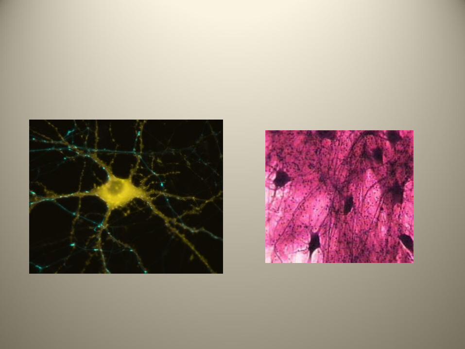
Neurons
• These generate and conduct the nerve impulses

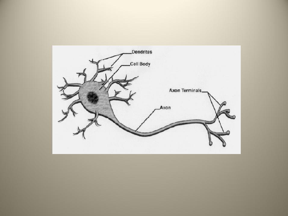
Neurons
• These generate and conduct the nerve impulses
• The dendrites respond to stimuli
Neurons
• These generate and conduct the nerve impulses
• The dendrites respond to stimuli • The nerve impulses are transmitted overlong
distances by the axons.
Support Cells
These are a mixed group of cells which support and insulate the neurons
Support Cells
These are a mixed group of cells which support and insulate the neurons Collectively they are called neuralgia (glial cells).
Support Cells
There are four types:• Astrocytes• Microglia• Ependymal cells• Oligodendrocytes
Muscle Tissue
• These cells are responsible for movement

Muscle Tissue
• These cells are responsible for movement • They are highly vascularized and cellular.
Muscle Tissue
• These cells are responsible for movement • They are highly vascularized and cellular• Muscle cells are filled with myofilaments
Muscle Tissue
Muscle tissue is divided into three types:• Skeletal• Cardiac• Smooth
Skeletal Muscle
This attaches to bone and is responsible for voluntary motion.
Skeletal Muscle
This attaches to bone and is responsible for voluntary motion.
The muscle cells (muscle fibers) are cylindrical cells that contain many nuclei.
Skeletal Muscle
This attaches to bone and is responsible for voluntary motion.
The muscle cells (muscle fibers) are cylindrical cells that contain many nuclei. The skeletal muscles are called striated because of the arrangement of the myofibrils.
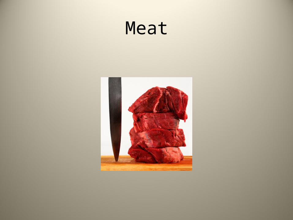
Meat
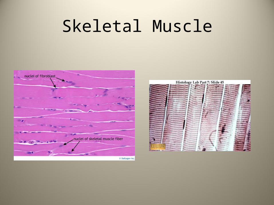
Skeletal Muscle
Cardiac Muscle
• This type is found only in the heart where its contractions propel blood through the body
Cardiac Muscle
• This type is found only in the heart where its contractions propel blood through the body
• It is striated but the cells are uninucleated
Cardiac Muscle
• This type is found only in the heart where its contractions propel blood through the body
• It is striated but the cells are uninucleated• This muscle in involuntary.
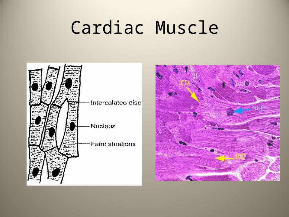
Cardiac Muscle
• The cells are branched and connect to each other with unique junctions called intercalated disks.
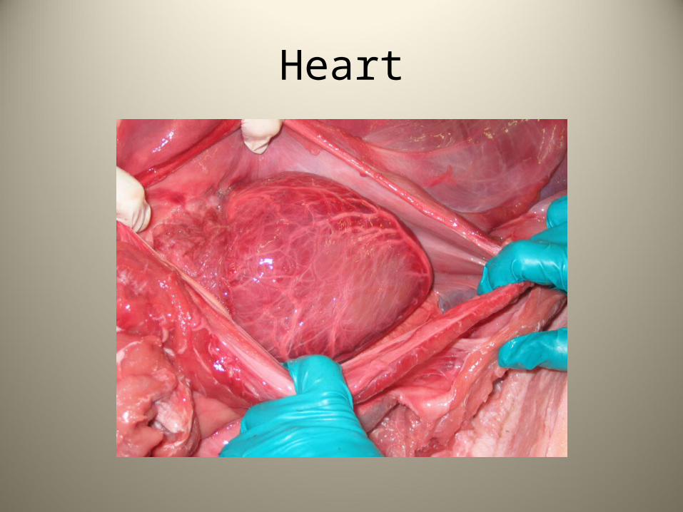
Heart
Cardiac Muscle
Smooth Muscle Tissue
• These cells have no visible striations
Smooth Muscle Tissue
• These cells have no visible striations • They are uninucleated and spindle shaped.
Smooth Muscle Tissue
• These cells have no visible striations • They are uninucleated and spindle shaped. • They are found lining the hollow organs such
as the intestines, respiratory and urinary tracts and blood vessels.
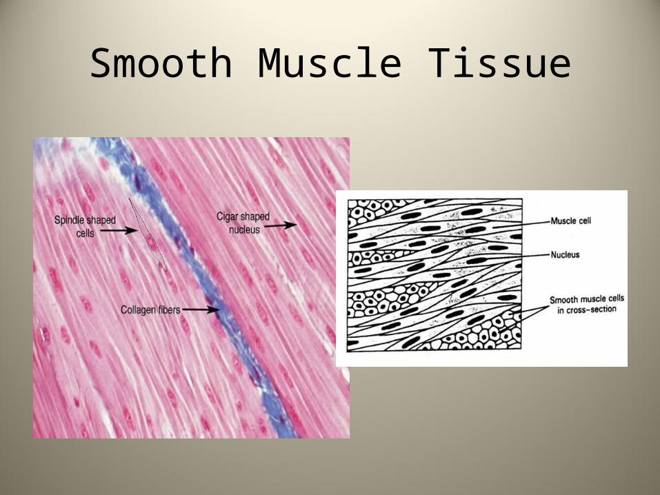
Smooth Muscle Tissue
• These cells have no visible striations • They are uninucleated and spindle shaped. • They are found lining the hollow organs such
as the intestines, respiratory and urinary tracts and blood vessels.
• They are involuntary
Smooth Muscle Tissue
Coverings and Membrane Linings
• The four tissues are incorporated into forming membranes which cover the body and its cavities.
Coverings and Membrane Linings
• The four tissues are incorporated into forming membranes which cover the body and its cavities.
There are three types:• Cutaneous• Mucous• Serous
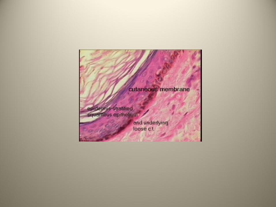
Cutaneous Membrane
• This covers the exterior of the body and is commonly known as the skin.
Cutaneous Membrane
• This covers the exterior of the body and is commonly known as the skin.
• It has a layer of keratinized stratified squamous epithelium (epidermis) with a layer of dense irregular connective tissue below it.
Cutaneous Membrane
• This covers the exterior of the body and is commonly known as the skin.
• It has a layer of keratinized stratified squamous epithelium (epidermis) with a layer of dense irregular connective tissue below it.
• Because it is exposed to the air it is considered a dry membrane.
Mucous Membrane
• These line the body cavities that are open to the exterior; examples include the digestive, respiratory and urinary tracts.
Mucous Membrane
• These line the body cavities that are open to the exterior; examples include the digestive, respiratory and urinary tracts.
• These are moist membranes
Mucous Membrane
• The epithelium can be stratified squamous, pseudostratified columnar or simple columnar epithelium.
Mucous Membrane
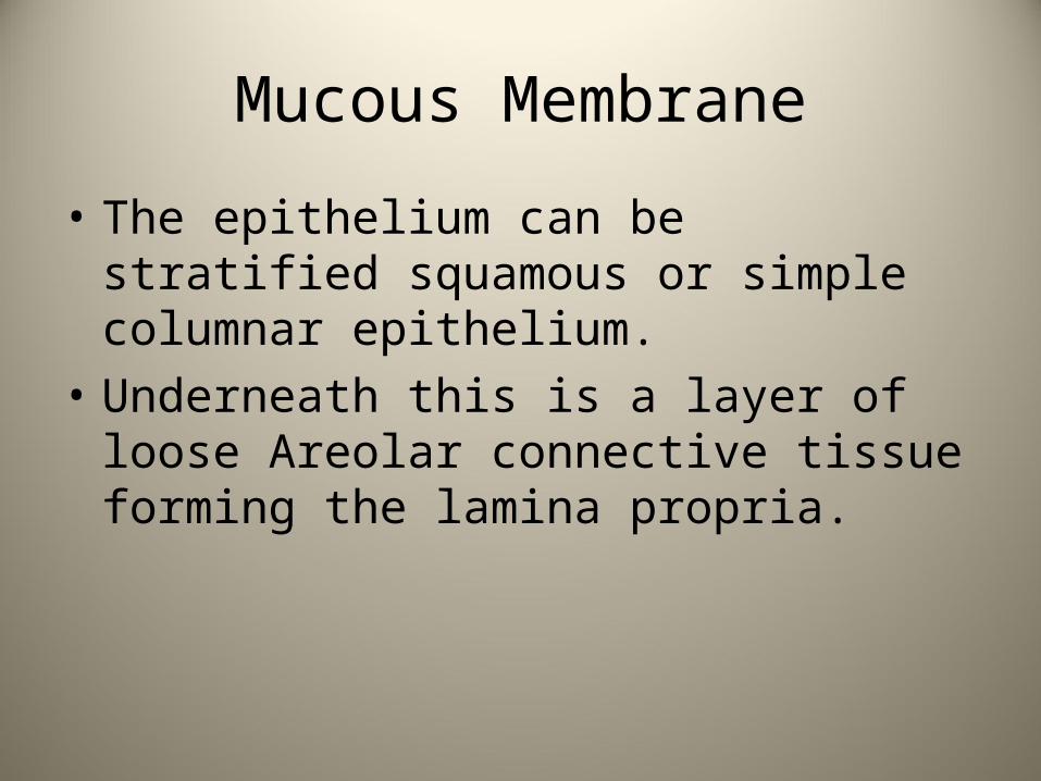
• The epithelium can be stratified squamous or simple columnar epithelium.
• Underneath this is a layer of loose Areolar connective tissue forming the lamina propria.
Mucous Membrane
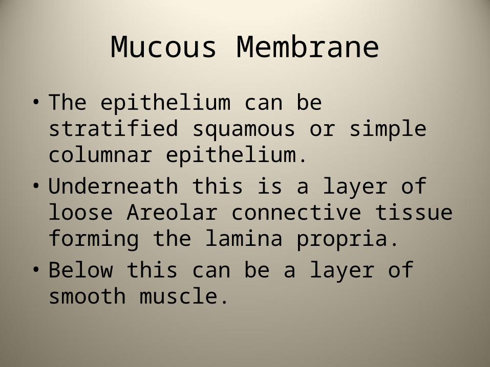
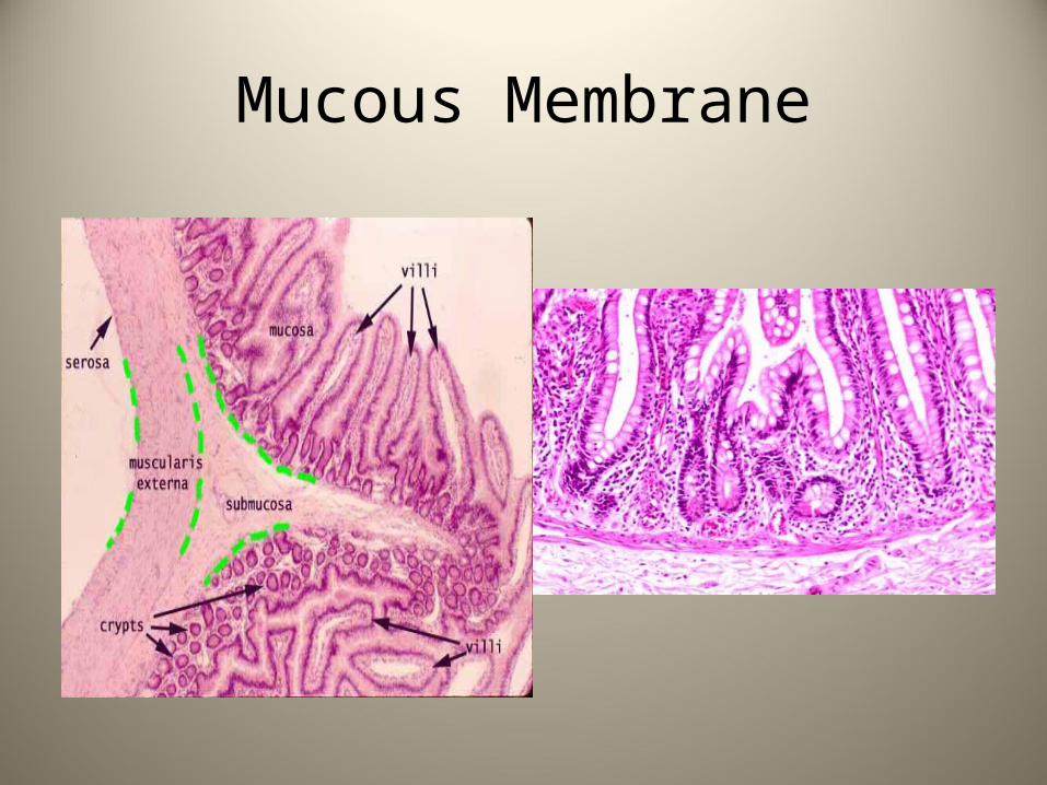
• The epithelium can be stratified squamous or simple columnar epithelium.
• Underneath this is a layer of loose Areolar connective tissue forming the lamina propria.
• Below this can be a layer of smooth muscle.
Mucous Membrane
Serous Membranes
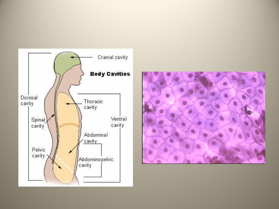
• These are the moist membranes that cover the body’s cavities
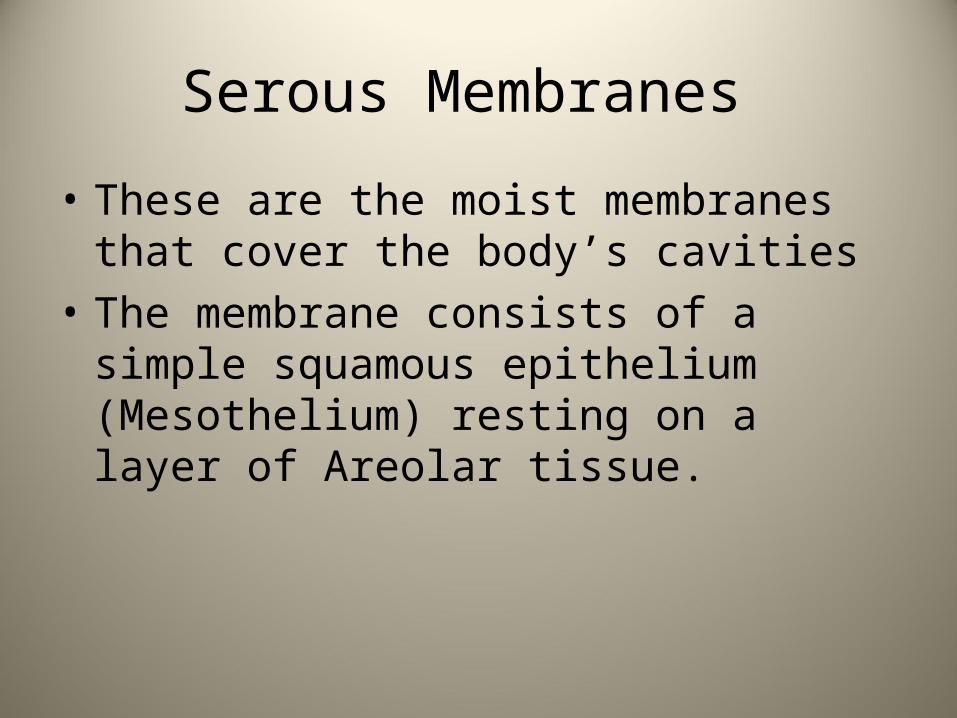
Serous Membranes
• These are the moist membranes that cover the body’s cavities
• The membrane consists of a simple squamous epithelium (Mesothelium) resting on a layer of Areolar tissue.
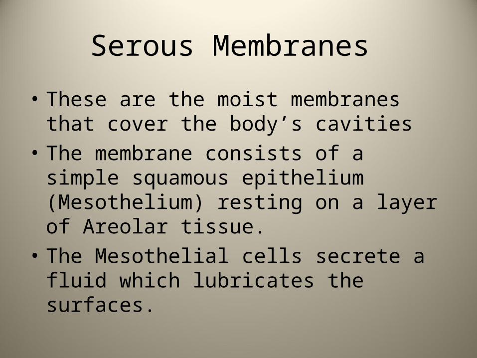
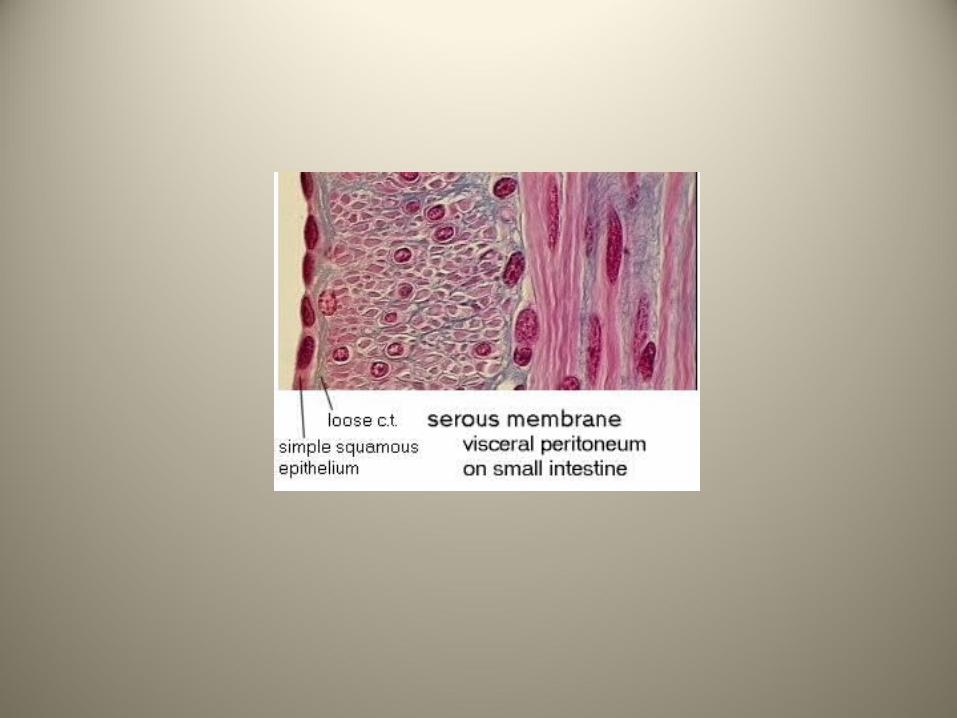
Serous Membranes
• These are the moist membranes that cover the body’s cavities
• The membrane consists of a simple squamous epithelium (Mesothelium) resting on a layer of Areolar tissue.
• The Mesothelial cells secrete a fluid which lubricates the surfaces.
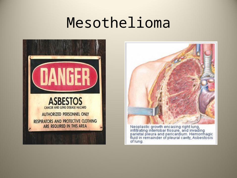
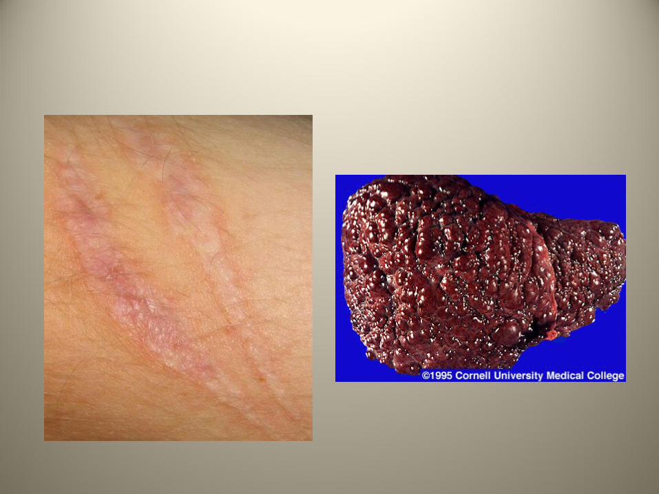
Mesothelioma
Tissue Repair
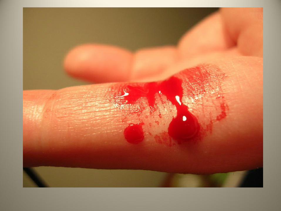
• Tissue injury is followed by a predictable series of steps which result in healing of the wound.
Tissue Repair
• Tissue injury is followed by a predictable series of steps which result in healing of the wound.
• These steps involve a nonspecific inflammatory response followed by repair which can result in either replacement of the tissue or fibrosis (scarring).
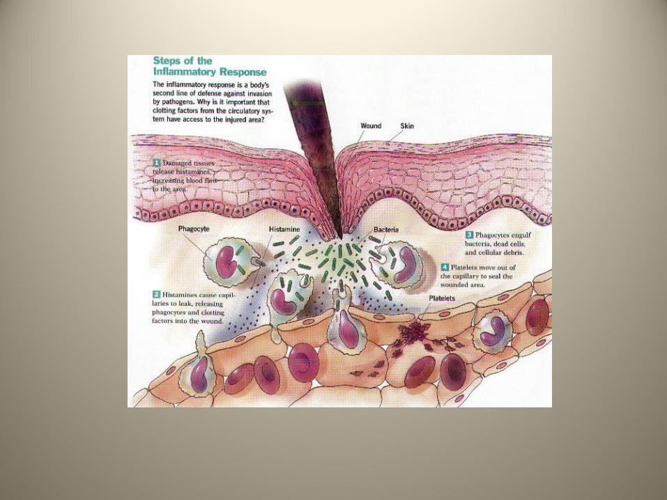
1. Inflammation
This occurs right after the injury and involves cells such as the macrophages, mast cells and lymphocytes (normal residents of the Areolar tissue) releasing factors which lead to increased vascular permeability and stimulation of cell division.
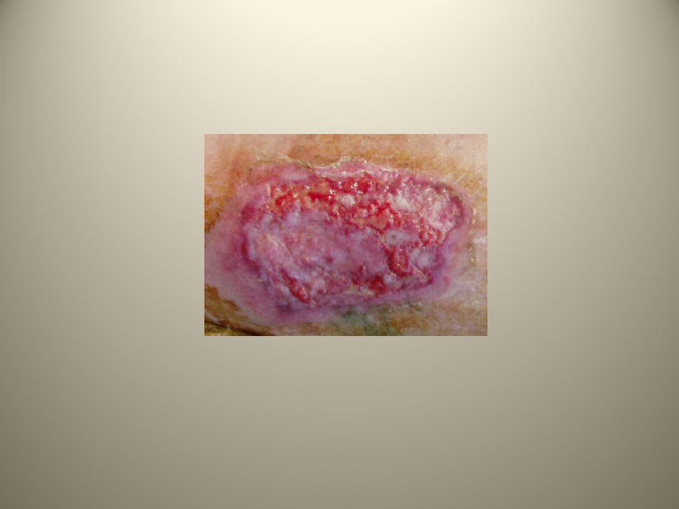
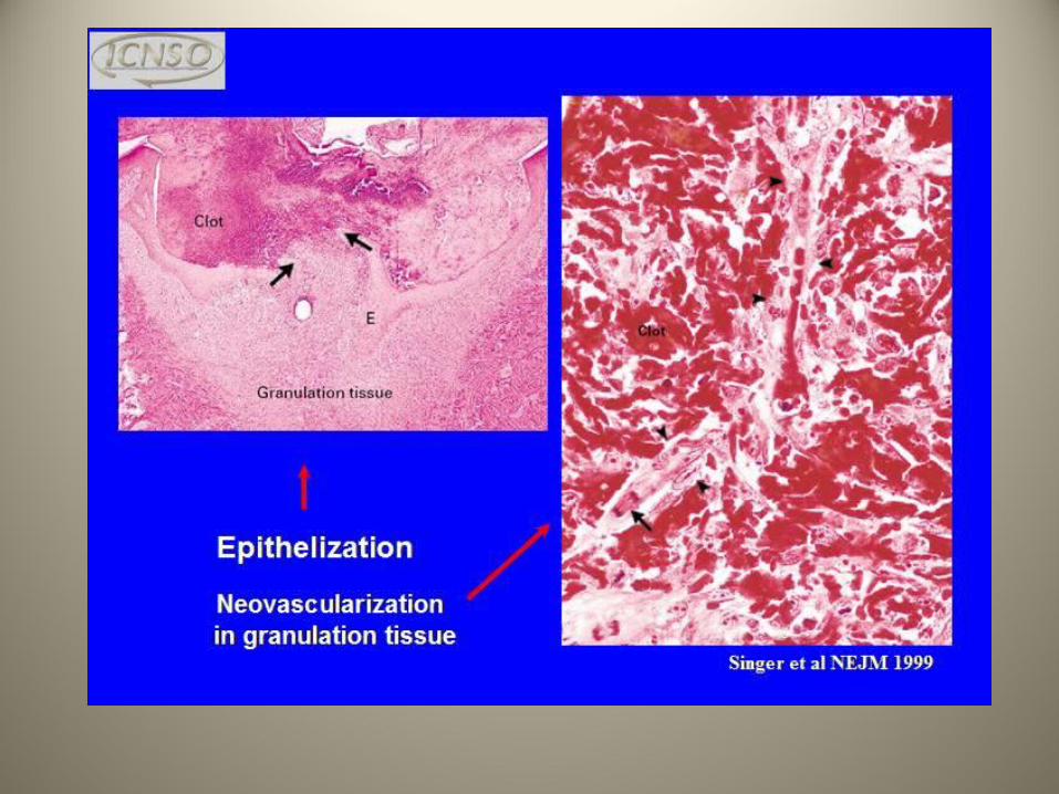
2. Organization
• Once the blood clot is formed, it is replaced by granulation tissue. This is a collection of capillaries and fibroblasts which lay down a new matrix.
3. Regeneration or Fibrosis
• Epithelial, bone, areolar, dense irregular and blood tissues regenerate.
3. Regeneration or Fibrosis
• Epithelial, bone, areolar, dense irregular and blood tissues regenerate.
• Smooth muscle and dense regular connective tissue have a moderate capacity to regenerate while skeletal muscle and cartilage are weak.
3. Regeneration or Fibrosis
• Epithelial, bone, areolar, dense irregular and blood tissues regenerate.
• Smooth muscle and dense regular connective tissue have a moderate capacity to regenerate while skeletal muscle and cartilage are weak.
• Cardiac and nervous tissues have none.