Embed Size (px)
Citation preview
ORIGINAL RESEARCHFUNCTIONAL
Time-Shift Homotopic Connectivity in Mesial TemporalLobe Epilepsy
Q. Xu, Z. Zhang, W. Liao, L. Xiang, F. Yang, Z. Wang, G. Chen, Q. Tan, Q. Jiao, and G. Lu
ABSTRACT
BACKGROUND AND PURPOSE: Voxel-mirrored intrinsic functional connectivity allows the depiction of interhemispheric homotopicconnections in the human brain, whereas time-shift intrinsic functional connectivity allows the detection of the extent of brain injury bymeasuring hemodynamic properties. We combined time-shift voxel-mirrored homotopic connectivity analyses to investigate the alter-ations in homotopic connectivity in mesial temporal lobe epilepsy and assessed the value of applying this approach to epilepsy lateral-ization and the prediction of surgical outcomes in mesial temporal lobe epilepsy.
MATERIALS AND METHODS: Resting-state functional MR imaging data were acquired from patients with unilateral mesial temporal lobeepilepsy (n � 62) (31 left- and 31 right-side) and healthy controls (n � 33). Dynamic interhemispheric homotopic architecture seeding from eachhemisphere was individually calculated by 0, 1, 2, and 3 repetition time time-shift voxel-mirrored homotopic connectivity. Voxel-mirroredhomotopic connectivity maps were compared between the patient and control groups by using 1-way ANOVA for each time-shift condition,separately. Group comparisons were further performed on the laterality of voxel-mirrored homotopic connectivity in each time-shift condition.Finally, we correlated the interhemispheric homotopic connection to the surgical outcomes in a portion of the patients (n � 20).
RESULTS: The patients with mesial temporal lobe epilepsy showed decreased homotopic connectivity in the mesial temporal structures,temporal pole, and striatum. Alterations of the bihemispheric homotopic connectivity were lateralized along with delays in the time-shiftin mesial temporal lobe epilepsy. The patients with unsuccessful surgical outcomes presented larger interhemispheric voxel-mirroredhomotopic connectivity differences.
CONCLUSIONS: This study showed whole patterns of dynamic alterations of interhemispheric homotopic connectivity in mesial tem-poral lobe epilepsy, extending the knowledge of abnormalities in interhemispheric connectivity in this condition. Time-shift voxel-mirrored homotopic connectivity has the potential for lateralization of unilateral mesial temporal lobe epilepsy and may have thecapability of predicting surgical outcomes in this condition.
ABBREVIATIONS: HC � healthy controls; mTLE � mesial temporal lobe epilepsy; ts � time-shift; VMHC � voxel-mirrored homotopic connectivity
Interhemispheric communication and coordination facilitate
information processing in the human brain.1,2 Thus homotopic
connections represent a fundamental characteristic of brain anat-
omy and function3,4 and have been considered an important in-
dicator for depicting the physiologic and pathologic features of
the brain. On the basis of resting-state functional MR imaging
measurements, an approach based on voxel-mirrored homotopic
connectivity (VMHC) quantifies the interhemispheric homo-
topic connections by measuring the functional connectivity be-
tween each voxel in 1 hemisphere and its mirrored counterpart.5
Zuo et al5 found age-related increases in interhemispheric func-
tional connectivity in the primary sensorimotor areas and de-
Received November 21, 2013; accepted after revision January 31, 2014.
From the Departments of Medical Imaging (Q.X., Z.Z., W.L., L.X., Q.J., G.L.), Neurol-ogy (F.Y., G.C.), and Neurosurgery (Q.T.), Jinling Hospital, Nanjing University Schoolof Medicine, Nanjing, China; Center for Cognition and Brain Disorders and the Af-filiated Hospital (W.L.), Hangzhou Normal University, Hangzhou, China; ZhejiangKey Laboratory for Research in Assessment of Cognitive Impairments (W.L.), Hang-zhou, China; Department of Medical Imaging (Z.W.), Nanjing Drum Tower Hospital,The Affiliated Hospital of Nanjing University Medical School, Nanjing, China; andDepartment of Medical Imaging (Q.J.), Taishan Medical College, TaiAn, China.
This work was supported by the Natural Science Foundation of China (grant nos.81271553, 81201155, 81201078, 81171328, 61131003, and 81020108022), Grants for YoungScholar of Jinling Hospital (grant nos. 2011060, 2011045, and 2011061), Chinese Key Grant(BWS11J063 and 10z026), and the China Postdoctoral Science Foundation (grant no.2013M532229).
Please address correspondence to Zhiqiang Zhang, MD, and Guangming Lu, MD,Department of Medical Imaging, Nanjing Jinling Hospital, 305#, Eastern ZhongshanRd, Nanjing 210002, China; e-mail: [email protected] (Z.Z.) or [email protected] (G.L.)
Indicates open access to non-subscribers at www.ajnr.org
Indicates article with supplemental on-line table.
http://dx.doi.org/10.3174/ajnr.A3934
1746 Xu Sep 2014 www.ajnr.org
creases in the higher order processing areas, which provided in-
sight into the evolution of brain development. Studies have also
revealed specific alterations of homotopic connection in a cohort
of brain diseases.6-8 Decreased VMHC in schizophrenia has been
suggested to reflect the substantial impairment of interhe-
miespheric coordination in these patients.6 Anderson et al7 found
homotopic connectivity alterations related to behavioral and de-
velopmental abnormalities in autism.7 More recently, studies
have further correlated functional homotopic connectivity with
microstructural impairment in multiple sclerosis8 and idiopathic
generalized epilepsy.9
In contrast to the brain disorders featuring abnormal connec-
tion pathways as mentioned above,6,8 mesial temporal lobe epi-
lepsy (mTLE) is a location-related disease characterized by hip-
pocampal sclerosis.10 Unilateral mTLE can cause bilateral and
distributed brain impairments due to seizure propagation via the
mesial temporal epileptic network.11-13 Resting-state fMRI stud-
ies have shown asymmetric connections between bihemi-
spheres14-16 and decreased connectivity between bilateral hip-
pocampi in mTLE.17,18 These findings suggest that there are intra-
and interhemispheric connection abnormalities in unilateral
mTLE. However, neither the homotopic alterations of whole-
brain functional connectivity nor the relationship between asym-
metric lesions and interhemispheric communication in this dis-
ease has been investigated.
fMRI-based VMHC provides a feasible way to observe the
whole-brain homotopic connectivity alterations in mTLE. How-
ever, the conventional nondirectional functional connectivity
measure, as used in VMHC, cannot detect the connection abnor-
malities resulting from deficits of the seed or target region. Re-
cently, Lv et al19 proposed a time-shift (ts) analysis for resting-
state functional connectivity. They quantified the temporal shift
correlation between time courses of each voxel and global mean
signal19 and correlated the time shifts with the extent and degree
of perfusion delay in patients with stroke.19,20 In addition, our
previous study used time-shift correlation analysis to demon-
strate the sequential effects of epileptic discharges on intrinsic
network connectivity in children with absence epilepsy.21 Thus,
time-shift delays in resting-state spontaneous connectivity were
assumed to reflect brain hemodynamics and could measure the
degree of brain injury; time-shift analysis also provides directional
information as a measure of functional connectivity.21 In the cur-
rent work, we combined the time-shift connectivity with the
VMHC technique and applied them to resting-state fMRI data
from patients with unilateral mTLE. We hypothesized that this
strategy would allow us to assess the whole-brain homotopic con-
nection impairments resulting from different hemispheres and
may potentially be a tool for epileptic focus lateralization and
surgical outcome prediction in mTLE.
MATERIALS AND METHODSSubjectsSixty-two patients with unilateral mTLE (31 left-sided and 31
right-sided) were recruited (demographic and clinical informa-
tion are detailed in the Table). Part of the patient population (23
patients with left-sided mTLE) was reported in our previous work
in which the Granger causal information of the hippocampus in
left-sided mTLE was estimated.13 Diagnosis of mTLE was per-
formed according to the International League Against Epilepsy
2001 classification based on a comprehensive evaluation, includ-
ing seizure history and semiology, neurologic examination, diag-
nostic MR imaging, and electroencephalography records in all
patients. Patients were selected on the basis of several criteria: 1)
Patients who were young and middle-aged were included (pa-
tients younger than 18 years or older than 50 years were exclud-
ed); 2) patients who exhibited identifiable structural MR imaging
abnormalities other than hippocampal sclerosis, such as cortical
dysplasia, vascular malformation, or tumor were excluded.
Twenty patients of the 62 underwent standard anterior temporal
lobectomy: Fourteen cases (8 left- and 6 right-sided mTLE) had
successful surgical outcomes, and 6 cases (4 left- and 2 right-sided
mTLE) had unsuccessful outcomes. A successful outcome was
defined as seizure-free with or without auras at the latest fol-
low-up that occurred at least 1 year after the surgery, which satis-
fies the International League Against Epilepsy Outcome Criteria 1
or 222 and Engel category 1A or 1B.23 No postoperative MR im-
aging data were used in this article.
Moreover, healthy controls (HC) (n � 33) were recruited from
the hospital staff. These controls did not have neurologic or psychi-
atric disorders at the time of the study. There were no differences in
ages and sexes between the controls and patients (Table).
This study was approved by the local Medical Ethics Commit-
tee, and written informed consent was obtained from all the
participants.
Data AcquisitionWe acquired functional and structural images in patients and HC
by using a Trio 3T scanner (Siemens, Erlangen, Germany). We
used foam padding to minimize head motion. We acquired rest-
ing-state functional images by using a single-shot, gradient-re-
called echo-planar imaging sequence (250 volumes, TR � 2000
ms, TE � 30 ms, flip angle � 90°, FOV � 240 � 240 mm2,
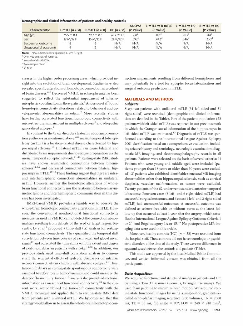
Demographic and clinical information of patients and healthy controls
Characteristic L-mTLE (n = 31) R-mTLE (n = 31) HC (n = 33)ANOVA(P Value)
L-mTLE vs R-mTLE(P Value)
L-mTLE vs HC(P Value)
R-mTLE vs HC(P Value)
Age (yr) 26.5 � 8.4 29.7 � 8.5 26.7 � 7.3 .231a .146c .903c .144c
Sex 19 M/12 F 16 M/15 F 21 M/12 F .592b .442d .846d .330d
Successful outcome 8 6 N/A N/A N/A N/A N/AUnsuccessful outcome 4 2 N/A N/A N/A N/A N/A
Note:—N/A indicates not applicable; L, left; R, right.a One-way analysis of variance.b Kruskal-Wallis ANOVA.c Two-sample t test.d �2 test.
AJNR Am J Neuroradiol 35:1746 –52 Sep 2014 www.ajnr.org 1747
intersection gap � 0.4 mm, voxel size � 3.75 � 3.75 � 4 mm3, 30
transverse sections aligned along the anterior/posterior commis-
sure). Subjects were instructed simply to rest with their eyes
closed, not to think about anything in particular, and not to fall
asleep. Subsequently, we acquired high-resolution 3D T1-
weighted anatomic images in a sagittal orientation by using a
magnetization-prepared rapid acquisi-
tion of gradient echo sequence (TR �
2300 ms, TE� 2.98 ms, flip angle � 9 °,
FOV � 256 � 256 mm2, voxel size � 1 �
1�1 mm3, 176 sections without intersec-
tion gap).
Data PreprocessingFunctional image preprocessing was per-
formed by using the DPARSF 2.2 (http://
www.restfmri.net) and SPM8 (Wellcome
Department of Imaging Neuroscience,
London, UK; http://www.fil.ion.ucl.ac.
uk/spm) toolkits. We excluded the first 10
images to ensure steady-state longitudinal
magnetization, and then we corrected the
remaining images for temporal differ-
ences and head motion. No translation or
rotation parameters in any given dataset
exceeded �1 mm or �1°. We then coreg-
istered individual 3D T1 images to func-
tional images. The 3D T1-weighted ana-
tomic images were segmented into gray
matter, white matter, and CSF by using
unified segmentation.24 Then, a nonlin-
ear spatial deformation was calculated
from the gray matter images to a gray
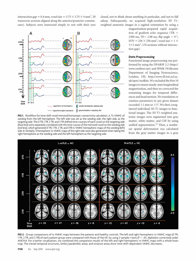
FIG 1. Workflow for time-shift voxel-mirrored homotopic connectivity calculation. A, Ts-VMHC ofseeding from the left hemisphere. The left side was set as the seeding side; the right side, as thetargeting side. The 0 TR, 1 TR, 2 TR, and 3 TR shifted time-courses of each voxel on the targeting side(blue line) were separately correlated with the time course of the mirrored voxel on the seeding side(red line), which generated 0 TR, 1 TR, 2 TR, and 3TR ts-VMHC hemispheric maps of the seeding (left)side. B, Similarly, 4 hemispheric ts-VMHC maps of the right side were also generated when taking theright hemisphere as the seeding side and the left hemisphere as the targeting side.
FIG 2. Group comparisons of ts-VMHC maps between the patients and healthy controls. The left and right hemispheric ts-VMHC maps (0 TR,1 TR, 2 TR, and 3 TR) of each patient group were compared with those of the HC by using 2-sample t tests (P � .05, AlphaSim corrected) underANOVA. For a better visualization, we combined the comparison results of the left and right hemispheric ts-VMHC maps with a whole-brainmap. The mesial temporal structures, limbic/paralimblic areas, and striatum areas show time-shift-dependent VMHC decreases.
1748 Xu Sep 2014 www.ajnr.org
matter template in Montreal Neurological Institute space. This
transformation was then applied to the functional images,
which were resectioned at a resolution of 3 � 3�3 mm3. The
data were bandpass-filtered (0.01– 0.08 Hz). Linear regression
was performed to remove the effect of 6 head-motion-averaged
signals from the CSF, white matter, and the global brain sig-
nal.25 The residual images were further coregistered to a sym-
metric template according to previous studies.5,8 The symmet-
ric template was created as follows: First, all participants’
normalized gray matter images were averaged to create a mean
normalized gray matter template. This template was then av-
eraged with its left-right mirrored version to generate a group-
specific symmetric template.5 Finally, we spatially smoothed
the images with an 8-mm full width at half maximum isotropic
Gaussian kernel.
Time-Shift Voxel-Mirrored Homotopic ConnectivityWe combined the time-shift analysis19 with voxel-mirrored ho-
motopic connectivity analysis.5 We first took the left hemisphere
as the seeding side and the right hemisphere as the targeting side.
The 0 TR, 1 TR, 2 TR, and 3 TR shifted time-courses of each voxel
on the targeting side were separately correlated with the time
course of the mirrored voxel on the seeding side, which generated
0 TR, 1 TR, 2 TR, and 3 TR ts-VMHC hemispheric maps of the
seeding (left) side. Similarly, 4 hemispheric ts-VMHC maps of the
right side were also generated when taking the right hemisphere as
the seeding side and the left hemisphere as the targeting side (Fig
1). The correlation maps were then converted to z scores by using
the Fisher r-to-z transformation.
Group Comparisons of ts-VMHCIn the second-level analyses, the left and
right hemispheric ts-VMHC maps (0 TR,
1 TR, 2 TR, and 3 TR) of each patient
group were compared with those of the
HC by using a 2-sample t test (AlphaSim
corrected P � .05, combined height thresh-
old P � .01, and extent threshold k � 20
voxels). For a better visualization, we com-
bined the comparison results of the left and
right hemispheric ts-VMHC maps with a
whole-brain map (Fig 2 and On-line Table).
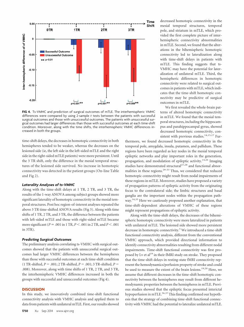
Laterality Analyses of ts-VMHCTo quantitatively identify the difference
between the left and right hemispheric ts-
VMHC and to further determine whether
the ts-VMHC could be used for mTLE lat-
eralization, we performed a voxelwise lat-
erality analysis. In each individual of all 3
participant groups, the right hemispheric
ts-VMHC map was subtracted from the
left hemispheric ts-VMHC map and the
results were divided by the summation of
both maps. Then the laterality (hemi-
spheric) maps of all 3 groups of partici-
pants were compared by using 1-way
ANOVA implemented in SPM8 in each
time-shift condition (P � .05, AlphaSim correction). Then, a post
hoc region of interest– based analysis was subsequently per-
formed. The ROIs were selected from the above ANOVA result in
3 TR time-shift conditions. The laterality values in the ROIs were
extracted for ANOVA among the 3 participant groups (Fig 3).
Predicting Surgical Outcomes of ts-VMHCWe performed a preliminary analysis to retrospectively investi-
gate the relationship between ts-VMHC and surgical outcomes of
mTLE. Twenty patients who underwent anterior temporal lobec-
tomy were included. The patients with right-sided mTLE (R-
mTLE) were left-right flipped to produce a homogeneous left-
sided mTLE dataset. We calculated the difference of VMHC
between bihemispheres by subtracting the connectivity values of
the contralateral side from those of the lesional side (ie, the epi-
leptogenic side). Subsequently, the interhemispheric differences
were compared by using 2-sample t tests between the patients
with successful surgical outcomes and those with unsuccessful
outcomes.
RESULTSGroup Comparison Results of ts-VMHCOn the basis of the comparison of the results of VMHC without
time-shift (ie, 0 TR shift), both patient groups showed decreased
homotopic connectivity in the regions of limbic/paralimbic areas
(mainly in the mesial temporal structures including the hip-
pocampus/parahippocampus, temporal pole, amygdala, and in-
sula) and striatum areas (mainly in the putamen and pallidum)
relative to the HC (P � .05 AlphaSim correction). Along with the
FIG 3. Laterality index analysis of ts-VMHC in mTLE. A, Group comparisons of voxelwise later-ality maps of ts-VMHC. Along with the delay of time shifts, a more significant difference ofVMHC laterality was found among the patient groups and healthy controls. B, Region of inter-est– based laterality analysis confirmed the above results. The region of interest was selectedfrom the group comparing the result of voxelwise laterality map of ts-VMHC (white circle).
AJNR Am J Neuroradiol 35:1746 –52 Sep 2014 www.ajnr.org 1749
time-shift delays, the decreases in homotopic connectivity in both
hemispheres tended to be weaker, whereas the decreases on the
lesioned side (ie, the left side in the left-sided mTLE and the right
side in the right-sided mTLE patients) were more persistent. Until
the 3 TR shift, only the difference in the mesial temporal struc-
tures of the lesioned side survived. No increase in homotopic
connectivity was detected in the patient groups (On-line Table
and Fig 2).
Laterality Analyses of ts-VMHCAlong with the time-shift delays at 1 TR, 2 TR, and 3 TR, the
results of the 1-way ANOVA among subject groups showed more
significant laterality of homotopic connectivity in the mesial tem-
poral structures. Post hoc region-of-interest analyses repeated the
above 3 TR time-shifted ANOVA results (Fig 3). Along with time
shifts of 1 TR, 2 TR, and 3 TR, the difference between the patients
with left-sided mTLE and those with right-sided mTLE became
more significant (P � .001 in 1 TR, P � .001 in 2 TR, and P � .001
in 3TR).
Predicting Surgical OutcomesThe preliminary analysis correlating ts-VMHC with surgical out-
comes showed that the patients with unsuccessful surgical out-
comes had larger VMHC differences between the hemispheres
than those with successful outcomes at each time-shift condition
(1 TR-shifted, P � .001; 2 TR-shifted, P � .001; 3 TR-shifted, P �
.008). Moreover, along with time shifts of 1 TR, 2 TR, and 3 TR,
the interhemispheric VMHC differences increased in both the
groups with successful and unsuccessful outcomes (Fig 4).
DISCUSSIONIn this study, we innovatively combined time-shift functional
connectivity analysis with VMHC analysis and applied them to
data from patients with unilateral mTLE. First, our results showed
decreased homotopic connectivity in themesial temporal structures, temporalpole, and striatum in mTLE, which pro-vided the first complete picture of inter-hemispheric connectivity abnormalitiesin mTLE. Second, we found that the alter-ations in the bihemispheric homotopicconnectivity led to lateralization alongwith time-shift delays in patients withmTLE. This finding suggests that ts-VMHC may have the potential for later-alization of unilateral mTLE. Third, thehemispheric differences in homotopicconnectivity were related to surgical out-comes in patients with mTLE, which indi-cates that the time-shift homotopic con-nectivity may be predictive of surgicaloutcomes in mTLE.
We first revealed the whole-brain pat-terns of altered homotopic connectivityin mTLE. We found that the mesial tem-poral structures, including the hippocam-pus and parahippocampal gyrus, showeddecreased homotopic connectivity, con-sistent with previous studies.14,15,17 Fur-
thermore, we found decreased homotopic connectivity in thetemporal pole, amygdala, insula, putamen, and pallidum. Theseregions have been regarded as key nodes in the mesial temporalepileptic networks and play important roles in the generation,propagation, and modulation of epileptic activity.14,26 Imagingstudies have demonstrated structural27,28 and functional abnor-malities in these regions.29-33 Thus, we considered that reducedhomotopic connectivity might result from nodal impairments ofthese regions in mTLE. Moreover, studies have proposed a varietyof propagation patterns of epileptic activity from the originatingfocus to the contralateral side; the limbic structures and basalganglia are the important nodes within the propagation path-way.34,35 Here we cautiously proposed another explanation, thattime-shift-dependent alterations of VMHC of these regionsmight represent propagations of epileptic activity.
Along with the time-shift delays, the decreases of the bihemi-spheric homotopic connectivity were more lateralized in patientswith unilateral mTLE. The lesioned side showed more persistentdecrease in homotopic connectivity.5 We introduced a time-shiftfunctional connectivity analysis, different from the conventionalVMHC approach, which provided directional information toidentify connectivity abnormalities resulting from different nodalimpairments. Time-shift functional connectivity was first pro-posed by Lv et al19 in their fMRI study on stroke. They proposedthat the time-shift delays in resting-state fMRI connectivity rep-resent the hemodynamics/perfusion property of stroke and couldbe used to measure the extent of the brain lesions.19,20 Here, weassume that different decreases in the time-shift homotopic con-nectivity between the hemispheres may result from different he-modynamic properties between the hemispheres in mTLE. Previ-ous studies showed that the epileptic focus presented interictalhypoperfusion in mTLE.36,37 This finding confirmed our hypoth-esis that the strategy of combining time-shift functional connec-tivity with VMHC had the potential to lateralize unilateral mTLE.
FIG 4. Ts-VMHC and prediction of surgical outcomes of mTLE. The interhemispheric VMHCdifferences were compared by using 2-sample t tests between the patients with successfulsurgical outcomes and those with unsuccessful outcomes. The patients with unsuccessful sur-gical outcomes had larger differences than those with successful outcomes at each time-shiftcondition. Moreover, along with the time shifts, the interhemispheric VMHC differences in-creased in both the groups.
1750 Xu Sep 2014 www.ajnr.org
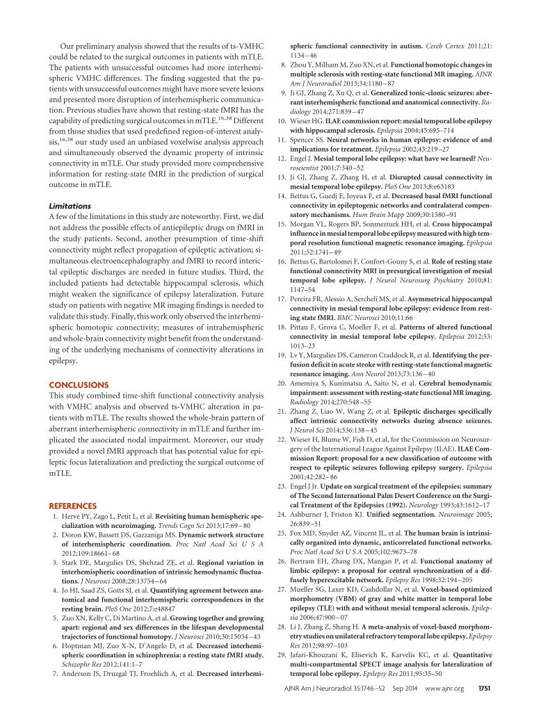
Our preliminary analysis showed that the results of ts-VMHCcould be related to the surgical outcomes in patients with mTLE.The patients with unsuccessful outcomes had more interhemi-spheric VMHC differences. The finding suggested that the pa-tients with unsuccessful outcomes might have more severe lesionsand presented more disruption of interhemispheric communica-tion. Previous studies have shown that resting-state fMRI has thecapability of predicting surgical outcomes in mTLE.16,38 Differentfrom those studies that used predefined region-of-interest analy-sis,16,38 our study used an unbiased voxelwise analysis approachand simultaneously observed the dynamic property of intrinsicconnectivity in mTLE. Our study provided more comprehensiveinformation for resting-state fMRI in the prediction of surgicaloutcome in mTLE.
LimitationsA few of the limitations in this study are noteworthy. First, we did
not address the possible effects of antiepileptic drugs on fMRI in
the study patients. Second, another presumption of time-shift
connectivity might reflect propagation of epileptic activation; si-
multaneous electroencephalography and fMRI to record interic-
tal epileptic discharges are needed in future studies. Third, the
included patients had detectable hippocampal sclerosis, which
might weaken the significance of epilepsy lateralization. Future
study on patients with negative MR imaging findings is needed to
validate this study. Finally, this work only observed the interhemi-
spheric homotopic connectivity; measures of intrahemispheric
and whole-brain connectivity might benefit from the understand-
ing of the underlying mechanisms of connectivity alterations in
epilepsy.
CONCLUSIONSThis study combined time-shift functional connectivity analysis
with VMHC analysis and observed ts-VMHC alteration in pa-
tients with mTLE. The results showed the whole-brain pattern of
aberrant interhemispheric connectivity in mTLE and further im-
plicated the associated nodal impairment. Moreover, our study
provided a novel fMRI approach that has potential value for epi-
leptic focus lateralization and predicting the surgical outcome of
mTLE.
REFERENCES1. Herve PY, Zago L, Petit L, et al. Revisiting human hemispheric spe-
cialization with neuroimaging. Trends Cogn Sci 2013;17:69 – 802. Doron KW, Bassett DS, Gazzaniga MS. Dynamic network structure
of interhemispheric coordination. Proc Natl Acad Sci U S A2012;109:18661– 68
3. Stark DE, Margulies DS, Shehzad ZE, et al. Regional variation ininterhemispheric coordination of intrinsic hemodynamic fluctua-tions. J Neurosci 2008;28:13754 – 64
4. Jo HJ, Saad ZS, Gotts SJ, et al. Quantifying agreement between ana-tomical and functional interhemispheric correspondences in theresting brain. PloS One 2012;7:e48847
5. Zuo XN, Kelly C, Di Martino A, et al. Growing together and growingapart: regional and sex differences in the lifespan developmentaltrajectories of functional homotopy. J Neurosci 2010;30:15034 – 43
6. Hoptman MJ, Zuo X-N, D’Angelo D, et al. Decreased interhemi-spheric coordination in schizophrenia: a resting state fMRI study.Schizophr Res 2012;141:1–7
7. Anderson JS, Druzgal TJ, Froehlich A, et al. Decreased interhemi-
spheric functional connectivity in autism. Cereb Cortex 2011;21:1134 – 46
8. Zhou Y, Milham M, Zuo XN, et al. Functional homotopic changes inmultiple sclerosis with resting-state functional MR imaging. AJNRAm J Neuroradiol 2013;34:1180 – 87
9. Ji GJ, Zhang Z, Xu Q, et al. Generalized tonic-clonic seizures: aber-rant interhemispheric functional and anatomical connectivity. Ra-diology 2014;271:839 – 47
10. Wieser HG. ILAE commission report: mesial temporal lobe epilepsywith hippocampal sclerosis. Epilepsia 2004;45:695–714
11. Spencer SS. Neural networks in human epilepsy: evidence of andimplications for treatment. Epilepsia 2002;43:219 –27
12. Engel J. Mesial temporal lobe epilepsy: what have we learned? Neu-roscientist 2001;7:340 –52
13. Ji GJ, Zhang Z, Zhang H, et al. Disrupted causal connectivity inmesial temporal lobe epilepsy. PloS One 2013;8:e63183
14. Bettus G, Guedj E, Joyeux F, et al. Decreased basal fMRI functionalconnectivity in epileptogenic networks and contralateral compen-satory mechanisms. Hum Brain Mapp 2009;30:1580 –91
15. Morgan VL, Rogers BP, Sonmezturk HH, et al. Cross hippocampalinfluence in mesial temporal lobe epilepsy measured with high tem-poral resolution functional magnetic resonance imaging. Epilepsia2011;52:1741– 49
16. Bettus G, Bartolomei F, Confort-Gouny S, et al. Role of resting statefunctional connectivity MRI in presurgical investigation of mesialtemporal lobe epilepsy. J Neurol Neurosurg Psychiatry 2010;81:1147–54
17. Pereira FR, Alessio A, Sercheli MS, et al. Asymmetrical hippocampalconnectivity in mesial temporal lobe epilepsy: evidence from rest-ing state fMRI. BMC Neurosci 2010;11:66
18. Pittau F, Grova C, Moeller F, et al. Patterns of altered functionalconnectivity in mesial temporal lobe epilepsy. Epilepsia 2012;53:1013–23
19. Lv Y, Margulies DS, Cameron Craddock R, et al. Identifying the per-fusion deficit in acute stroke with resting-state functional magneticresonance imaging. Ann Neurol 2013;73:136 – 40
20. Amemiya S, Kunimatsu A, Saito N, et al. Cerebral hemodynamicimpairment: assessment with resting-state functional MR imaging.Radiology 2014;270:548 –55
21. Zhang Z, Liao W, Wang Z, et al. Epileptic discharges specificallyaffect intrinsic connectivity networks during absence seizures.J Neurol Sci 2014;336:138 – 45
22. Wieser H, Blume W, Fish D, et al, for the Commission on Neurosur-gery of the International League Against Epilepsy (ILAE). ILAE Com-mission Report: proposal for a new classification of outcome withrespect to epileptic seizures following epilepsy surgery. Epilepsia2001;42:282– 86
23. Engel J Jr. Update on surgical treatment of the epilepsies: summaryof The Second International Palm Desert Conference on the Surgi-cal Treatment of the Epilepsies (1992). Neurology 1993;43:1612–17
24. Ashburner J, Friston KJ. Unified segmentation. Neuroimage 2005;26:839 –51
25. Fox MD, Snyder AZ, Vincent JL, et al. The human brain is intrinsi-cally organized into dynamic, anticorrelated functional networks.Proc Natl Acad Sci U S A 2005;102:9673–78
26. Bertram EH, Zhang DX, Mangan P, et al. Functional anatomy oflimbic epilepsy: a proposal for central synchronization of a dif-fusely hyperexcitable network. Epilepsy Res 1998;32:194 –205
27. Mueller SG, Laxer KD, Cashdollar N, et al. Voxel-based optimizedmorphometry (VBM) of gray and white matter in temporal lobeepilepsy (TLE) with and without mesial temporal sclerosis. Epilep-sia 2006;47:900 – 07
28. Li J, Zhang Z, Shang H. A meta-analysis of voxel-based morphom-etry studies on unilateral refractory temporal lobe epilepsy. EpilepsyRes 2012;98:97–103
29. Jafari-Khouzani K, Elisevich K, Karvelis KC, et al. Quantitativemulti-compartmental SPECT image analysis for lateralization oftemporal lobe epilepsy. Epilepsy Res 2011;95:35–50
AJNR Am J Neuroradiol 35:1746 –52 Sep 2014 www.ajnr.org 1751
30. Kim BJ, Hong SB, Seo DW. Differences in ictal hyperperfusion oflimbic-related structures between mesial temporal and neocorticalepilepsy. Epilepsy Res 2008;81:167–75
31. Semah F, Baulac M, Hasboun D, et al. Is interictal temporal hypo-metabolism related to mesial temporal sclerosis? A positron emis-sion tomography/magnetic resonance imaging confrontation. Epi-lepsia 1995;36:447–56
32. Zhang Z, Lu G, Zhong Y, et al. fMRI study of mesial temporal lobeepilepsy using amplitude of low-frequency fluctuation analysis.Hum Brain Mapp 2010;31:1851– 61
33. Liao W, Zhang Z, Pan Z, et al. Altered functional connectivity andsmall-world in mesial temporal lobe epilepsy. PloS One 2010;5:e8525
34. Kucker S, Tollner K, Piechotta M, et al. Kindling as a model of tem-
poral lobe epilepsy induces bilateral changes in spontaneous stria-tal activity. Neurobiol Dis 2010;37:661–72
35. Pan J, Spencer D, Kuzniecky R, et al. Metabolic networks in epilepsyby MR spectroscopic imaging. Acta Neurol Scand 2012;126:411–20
36. Tae WS, Joo EY, Kim JH, et al. Cerebral perfusion changes in mesialtemporal lobe epilepsy: SPM analysis of ictal and interictal SPECT.Neuroimage 2005;24:101–10
37. Marques LH, Ferraz-Filho JR, Lins-Filho ML, et al. Interictal SPECTin the presurgical evaluation in epileptic patients with normal MRIor bilateral mesial temporal sclerosis. Arq Neuropsiquiatr 2009;67:639 – 42
38. Negishi M, Martuzzi R, Novotny EJ, et al. Functional MRI connec-tivity as a predictor of the surgical outcome of epilepsy. Epilepsia2011;52:1733– 40
1752 Xu Sep 2014 www.ajnr.org