Embed Size (px)
Citation preview

Thyroid Dysfunction and the Eye January 25, 2014
Greg A Caldwell, OD, FAAO [email protected] 814-931-2030 cell 1
Thyroid Dysfunction and the Eye
Greg Caldwell OD, FAAO The Denver Metro Optometric Society Russell Pavilion at St. Joseph Hospital
Denver, CO January 25, 2014
Disclosure Statement (next slide)
Disclosures
$ Greg A. Caldwell, OD, FAAO will mention many products, instruments and companies during our discussion, I don’t have any financial interest in any of these products, instruments or companies.
$ In the past 12 months I have lectured or participated in a focus group which I received a honorarium for: ¬ Allergan, Alcon, Valeant and SARcode Bioscience
$ All of these cases have entered/referred to my practice
Learning Objectives
$ Enhance clinical understanding of thyroid dysfunction and the ocular associations
$ Enhance clinical diagnosis of ocular manifestations of thyroid disease
$ Enrich clinical management and treatment of ocular manifestations thyroid eye disease
$ Increase comfort level when ordering or interpreting laboratory tests in thyroid diseases
$ Gain confidence in working closer with the endocrinologist, oculoplastic surgeon and orbital specialist
Thyroid Disease and
Thyroid Eye Disease
Thyroid $ Thyroid is an endocrine gland $ Two types of glands
¬ Endocrine ¬ Exocrine
$ Endocrine system is a control system of ductless endocrine glands that secrete hormones (chemical messenger) that circulate within the body via the bloodstream or lymph system to affect distant organs
¬ Hypothalamus ¬ Pituitary gland ¬ Thyroid ¬ Parathyroid glands
¬ Pancreas ¬ Adrenal glands ¬ Gonads (testes and ovaries) ¬ Pineal gland
Thyroid
$ Exocrine glands contain ducts. Ducts are tubes leading from a gland to its target organ ¬ Digestive glands have ducts for releasing the digestive enzymes ¬ Salivary glands, sweat glands and glands within the
gastrointestinal tract
$ Pancreas is both endocrine and exocrine ¬ Exocrine (ducted gland) secreting digestive enzymes into the small
intestine. ¬ Endocrine (ductless gland) in that the islets of Langerhans secrete
insulin and glucagon to regulate the blood sugar level.
Thyroid Dysfunction and the Eye January 25, 2014
Greg A Caldwell, OD, FAAO [email protected] 814-931-2030 cell 2
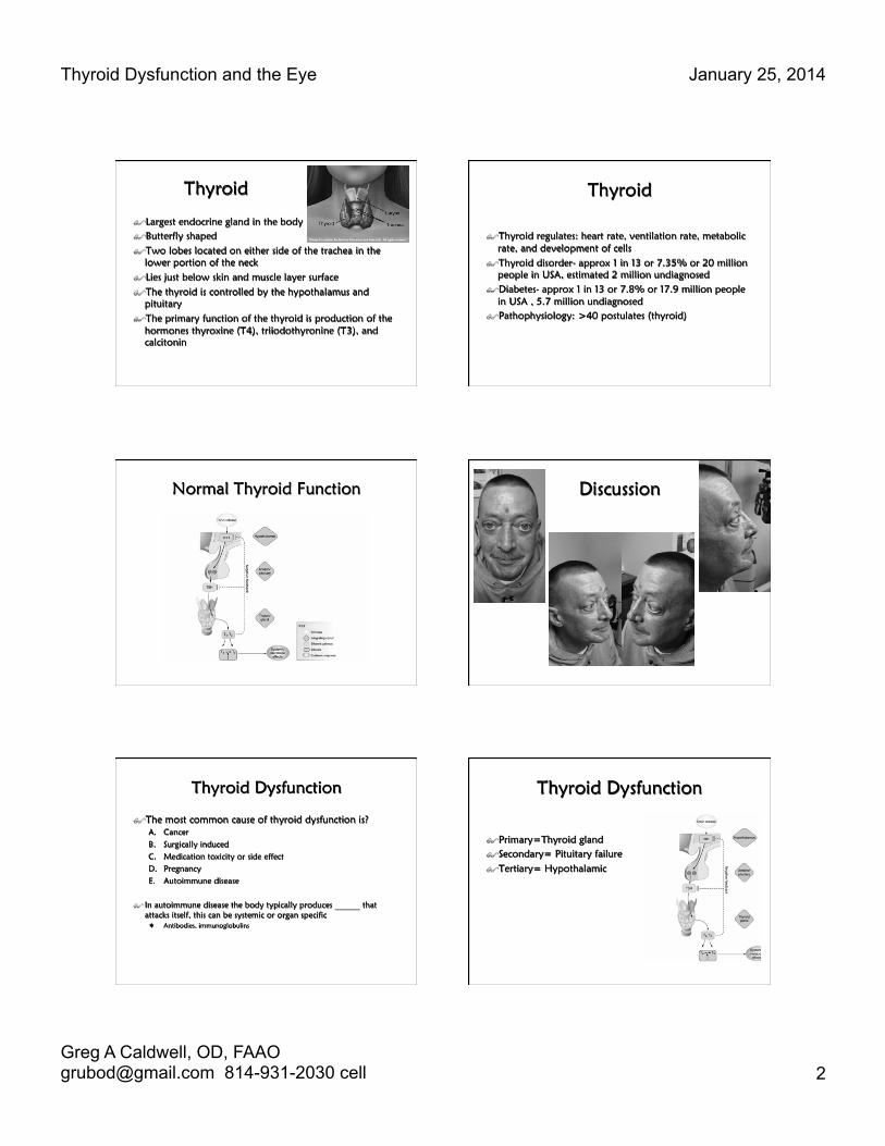
Thyroid
$ Largest endocrine gland in the body $ Butterfly shaped $ Two lobes located on either side of the trachea in the
lower portion of the neck $ Lies just below skin and muscle layer surface $ The thyroid is controlled by the hypothalamus and
pituitary $ The primary function of the thyroid is production of the
hormones thyroxine (T4), triiodothyronine (T3), and calcitonin
Thyroid
$ Thyroid regulates: heart rate, ventilation rate, metabolic rate, and development of cells
$ Thyroid disorder- approx 1 in 13 or 7.35% or 20 million people in USA, estimated 2 million undiagnosed
$ Diabetes- approx 1 in 13 or 7.8% or 17.9 million people in USA , 5.7 million undiagnosed
$ Pathophysiology: >40 postulates (thyroid)
Normal Thyroid Function Discussion
Thyroid Dysfunction
$ The most common cause of thyroid dysfunction is? A. Cancer B. Surgically induced C. Medication toxicity or side effect D. Pregnancy E. Autoimmune disease
$ In autoimmune disease the body typically produces ______ that attacks itself, this can be systemic or organ specific ¬ Antibodies, immunoglobulins
Thyroid Dysfunction
$ Primary=Thyroid gland $ Secondary= Pituitary failure $ Tertiary= Hypothalamic

Thyroid Dysfunction and the Eye January 25, 2014
Greg A Caldwell, OD, FAAO [email protected] 814-931-2030 cell 3
Antibodies of Thyroid Dysfunction
$ TSH Receptor Antibodies ¬ Stimulating TSH receptor antibody
2 Thyroid Stimulating Immunoglobulin (TSI)
¬ Thyroid blocking antibody (TBAb)
$ Thyroid Peroxidase Antibodies (TPOAb) ¬ TPO is found in thyroid follicle cells where it converts the thyroid
hormone T4 to T3 ¬ TPOAb contributes to thyroid cellular destruction
$ Most autoimmune thyroid dysfunctions have a combination of thyroid antibodies, however depending on which AB is more abundant results in the outcome of the disease
Hyperthyroid
$ TSI attacks the thyroid
$ T3 and T4 increase $ TSH decreases
Hypothyroid
$ TBAb attacks the thyroid
$ T3 and T4 decrease $ TSH increases
Thyroid Dysfunction Hyperthyroidism
(Thyrotoxicosis)
$ Primary-autoimmune ¬ Graves
2 Graves-Basedow or von Basedow’s
$ Secondary/Tertiary ¬ Excess thyroid medication for treatment
of hypo or goiter ¬ Toxic multinodular goiter ¬ Toxic adenoma ¬ Excess iodine ¬ Thyroiditis (inflammatory induced)
¬ Excess hormone production ectopic tissue
¬ Thyroid carcinoma
Hypothyroidism (most common organ-specific autoimmune disorder)
$ Primary-autoimmune ¬ Chronic autoimmune thyroiditis
2 Hashimoto's thyroiditis
¬ Autoimmune atrophic thyroiditis 2 Primary myxedema 2 Opposite of Graves disease
¬ Postpartum thyroiditis
$ Secondary/Tertiary ¬ Lithium medication ¬ Pregnancy ¬ Surgically induced ¬ Disorders of the pituitary gland
or hypothalamus
GRAVE’S (Hyperthyoidism)
$ A multisystem disorder consisting of a triad ¬ Hyperthyroidism with diffuse hyperplasia of the thyroid gland ¬ Infiltrative dermopathy ¬ Infiltrative ophthalmopathy
$ Prevalence: ¬ 20-40 year old female (F:M = 7:1) ¬ Genetic link
$ Etiology: ¬ Autoimmune disease: hypersensitivity reaction with thyroid
stimulation by the circulation of abnormal thyroid-stimulating immunoglobulins (TSI)
Hashimoto's Thyroiditis (Hypothyroidism)
$ The most common cause of hypothyroidism in the United States
$ It is named after the first doctor who described this condition, Dr. Hakaru Hashimoto, in 1912
$ Autoimmune disease $ Goiter formation $ 5-10 times more common in women than in men $ The underlying cause of the autoimmune process still is
unknown ¬ Anti-TPO ab and Anti-TB recp ab present

Thyroid Dysfunction and the Eye January 25, 2014
Greg A Caldwell, OD, FAAO [email protected] 814-931-2030 cell 4
Autoimmune atrophic thyroiditis (Hypothyroidism)
$ Atrophic thyroiditis is similar to Hashimoto's thyroiditis $ A goiter is not present
Postpartum Thyroiditis (Hypothyroidism)
$ These women develop antibodies to their own thyroid during pregnancy, causing an inflammation of the thyroid after delivery
Systemic Manifestations of Hyperthyroid (Primary or Secondary)
$ Symptoms ¬ Nervousness ¬ Heat intolerance ¬ Sweating ¬ Fatigue ¬ Palpitation ¬ Insomnia ¬ Early waking ¬ Alopecia ¬ Vitiligo ¬ Brittle nails
$ Signs ¬ Sweating ¬ Muscle Weakness ¬ Emotionally labile ¬ Tremor ¬ Tachycardia ¬ Arrhythmia ¬ Hypertension ¬ Brisk tendon reflex ¬ Diabetes ¬ ↑Triglycerides & Ca, ↓CHO ¬ Microcyticanemia ¬ Possible goiter ¬ Myxedema
Systemic Manifestations of Hypothyroid (Primary or Secondary)
$ Symptoms ¬ Cold intolerance ¬ Weakness ¬ Reduced energy ¬ Lethargy ¬ Muscle cramps ¬ Constipation ¬ Increased sleeping ¬ Weight gain ¬ Reduced appetite ¬ Joint stiffness
$ Signs ¬ Cool, scaling skin ¬ Puffy hands and face ¬ Deep voice ¬ Myotonia ¬ Delirium ¬ Bradycardia ¬ Slow reflexes ¬ Obesity ¬ Hypothermia ¬ Myxedema
Thyroid Eye Disease (TED)
$ Other names used ¬ Grave’s disease ¬ Grave's ophthalmopathy ¬ Grave's orbitopathy ¬ Exophthalmos in Graves Disease ¬ Thyroid Associated Orbitopathy (TAO) ¬ Thyroid Orbitopathy ¬ Ophthalmic Graves Disease ¬ Inflammatory Eye Disease ¬ Endocrine Orbitopathy
Why is this so confusing?
$ Thyroid Eye Disease ¬ Is often seen in conjunction with Graves' Disease (hyperthyroid) ¬ Is seen in people with no other evidence of thyroid dysfunction ¬ Is seen in patients who have Hashimoto's Disease (hypothyroid)
$ Most thyroid patients, however, will not develop thyroid eye disease

Thyroid Dysfunction and the Eye January 25, 2014
Greg A Caldwell, OD, FAAO [email protected] 814-931-2030 cell 5
Why is this so confusing? $ The eye symptoms usually occur at the same time as the thyroid
disease ¬ However they may precede or follow the obvious symptoms of the thyroid
abnormality $ The incidence of thyroid eye disease associated with thyroid
dysfunction is higher and more severe in smokers ¬ There is no way to predict which thyroid patients will be affected
Why is this so confusing? $ While eye disease may be brought on by thyroid dysfunction
¬ Successful treatment of the thyroid gland does not guarantee that the eye disease will improve
¬ No particular thyroid treatment can guarantee that the eyes will not continue to deteriorate
¬ Once inflamed, the eye disease may remain active from several months to as long as three years
¬ There may be a gradual or, in some cases, a complete improvement
Thyroid Eye Disease $ Commonly known as Graves' ophthalmopathy $ About 80% of all patients with TED have the autoimmune hyperthyroid
disorder known as Graves' disease $ Another 10% of all cases are seen in patients with autoimmune
hypothyroidism, either Hashimoto's thyroiditis, atrophic thyroiditis or Hashitoxicosis
$ Another 10% of all cases are seen in people with normal thyroid function ¬ When thyroid function is normal, the eye condition is referred to as euthyroid
Graves' disease ¬ Euthyroid is a term meaning that thyroid function tests are normal. Most people
with euthyroid Graves' disease develop a thyroid disorder within eighteen months of the emergence of the eye disorder
¬ But some people with euthyroid Graves' disease never develop thyroid dysfunction
Thyroid Eye Disease $ What causes the Thyroid Eye Disease signs and symptoms?
$ The high and low levels of T3 and T4
$ The antibodies that are attacking the thyroid gland
Thyroid Eye Disease
$ Thyroid Eye Disease has 2 phases
¬ A phase secondary to abnormal thyroid hormone levels 2 Increased or decreased FT3 and FT4 levels 2 Once these levels are normalized, ocular symptoms will resolve
¬ Congestive Autoimmune form of Thyroid Eye Disease 2 Active phase-stimulating or blocking TRAb are causing ocular activity 2 Plateau phase-reduced activity 2 Resolution phase-symptoms regress and eyes return to normal
Phase secondary to abnormal thyroid hormone levels (T3/T
4)
(Thyroid Eye Disease)
$ Hyperthyroidism eye symptoms ¬ Excess hormone acting on the nerves
that supply the eye ¬ Usually spastic and include staring ¬ Dryness ¬ Eyelid retraction
$ Hypothyroidism eye symptoms ¬ Deficient hormone causing venous
congestion, impaired circulation and fluid stagnation
¬ Periorbital edema
$ This form of TED resolves within a few weeks after thyroid hormone levels (FT4 and FT3) are corrected and brought back into the normal range
$ The pituitary hormone TSH can stay low or suppressed for many months during the course of treatment for hyperthyroidism and doesn't mean that the patient is still hyperthyroid
$ TSH also lags at least 6 weeks behind thyroid hormone levels and often remains elevated longer in people who have been hypothyroid
$ Relying on the TSH level can be misleading and in treating TED

Thyroid Dysfunction and the Eye January 25, 2014
Greg A Caldwell, OD, FAAO [email protected] 814-931-2030 cell 6
Congestive Autoimmune form of Thyroid Eye Disease (Active phase, Plateau phase, Resolution phase)
$ Caused by both stimulating and blocking TSH receptor antibodies (TRAb) and also immune system chemicals known as cytokines
$ Secondary targets appear to be TSH receptor antigens (epitopes) located on orbital fibroblasts as well as dermal fibroblasts
$ Active “inflammatory” phase of TED varies ¬ Symptoms resolve quickly although on average the active phase lasts
about 12-18 months ¬ TRAb levels are high, patients are smokers, nutrient deficiencies are
present, or the patient continues to be exposed to environmental triggers such as excess dietary iodine, the active phase can last as long as 5 years
¬ Avoid any lid, muscle or orbital surgery
$ Plateau phase and Resolution “Passive” phase ¬ An individual may be left with structural changes, such as eye protrusion, eyelid
retraction, and in some cases, double vision ¬ There are corrective procedures that can be performed to address these problems
Euthyroid Graves' disease
$ If thyroid function is normal. How does one develop thyroid eye disease?
Similar receptors are found in the skin, fat and muscle of the orbit General Ocular Symptoms
$ Prominent eyes, stare $ Pain $ Lacrimation $ Eyelid swelling $ Foreign-body sensation $ Double vision $ Photophobia $ Decreased vision in one or both eyes
NOSPECS: Grading System
$ 1969 by S.C. Werner ¬ Class 0: No signs or symptoms ¬ Class 1: Only signs, upper lid retraction ¬ Class 2: Soft Tissue involvement with symptoms ¬ Class 3: Proptosis ¬ Class 4: EOM involvement ¬ Class 5: Corneal Involvement ¬ Class 6: Sight Loss
$ Within classes 2 to 6 the investigator has to differentiate the severity grades 0, A, B, C
$ NOSPECS, classifies severity but not the activity or stage (active/inflammatory or passive/congestive)
$ Class 2-6 document severity ¬ 0: absent ¬ A: minimal ¬ B: moderate ¬ C: marked
NOSPECS: Grading System $ 0: No symptoms or signs $ 1: Only signs (upper lid retraction without lid lag or proptosis) $ 2: Soft tissue involvement with symptoms (excess lacrimation, sandy sensation,
retrobulbar discomfort) ¬ Grade 0: absent ¬ Grade A: minimal (edema of lids, injection, sandy feeling) ¬ Grade B: moderate (edema of lids, injection, chemosis, FBS, pain behind eyes) ¬ Grade C: marked
$ 3: Proptosis associated with classes 2-6 only ¬ Grade 0: absent ¬ Grade A: minimal: 21mm -23mm ¬ Grade B: moderate: 24mm -27mm ¬ Grade C: marked: 28mm or more ¬ Specify if inequality of >3 mm between eyes, or if progression of >3 mm under observation

Thyroid Dysfunction and the Eye January 25, 2014
Greg A Caldwell, OD, FAAO [email protected] 814-931-2030 cell 7
NOSPECS: Grading System $ 4: EOM involvement (usually with diplopia)
¬ 0: absent ¬ A: minimal (limitation of motion, patient reports diplopia but no obvious restriction ¬ B: moderate (evident restriction of motion) ¬ C: marked (position of globe is fixed)
$ 5: Corneal involvement (due to proptosis, incomplete closure, lagophthalmos) ¬ 0: absent ¬ a: minimal (staining) ¬ b: moderate (ulceration) ¬ c: marked (clouding, necrosis, perforation)
$ 6: Sight loss (due to optic nerve involvement) ¬ 0: absent ¬ A: minimal (disc pallor or edema, or VF defect, vision 20/20-20/60) ¬ B: moderate (same as A but VA 20/70-20/200) ¬ C: marked (blindness, VA < 20/200)
LEMO Classification
$ 1991-Boergen and Pickardt $ Complements NOSPECS $ 4 finding-categories
¬ Lid ¬ Exophthalmos ¬ Muscular ¬ Optic nerve
$ Grade between 0 and 4 depending on severity $ LEMO, classifies severity but not the activity or stage
(active/inflammatory or passive/congestive)
LEMO Classification
Lid (L) $ 0: missing $ 1: lid edema only $ 2: real retraction (impaired lid
closing) $ 3: retraction and upper lid edema $ 4: retraction and global lid edema
Exophthalmos (E) $ 0: missing $ 1: eye closing not impaired $ 2: conjunctival injection in the
morning $ 3: persistent conjunctival injection $ 4: corneal complications
LEMO Classification
Muscular (M) $ 0: missing $ 1: detectable in imaging only $ 2: Pseudoparesis $ 3: Pseudoparalysis
Optic Nerve (O) $ 0: missing $ 1: regarding color vision only
or detected via VEP $ 2: peripheral scotoma $ 3: central scotoma
L1E1M2O0 Endocrine ophthalmopathy with lid edema, exophthalmos , pseudoparesis of external eye muscles, and no optic nerve involvement
Grading Scales
$ New grading scales are trying to be developed to not only grade the severity but also help to determine if inflammatory or passive stage
Lid Involvement
$ Lid Retraction $ Lid Lag $ Lagophthalmus

Thyroid Dysfunction and the Eye January 25, 2014
Greg A Caldwell, OD, FAAO [email protected] 814-931-2030 cell 8
Lid Retraction $ Scleral show in primary gaze $ Occurs in ~90% of Grave’s patients
¬ Excess stimulation of Muller’s muscle ¬ Fibrotic inferior rectus ¬ Mechanical restriction or infiltration
of levator ¬ Increased orbital volume causes
exophthalmos
$ Normal Lid Position ¬ Upper lid intersects cornea at the 2
and 10 o’clock positions 2 ~2 mm below the limbus
¬ Lower lid coincident or 1-2mm below the limbus
Eyelid Lag: von Graefe’s Sign
$ Immobility or lagging of upper eyelid on downward gaze
$ Fibrosis of the inferior rectus muscle may induce lower lid retraction
Lagophthalmos
$ Inability to form a complete lid closure with a normal blink due to Exophthalmos/ Proptosis
$ Often leads to corneal exposure
Soft Tissue Involvement
$ Conjunctiva $ Chemosis $ Periorbital edema
Conjunctiva $ Conjunctival and episcleral
injection ¬ Especially near the horizontal recti
insertions $ Chemosis
¬ Edema of the conjunctiva and caruncle
$ Superior Limbic Keratoconjunctivitis ¬ 65% correlation between SLK and
systemic thyroid disease ¬ Rheumatoid arthritis ¬ Sjögren’s syndrome
“If it is Red think TED” Dr. Andy Morgenstern 12-7-2013, OMS-Contemporary Resort
Thyroid Dysfunction and the Eye January 25, 2014
Greg A Caldwell, OD, FAAO [email protected] 814-931-2030 cell 9
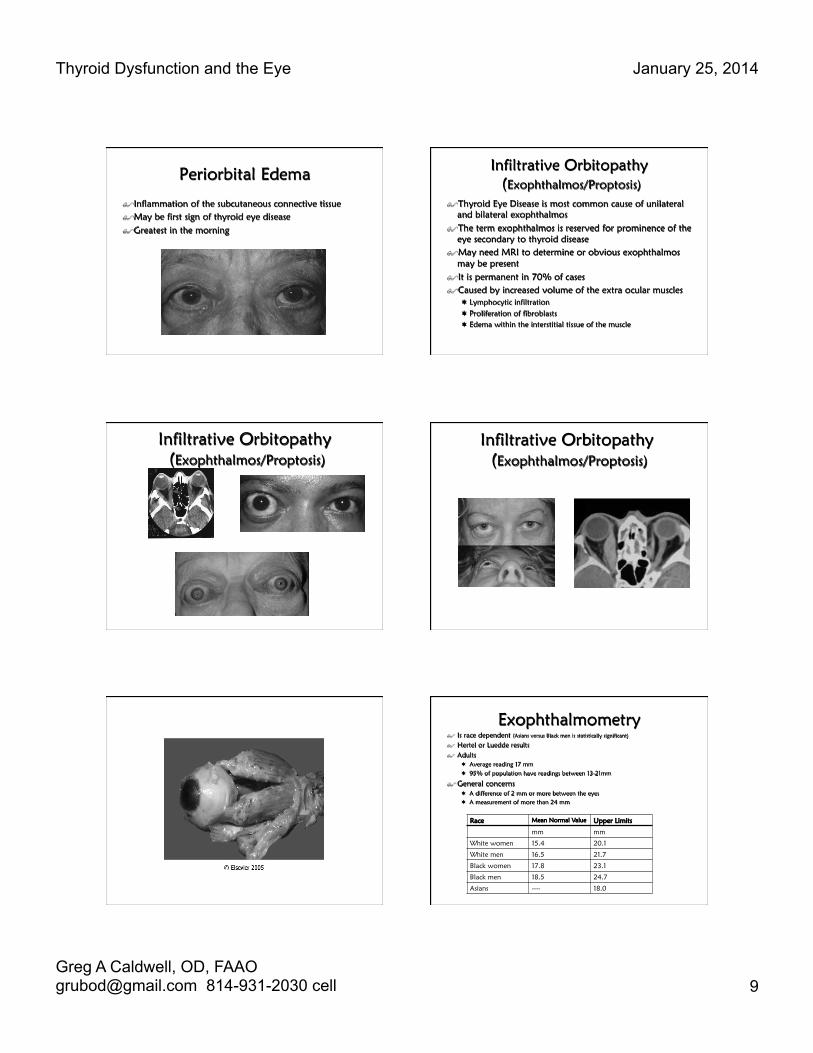
Periorbital Edema
$ Inflammation of the subcutaneous connective tissue $ May be first sign of thyroid eye disease $ Greatest in the morning
Infiltrative Orbitopathy (Exophthalmos/Proptosis)
$ Thyroid Eye Disease is most common cause of unilateral and bilateral exophthalmos
$ The term exophthalmos is reserved for prominence of the eye secondary to thyroid disease
$ May need MRI to determine or obvious exophthalmos may be present
$ It is permanent in 70% of cases $ Caused by increased volume of the extra ocular muscles
¬ Lymphocytic infiltration ¬ Proliferation of fibroblasts ¬ Edema within the interstitial tissue of the muscle
Infiltrative Orbitopathy (Exophthalmos/Proptosis)
Infiltrative Orbitopathy (Exophthalmos/Proptosis)
Exophthalmometry $ Is race dependent (Asians versus Black men is statistically significant)
$ Hertel or Luedde results $ Adults
¬ Average reading 17 mm ¬ 95% of population have readings between 13-21mm
$ General concerns ¬ A difference of 2 mm or more between the eyes ¬ A measurement of more than 24 mm
Race Mean Normal Value Upper Limits
mm mm
White women 15.4 20.1
White men 16.5 21.7
Black women 17.8 23.1
Black men 18.5 24.7
Asians ---- 18.0

Thyroid Dysfunction and the Eye January 25, 2014
Greg A Caldwell, OD, FAAO [email protected] 814-931-2030 cell 10
Restrictive Myopathy
$ Secondary to edema and fibrosis of EOM’s $ Inferior Rectus (IR) muscle is most commonly involved $ Occurs in 30-50% of patients $ Diplopia may be transient but in 50% it’s permanent
IOP in Thyroid Eye Disease
$ A rise in IOP has been reported with TED $ I would have higher suspicion when you see
¬ Periorbital edema ¬ Exophthalmos, proptosis ¬ Restrictive myopathy
$ Some literature reports IOP in up gaze to be part of the diagnoses of thyroid dysfunction
Restrictive Myopathy
Obvious restrictive myopathy but also note the periorbital edema, and conjunctival hyperemia
Corneal Exposure
$ Exposure keratopathy secondary to exophthalmos and lagophthalmos
$ Significant threat to visual function
Optic Neuropathy $ Affects 5% of patients $ Usually mild to moderate
exophthalmos and shallow orbits $ Enlargement of the recti muscles
compresses ONH or its blood supply at the apex of the orbit
$ Compression MAY occur without significant proptosis
$ Compressive and/or ischemic and/or toxic
Treatment of Thyroid Eye Disease $ Depends on what phase of the disease we are in:
¬ Phase secondary to abnormal thyroid hormone levels ¬ Active “inflammatory” phase ¬ Plateau phase and Resolution “Passive” phase
$ Depends on what orbital tissue or structures are involved $ Depends on the risk of vision loss $ Depends if primary, secondary or tertiary thyroid dysfunction $ Management consists of:
¬ Control of inflammation
¬ Prevention of ocular and visual damage ¬ Addressing ocular motor abnormalities ¬ Improving cosmetic disfigurement
$ Patient education is essential $ Communication with an endocrinologist or internist will ensure
proper patient care
Thyroid Dysfunction and the Eye January 25, 2014
Greg A Caldwell, OD, FAAO [email protected] 814-931-2030 cell 11
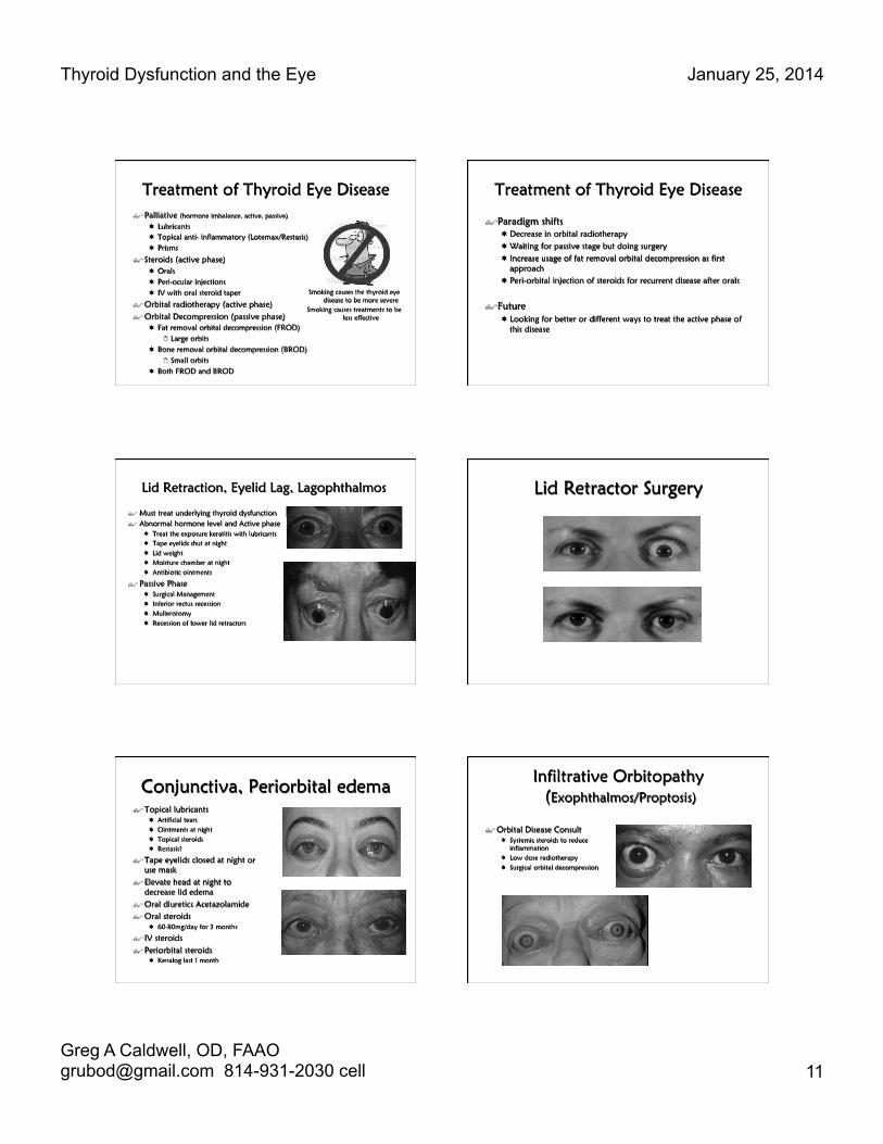
Treatment of Thyroid Eye Disease $ Palliative (hormone imbalance, active, passive)
¬ Lubricants ¬ Topical anti- inflammatory (Lotemax/Restasis) ¬ Prisms
$ Steroids (active phase) ¬ Orals ¬ Peri-ocular injections ¬ IV with oral steroid taper
$ Orbital radiotherapy (active phase) $ Orbital Decompression (passive phase)
¬ Fat removal orbital decompression (FROD) 2 Large orbits
¬ Bone removal orbital decompression (BROD) 2 Small orbits
¬ Both FROD and BROD
Smoking causes the thyroid eye disease to be more severe
Smoking causes treatments to be less effective
Treatment of Thyroid Eye Disease
$ Paradigm shifts ¬ Decrease in orbital radiotherapy ¬ Waiting for passive stage but doing surgery ¬ Increase usage of fat removal orbital decompression as first
approach ¬ Peri-orbital injection of steroids for recurrent disease after orals
$ Future ¬ Looking for better or different ways to treat the active phase of
this disease
Lid Retraction, Eyelid Lag, Lagophthalmos
$ Must treat underlying thyroid dysfunction $ Abnormal hormone level and Active phase
¬ Treat the exposure keratitis with lubricants ¬ Tape eyelids shut at night ¬ Lid weight ¬ Moisture chamber at night ¬ Antibiotic ointments
$ Passive Phase ¬ Surgical Management ¬ Inferior rectus recession ¬ Mullerotomy ¬ Recession of lower lid retractors
Lid Retractor Surgery
Conjunctiva, Periorbital edema $ Topical lubricants
¬ Artificial tears ¬ Ointments at night ¬ Topical steroids ¬ Restasis?
$ Tape eyelids closed at night or use mask
$ Elevate head at night to decrease lid edema
$ Oral diuretics Acetazolamide $ Oral steroids
¬ 60-80mg/day for 3 months
$ IV steroids $ Periorbital steroids
¬ Kenalog last 1 month
Infiltrative Orbitopathy (Exophthalmos/Proptosis)
$ Orbital Disease Consult ¬ Systemic steroids to reduce
inflammation ¬ Low dose radiotherapy ¬ Surgical orbital decompression

Thyroid Dysfunction and the Eye January 25, 2014
Greg A Caldwell, OD, FAAO [email protected] 814-931-2030 cell 12
Restrictive Myopathy $ Non-surgical (while waiting for stability)
¬ Teach proper head position to alleviate diplopia
¬ Prism in spectacle correction (Fresnel or ground in)
¬ Oral steroids ¬ Botulinum toxin injection
$ Surgical Consult ¬ Recession of the rectus muscle/s involved ¬ Diplopia in primary gaze, reading gaze or both ¬ Stable angle of deviation for at least 6 months ¬ No evidence of active disease ¬ Binocular vision in at least primary and reading
positions
Corneal Exposure
$ Manage the corneal defect as first line ¬ Lubricating and antibiotic ¬ Lid taping ¬ Moisture barrier
$ Orbital Disease Consult ¬ High dose oral steroids
2 120-140mg /day x 7 days
¬ Orbital decompression
Optic Neuropathy $ Systemic Steroids
¬ If rapidly progressive and painful in the early stage of the disease
¬ Only if no contraindications ¬ Prednisolone 80-100mg, expect
results within 48hrs. Taper dose and d/c within 3 mo
$ IV Methylprednisolone $ Radiotherapy: if contraindication
to steroid $ Orbital decompression
Orbital Decompression
$ Not effective if no medical treatment ¬ Two-wall decompression
2 3-6 mm retro-placement of the globe
¬ Three-wall decompression 2 6-10mm retro-placement
¬ Four-wall decompression 2 10-16mm retro-placement
Orbital Decompression (Surgical/Cosmetic)
Thyroid Eye Disease and Depression
$ When facial disfigurement occurs, thyroid eye disease is equivalent to the diagnosis of cancer and AIDS
Thyroid Dysfunction and the Eye January 25, 2014
Greg A Caldwell, OD, FAAO [email protected] 814-931-2030 cell 13
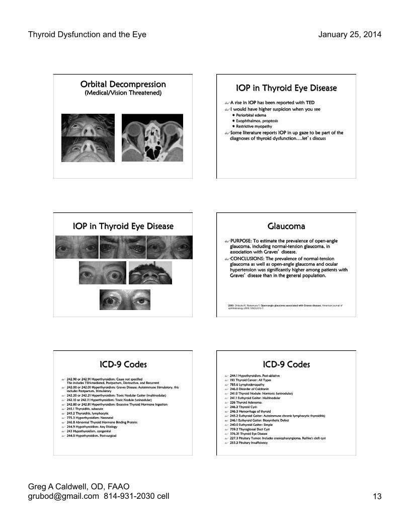
Orbital Decompression (Medical/Vision Threatened) IOP in Thyroid Eye Disease
$ A rise in IOP has been reported with TED $ I would have higher suspicion when you see
¬ Periorbital edema ¬ Exophthalmos, proptosis ¬ Restrictive myopathy
$ Some literature reports IOP in up gaze to be part of the diagnoses of thyroid dysfunction….let’s discuss
IOP in Thyroid Eye Disease Glaucoma
$ PURPOSE: To estimate the prevalence of open-angle glaucoma, including normal-tension glaucoma, in association with Graves’ disease.
$ CONCLUSIONS: The prevalence of normal-tension glaucoma as well as open-angle glaucoma and ocular hypertension was significantly higher among patients with Graves’ disease than in the general population.
2000: Ohtsuka K; Nakamura Y, Open-angle glaucoma associated with Graves disease. American journal of ophthalmology 2000;129(5):613-7.
ICD-9 Codes
$ 242.90 or 242.91 Hyperthyroidism: Cause not specified This includes TSH-mediated, Postpartum, Destructive, and Recurrent
$ 242.00 or 242.01 Hyperthyroidism: Graves Disease; Autoimmune Stimulatory. this includes Postpartum, Stimulatory
$ 242.20 or 242.21 Hyperthyroidism: Toxic Nodular Goiter (multinodular) $ 242.10 or 242.11 Hyperthyroidism: Toxic Nodule (uninodular) $ 242.80 or 242.81 Hyperthyroidism: Excessive Thyroid Hormone Ingestion $ 245.1 Thyroiditis, subacute $ 245.2 Thyroiditis, lymphocytic $ 775.3 Hyperthyroidism: Neonatal $ 246.8 Abnormal Thyroid Hormone Binding Protein: $ 244.9 Hypothyroidism: Any Etiology $ 243 Hypothyroidism, congenital $ 244.0 Hypothyroidism, Post-surgical
ICD-9 Codes $ 244.1 Hypothyroidism, Post-ablative $ 193 Thyroid Cancer: All Types $ 785.6 Lymphadenopathy $ 246.0 Disorder of Calcitonin $ 241.0 Thyroid Nodule: Nontoxic (uninodular) $ 241.1 Euthyroid Goiter: Multinodular $ 226 Thyroid Adenoma: $ 246.2 Thyroid Cyst: $ 246.3 Hemorrhage of thyroid $ 245.2 Euthyroid Goiter: Autoimmune chronic lymphocytic thyroiditis) $ 246.1 Euthyroid Goiter: Biosynthetic Defect $ 240.0 Euthyroid Goiter: Simple $ 759.2 Thyroglossal Duct Cyst $ 376.21 Thyroid Eye Disease $ 227.3 Pituitary Tumor: Includes craniopharyngioma, Rathke's cleft cyst $ 253.2 Pituitary Insufficiency

Thyroid Dysfunction and the Eye January 25, 2014
Greg A Caldwell, OD, FAAO [email protected] 814-931-2030 cell 14
Laboratory Testing $ Thyroid Hormone Levels
¬ Serum TSH concentration Serum total T4 (Thyroxine) ¬ Serum total T3 (Triiodithyronine) ¬ Estimation of the serum free T4 (or T3) concentration ¬ Thyroglobulin (Tg) level
$ Anti-thyroid antibodies ¬ Thyrotropin receptor antibodies (TSI) ¬ TSH binding inhibiting immunoglobulins (TBII) ¬ Anti-TPO antibodies ¬ Thyroglobulin (Tg) Antibodies (TgAb)
$ Commonly used thyroid tests ¬ Resin T3 uptake test ¬ Sensitive serum TSH test (Thyroid stimulating hormone) ¬ TRH stimulation test (Thyroid releasing hormone) ¬ Thyroid (T3) suppression test
¬ Sonography ¬ Needle Biopsy ¬ Thyroid Scan
Laboratory Testing $ Hypothyroid
¬ Low FT4, High TSH, indicates primary check antibodies ¬ Low FT4, Low TSH, indicates secondary or tertiary, TRH stimulation,
MRI ¬ Hashimoto’s (primary disease)
2 Most common 2 Low FT4, High TSH, High Anti-TPO Ab, High levels of Thyroglobulin (Tg)
Antibodies (TgAb), Anti-TB Recp Ab (approx 10% present)
¬ Autoimmune atrophic thyroiditis 2 Low FT4, High TSH, Low Anti-TPO Ab, Low levels of Thyroglobulin (Tg)
Antibodies (TgAb), Anti-TB Recp Ab (approx 60% present)
¬ Treatment: Levothyroxine (Synthroid, Levothroid, Levoxyl, Unithroid)
$ Hyperthyroid ¬ High FT4, Low TSH ¬ TSI present
Sign’s in Thyroid Eye Disease
$ Dalrymple’s sign: Lid retraction $ von Graefe’s sign: Upper lid lag
on downward gaze $ Griffith’s sign: Lower lid lag on
downward gaze $ Boston’s sign: Jerky irregular
movement of the upper lid on downward gaze
$ Jellinek’s sign: Increased pigmentation of the lids
$ Stellwag’s sign: Infrequent blinking $ Kocher’s sign: Increased lid
retraction with visual fixation
$ Enroth’s sign: Puffy swelling of the lids
$ Rosenbach’s sign: Tremor of closed lids
$ Mobius’ sign: Weakness of convergence
$ Ballet’s sign: Palsy of one or more extraocular muscles
$ Suker’s sign: Weakness of fixation on lateral gaze
$ Cowen’s sign: Jerky papillary contraction to consensual light
$ Knies’ sign: Unequal dilatation of the pupils
$ Jeffrey’s sign: Absence of forehead wrinkling on upward gaze
Thank-You and
Hope You Enjoyed
Greg Caldwell, OD, FAAO [email protected]