Embed Size (px)
Citation preview
TOUCH MEDICAL MEDIA 85
Original Research Thyroid Disorders
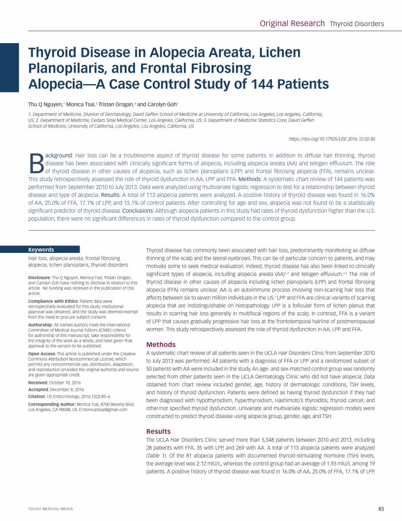
Thyroid Disease in Alopecia Areata, Lichen Planopilaris, and Frontal Fibrosing Alopecia—A Case Control Study of 144 PatientsThu Q Nguyen,1 Monica Tsai,2 Tristan Grogan,3 and Carolyn Goh1
1. Department of Medicine, Division of Dermatology, David Geffen School of Medicine at University of California, Los Angeles, Los Angeles, California, US; 2. Department of Medicine, Cedars Sinai Medical Center, Los Angeles, California, US; 3. Department of Medicine Statistics Core, David Geffen School of Medicine, University of California, Los Angeles, Los Angeles, California, US
Background: Hair loss can be a troublesome aspect of thyroid disease for some patients. In addition to diffuse hair thinning, thyroid disease has been associated with clinically significant forms of alopecia, including alopecia areata (AA) and telogen effluvium. The role of thyroid disease in other causes of alopecia, such as lichen planopilaris (LPP) and frontal fibrosing alopecia (FFA), remains unclear.
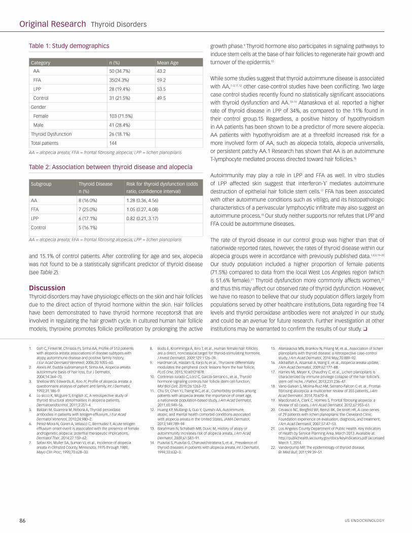
This study retrospectively assessed the role of thyroid dysfunction in AA, LPP and FFA. Methods: A systematic chart review of 144 patients was performed from September 2010 to July 2013. Data were analyzed using multivariate logistic regression to test for a relationship between thyroid disease and type of alopecia. Results: A total of 113 alopecia patients were analyzed. A positive history of thyroid disease was found in 16.0% of AA, 25.0% of FFA, 17.1% of LPP, and 15.1% of control patients. After controlling for age and sex, alopecia was not found to be a statistically significant predictor of thyroid disease. Conclusions: Although alopecia patients in this study had rates of thyroid dysfunction higher than the U.S. population, there were no significant differences in rates of thyroid dysfunction compared to the control group.
Keywords
Hair loss, alopecia areata, frontal fibrosing alopecia, lichen planopilaris, thyroid disorders
Disclosure: Thu Q Nguyen, Monica Tsai, Tristan Grogan, and Carolyn Goh have nothing to disclose in relation to this article. No funding was received in the publication of this article.
Compliance with Ethics: Patient data were retrospectively evaluated for this study. Institutional approval was obtained, and the study was deemed exempt from the need to procure subject consent.
Authorship: All named authors meet the International Committee of Medical Journal Editors (ICMJE) criteria for authorship of this manuscript, take responsibility for the integrity of the work as a whole, and have given final approval to the version to be published.
Open Access: This article is published under the Creative Commons Attribution Noncommercial License, which permits any noncommercial use, distribution, adaptation, and reproduction provided the original author(s) and source are given appropriate credit.
Received: October 10, 2016
Accepted: December 8, 2016
Citation: US Endocrinology, 2016;12(2):85–6
Corresponding Author: Monica Tsai, 8700 Beverly Blvd, Los Angeles, CA 90048, US. E:[email protected]
Thyroid disease has commonly been associated with hair loss, predominantly manifesting as diffuse
thinning of the scalp and the lateral eyebrows. This can be of particular concern to patients, and may
motivate some to seek medical evaluation. Indeed, thyroid disease has also been linked to clinically
significant types of alopecia, including alopecia areata (AA)1–3 and telogen effluvium.4–6 The role of
thyroid disease in other causes of alopecia including lichen planopilaris (LPP) and frontal fibrosing
alopecia (FFA) remains unclear. AA is an autoimmune process involving non-scarring hair loss that
affects between six to seven million individuals in the US.7 LPP and FFA are clinical variants of scarring
alopecia that are indistinguishable on histopathology. LPP is a follicular form of lichen planus that
results in scarring hair loss generally in multifocal regions of the scalp. In contrast, FFA is a variant
of LPP that causes gradually progressive hair loss at the frontotemporal hairline of postmenopausal
women. This study retrospectively assessed the role of thyroid dysfunction in AA, LPP and FFA.
MethodsA systematic chart review of all patients seen in the UCLA Hair Disorders Clinic from September 2010
to July 2013 was performed. All patients with a diagnosis of FFA or LPP and a randomized subset of
50 patients with AA were included in the study. An age- and sex-matched control group was randomly
selected from other patients seen in the UCLA Dermatology Clinic who did not have alopecia. Data
obtained from chart review included gender, age, history of dermatologic conditions, TSH levels,
and history of thyroid dysfunction. Patients were defined as having thyroid dysfunction if they had
been diagnosed with hypothyroidism, hyperthyroidism, Hashimoto’s thyroiditis, thyroid cancer, and
other/not specified thyroid dysfunction. Univariate and multivariate logistic regression models were
constructed to predict thyroid disease using alopecia group, gender, age, and TSH.
ResultsThe UCLA Hair Disorders Clinic served more than 5,548 patients between 2010 and 2013, including
28 patients with FFA, 35 with LPP, and 269 with AA. A total of 113 alopecia patients were analyzed
(Table 1). Of the 81 alopecia patients with documented thyroid-stimulating hormone (TSH) levels,
the average level was 2.12 mIU/L, whereas the control group had an average of 1.93 mIU/L among 19
patients. A positive history of thyroid disease was found in 16.0% of AA, 25.0% of FFA, 17.1% of LPP,
Nguyen_FINAL.indd 85 05/01/2017 17:32
https://doi.org/10.17925/USE.2016.12.02.85
https://doi.org/10.17925/USE.2016.12.02.85
US ENDOCRINOLOGY86
Original Research Thyroid Disorders
and 15.1% of control patients. After controlling for age and sex, alopecia
was not found to be a statistically significant predictor of thyroid disease
(see Table 2).
DiscussionThyroid disorders may have physiologic effects on the skin and hair follicles
due to the direct action of thyroid hormone within the skin. Hair follicles
have been demonstrated to have thyroid hormone receptors8 that are
involved in regulating the hair growth cycle. In cultured human hair follicle
models, thyroxine promotes follicle proliferation by prolonging the active
growth phase.9 Thyroid hormone also participates in signaling pathways to
induce stem cells at the base of hair follicles to regenerate hair growth and
turnover of the epidermis.10
While some studies suggest that thyroid autoimmune disease is associated
with AA,1–3,11,12 other case-control studies have been conflicting. Two large
case control studies recently found no statistically significant associations
with thyroid dysfunction and AA.13–14 Atanaskova et al. reported a higher
rate of thyroid disease in LPP of 34%, as compared to the 11% found in
their control group.15 Regardless, a positive history of hypothyroidism
in AA patients has been shown to be a predictor of more severe alopecia.
AA patients with hypothyroidism are at a threefold increased risk for a
more involved form of AA, such as alopecia totalis, alopecia universalis,
or persistent patchy AA.1 Research has shown that AA is an autoimmune
T-lymphocyte mediated process directed toward hair follicles.16
Autoimmunity may play a role in LPP and FFA as well. In vitro studies
of LPP affected skin suggest that interferon-ϒ mediates autoimmune
destruction of epithelial hair follicle stem cells.17 FFA has been associated
with other autoimmune conditions such as vitiligo, and its histopathologic
characteristics of a perivascular lymphocytic infiltrate may also suggest an
autoimmune process.18 Our study neither supports nor refutes that LPP and
FFA could be autoimmune diseases.
The rate of thyroid disease in our control group was higher than that of
nationwide reported rates, however, the rates of thyroid disease within our
alopecia groups were in accordance with previously published data.1,8,9,19–20
Our study population included a higher proportion of female patients
(71.5%) compared to data from the local West Los Angeles region (which
is 51.6% female).21 Thyroid dysfunction more commonly affects women,22
and thus this may affect our observed rate of thyroid dysfunction. However,
we have no reason to believe that our study population differs largely from
populations served by other healthcare institutions. Data regarding free T4
levels and thyroid peroxidase antibodies were not analyzed in our study,
and could be an avenue for future research. Further investigation at other
institutions may be warranted to confirm the results of our study. q
Table 1: Study demographics
Category n (%) Mean Age
AA 50 (34.7%) 43.2
FFA 35(24.3%) 59.2
LPP 28 (19.4%) 53.5
Control 31 (21.5%) 49.5
Gender
Female 103 (71.5%)
Male 41 (28.4%)
Thyroid Dysfunction 26 (18.1%)
Total patients 144
AA = alopecia areata; FFA = frontal fibrosing alopecia; LPP = lichen planopilaris
Table 2: Association between thyroid disease and alopecia
Subgroup Thyroid Disease
n (%)
Risk for thyroid dysfunction (odds
ratio, confidence interval)
AA 8 (16.0%) 1.28 (0.36, 4.56)
FFA 7 (25.0%) 1.05 (0.27, 4.08)
LPP 6 (17.1%) 0.82 (0.21, 3.17)
Control 5 (16.1%)
AA = alopecia areata; FFA = frontal fibrosing alopecia; LPP = lichen planopilaris
1. Goh C, Finkel M, Christos PJ, Sinha AA, Profile of 513 patients with alopecia areata: associations of disease subtypes with atopy, autoimmune disease and positive family history, J Eur Acad Dermatol Venereol, 2006;20:1055–60.
2. Alexis AF, Dudda-subramanya R, Sinha AA, Alopecia areata: autoimmune basis of hair loss, Eur J Dermatol, 2004;14:364–70.
3. Shellow WV, Edwards JE, Koo JY, Profile of alopecia areata: a questionnaire analysis of patient and family, Int J Dermatol, 1992;31:186–9
4. Lo sicco K, Mcguire S, English JC, A retrospective study of thyroid structural abnormalities in alopecia patients, Dermatoendocrinol, 2011;3:251–4.
5. Baldari M, Guarrera M, Rebora A, Thyroid peroxidase antibodies in patients with telogen effluvium, J Eur Acad Dermatol Venereol, 2010;24:980–2.
6. Perez-Mora N, Goren A, Velasco C, Bermudez F, Acute telogen effluvium onset event is associated with the presence of female androgenetic alopecia: potential therapeutic implications, Dermatol Ther, 2014;27:159–62.
7. Safavi KH, Muller SA, Suman VJ, et al., Incidence of alopecia areata in Olmsted County, Minnesota, 1975 through 1989, Mayo Clin Proc, 1995;70:628–33.
8. Bodo E, Kromminga A, Biro T, et al., Human female hair follicles are a direct, nonclassical target for thyroid-stimulating hormone, J Invest Dermatol, 2009;129:1126–39.
9. Hardman JA, Haslam IS, Farjo N, et al., Thyroxine differentially modulates the peripheral clock: lessons from the hair follicle, PLoS One, 2015;10:e0121878.
10. Contreras-Jurado C, Lorz C, Garcia-Serrano L, et al., Thyroid hormone signaling controls hair follicle stem cell function, Mol Biol Cell, 2015;26:1263–72.
11. Chu SY, Chen YJ, Tseng WC, et al., Comorbidity profiles among patients with alopecia areata: the importance of onset age, a nationwide population-based study, J Am Acad Dermatol, 2011;65:949–56.
12. Huang KP, Mullangi S, Guo Y, Qureshi AA, Autoimmune, atopic, and mental health comorbid conditions associated with alopecia areata in the United States, JAMA Dermatol, 2013;149:789–94
13. Barahmani N, Schabath MB, Duvic M, History of atopy or autoimmunity increases risk of alopecia areata, J Am Acad Dermatol, 2009;61:581–91.
14. Puavilai S, Puavilai G, Charuwichitratana S, et al., Prevalence of thyroid diseases in patients with alopecia areata, Int J Dermatol, 1994;33:632–3.
15. Atanaskova MN, Brankov N, Piliang M, et al., Association of lichen planopilaris with thyroid disease: a retrospective case-control study, J Am Acad Dermatol, 2014 May;70:889–92.
16. Alkhalifah A, Alsantali A, Wang E, et al., Alopecia areata update, J Am Acad Dermatol, 2009;62:177–88.
17. Harries MJ, Meyer K, Chaudhry C, et al., Lichen planopilaris is characterized by immune privilege collapse of the hair follicle’s stem cell niche, J Pathol, 2013;231:236–47.
18. Vano-Galvan S, Molina-Ruiz AM, Serrano-Falcon C et. al., Frontal-fibrosing alocepcia: a multicenter review of 355 patients, J Am Acad Dermatol, 2014;70:670–8.
19. Macdonald A, Clark C, Holmes S, Frontal fibrosing alopecia: a review of 60 cases, J Am Acad Dermatol, 2012;67:955–61.
20. Cevasco NC, Bergfeld WF, Remzi BK, De knott HR, A case-series of 29 patients with lichen planopilaris: the Cleveland Clinic Foundation experience on evaluation, diagnosis, and treatment, J Am Acad Dermatol, 2007;57:47–53.
21. Los Angeles County Department of Public Health. Key Indicators of Health by Service Planning Area. March 2013. Available at: http://publichealth.lacounty.gov/docs/keyindicators.pdf (accessed March 1, 2014.
22. Vanderpump MP, The epidemiology of thyroid disease, Br Med Bull, 2011;99:39–51.
Nguyen_FINAL.indd 86 05/01/2017 17:32