Embed Size (px)
Citation preview
�������� ����� ��
The Autoimmune Basis of Alopecia Areata: A Comprehensive Review
Naseeha Islam, Patrick S.C. Leung, Arthur Huntley, M. Eric Gershwin
PII: S1568-9972(14)00226-2DOI: doi: 10.1016/j.autrev.2014.10.014Reference: AUTREV 1632
To appear in: Autoimmunity Reviews
Received date: 2 October 2014Accepted date: 5 October 2014
Please cite this article as: Islam Naseeha, Leung Patrick S.C., Huntley Arthur, EricGershwin M, The Autoimmune Basis of Alopecia Areata: A Comprehensive Review,Autoimmunity Reviews (2014), doi: 10.1016/j.autrev.2014.10.014
This is a PDF file of an unedited manuscript that has been accepted for publication.As a service to our customers we are providing this early version of the manuscript.The manuscript will undergo copyediting, typesetting, and review of the resulting proofbefore it is published in its final form. Please note that during the production processerrors may be discovered which could affect the content, and all legal disclaimers thatapply to the journal pertain.
ACC
EPTE
D M
ANU
SCR
IPT
ACCEPTED MANUSCRIPT
1
The Autoimmune Basis of Alopecia Areata: A Comprehensive Review
Naseeha Islam 1, Patrick SC Leung1, Arthur Huntley2, M. Eric Gershwin1
1 Division of Rheumatology/Allergy and Clinical Immunology, University of California, Davis CA
95616.
2 Department of Dermatology, University of California, Davis CA 95616.
To whom correspondence should be addressed:
M. Eric Gershwin, M.D., Division of Rheumatology, Allergy and Clinical Immunology, University
of California at Davis School of Medicine, 451 Health Sciences Drive, Suite 6510, Davis, CA
95616; Telephone: 530-752-2884; Fax: 530-752-4669; Email: [email protected]
List of Abbreviations:
AA: Alopecia areata; BECs: biliary epithelial cells; CMV: cytomegalovirus; CRH: corticotropin-
releasing hormone; DNCB: dinitrochlorobenzene; DPCP: dephnylcyclopropenone; HF: hair
follicle; HLA: human leukocyte antigen; GWAS: genome wide association studies; SNP: single
nucleotide polymorphism; HPA: hypothalamic-pituitary-adrenal; NK: natural killer, ORS: outer
root sheath; PBC: primary biliary cirrhosis; SADBE: squaric acid dibutyl ester.
ACC
EPTE
D M
ANU
SCR
IPT
ACCEPTED MANUSCRIPT
2
Abstract
Alopecia areata (AA) is a common, non-scarring dermatologic condition regularly distinguished
by patches of hair loss on the scalp also manifesting in other, severe forms, including alopecia
totalis (total loss of hair on the scalp) and alopecia universalis (complete loss of hair on the
scalp and body). AA is a clinically heterogeneous disease with greatly varying yet typical
symptoms, but the etiology for AA remains an enigma. However, clinical and experimental
studies have pointed to autoimmune involvement, specifically regarding immune privilege sites
of the hair follicles and the infiltration of CD4+ and CD8+ T cells and a predominant Th1
cytokine profile. Environmental insults, such as viral infections, trauma and genetic
predisposition are also believed to contribute to the disease process. Multiple treatment
options including the use of broad acting corticosteroids appear to be relative effective in mild
cases, however the clinical management of more severe forms of AA is much more difficult.
Recent studies suggest that intervention of the JAK pathway may have a potential therapeutic
efficacy for AA.
Key words: alopecia areata, autoimmunity, corticosteroids, environment, HLA, hair follicle,
immune privilege
ACC
EPTE
D M
ANU
SCR
IPT
ACCEPTED MANUSCRIPT
3
1. Introduction
Autoimmunity can be the underlying cause of a broad spectrum of human disorders. As such,
autoimmune diseases are difficult to diagnose and are often based on a multitude of
clinical/laboratory markers [1, 2]. For example, patients with autoimmune diseases often have
a positive family history for the same disease, or for other diseases known to have an
autoimmune etiology. Immunologically, there are prominent mononuclear cells infiltrations in
the affected organ or tissue, high titer of serum autoantibodies and the presence of
preferential usage of certain HLA haplotypes. The deposition of antigen-antibody complexes in
the affected organ or tissue is evident often leads to pathological damage; clinical symptoms
are often improved with the use of immunosuppressive agents [3, 4]. Autoimmune diseases are
conventionally treated by immunologists according to the type of organ involvement, and are
classified as organ specific or systemic autoimmune diseases. Autoimmune diseases are
common, affecting 8% of the American population [5]. Some autoimmune diseases are rare
while others are highly common. Although autoimmune diseases are more common in females,
some autoimmune disorders such as alopecia are more prevalent in males. In this article, we
will discuss the natural history, epidemiology, etiology, contribution of the autoimmune
responses and the clinical treatment of one of the most common autoimmune disorders,
alopecia areata (AA).
2. Natural History/Clinical Features of AA
AA is extremely unpredictable and varies greatly from person to person, causing the disease to
have a wide spectrum of features (Table 1). This extensive range of symptoms makes AA
difficult to diagnose, and no regulated consistencies of disease analysis have been agreed upon.
ACC
EPTE
D M
ANU
SCR
IPT
ACCEPTED MANUSCRIPT
4
The most common clinical presentation of AA is a circumscribed area of bald skin on the scalp,
which is usually isolated from other patches, as seen in 90% of clinical diagnoses (Figure 1).
There is typically no scarring or inflammation on the scalp itself. Remaining hairs take on an
‘exclamation point’ resemblance, consisting of short, thin hairs tapering to the end. These hair-
loss characteristics, however, may also be found in other patterns, such as reticular thinning or
diffusion. Such extensive patterns may include the distribution-ophiasis form usually with a
band of hair loss around the scalp, or its complete opposite—a scalp-sisapho pattern that
includes hair loss on the central part of the scalp [6]. Since AA is so dynamic, patches may be
seen other areas of the body such as elbow, arms and thigh (Figure 2). In addition, the disease
may involve facial hair, including eyelashes, eyebrows, and the beard area. As previously
mentioned, alopecia totalis includes total hair loss on the scalp, and alopecia universalis is
diagnosed with 100% loss of both scalp and body hair. Pitting of the nails is also common in AA
patients, seen in anywhere between 10%-66% of cases [7]. AA is a systemic disease because, in
addition to the hair follicles, it can also affect the nails and the eyes.
AA may also be responsible for the unfortunate sensation known as ‘overnight graying’, as it
usually only causes darker hairs to fall out. Because alopecia is a non-scarring disease, hair
regrowth is possible at diseased hair follicles. Non-pigmented hairs are usually spared by
disease spreading, and grow back prior to normal pigmented hairs [8]. Less serious forms of AA
can almost guarantee remission over a patient-dependent period of time, but even extreme
cases such as alopecia universalis may be overcome at some point. Remission is fundamentally
dependent on the prognostic factors presented by the individual. Poor prognosis that may
foreshadow the prolonging of AA include early age of disease onset, a positive family history,
ACC
EPTE
D M
ANU
SCR
IPT
ACCEPTED MANUSCRIPT
5
atopy, etc. [9]. Otherwise, spontaneous regrowth of hair in the isolated bald patches can be
expected.
Immunohistologically, AA is characterized by the presence of lymphocytic infiltration,
comprised of T-cells, adjoining the HF site [10, 11]. CD4+ T-cells constitute the majority of
lymphocytes in the HF area, whereas CD8+ T-cells are commonly seen in the follicular
epithelium. These immunological findings reflect the potential contribution of autoimmune
responses to AA, as opposed to other dermatological conditions on the scalp [12]. Apart from
AA, multiple other dermatological conditions with similar symptoms exist, often making it
difficult to distinguish one disease from another [13, 14] (Table 1).
3. Epidemiology
AA is a relatively frequent disease with a prevalence of 0.1%-0.2% worldwide [15, 16]. Among
different races and ethnic groups, the prevalence can range, from 0.9% to 6.9% [17]. Notably,
individuals with Down’s syndrome seem to have a slightly higher incidence [12]. In the United
States, one study reported that about 14.5 million patients suffer from AA, constituting about
2% of the national population [17], while another study suggested that only about 5.3 million in
the U.S. are clinically affected [18]. Overall, AA seems to account for about 0.7%-3% of all
patients in the United States [19], and about 2% in the United Kingdom [12] (Table 2).
The likelihood of diagnosis of AA during one’s lifetime is thought to be around 1.7%,
regardless of demographical location, affecting both genders and all age groups [13, 15, 20, 21].
Although both men and women are affected, men seem to be more frequently associated with
the more severe cases of alopecia than women [17]. The onset of AA is more likely early in life.
ACC
EPTE
D M
ANU
SCR
IPT
ACCEPTED MANUSCRIPT
6
For example, various studies have reported that the peak incidence of AA occurs prior to 20
years of age, with about 60% of cases experiencing their first manifestation of hair loss during
their late childhood/early adulthood [12, 17, 22]. Others studies report that only 44% of
patients manifest disease onset before their 20s, but still agree that less than half of AA
patients are diagnosed after age 40 [15]. In fact, study of a cohort of Asian AA patients reported
that 85.5% of the AA patients had their first episode of AA before the age of 40 years [19, 23].
Of note, diagnoses of AA in prepubescent individuals seems to indicate poor prognosis [12, 22].
The epidemiology of AA also reflects the contribution of heredity factors. Of the 0.1% of the
human population that developed AA, 10% to 42% reported a positive family history of AA,
usually involving at least one first-degree relative [13, 17, 24]. The age of onset of AA also
reflects familial history, with nearly 40% of cases having a positive familial history if diagnosed
before age 30, but only about 7% if onset occurred later on in life [24]. Twin studies tend to
show a concordance rate of about 55% [24], implicating the contribution of heredity factors in
AA (Table 2).
4. Etiology
The etiology of AA is still unclear. Herein, we will examine the current literature with respect to
genetic susceptibility, environmental factors, and the contribution of autoimmune responses
within the hair follicle autoimmune privilege site in AA.
4.1 Genetic susceptibility
ACC
EPTE
D M
ANU
SCR
IPT
ACCEPTED MANUSCRIPT
7
The genetic basis of AA is strongly supported by its observed heritability in first degree
relatives, twin studies and genetic linkage analysis of AA families. In AA patients, first-degree
relatives are associated with the disease in about 10% to 42% of cases. Among them, 7% of
patients have at least one parent with AA, 3% have at least one sibling with the disease, and a
minority of 2% have a child who also suffers from AA. In addition, there is a very high
concordance in monozygotic twins [25]. A genome wide search for linkage of 20 families with
AA consisting 102 affected and 118 unaffected individuals demonstrated the association of HLA
with AA [26]. Of the HLA class II genes that are known to have associations with AA, the DQ3,
DQ7, DR4, DR5, and DPW4 alleles are particularly involved. In addition to class II alleles, AA also
includes the involvement of particular class I HLA alleles as well, albeit less frequently. For
example, A1, B13, B18, B52-Cw*0704, B27, B40, B44, B62 are involved. Along with these HLA
alleles that potentially contribute to the diagnosis of AA, certain regions of the human genome
are associated with the more severe disease. DQ3, DR11, and DQ7 are suggested to predispose
to alopecia totalis and alopecia universalis. Another notable genetic association is DRB1*04 in
patients that suffer from AA, and a reduction in DRB1*03 alleles. These alleles are more or less
specific to various ethnic identities, and therefore genetic associations depend on the patient’s
particular geographical location and ethnic background [17, 20, 27, 28] (Table 3).
In a GWAS study of 1,054 AA cases and 3,278 controls, Petukhova et al [18] reported that eight
genomic regions, including loci encoding genes in both innate and adaptive immunity are
associated with AA. These include loci on chromosome 2q33.2 containing the CTLA4 gene,
chromosome 4q27 containing IL-2/IL-21 genes, chromosome 6p21.32 containing the HLA
segment, chromosome 6q25.1 containing the ULBP genes, chromosome 10p15.1 containing IL-
ACC
EPTE
D M
ANU
SCR
IPT
ACCEPTED MANUSCRIPT
8
2RA (CD25), chromosome 12q13 containing Eos (IKZF4) and ERBB3 genes, chromosome 9q31.1
containing the syntaxin 17 gene and chromosome 11q13, upstream from peroxiredoxin 5
(PRDX5). A follow up study of this GWAS on five of these loci including IL-2/IL-21, ULBP,
IKZF4/ERBB3,syntaxin 17, PRDX5 confirmed their validity, in addition IL-13 and KIAA0350 were
also identified as susceptibility loci in AA [29]. Using a genome wide pooling approach, a SNP
corresponding to an intronic region of the SPATA5 (spermatogenesis-associated protein 5) gene
on chromosome 4 was identified as a potential susceptibility locus for AA [30]. More recently,
additional loci within the IL-4 intron 3, promoter regions of Foxp3 and ICSOSLG genes have
been found to be associated with AA [31, 32].
4.2 Environmental Factors
Clinical and experimental studies showed that environmental insults such as emotional/physical
stressors, hormones and infections contribute to autoimmunity [33-36]. Stress hormones, in
their efforts to maintain dermatological homeostasis in reaction to their environment, are
known to affect AA [19]. These particular stressors in relation to diagnosed hair loss include
exposure to ultraviolet light, natural and chemical bodily offenses, physical injury, and
emotional distress. The human hair follicle (HF) appears to parallel the basic functionality of the
hypothalamic-pituitary-adrenal (HPA) axis in the skin, incorporating the specific stress
hormones that are accepted as prospective origins of alopecia [37]. Corticotropin-releasing
hormone (CRH) is widely known to be a significant stress-induced component of the larger HPA
axis [37]. Studies have shown that CRH and its receptors, CRH-R1, are expressed within the
outer root sheath (ORS) of the HF, and the gene transcriptions of both hormone and receptor
take place at the HF site [38]. Clinical observations document that in the affected

ACC
EPTE
D M
ANU
SCR
IPT
ACCEPTED MANUSCRIPT
9
dermatological lesions of AA patients, there appeared a distinct association with CRH-2B (a
subset of the CRH). This is in contrast to the much weaker signals of CRH-2B in both healthy
subjects and the unaffected skin in AA [39]. The diseased areas, especially the epidermis and
sebaceous glands of the HF, also have increased expression of CRH, adreno-corticotropic
hormone, and α-melanocyte-stimulating hormone when compared to healthy individuals [39-
41].
The association of cytomegalovirus (CMV) was initially proposed by Skinner based upon by the
presence of CMV DNA sequences in skin biopsies of patients with AA [42]. However, this has
been refuted [43]. Other viruses, including hepatitis B, hepatitis C, Epstein-Barr, and swine flu
have been also been suggested to trigger AA [44-48]. Theories have also been put forward
regarding seasonal associations, with evidence of increased disease relapses between the
months of February and March. This may also be a result of the high multitudes of viruses in
early spring, supporting the hypothesis that AA may be an effect of certain viral infections.
4.3 Contribution of autoimmune responses in AA
Autoimmunity generally arises from defects in immune tolerance, which results in the
generation and proliferation of autoreactive T cells and autoantibodies [49-51]. We discuss
primary biliary cirrhosis (PBC) because it is also an organ specific disease and has many
analogies to the specificity of AA [52-60]. PBC is a chronic cholestatic liver disease histologically
characterized by the immune mediated destruction of biliary epithelial cells (BECs) and small
bile ducts with portal inflammatory cell infiltration. Damaged BECs lead to the development of
fibrosis, cirrhosis and liver failure. Serologically, over 95% of patients with PBC also have high
ACC
EPTE
D M
ANU
SCR
IPT
ACCEPTED MANUSCRIPT
10
titers of antimitochondrial antibodies [52, 56, 61]. Extensive efforts in defining the target
mitochondrial autoantigens, T and B cell epitopes, the innate and adaptive immune responses,
the immunobiology of the biliary epithelium, and the pathology of biliary cells destruction have
greatly advanced the knowledge of the molecular mechanisms in the pathogenesis of PBC [54,
59, 61-68]. These have led to therapeutic designs in muse models and clinical trials in patients
with PBC [69-72].
Autoimmunity in AA is strongly supported by clinical observations that patients with AA are
often diagnosed with one or more other autoimmune disorders, including vitiligo, lupus
erythematosus, myasthenia gravis, scleroderma, ulcerative colitis, Type I diabetes, thyroiditis,
celiac disease, and rheumatoid arthritis [48, 73]. In addition, the effectiveness and efficacy of
various immunosuppressive agents including cyclosporine and systemic corticosteroids;
immunotherapy drugs, particularly contact synthesizers, such as dinitrochlorobenzene (DNCB),
squaric acid dibutyl ester (SADBE), and dephnylcyclopropenone (DPCP) [74] also suggest an
autoimmune mechanism. Further, the human leukocyte antigens (HLA) has been reported to
play a major role in the etiology of autoimmunity [75, 76]. Confirmation of this specific
hypothesis in AA lies in the increased expression of specific HLAs in AA patients such as HLA-DR,
HLA-A, HLA-B, and HLA-C, which are rarely seen in healthy individuals [11, 77] as well as the
identification of a number of genetic risk factors within various innate and adaptive immunity
gene loci [18].
The most widely accepted hypothesis for the effector mechanism of AA is the destruction of the
HF, an immune privilege site. Within the HF itself, individuals without AA maintain immune
ACC
EPTE
D M
ANU
SCR
IPT
ACCEPTED MANUSCRIPT
11
privilege in multiple ways, such as omitting MHC class 1 in the proximal outer root sheath
(ORS). Conversely, patients identified with AA have a strong association with those same MHC
class 1 alleles. Autoreactive cytotoxic T cells target specific autoantigens, especially
melanogenesis-associated peptides expressed by anagen HFs that produce the melanin
pigment [78, 79]. This is consistent with sparing of unpigmented hairs, and regrowth of initially
white or grey hairs following onset of AA [80]. Additionally, it has been proposed that follicular
melanocytes are also affected. The general exposure of these autoantigens in the follicles of
diseased individuals then attracts a multitude of lymphocytes in the hair bulb area [21]. The
repertoire of cells at the HF is comprised of natural killer (NK) cells, 20%-40% CD8+ T-cells, and
60%-80% CD4+ T-cells [81, 82]. Another associated cytokine, IFN-, has been observed to
potentially upregulate MHC class 1 alleles, increasing the possibility of the destruction of the HF
immune privilege [83]. Chemokines are involved with the development of autoimmune
interactions [84-87], involving Th1 chemokines (specifically CXCL9/MIG and CXCL10/IP-10) that
are more prominent in AA patients than healthy individuals [88]. Other cytokines that may
potentially be associated are IL-1, IL-2, IL-4, and IL-10, along with macrophage migration
inhibitory factor and tumor necrosis factor (TNF, including the B-cell activating factor subset)
[16]. Apart from research of AA-involved cytokines and chemokines, studies in the chemotaxis
of lymphocyte accumulations demonstrated distinct activity of CXCL10 in regards to histological
outcomes of acute AA [89, 90].
With the growing interest of the NK cells in autoimmunity [91-93], its role in the pathogenesis
of AA has also been investigated. NKG2D is expressed in these NK cells, although they also are
components of various T-cells. These cells attack by recognizing certain glycoproteins and CMV
ACC
EPTE
D M
ANU
SCR
IPT
ACCEPTED MANUSCRIPT
12
UL16 proteins, and MHC class I related proteins MICA/MICB [94]. In AA patients, the outer root
sheath presents MICA proteins, leading NKG2D+ NK cells to target the HF [82]. The HF immune
privilege of AA is maintained by being MHI class I negative, MICA negative with low expression
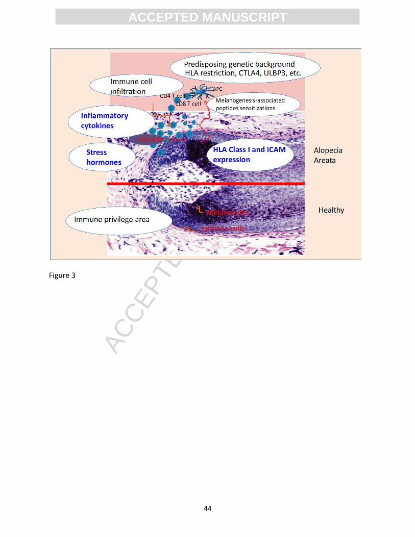
of NKG2D NK cells. The breaking of tolerance in HF and subsequent change in
cytokine/chemokine profiles leads to infiltration of lymphocytes [18, 19] (Figure 3).
The C3H/HeJ mouse is a spontaneous mouse model of AA, which manifests the clinical
pathological features of human AA including infiltration of CD8+NKG2+ T cells around the
epithelial layers of HFs [95, 96]. Interestingly, adoptive transfer of CD8+NKG2D+ cells from
alopecia C3H/HeJ mice induces AA in healthy recipient mice whereas transfer of lymph node
cells lacking NKG2D+ had no effect. C3H/HeJ mice also develop characteristics of AA when
injected with IFN-y, prompting MHC class I and II manifestations at HF. Global transcriptional
profiling analyses of alopecia skin tissues from both human and C3H/HeJ mice identified three
gene expression signatures including IFN response genes, cytotoxic T cell specific transcripts,
and c cytokines. Administration of blocking antibodies to either IL-2 or IL-15R inhibit
accumulation of CD8+NKG2D+ cells in T skin, abrogate MHC regulation and prevent AA in a skin
graft model of AA. Interestingly, systemic administration of Janus kinase (JAK) inhibitors
including ruxolitinib and tofacitinib were able to inhibit downstream effects of type 1 cytokines
and eliminated the IFN signature and prevented the development of AA in the C3H/HeJ mice
model of AA. Furthermore, topical administration of ruxolitinib and tofacitinib markedly
reduced CD8+NKG2D+ cells in treated skin. More importantly, the clinical response of a small
number of AA patients receiving 20mg ruxolitinib twice a day orally had near complete hair re-
growth within three to five months of treatment [96].
ACC
EPTE
D M
ANU
SCR
IPT
ACCEPTED MANUSCRIPT
13
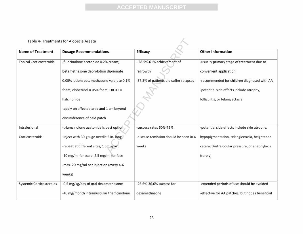
5. Clinical Treatments
The onset of hair loss, especially in women and children, may cause psychological distress [22,
97]. Treatment options are limited and still being developed [12]. AA patients with only mild
forms of the disease are normally advised to avoid unnecessary medication, as spontaneous
regrowth is probable [22]. Broad-acting Intralesional corticosteroids are relatively effective
(Table 4). However, the severe forms of alopecia are much harder to treat, and the only
potential medications known to have efficacy with handling these diseases are allergic contact
sensitizers through immunotherapy. This method of management works by inducing mild
dermatitis on the affected skin, therefore averting the diseased HF lymphocytes from the scalp
to the newly irritated area on the epidermis [98]. Unfortunately, the efficacy of this treatment
has not always been recapitulated. Other treatments have also been suggested, including
various formulations of corticosteroids, minoxidil, anthralin, topical immunotherapy, and
sulfasalazine [74] . Biologics are actively sought as possible therapeutics in autoimmune
diseases as in the case of primary biliary cirrhosis [69-71, 99]. Medications for AA are still being
developed and researched, and hopefully will provide a suitable treatment for alopecia in the
future.
6. Future Directions
AA is a relatively common dermatological and autoimmune disorder. Although AA is not life
threatening, it can cause severe emotional and psychological distress. Data from
epidemiological studies, clinical observations, immunological and pathological studies together
with familial and molecular genetics studies indicate that autoimmune disorders are cumulative
ACC
EPTE
D M
ANU
SCR
IPT
ACCEPTED MANUSCRIPT
14
results of immunological, environmental, and genetic factors [57, 100-103]. Continuous
advances in dissecting underlying mechanisms will likely make inroads towards better
treatments. At present, a multitude of questions remains to be answered in AA: how do genetic
factors, such as HLA associations and MHC allele expressions, affect disease vulnerability or
prognosis? What are the factors that regulate the HF immune privilege and the AA process?
How do we apply understanding of the biology of HF to the pathogenesis of AA? How do AA
autoantigens play a role in the pathogenesis of the disease? In order to answer these
impending inquiries, new research strategies to further understand the etiology and
pathogenesis of AA must be developed.
ACC
EPTE
D M
ANU
SCR
IPT
ACCEPTED MANUSCRIPT
15
Take Home Messages
Alopecia areata is a non-scarring hair loss disorder that has a relatively high prevalence
with no discrimination as to age, gender, or ethnicity.
Etiology for this disease may be attributed to genetic predispositions, environmental
factors, or autoimmune conditions specific to the patient.
The destruction of the hair follicle immune privilege site may contribute to the
pathogenesis of the disease, resulting in a predominantly CD8+ T-cell infiltrate at location of the
follicle.
No current treatments have been widely accepted to have complete and satisfactory
efficacy. Current clinical management of AA are primarily directed towards the symptoms and
psychological distress .
ACC
EPTE
D M
ANU
SCR
IPT
ACCEPTED MANUSCRIPT
16
Figure legends.
Figure 1. Clinical manifestation of alopecia in the scalp. Alopecia can presented in a wide
spectrum characterized by focal patches of hair loss on the scalp to extensive involvement of
the scalp (A, B, C, D) and total loss of hair over the entire scalp and involving the eyebrows (E).
Alopecia is also seen in infant. In this age group, it is more common to see patchy alopecia
resulting from tinea capitus (F).
Figure 2. Clinical manifestation of alopecia other parts such as in elbow (A), arms (B), beard
area (C) and thigh (D).
Figure 3. Immunological cascade in the hair follicle in patients with alopecia areata.
External stressors and inflammatory cytokines/chemokines lead to strong association of MHC
class I, infiltration of autoreactive CD8+ and CD4+ T cells targeting the melanogenesis
associated peptides on HFs and CD8+NKG2D+cells within the immune privilege area of the hair
follicle leads to immune mediated destruction of the hair bulb area and pathological
development of AA.
ACC
EPTE
D M
ANU
SCR
IPT
ACCEPTED MANUSCRIPT
17
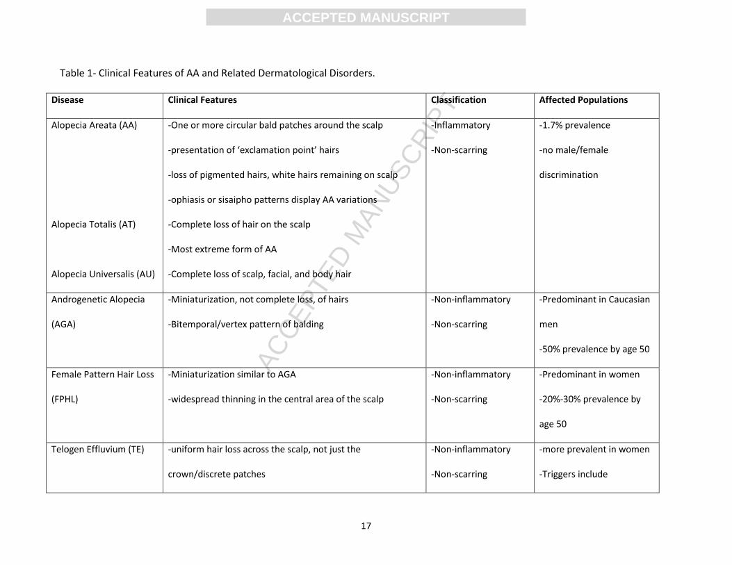
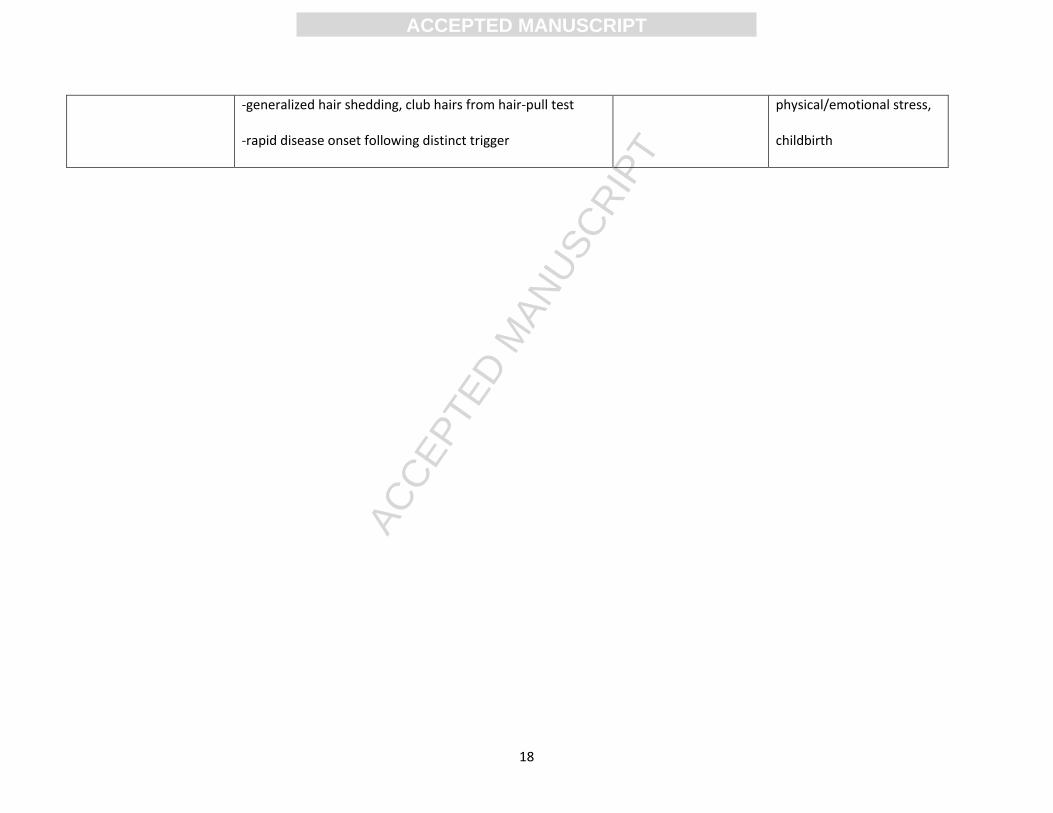
Table 1- Clinical Features of AA and Related Dermatological Disorders.
Disease Clinical Features Classification Affected Populations
Alopecia Areata (AA)
Alopecia Totalis (AT)
Alopecia Universalis (AU)
-One or more circular bald patches around the scalp
-presentation of ‘exclamation point’ hairs
-loss of pigmented hairs, white hairs remaining on scalp
-ophiasis or sisaipho patterns display AA variations
-Complete loss of hair on the scalp
-Most extreme form of AA
-Complete loss of scalp, facial, and body hair
-Inflammatory
-Non-scarring
-1.7% prevalence
-no male/female
discrimination
Androgenetic Alopecia
(AGA)
-Miniaturization, not complete loss, of hairs
-Bitemporal/vertex pattern of balding
-Non-inflammatory
-Non-scarring
-Predominant in Caucasian
men
-50% prevalence by age 50
Female Pattern Hair Loss
(FPHL)
-Miniaturization similar to AGA
-widespread thinning in the central area of the scalp
-Non-inflammatory
-Non-scarring
-Predominant in women
-20%-30% prevalence by
age 50
Telogen Effluvium (TE) -uniform hair loss across the scalp, not just the
crown/discrete patches
-Non-inflammatory
-Non-scarring
-more prevalent in women
-Triggers include
ACC
EPTE
D M
ANU
SCR
IPT
ACCEPTED MANUSCRIPT
18
-generalized hair shedding, club hairs from hair-pull test
-rapid disease onset following distinct trigger
physical/emotional stress,
childbirth
ACC
EPTE
D M
ANU
SCR
IPT
ACCEPTED MANUSCRIPT
19
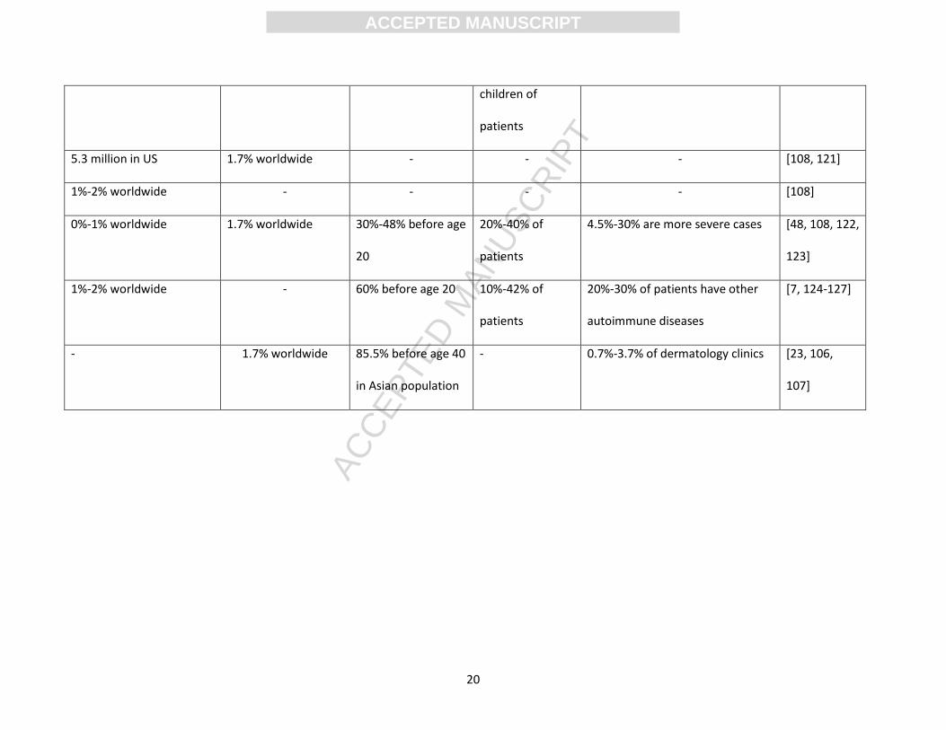
Table 2 - Epidemiology of Alopecia Areata
Disease Prevalence Disease Incidence Age Family History Miscellaneous References
Higher prevalence with
Downs syndrome
1.7% worldwide 60% before age 20 20% of patients More prevalent with Downs
syndrome
[104, 105]
0.1%-0.2% worldwide
1.7% worldwide 60% before age 20;
Peak: 30-59 years
8.7%-20% of
patients
Dermatology clinics: 2%-3%
(US/UK); 3.8% (China); 0.7%
(India)
[23, 106-111]
0.1%-0.2% worldwide - 44% before age 20;
30% after age 40
- 0.7%-3.8% of dermatology clinics [108, 112,
113]
0.1% worldwide;
2% of US;
8.8% with Downs
Syndrome
1.7% worldwide 60% before age 20;
70% between ages
10-25
10%-42% of
patients
3%-42% heritability of disease;
Severity distribution 63% men,
36% women
[7, 108, 114-
118]
1%-2% worldwide - - 55% twin
concordance;
5%-6%
recurrence in
- [108, 119,
120]
ACC
EPTE
D M
ANU
SCR
IPT
ACCEPTED MANUSCRIPT
20
children of
patients
5.3 million in US 1.7% worldwide - - - [108, 121]
1%-2% worldwide - - - - [108]
0%-1% worldwide 1.7% worldwide 30%-48% before age
20
20%-40% of
patients
4.5%-30% are more severe cases [48, 108, 122,
123]
1%-2% worldwide - 60% before age 20 10%-42% of
patients
20%-30% of patients have other
autoimmune diseases
[7, 124-127]
- 1.7% worldwide 85.5% before age 40
in Asian population
- 0.7%-3.7% of dermatology clinics [23, 106,
107]
ACC
EPTE
D M
ANU
SCR
IPT
ACCEPTED MANUSCRIPT
21
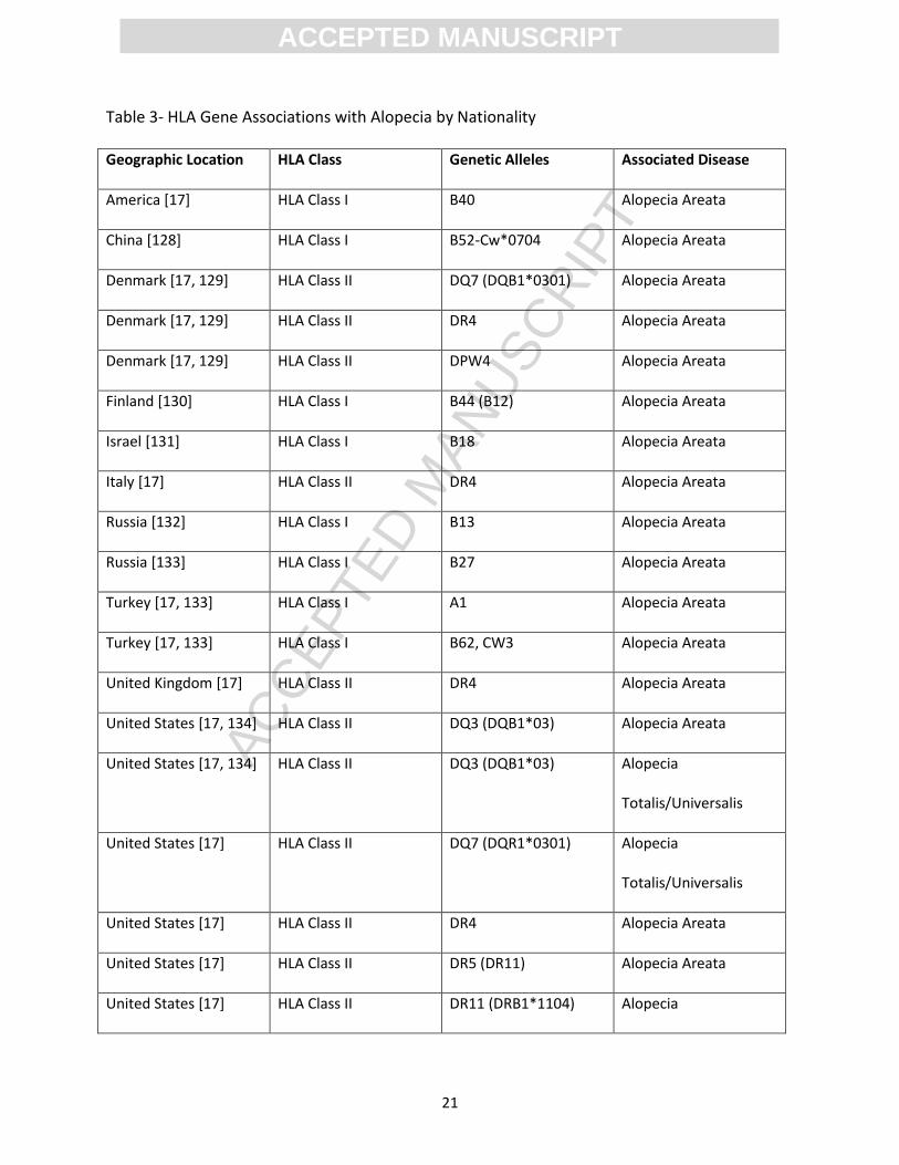
Table 3- HLA Gene Associations with Alopecia by Nationality
Geographic Location HLA Class Genetic Alleles Associated Disease
America [17] HLA Class I B40 Alopecia Areata
China [128] HLA Class I B52-Cw*0704 Alopecia Areata
Denmark [17, 129] HLA Class II DQ7 (DQB1*0301) Alopecia Areata
Denmark [17, 129] HLA Class II DR4 Alopecia Areata
Denmark [17, 129] HLA Class II DPW4 Alopecia Areata
Finland [130] HLA Class I B44 (B12) Alopecia Areata
Israel [131] HLA Class I B18 Alopecia Areata
Italy [17] HLA Class II DR4 Alopecia Areata
Russia [132] HLA Class I B13 Alopecia Areata
Russia [133] HLA Class I B27 Alopecia Areata
Turkey [17, 133] HLA Class I A1 Alopecia Areata
Turkey [17, 133] HLA Class I B62, CW3 Alopecia Areata
United Kingdom [17] HLA Class II DR4 Alopecia Areata
United States [17, 134] HLA Class II DQ3 (DQB1*03) Alopecia Areata
United States [17, 134] HLA Class II DQ3 (DQB1*03) Alopecia
Totalis/Universalis
United States [17] HLA Class II DQ7 (DQR1*0301) Alopecia
Totalis/Universalis
United States [17] HLA Class II DR4 Alopecia Areata
United States [17] HLA Class II DR5 (DR11) Alopecia Areata
United States [17] HLA Class II DR11 (DRB1*1104) Alopecia

ACC
EPTE
D M
ANU
SCR
IPT
ACCEPTED MANUSCRIPT
22
Totalis/Universalis
ACC
EPTE
D M
ANU
SCR
IPT
ACCEPTED MANUSCRIPT
23
Table 4- Treatments for Alopecia Areata
Name of Treatment Dosage Recommendations Efficacy Other Information
Topical Corticosteroids
-fluocinolone acetonide 0.2% cream;
betamethasone deprolotion diprionate
0.05% lotion; betamethasone valerate 0.1%
foam; clobetasol 0.05% foam; OR 0.1%
halcinonide
-apply on affected area and 1 cm beyond
circumference of bald patch
- 28.5%-61% achievement of
regrowth
-37.5% of patients did suffer relapses
-usually primary stage of treatment due to
convenient application
-recommended for children diagnosed with AA
-potential side effects include atrophy,
folliculitis, or telangiectasia
Intralesional
Corticosteroids
-triamcinolone acetonide is best option
-inject with 30-gauge needle 5 in. long
-repeat at different sites, 1 cm apart
-10 mg/ml for scalp, 2.5 mg/ml for face
-max. 20 mg/ml per injection (every 4-6
weeks)
-success rates 60%-75%
-disease remission should be seen in 4
weeks
-potential side effects include skin atrophy,
hypopigmentation, telangiectasia, heightened
cataract/intra-ocular pressure, or anaphylaxis
(rarely)
Systemic Corticosteroids -0.5 mg/kg/day of oral dexamethasone
-40 mg/month intramuscular triamcinolone
-26.6%-36.6% success for
dexamethasone
-extended periods of use should be avoided
-effective for AA patches, but not as beneficial
ACC
EPTE
D M
ANU
SCR
IPT
ACCEPTED MANUSCRIPT
24
acetonide
-prednisolone 200 mg/week for 3 months
-5 mg betamethasone twice a week/6
months
-60% success prednisolone
-75% success betamethasone
for more extensive forms of the disease (AU,
etc.)
Minoxidil -applied twice a day
-2% or 5% solution (2% less effective)
-combination with corticosteroids promotes
efficacy
-foam form less likely to stimulate side
effects
-effective in extensive AA (but not AT
or AU)
-potential side effects include dermatitis or
pruritus
-studies show that 3% female consumers grew
undesired facial hair
Anthralin -short-contact cream, 0.5%-1%
-applied for 20-30 min. daily, 2-3 weeks
-efficacy not well established -may be good choice for children with AA
-potential side effects include folliculitis,
regional lymphadenopathy, or irritation
Topical
Immunotherapy/contact
sensitizers
-dinitrochlorobenzene (DNCB), diphenyl-
cyclo-propenone (DPCP) OR squaric acid
dibutyl ester (SADBE)
-2% solution is advised
-apply onto 4 cm^2 of affected scalp, left on
for 1-2 days
-36% acceptable regrowth with DNCB
-50%-60% success with DPCP/SADBE
-physical contact with allergen should be
avoided by using gloves and aprons
-no data on safety during pregnancy
-allergic reactions may occur, due to the
mechanisms of the contact sensitizers
ACC
EPTE
D M
ANU
SCR
IPT
ACCEPTED MANUSCRIPT
25
Sulfasalizine -0.5g twice daily/month, then 1 g twice
daily/month, then 1.5 g twice daily/3
months
-moderate disease remission in 23%-
25.6% of cases
-potential side effects may include
gastrointestinal distress, fever, rash,
hepatotoxicity, headache, or hematological
abnormalities
ACC
EPTE
D M
ANU
SCR
IPT
ACCEPTED MANUSCRIPT
26
References
[1] Parks CG, Miller FW, Pollard KM, Selmi C, Germolec D, Joyce K, et al. Expert Panel
Workshop Consensus Statement on the Role of the Environment in the Development of
Autoimmune Disease. International journal of molecular sciences. 2014;15:14269-97.
[2] Pengo V, Banzato A, Denas G, Jose SP, Bison E, Hoxha A, et al. Correct laboratory
approach to APS diagnosis and monitoring. Autoimmunity reviews. 2013;12:832-4.
[3] Ruiz-Irastorza G, Danza A, Perales I, Villar I, Garcia M, Delgado S, et al. Prednisone in
lupus nephritis: how much is enough? Autoimmunity reviews. 2014;13:206-14.
[4] Doria A, Gatto M, Zen M, Iaccarino L, Punzi L. Optimizing outcome in SLE: treating-to-
target and definition of treatment goals. Autoimmunity reviews. 2014;13:770-7.
[5] Fairweather D, Rose NR. Women and autoimmune diseases. Emerging infectious
diseases. 2004;10:2005-11.
[6] Alkhalifah A, Alsantali A, Wang E, McElwee KJ, Shapiro J. Alopecia areata update: part I.
Clinical picture, histopathology, and pathogenesis. J Am Acad Dermatol. 2010;62:177-
88, quiz 89-90.
[7] Madani S, Shapiro J. Alopecia areata update. J Am Acad Dermatol. 2000;42:549-66; quiz
67-70.
[8] Wade MS, Sinclair RD. Persistent depigmented regrowth after alopecia areata. J Am
Acad Dermatol. 2002;46:619-20.
[9] Goh C, Finkel M, Christos PJ, Sinha AA. Profile of 513 patients with alopecia areata:
associations of disease subtypes with atopy, autoimmune disease and positive family
history. J Eur Acad Dermatol Venereol. 2006;20:1055-60.
ACC
EPTE
D M
ANU
SCR
IPT
ACCEPTED MANUSCRIPT
27
[10] Sperling LC, Lupton GP. Histopathology of non-scarring alopecia. J Cutan Pathol.
1995;22:97-114.
[11] Gilhar A, Paus R, Kalish RS. Lymphocytes, neuropeptides, and genes involved in alopecia
areata. J Clin Invest. 2007;117:2019-27.
[12] MacLean KJ, Tidman MJ. Alopecia areata: more than skin deep. Practitioner.
2013;257:29-32, 3.
[13] Mounsey AL, Reed SW. Diagnosing and treating hair loss. Am Fam Physician.
2009;80:356-62.
[14] Vary JC, O'Connor KM. Common dermatologic conditions. Med Clin North Am.
2014;98:445-85.
[15] Jabbari A, Petukhova L, Cabral RM, Clynes R, Christiano AM. Genetic basis of alopecia
areata: a roadmap for translational research. Dermatol Clin. 2013;31:109-17.
[16] Gregoriou S, Papafragkaki D, Kontochristopoulos G, Rallis E, Kalogeromitros D,
Rigopoulos D. Cytokines and other mediators in alopecia areata. Mediators Inflamm.
2010;2010:928030.
[17] Alzolibani AA. Epidemiologic and genetic characteristics of alopecia areata (part 1). Acta
Dermatovenerol Alp Pannonica Adriat. 2011;20:191-8.
[18] Petukhova L, Duvic M, Hordinsky M, Norris D, Price V, Shimomura Y, et al. Genome-wide
association study in alopecia areata implicates both innate and adaptive immunity.
Nature. 2010;466:113-7.
[19] Ito T. Recent advances in the pathogenesis of autoimmune hair loss disease alopecia
areata. Clin Dev Immunol. 2013;2013:348546.
ACC
EPTE
D M
ANU
SCR
IPT
ACCEPTED MANUSCRIPT
28
[20] Alexis AF, Dudda-Subramanya R, Sinha AA. Alopecia areata: autoimmune basis of hair
loss. Eur J Dermatol. 2004;14:364-70.
[21] Gilhar A, Kalish RS. Alopecia areata: a tissue specific autoimmune disease of the hair
follicle. Autoimmun Rev. 2006;5:64-9.
[22] Dombrowski NC, Bergfeld WF. Alopecia areata: what to expect from current treatments.
Cleve Clin J Med. 2005;72:758, 60-1, 65-6 passim.
[23] Tan E, Tay YK, Goh CL, Chin Giam Y. The pattern and profile of alopecia areata in
Singapore--a study of 219 Asians. Int J Dermatol. 2002;41:748-53.
[24] Dudda-Subramanya R, Alexis AF, Siu K, Sinha AA. Alopecia areata: genetic complexity
underlies clinical heterogeneity. Eur J Dermatol. 2007;17:367-74.
[25] Scerri L, Pace JL. Identical twins with identical alopecia areata. J Am Acad Dermatol.
1992;27:766-7.
[26] Martinez-Mir A, Zlotogorski A, Gordon D, Petukhova L, Mo J, Gilliam TC, et al.
Genomewide scan for linkage reveals evidence of several susceptibility loci for alopecia
areata. American journal of human genetics. 2007;80:316-28.
[27] McElwee KJ, Gilhar A, Tobin DJ, Ramot Y, Sundberg JP, Nakamura M, et al. What causes
alopecia areata? Exp Dermatol. 2013;22:609-26.
[28] Megiorni F, Pizzuti A, Mora B, Rizzuti A, Garelli V, Maxia C, et al. Genetic association of
HLA-DQB1 and HLA-DRB1 polymorphisms with alopecia areata in the Italian population.
The British journal of dermatology. 2011;165:823-7.
[29] Jagielska D, Redler S, Brockschmidt FF, Herold C, Pasternack SM, Garcia Bartels N, et al.
Follow-up study of the first genome-wide association scan in alopecia areata: IL13 and
ACC
EPTE
D M
ANU
SCR
IPT
ACCEPTED MANUSCRIPT
29
KIAA0350 as susceptibility loci supported with genome-wide significance. The Journal of
investigative dermatology. 2012;132:2192-7.
[30] Forstbauer LM, Brockschmidt FF, Moskvina V, Herold C, Redler S, Herzog A, et al.
Genome-wide pooling approach identifies SPATA5 as a new susceptibility locus for
alopecia areata. Eur J Hum Genet. 2012;20:326-32.
[31] Conteduca G, Rossi A, Megiorni F, Parodi A, Ferrera F, Tardito S, et al. Single nucleotide
polymorphisms in the promoter regions of Foxp3 and ICOSLG genes are associated with
Alopecia areata. Clinical and experimental medicine. 2014;14:91-7.
[32] Kalkan G, Karakus N, Bas Y, Takci Z, Ozuguz P, Ates O, et al. The association between
Interleukin (IL)-4 gene intron 3 VNTR polymorphism and alopecia areata (AA) in Turkish
population. Gene. 2013;527:565-9.
[33] Perricone C, Colafrancesco S, Mazor RD, Soriano A, Agmon-Levin N, Shoenfeld Y.
Autoimmune/inflammatory syndrome induced by adjuvants (ASIA) 2013: Unveiling the
pathogenic, clinical and diagnostic aspects. Journal of autoimmunity. 2013;47:1-16.
[34] Pillai S. Rethinking mechanisms of autoimmune pathogenesis. Journal of autoimmunity.
2013;45:97-103.
[35] Selmi C, Leung PS, Sherr DH, Diaz M, Nyland JF, Monestier M, et al. Mechanisms of
environmental influence on human autoimmunity: a National Institute of Environmental
Health Sciences expert panel workshop. Journal of autoimmunity. 2012;39:272-84.
[36] Costenbader KH, Gay S, Alarcon-Riquelme ME, Iaccarino L, Doria A. Genes, epigenetic
regulation and environmental factors: which is the most relevant in developing
autoimmune diseases? Autoimmunity reviews. 2012;11:604-9.
ACC
EPTE
D M
ANU
SCR
IPT
ACCEPTED MANUSCRIPT
30
[37] Ito N, Ito T, Kromminga A, Bettermann A, Takigawa M, Kees F, et al. Human hair follicles
display a functional equivalent of the hypothalamic-pituitary-adrenal axis and synthesize
cortisol. Faseb j. 2005;19:1332-4.
[38] Arck PC, Handjiski B, Peters EM, Peter AS, Hagen E, Fischer A, et al. Stress inhibits hair
growth in mice by induction of premature catagen development and deleterious
perifollicular inflammatory events via neuropeptide substance P-dependent pathways.
Am J Pathol. 2003;162:803-14.
[39] Katsarou-Katsari A, Singh LK, Theoharides TC. Alopecia areata and affected skin CRH
receptor upregulation induced by acute emotional stress. Dermatology. 2001;203:157-
61.
[40] Kim HS, Cho DH, Kim HJ, Lee JY, Cho BK, Park HJ. Immunoreactivity of corticotropin-
releasing hormone, adrenocorticotropic hormone and alpha-melanocyte-stimulating
hormone in alopecia areata. Exp Dermatol. 2006;15:515-22.
[41] Harbuz MS, Richards LJ, Chover-Gonzalez AJ, Marti-Sistac O, Jessop DS. Stress in
autoimmune disease models. Ann N Y Acad Sci. 2006;1069:51-61.
[42] Skinner RB, Jr., Light WH, Bale GF, Rosenberg EW, Leonardi C. Alopecia areata and
presence of cytomegalovirus DNA. JAMA : the journal of the American Medical
Association. 1995;273:1419-20.
[43] Garcia-Hernandez MJ, Torres MJ, Palomares JC, Rodriguez-Pichardo A, Aznar J, Camacho
F. No evidence of cytomegalovirus DNA in alopecia areata. The Journal of investigative
dermatology. 1998;110:185.
ACC
EPTE
D M
ANU
SCR
IPT
ACCEPTED MANUSCRIPT
31
[44] Gamal N, Brodosi L, Misciali C, Patrizi A, Vukatana G, Malavolta N, et al. Alopecia
universalis after discontinuation of pegylated interferon and ribavirin combination
therapy for hepatitis C: a case report. Annals of hepatology. 2014;13:293-6.
[45] Geier DA, Geier MR. A case-control study of serious autoimmune adverse events
following hepatitis B immunization. Autoimmunity. 2005;38:295-301.
[46] Ito T. Advances in the management of alopecia areata. J Dermatol. 2012;39:11-7.
[47] Rodriguez TA, Duvic M. Onset of alopecia areata after Epstein-Barr virus infectious
mononucleosis. Journal of the American Academy of Dermatology. 2008;59:137-9.
[48] Shellow WV, Edwards JE, Koo JY. Profile of alopecia areata: a questionnaire analysis of
patient and family. Int J Dermatol. 1992;31:186-9.
[49] Fenoglio D, Bernuzzi F, Battaglia F, Parodi A, Kalli F, Negrini S, et al. Th17 and regulatory
T lymphocytes in primary biliary cirrhosis and systemic sclerosis as models of
autoimmune fibrotic diseases. Autoimmunity reviews. 2012;12:300-4.
[50] Gianchecchi E, Delfino DV, Fierabracci A. Recent insights into the role of the PD-1/PD-L1
pathway in immunological tolerance and autoimmunity. Autoimmunity reviews.
2013;12:1091-100.
[51] Romo-Tena J, Gomez-Martin D, Alcocer-Varela J. CTLA-4 and autoimmunity: new
insights into the dual regulator of tolerance. Autoimmunity reviews. 2013;12:1171-6.
[52] Chen RC, Naiyanetr P, Shu SA, Wang J, Yang GX, Kenny TP, et al. Antimitochondrial
antibody heterogeneity and the xenobiotic etiology of primary biliary cirrhosis.
Hepatology. 2013;57:1498-508.
ACC
EPTE
D M
ANU
SCR
IPT
ACCEPTED MANUSCRIPT
32
[53] Kawata K, Tsuda M, Yang GX, Zhang W, Tanaka H, Tsuneyama K, et al. Identification of
potential cytokine pathways for therapeutic intervention in murine primary biliary
cirrhosis. PloS one. 2013;8:e74225.
[54] Lleo A, Zhang W, McDonald WH, Seeley EH, Leung PS, Coppel RL, et al. Shotgun
proteomics: Identification of unique protein profiles of apoptotic bodies from biliary
epithelial cells. Hepatology. 2014.
[55] Tsuda M, Zhang W, Yang GX, Tsuneyama K, Ando Y, Kawata K, et al. Deletion of
interleukin (IL)-12p35 induces liver fibrosis in dominant-negative TGFbeta receptor type
II mice. Hepatology. 2013;57:806-16.
[56] Wang J, Budamagunta MS, Voss JC, Kurth MJ, Lam KS, Lu L, et al. Antimitochondrial
antibody recognition and structural integrity of the inner lipoyl domain of the E2 subunit
of pyruvate dehydrogenase complex. Journal of immunology. 2013;191:2126-33.
[57] Wang J, Yang GX, Tsuneyama K, Gershwin ME, Ridgway WM, Leung PS. Animal models
of primary biliary cirrhosis. Seminars in liver disease. 2014;34:285-96.
[58] Yang CY, Leung PS, Yang GX, Kenny TP, Zhang W, Coppel R, et al. Epitope-specific anti-
nuclear antibodies are expressed in a mouse model of primary biliary cirrhosis and are
cytokine-dependent. Clinical and experimental immunology. 2012;168:261-7.
[59] Yang CY, Ma X, Tsuneyama K, Huang S, Takahashi T, Chalasani NP, et al. IL-12/Th1 and IL-
23/Th17 biliary microenvironment in primary biliary cirrhosis: implications for therapy.
Hepatology. 2014;59:1944-53.
[60] Bowlus CL, Gershwin ME. The diagnosis of primary biliary cirrhosis. Autoimmunity
reviews. 2014;13:441-4.
ACC
EPTE
D M
ANU
SCR
IPT
ACCEPTED MANUSCRIPT
33
[61] Zhang J, Zhang W, Leung PS, Bowlus CL, Dhaliwal S, Coppel RL, et al. Ongoing Activation
of Autoantigen-Specific B cells in Primary Biliary Cirrhosis. Hepatology. 2014.
[62] Ando Y, Yang GX, Kenny TP, Kawata K, Zhang W, Huang W, et al. Overexpression of
microRNA-21 is associated with elevated pro-inflammatory cytokines in dominant-
negative TGF-beta receptor type II mouse. Journal of autoimmunity. 2013;41:111-9.
[63] Chang CH, Chen YC, Yu YH, Tao MH, Leung PS, Ansari AA, et al. Innate immunity drives
xenobiotic-induced murine autoimmune cholangitis. Clinical and experimental
immunology. 2014;177:373-80.
[64] Leung PS, Wang J, Naiyanetr P, Kenny TP, Lam KS, Kurth MJ, et al. Environment and
primary biliary cirrhosis: electrophilic drugs and the induction of AMA. Journal of
autoimmunity. 2013;41:79-86.
[65] Lleo A, Liao J, Invernizzi P, Zhao M, Bernuzzi F, Ma L, et al. Immunoglobulin M levels
inversely correlate with CD40 ligand promoter methylation in patients with primary
biliary cirrhosis. Hepatology. 2012;55:153-60.
[66] Lleo A, Oertelt-Prigione S, Bianchi I, Caliari L, Finelli P, Miozzo M, et al. Y chromosome
loss in male patients with primary biliary cirrhosis. Journal of autoimmunity. 2013;41:87-
91.
[67] Rong GH, Yang GX, Ando Y, Zhang W, He XS, Leung PS, et al. Human intrahepatic biliary
epithelial cells engulf blebs from their apoptotic peers. Clinical and experimental
immunology. 2013;172:95-103.
ACC
EPTE
D M
ANU
SCR
IPT
ACCEPTED MANUSCRIPT
34
[68] Wang Q, Selmi C, Zhou X, Qiu D, Li Z, Miao Q, et al. Epigenetic considerations and the
clinical reevaluation of the overlap syndrome between primary biliary cirrhosis and
autoimmune hepatitis. Journal of autoimmunity. 2013;41:140-5.
[69] Dhirapong A, Yang GX, Nadler S, Zhang W, Tsuneyama K, Leung P, et al. Therapeutic
effect of cytotoxic T lymphocyte antigen 4/immunoglobulin on a murine model of
primary biliary cirrhosis. Hepatology. 2013;57:708-15.
[70] Tanaka H, Yang GX, Iwakoshi N, Knechtle SJ, Kawata K, Tsuneyama K, et al. Anti-CD40
ligand monoclonal antibody delays the progression of murine autoimmune cholangitis.
Clinical and experimental immunology. 2013;174:364-71.
[71] Tsuda M, Moritoki Y, Lian ZX, Zhang W, Yoshida K, Wakabayashi K, et al. Biochemical and
immunologic effects of rituximab in patients with primary biliary cirrhosis and an
incomplete response to ursodeoxycholic acid. Hepatology. 2012;55:512-21.
[72] Tanaka H, Zhang W, Yang GX, Ando Y, Tomiyama T, Tsuneyama K, et al. Successful
Immunotherapy of Autoimmune Cholangitis by Adoptive Transfer of Foxp3 Regulatory T
cells. Clinical and experimental immunology. 2014.
[73] Sipetic S, Vlajinac H, Kocev N, Marinkovic J, Radmanovic S, Denic L. Family history and
risk of type 1 diabetes mellitus. Acta Diabetol. 2002;39:111-5.
[74] Seetharam KA. Alopecia areata: an update. Indian J Dermatol Venereol Leprol.
2013;79:563-75.
[75] Avidan N, Le Panse R, Berrih-Aknin S, Miller A. Genetic basis of myasthenia gravis - A
comprehensive review. Journal of autoimmunity. 2014;52C:146-53.
ACC
EPTE
D M
ANU
SCR
IPT
ACCEPTED MANUSCRIPT
35
[76] Hasham A, Zhang W, Lotay V, Haggerty S, Stefan M, Concepcion E, et al. Genetic analysis
of interferon induced thyroiditis (IIT): evidence for a key role for MHC and apoptosis
related genes and pathways. Journal of autoimmunity. 2013;44:61-70.
[77] Carter DM, Jegasothy BV. Alopecia areata and Down syndrome. Arch Dermatol.
1976;112:1397-9.
[78] Paus R, Slominski A, Czarnetzki BM. Is alopecia areata an autoimmune-response against
melanogenesis-related proteins, exposed by abnormal MHC class I expression in the
anagen hair bulb? Yale J Biol Med. 1993;66:541-54.
[79] Gilhar A, Landau M, Assy B, Shalaginov R, Serafimovich S, Kalish RS. Melanocyte-
associated T cell epitopes can function as autoantigens for transfer of alopecia areata to
human scalp explants on Prkdc(scid) mice. J Invest Dermatol. 2001;117:1357-62.
[80] Alzolibani AA, Zari S, Ahmed AA. Epidemiologic and genetic characteristics of alopecia
areata (part 2). Acta Dermatovenerol Alp Pannonica Adriat. 2012;21:15-9.
[81] Todes-Taylor N, Turner R, Wood GS, Stratte PT, Morhenn VB. T cell subpopulations in
alopecia areata. J Am Acad Dermatol. 1984;11:216-23.
[82] Ito T, Ito N, Saatoff M, Hashizume H, Fukamizu H, Nickoloff BJ, et al. Maintenance of hair
follicle immune privilege is linked to prevention of NK cell attack. J Invest Dermatol.
2008;128:1196-206.
[83] Arca E, Musabak U, Akar A, Erbil AH, Tastan HB. Interferon-gamma in alopecia areata.
Eur J Dermatol. 2004;14:33-6.
[84] Antonelli A, Ferrari SM, Giuggioli D, Ferrannini E, Ferri C, Fallahi P. Chemokine (C-X-C
motif) ligand (CXCL)10 in autoimmune diseases. Autoimmunity reviews. 2014;13:272-80.
ACC
EPTE
D M
ANU
SCR
IPT
ACCEPTED MANUSCRIPT
36
[85] Cordiglieri C, Marolda R, Franzi S, Cappelletti C, Giardina C, Motta T, et al. Innate
immunity in myasthenia gravis thymus: Pathogenic effects of Toll-like receptor 4
signaling on autoimmunity. Journal of autoimmunity. 2014;52:74-89.
[86] Cufi P, Dragin N, Ruhlmann N, Weiss JM, Fadel E, Serraf A, et al. Central role of
interferon-beta in thymic events leading to myasthenia gravis. Journal of autoimmunity.
2014;52:44-52.
[87] Lee EY, Lee ZH, Song YW. The interaction between CXCL10 and cytokines in chronic
inflammatory arthritis. Autoimmunity reviews. 2013;12:554-7.
[88] Benoit S, Toksoy A, Goebeler M, Gillitzer R. Selective expression of chemokine monokine
induced by interferon-gamma in alopecia areata. The Journal of investigative
dermatology. 2003;121:933-5.
[89] Subramanya RD, Coda AB, Sinha AA. Transcriptional profiling in alopecia areata defines
immune and cell cycle control related genes within disease-specific signatures.
Genomics. 2010;96:146-53.
[90] Ito T, Hashizume H, Shimauchi T, Funakoshi A, Ito N, Fukamizu H, et al. CXCL10 produced
from hair follicles induces Th1 and Tc1 cell infiltration in the acute phase of alopecia
areata followed by sustained Tc1 accumulation in the chronic phase. J Dermatol Sci.
2013;69:140-7.
[91] Hudspeth K, Pontarini E, Tentorio P, Cimino M, Donadon M, Torzilli G, et al. The role of
natural killer cells in autoimmune liver disease: a comprehensive review. Journal of
autoimmunity. 2013;46:55-65.

ACC
EPTE
D M
ANU
SCR
IPT
ACCEPTED MANUSCRIPT
37
[92] Rival C, Setiady Y, Samy ET, Harakal J, Tung KS. The unique neonatal NK cells: a critical
component required for neonatal autoimmune disease induction by maternal
autoantibody. Frontiers in immunology. 2014;5:242.
[93] Tian Z, Gershwin ME, Zhang C. Regulatory NK cells in autoimmune disease. Journal of
autoimmunity. 2012;39:206-15.
[94] Bauer S, Groh V, Wu J, Steinle A, Phillips JH, Lanier LL, et al. Activation of NK cells and T
cells by NKG2D, a receptor for stress-inducible MICA. Science. 1999;285:727-9.
[95] Sundberg JP, Cordy WR, King LE, Jr. Alopecia areata in aging C3H/HeJ mice. J Invest
Dermatol. 1994;102:847-56.
[96] Xing L, Dai Z, Jabbari A, Cerise JE, Higgins CA, Gong W, et al. Alopecia areata is driven by
cytotoxic T lymphocytes and is reversed by JAK inhibition. Nature medicine. 2014.
[97] Wasserman D, Guzman-Sanchez DA, Scott K, McMichael A. Alopecia areata. Int J
Dermatol. 2007;46:121-31.
[98] Weise K, Kretzschmar L, John SM, Hamm H. Topical immunotherapy in alopecia areata:
anamnestic and clinical criteria of prognostic significance. Dermatology. 1996;192:129-
33.
[99] Invernizzi P, Gershwin ME. New therapeutics in primary biliary cirrhosis: will there ever
be light? Liver international : official journal of the International Association for the
Study of the Liver. 2014;34:167-70.
[100] Berrih-Aknin S. Myasthenia Gravis: paradox versus paradigm in autoimmunity. Journal of
autoimmunity. 2014;52:1-28.
ACC
EPTE
D M
ANU
SCR
IPT
ACCEPTED MANUSCRIPT
38
[101] Cornec D, Jamin C, Pers JO. Sjogren's syndrome: where do we stand, and where shall we
go? Journal of autoimmunity. 2014;51:109-14.
[102] Huang W, Kachapati K, Adams D, Wu Y, Leung PS, Yang GX, et al. Murine autoimmune
cholangitis requires two hits: cytotoxic KLRG1(+) CD8 effector cells and defective T
regulatory cells. Journal of autoimmunity. 2014;50:123-34.
[103] Kivity S, Arango MT, Ehrenfeld M, Tehori O, Shoenfeld Y, Anaya JM, et al. Infection and
autoimmunity in Sjogren's syndrome: a clinical study and comprehensive review. Journal
of autoimmunity. 2014;51:17-22.
[104] Messenger AG, McKillop J, Farrant P, McDonagh AJ, Sladden M. British Association of
Dermatologists' guidelines for the management of alopecia areata 2012. Br J Dermatol.
2012;166:916-26.
[105] Delamere FM, Sladden MM, Dobbins HM, Leonardi-Bee J. Interventions for alopecia
areata. Cochrane Database Syst Rev. 2008:Cd004413.
[106] McMichael AJ, Pearce DJ, Wasserman D, Camacho FT, Fleischer AB, Jr., Feldman SR, et
al. Alopecia in the United States: outpatient utilization and common prescribing
patterns. J Am Acad Dermatol. 2007;57:S49-51.
[107] Sharma VK, Dawn G, Kumar B. Profile of alopecia areata in Northern India. Int J
Dermatol. 1996;35:22-7.
[108] Safavi KH, Muller SA, Suman VJ, Moshell AN, Melton LJ, 3rd. Incidence of alopecia areata
in Olmsted County, Minnesota, 1975 through 1989. Mayo Clin Proc. 1995;70:628-33.
ACC
EPTE
D M
ANU
SCR
IPT
ACCEPTED MANUSCRIPT
39
[109] Al-Mutairi N, Eldin ON. Clinical profile and impact on quality of life: seven years
experience with patients of alopecia areata. Indian J Dermatol Venereol Leprol.
2011;77:489-93.
[110] Bhat YJ, Manzoor S, Khan AR, Qayoom S. Trace element levels in alopecia areata. Indian
J Dermatol Venereol Leprol. 2009;75:29-31.
[111] Muller SA, Winkelmann RK. ALOPECIA AREATA. AN EVALUATION OF 736 PATIENTS. Arch
Dermatol. 1963;88:290-7.
[112] Dhirapong A, Lleo A, Yang GX, Tsuneyama K, Dunn R, Kehry M, et al. B cell depletion
therapy exacerbates murine primary biliary cirrhosis. Hepatology. 2011;53:527-35.
[113] Safavi K. Prevalence of alopecia areata in the First National Health and Nutrition
Examination Survey. Arch Dermatol. 1992;128:702.
[114] Schwartz RA, Janniger CK. Alopecia areata. Cutis. 1997;59:238-41.
[115] Tazi-Ahnini R, di Giovine FS, McDonagh AJ, Messenger AG, Amadou C, Cox A, et al.
Structure and polymorphism of the human gene for the interferon-induced p78 protein
(MX1): evidence of association with alopecia areata in the Down syndrome region. Hum
Genet. 2000;106:639-45.
[116] Price VH. Alopecia areata: clinical aspects. J Invest Dermatol. 1991;96:68s.
[117] Sawaya ME, Hordinsky MK. Advances in alopecia areata and androgenetic alopecia. Adv
Dermatol. 1992;7:211-26; discussion 27.
[118] Price VH, Colombe BW. Heritable factors distinguish two types of alopecia areata.
Dermatol Clin. 1996;14:679-89.
ACC
EPTE
D M
ANU
SCR
IPT
ACCEPTED MANUSCRIPT
40
[119] Blaumeiser B, van der Goot I, Fimmers R, Hanneken S, Ritzmann S, Seymons K, et al.
Familial aggregation of alopecia areata. J Am Acad Dermatol. 2006;54:627-32.
[120] Jackow C, Puffer N, Hordinsky M, Nelson J, Tarrand J, Duvic M. Alopecia areata and
cytomegalovirus infection in twins: genes versus environment? J Am Acad Dermatol.
1998;38:418-25.
[121] Cooper GS, Bynum ML, Somers EC. Recent insights in the epidemiology of autoimmune
diseases: improved prevalence estimates and understanding of clustering of diseases. J
Autoimmun. 2009;33:197-207.
[122] Price VH. Treatment of hair loss. N Engl J Med. 1999;341:964-73.
[123] McDonagh AJ, Messenger AG. The pathogenesis of alopecia areata. Dermatol Clin.
1996;14:661-70.
[124] Crowder JA, Frieden IJ, Price VH. Alopecia areata in infants and newborns. Pediatr
Dermatol. 2002;19:155-8.
[125] Norris D. Alopecia areata: current state of knowledge. J Am Acad Dermatol.
2004;51:S16-7.
[126] Barahmani N, de Andrade M, Slusser J, Zhang Q, Duvic M. Interleukin-1 receptor
antagonist allele 2 and familial alopecia areata. J Invest Dermatol. 2002;118:335-7.
[127] Hordinsky M, Ericson M. Autoimmunity: alopecia areata. J Investig Dermatol Symp Proc.
2004;9:73-8.
[128] Xiao FL, Zhou FS, Liu JB, Yan KL, Cui Y, Gao M, et al. Association of HLA-DQA1 and DQB1
alleles with alolpecia areata in Chinese Hans. Archives of dermatological research.
2005;297:201-9.
ACC
EPTE
D M
ANU
SCR
IPT
ACCEPTED MANUSCRIPT
41
[129] Morling N, Frentz G, Fugger L, Georgsen J, Jakobsen B, Odum N, et al. DNA
polymorphism of HLA class II genes in alopecia areata. Disease markers. 1991;9:35-42.
[130] Kianto U, Reunala T, Karvonen J, Lassus A, Tiilikainen A. HLA-B12 in alopecia areata.
Archives of dermatology. 1977;113:1716.
[131] Hacham-Zadeh S, Brautbar C, Cohen CA, Cohen T. HLA and alopecia areata in Jerusalem.
Tissue antigens. 1981;18:71-4.
[132] Averbakh EV, Pospelov LE. [HLA antigens of patients with alopecia areata]. Vestnik
dermatologii i venerologii. 1986:24-6.
[133] Kavak A, Baykal C, Ozarmagan G, Akar U. HLA in alopecia areata. International journal of
dermatology. 2000;39:589-92.
[134] Colombe BW, Lou CD, Price VH. The genetic basis of alopecia areata: HLA associations
with patchy alopecia areata versus alopecia totalis and alopecia universalis. The journal
of investigative dermatology Symposium proceedings / the Society for Investigative
Dermatology, Inc [and] European Society for Dermatological Research. 1999;4:216-9.
ACC
EPTE
D M
ANU
SCR
IPT
ACCEPTED MANUSCRIPT
42
Figure 1
ACC
EPTE
D M
ANU
SCR
IPT
ACCEPTED MANUSCRIPT
43
Figure 2
ACC
EPTE
D M
ANU
SCR
IPT
ACCEPTED MANUSCRIPT
44
Figure 3


![CASE OF ALOPECIA AREATA ORIGINATED FROM DENTAL FOCUS · like alopecia areata (Ormsby 1948, Roxburgh 1950), before progressing to development of lesions and sores. [18, 19, 20] Alopecia](https://img.pdfslide.us/doc/110x75/5c8baa0709d3f2016f8cacb0/case-of-alopecia-areata-originated-from-dental-focus-like-alopecia-areata-ormsby.jpg)