Embed Size (px)
Citation preview
Page 1/20
IFITM3 in�uences the invasion and metastasis ofhepatocellular carcinoma by regulating NCAPGthrough phosphorylationWeiwei Liu ( [email protected] )
Nanchang University Second A�liated HospitalRongguiyi Zhang
Department of Hepatobiliary Surgery, the Second A�liated Hospital of Nanchang University,1 MindleRoad,Nanchang Jiangxi 330006,P.R.ChinaEnliang Li
Department of Hepatobiliary Surgery, the Second A�liated Hospital of Nanchang University,1 MindleRoad,Nanchang Jiangxi 330006,P.R.ChinaJiakun Wang
Department of Hepatobiliary Surgery, the Second A�liated Hospital of Nanchang University,1 MindleRoad,Nanchang Jiangxi 330006,P.R.ChinaLinquan Wu
Department of Hepatobiliary Surgery, the Second A�liated Hospital of Nanchang University,1 MindleRoad,Nanchang Jiangxi 330006,P.R.China
Research
Keywords: IFITM3, NCAPG, HCC, invasion and metastasis, phosphorylation modi�cation.
Posted Date: November 19th, 2020
DOI: https://doi.org/10.21203/rs.3.rs-110576/v1
License: This work is licensed under a Creative Commons Attribution 4.0 International License. Read Full License
Page 2/20
AbstractBackground:Several studies have demonstrated that the expressions of IFITM3 and NCAPG are closelyrelated to the prognosis of various tumors. However, the mechanism of action of these two is not yetclear. In this study, we have explored the mechanism of action of IFITM3 and NCAPG in the promotion ofthe invasion and metastasis of hepatocellular carcinoma (HCC).
Methods: Specimens of liver cancer and adjacent tissues from 55 HCC patients at the Department ofHepatobiliary Surgery, Second A�liated Hospital of Nanchang University were collected, and theexpressions of NCAPG and IFITM3 were determined by qRT-PCR and Western blotting. Through theanalysis of multiple databases, the relationship between IFITM3 and NCAPG was established by the CO-IP method. SiRNA and plasmids were used to downregulate and upregulate IFITM3, and the expression ofSTAT3/CDK1, NCAPG mRNA, and protein was observed. After downregulating and upregulating theexpression of IFITM3 and NCAPG, the ability of HCC cells to invade and metastasize was determinedusing a scratch test and Transwell assays. After using pathway inhibitors and activators, the expressionof NCAPG was observed.
Results: According to the database, both IFITM3 and NCAPG were highly expressed in liver hepatocellularcarcinoma. We also con�rmed that IFITM3 and NCAPG were upregulated in HCC tissues and cells.Furthermore, the bioinformatics analysis and CO-IP indicated that there was a protein interaction betweenIFITM3 and NCAPG, and that IFITM3 could regulate NCAPG by phosphorylating it. We further con�rmedour observations by retrospective experiments. The reuse of pathway inhibitors and activators indicatedthat IFITM3 could regulate NCAPG through STAT3/CDK1 to promote the invasion and metastasis of HCC.Finally, the animal experiments con�rmed that the results were also reproducible in vivo.
Conclusion: IFITM3 can regulate NCAPG through STAT3/CDK1 to promote the invasion and metastasisof HCC.
1. BackgroundLiver cancer is a common malignancy, and hepatocellular carcinoma (HCC) is the most common type ofliver cancer [1, 2]. HCC has a poor prognosis, as it metastasizes rapidly, and by the time diagnosis ismade, most of it is already in the advanced stage of cancer [3, 4]. Therefore, the identi�cation of thebiomarkers of liver cancer and understanding the mechanism of invasion and metastasis of liver canceris imperative.
Interferon-induced transmembrane protein 3 (IFITM3) is a member of interferon-stimulating gene (ISG)family [5, 6]. Previous studies have reported that IFITM3 is closely related to several viral infections [7–9].For example, studies have shown that restriction of viral entry by IFITM3 inhibits the infectivity of irisvirus and Noda virus [10]. Sun et al. have also shown that IFITM3 prevents acute in�uenza in mice [11].Recent research has also reported that IFITM3 is closely related to the development of COVID-19 disease[12, 13]. The BAT SARS WIV1 coronavirus uses the ACE2 in a variety of animals as the receptor and
Page 3/20
evades IFITM3 through the activation of the membrane fusion protein TMPRSS2[14]. Moreover, theexpression of IFITM3 is signi�cantly positively correlated with the prognosis of several tumors[13, 15–17]. For example, IFITM3 upregulates the expression of c-myc through the ERK1/2 signaling pathway topromote the proliferation of liver cancer cells[8]. Although IFITM3 has been proven to be a key geneaffecting the progression of several disease, its in�uence in liver cancer has been studied relativelyscarcely, and its speci�c mechanism remains to be understood.
The non-SMC condensin I complex subunit G (NCAPG), an antigen that organizes the coiled topology of asingle chromatid, is overexpressed in various types of cancer[18–20]. It helps reorganize chromatin intorod-shaped mitotic chromosomes and ensures the separation of sister chromatids during cell division[21,22]. Initially, NCAPG was identi�ed as a breeding gene in dairy cows. Subsequent research reported thatNCAPG was associated with the progression of various diseases[23, 24]. For example, NCAPG wasidenti�ed as a risk factor for psoriasis. It is also involved as a key gene in the progression of varioustumors[25–27]. For example, NCAPG is overexpressed in colorectal cancer and prostate cancer tissuesand has a close relationship with its prognosis. Recent research has reported that NCAPG plays anextremely important role in HCC, but the speci�c mechanism of action is unknown.
Protein phosphorylation is an important step in the post-translational modi�cation of proteins and playsan important role in determining the activity of enzymes and other important functional molecules, thedelivery of secondary messengers, and the cascade of enzymes[28, 29]. STAT3 is a member of the STATfamily. It is an important factor that is involved in the modi�cation of phosphorylation and is animportant nuclear transcription factor[30, 31]. STAT3 can also be activated by HBV, HCV, and variousoncogene proteins[32]. An unregulated STAT/SOCS signal can also lead to the activation of STAT3,which then regulates the transcription of downstream genes[31, 33, 34]. Hepatocellular carcinoma (HCC)tissues show a signi�cant overexpression of STAT3, which can result in malignant transformation ofhepatocytes, causing cancer. Furthermore, activation of the STAT3 signal and c-Myc, EGFR, TGF, survivin,and VEGF is closely related to the occurrence and development of HCC[32, 35]. Cyclin-dependent kinases(CDK) represent a Ser/Thr kinase system that corresponds to the cell cycle process. It is not onlyregulated by phosphorylation and dephosphorylation but is also affected by oncogenes and tumorsuppressor genes. Therefore, identifying and clarifying the relationship between STAT3/CDK1 and HCC iscrucial for the diagnosis and treatment of liver cancer.
In our research, we observed that both IFITM3 and NCAPG played an irreplaceable role in the invasionand metastasis of HCC, and IFITM3 could positively regulate NCAPG. Importantly, we discovered for the�rst time that IFITM3 could in�uence the invasion and metastasis of HCC by changing the level ofNCAPG phosphorylation modi�cation.
2. Materials And Methods2.1. Tissue specimen

Page 4/20
The study was carried out on 55 patients with HCC diagnosed between 2015 and 2019. Only thosepatients who had undergone hepatectomy without any treatment before surgery, including radiotherapyor chemotherapy, were included in the study. The liver cancer and adjacent tissue specimens were placedin liquid nitrogen immediately upon collection. The study was approved by the Ethics Review Committeeof the Second A�liated Hospital of Nanchang University. The procedure followed the ethical standards ofthe Human Experiment Responsibility Committee (institution and country) and the 1975 “HelsinkiDeclaration” (revised in 2008). Informed consent was obtained from all the patients before enrollment.2.2. Cell culture
The hepatocyte cell line (HL-7702) and four HCC cell lines (SMMC7721, MHCC97H, HCCLM3, and Huh-7)used in this study were purchased from the Shanghai Institute of Cell Biology (Shanghai, China). All celllines were cultured in Dulbecco’s Modi�ed Eagle Medium (DMEM) (Solarbio, Beijing, China) supplementedwith 10% FBS (Biological Industries, Beit-Haemek, Israel), 100 µg/mL streptomycin, and 100 U/mLpenicillin, and incubated under 5% carbon dioxide conditions. All the experiments used cells in thelogarithmic phase of growth.2.3. Cell transfection
The SiRNA and plasmids were obtained from Ruibo (Guangzhou), Transfect the SiRNA into the cells �rst,and then perform qRT-PCR to select the one with the best effect(Fig S1).and the HCC cells weretransfected using Lipofectamine 3000 (Thermo Fisher Scienti�c, Inc), according to the manufacturer’sinstructions. All the transfected cells were incubated in complete medium for at least 24 h prior totransfection and were rinsed with phosphate buffered saline (PBS, pH 7.4) before transient transfection.2.4. RNA extraction and qRT-PCR
Total RNA was isolated from the cells using Trizol reagent (Invitrogen), according to the manufacturer’sinstructions. Reverse transcription (RT) and qRT-PCR were performed using PrimeScript RT kit (Dalian,China, Treasure) and SYBR Prime Script RT PCR kit (Dalian, China, Treasure). The sequences of IFITM3,NCAPG, STAT3, CDK1, and GAPDH are provided in the supplementary information. The result wascalculated using the 2−ΔΔCt method.2.5. Western blotting (WB)
The proteins were separated by polyacrylamide sodium dodecyl sulphate gel electrophoresis (SDS-PAGE),transferred to a nitrocellulose membrane (Amersham, USA), and blocked with 5% skim milk at roomtemperature. The membrane was then treated with reagents containing the rabbit polyclonal antibodyIFITM3 (ab109429, 1:1000, Abcam, Cambridge, UK), NCAPG rabbit polyclonal antibody (ab226805,1:2000, Abcam, Cambridge, UK), the STAT3 polyclonal rabbit antibody (ab68153, 1:1000, Abcam,Cambridge, UK), CDK1 rabbit polyclonal antibody (ab18, 1:10000, Abcam, Cambridge, UK), and GAPDHrabbit polyclonal antibody (ab9485, 1:2500, Abcam, Cambridge, UK). The treated membrane wasincubated overnight at 4 ℃, after which it was washed thrice with PBST buffer (PBS buffer containing0.1% Tween-20) for 10 min. Horse peroxidase-labeled anti-rabbit IgG secondary antibody (ab6721,1:2000, Abcam, Cambridge, UK) was added to the membrane, followed by incubation for 1 h at room
Page 5/20
temperature. The membrane was washed thrice with PBST buffer for 10 min. A photometer (GE, USA)was used to detect immune activity.2.6. Transwell migration analysis
Tests for cell migration and invasion were performed in a Transwell chamber (Corning Inc., Corning, NY,USA) with a polycarbonate membrane. In the Transwell migration analysis, 1 × 10⁵ cells of both theexperimental cell lines MHCC-97H and HCC-LM3 were seeded into the upper chamber of DMEM withoutserum, while 10% FBS was added to the lower chamber. After incubation for about 18–36 h, the cells inthe upper chamber were wiped off, and those in the lower chamber were stained with 1% crystal violet at25 ℃ for 1 min. The stained cells were observed and counted under an optical microscope (Nikon). Theprocedure of the Transwell invasion assay was the same as above, except that the upper chamber wascoated with 20 µg extracellular matrix gel (Sigma-Aldrich; Merck KGaA).2.7. Scratch test
NHCC-97H and HCC-LM3 cells were used for the scratch experiment. First, the cells were seeded in a six-well plate and transfected in the six-well plate. While transfecting and changing the medium, the six-wellplate was scratched using a 200 µl sterile tip, marked as 0 h, and a picture was taken. After 24 h, anotherpicture was taken at the same marked location. The blank distance of the two pictures was comparedstatistically.2.8. RNA Seq
In addition to analyzing gene expression level, RNA Seq can also �nd new transcripts, SNP, splicevariants, and provide allele speci�c gene expression. It can quickly obtain almost all transcripts sequenceinformation of a speci�c tissue or organ of a species in a certain state. It has been widely used in basicresearch, clinical diagnosis and drug development. The experimental �ow is total RNA extraction mRNAseparation library building reagent quantitative Du library recovery bridge ampli�cation computersequencing; project analysis process is data output data = data De hybridization transcriptome splicingSSR Analysis and SNP analysis gene function annotation gene expression difference analysis differentialgene expression pattern clustering differential gene enrichment analysis2.9. Co-immunoprecipitation (CO-IP)
The cells were �rst transfected and then harvested to prepare the protein samples, using the followingsteps: 1. Cells were harvested 24–48 h after transfection by adding an appropriate amount of cell lysisbuffer (containing protease inhibitors), lysing them on ice for 30 min, and then centrifuging at 12000 rpmfor 30 min. 2. A microcentrifuge tube was taken for Western blot analysis, 1 µg of the correspondingantibody was added to the remaining lysate, which was added to the cell lysate, and then incubated at4 °C overnight. 3. An aliquot of 10 µL of protein A agarose beads was taken, and an appropriate amountof lysis buffer was used. The solution was washed thrice by centrifuging at 3,000 rpm for 3 min eachtime. 4. An aliquot of 10 µL of protein A agarose beads pretreated with the cell lysate was incubatedovernight with the antibody and then incubated at 4 ℃ for 2–4 h for coupling of the Protein A agarosebeads. 5. After the immunoprecipitation reaction, the mixture was centrifuged at 3,000 rpm for 3 min at
Page 6/20
4 °C to settle the agarose beads at the bottom of the tube, after which the supernatant was carefullyaspirated, and agarose beads were washed 3–4 times with 1 mL of lysis buffer. Finally, 15 µL of 2x SDSloading buffer was added and boiled for 5 min. 6. The proteins were then separated by SDS-PAGE andsubjected to Western blot analysis.2.10. In vivo experiments
For the in vivo invasion and metastasis assays, �rst we divided the nude mice into seven groups and theninjected HCC-LM3 cells from different treatments into the tail vein. Afterward, 1 × 10⁷ cells in 100 µL PBSwere injected subcutaneously into the sides of male BALB/c-nu/nu mice (6–8 weeks old; n = 6 per group)(Hunan SJA Experimental Laboratory Animal Company) (Hunan, China). After six weeks, the mice weresacri�ced (Using cervical dislocation method) and lung tissues were collected for HE staining andimmunohistochemistry (IHC). All the animal experiments were approved by the Animal Experiment EthicsCommittee of the Second A�liated Hospital of Nanchang University and were carried out in accordancewith the guidance of the British Animal (Scienti�c Procedures) Act, 1986, and the EU Directive2010/63/EU.
2.11. HE staining and immunohistochemistry (IHC)
The prepared tissues were �xed with tissue �xative, placed in para�n blocks, and sectioned using amicrotome. The tissue sections were �rst dewaxed with xylene and then dehydrated with gradientethanol. The sections were stained with H&E to identify any changes in their morphology and thenrehydrated and microwaved in sodium citrate buffer (10 mmol/L, pH 6.0) to recover the antigen. Thesections were incubated with 0.3% hydrogen peroxide/PBS for 30 min and then blocked with serum.Subsequently, the tissue was incubated overnight at 4 °C with 1:200 diluted rabbit monoclonal antibodyNCAPG (ab226805, Abcam, Cambridge, MA, USA). Subsequently, it was washed with PBS every 5 min forthree times and then incubated with the secondary antibody at 37 °C for 30 min. The sections werestained with diaminobenzidine (DAB) and hematoxylin dye, after which the excess dye was washed underrunning water, rehydrated with gradient alcohol, and sealed with neutral resin. Finally, the sections wereobserved under an inverted microscope and images were taken.
2.12. Statistical Analysis
Statistical analysis was performed using GraphPad Prism 6.0 (GraphPad Software Inc.). All the data wereexpressed as mean ± standard deviation (SD). The two groups were compared using the t-test. Pearson’sχ2 test was used to analyze the relationship between IFITM3 expression and NCAPG. All the experimentsin the study were repeated thrice. P values of < 0.05 or < 0.01 were considered statistically signi�cant.
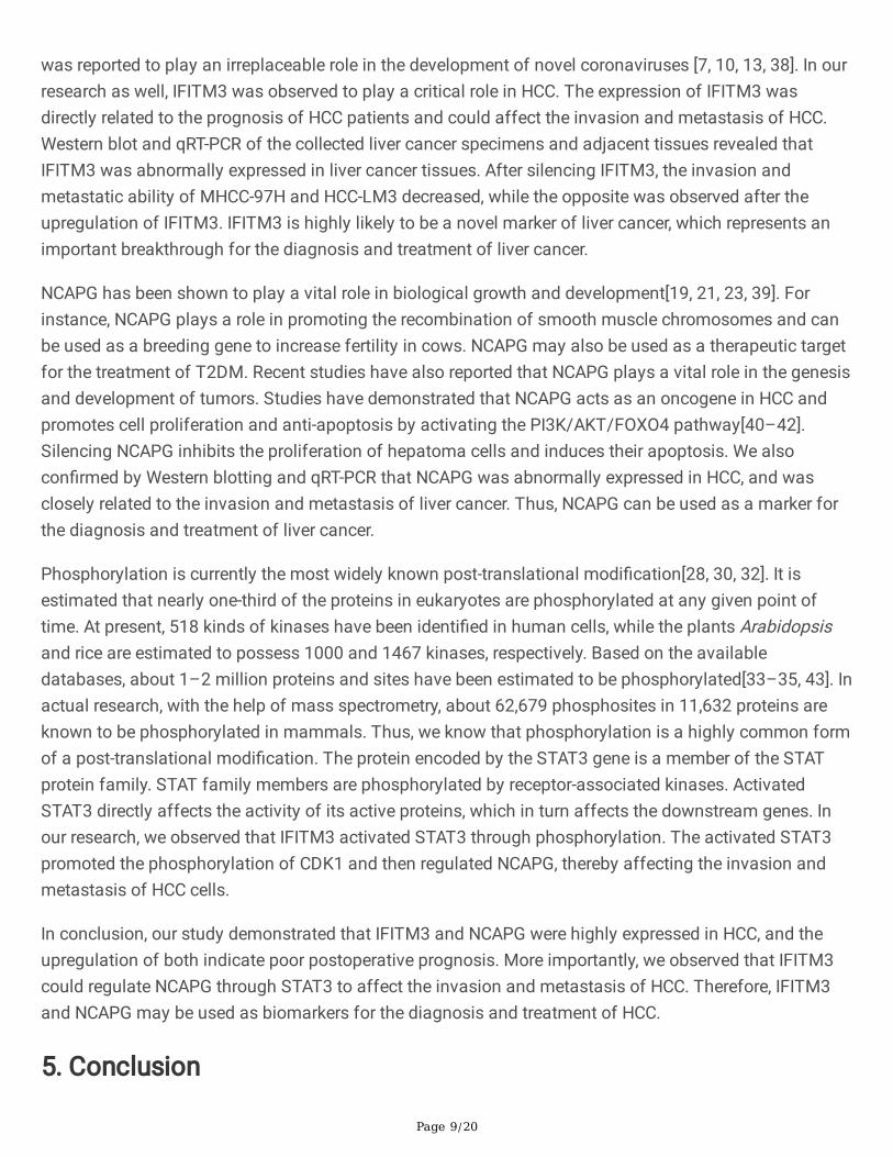
3. Results3.1. High expression of IFITM3 and NCAPG is associated with poor HCC prognosis.
Page 7/20
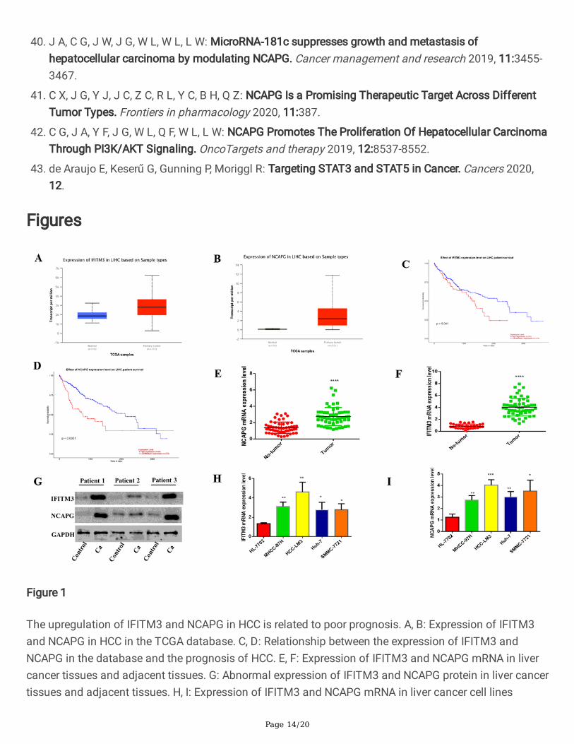
In order to evaluate the expression of IFITM3 and NCAPG in HCC, we �rst analyzed the documentedexpression of IFITM3 and NCAP in HCC from the TCGA database. We observed that both IFITM3 andNCAPG were highly expressed in HCC, and both high and low levels of expression were associated withpoor HCC prognosis (Fig. 1A-1D). We then performed PCR and Western blotting on the liver cancerspecimens and the adjacent tissues from the 55 patients enrolled in our study (Fig. 1E,1F,1G), andobserved that the results were consistent with those in the database. We also performed PCR analysis onliver cell line HL-7702 and the hepatoma cell lines MHCC-97H, HCC-LM3, SMCC-7721, and Huh-7, andthese results also indicated that the expression of IFITM3 and NCAPG in HCC cells was upregulated(Fig. 1H and1I). These results suggest that the expression levels of IFITM3 and NCAPG are upregulated inHCC tissue cells, which affects the prognosis of HCC patients.
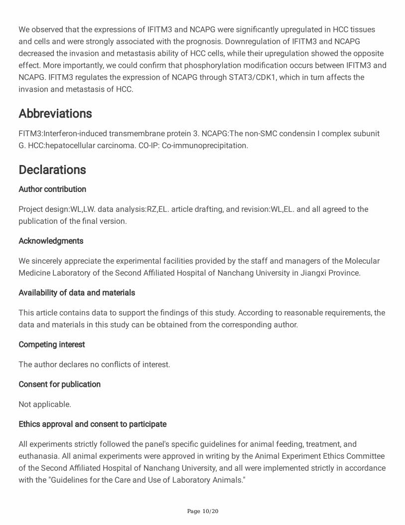
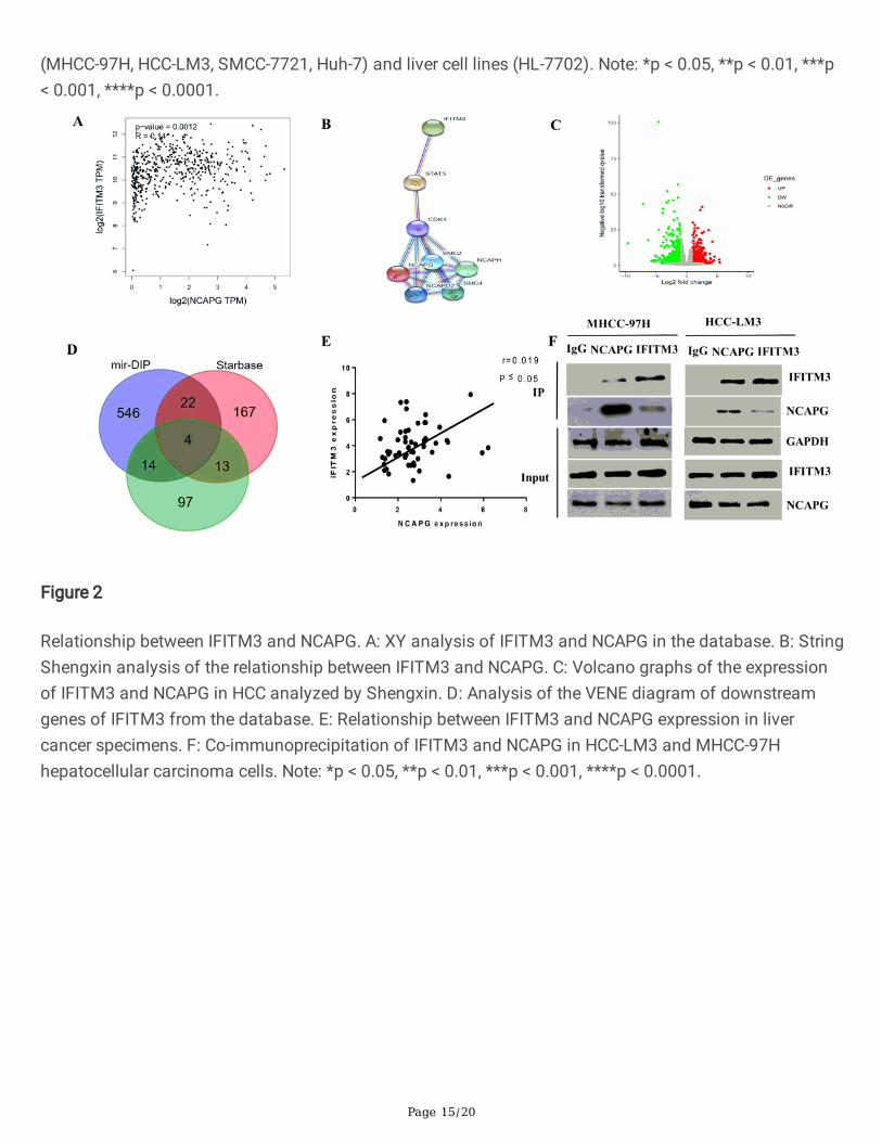
3.2. The relationship between IFITM3 and NCAPG in HCCIn order to further understand the relationship between IFITM3 and NCAPG, we carried out bioinformaticsanalysis on multiple databases available online and observed that both IFITM3 and NCAPG were highlyexpressed in liver cancer tissues, with a positive correlation between the two (Fig. 2A). This wasconsistent with the results of qRT-PCR analysis on the tissues (Fig. 2E). Furthermore, we constructedvolcano maps of RNA Seq in HCC tissues and adjacent tissues, and found that IFITM3 and ncapg werehighly expressed (Fig. 2C), and predicted the target genes downstream of IFITM3 using the mirDIP andstarBase databases (Fig. 2D). We then predicted on the String website that there may be aphosphorylation modi�cation between IFITM3 and NCAPG (Fig. 2B). Finally, using CO-IP, we con�rmedthe presence of protein interactions between IFITM3 and NCAPG (Fig. 2F).
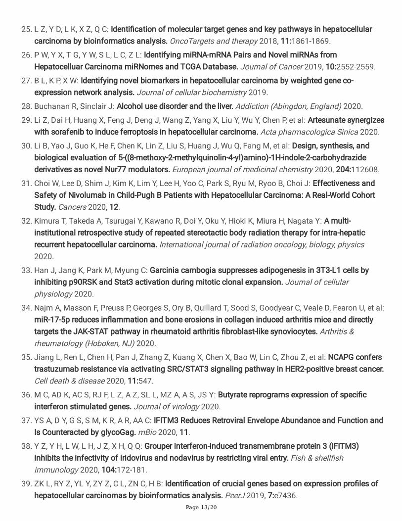
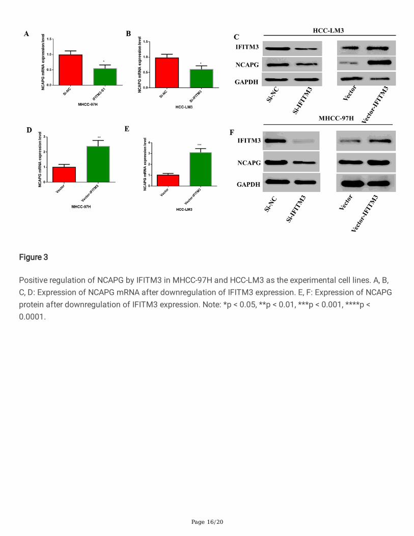
3.3. IFITM3 can positively regulate NCAPGAfter understanding the relationship between IFITM3 and NCAPG, in order to further regulate therelationship between the two, we transfected the interfering fragments of IFITM3 (i.e., SiRNA) andplasmids into MHCC-97H and HCC-LM3, and observed the mRNA and protein expressions of NCAPG. Our�ndings con�rmed that IFITM3 caused positive regulation of NCAPG; thus, downregulation of IFITM3reduced the mRNA and protein levels of NCAPG (Fig. 3A-3C), and upregulation of IFITM3 increased themRNA and protein levels of NCAPG(Fig. 3D-3F). This indicated that IFITM3 regulated the expression ofNCAPG positively.
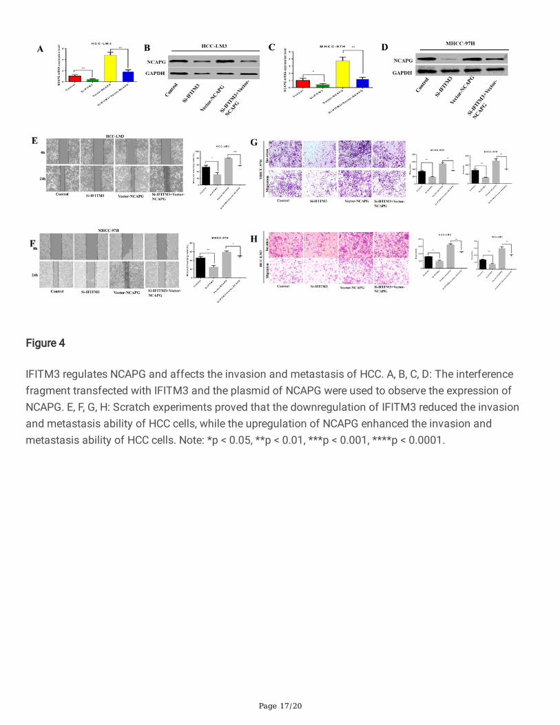
3.4. IFITM3 regulates NCAPG and affects the invasion and metastasis of HCCAfter determining that IFITM3 positively regulates NCAPG, we conducted a retrospective experiment tocorroborate our experimental results further. We transfected the interfering fragments and plasmids ofIFITM3, interfering fragments and plasmids of NCAPG, interfering fragments of IFITM3 plus plasmids ofNCAPG, and plasmids of IFITM3 plus interference fragments of NCAPG simultaneously, and comparedthem with the control group. We observed that when the expression was IFITM3 was upregulated, NCAPGexpression also enhanced, and NCAPG was upregulated, IFITM3 was also enhanced (Fig. 4A-4D).Simultaneous transfection of the IFITM3 interference fragment and NCAPG plasmid or the IFITM3plasmid and NCAPG interference fragment resulted in levels of NCAPG expression that were close tothose of the control group. Regarding the relationship between IFITM3 and NCAPG in terms of the
Page 8/20
invasion and metastasis ability of HCC cells, we observed through cell scratch experiments and Transwellexperiments that the expression of IFITM3 and NCAPG was positively correlated with the ability of cellinvasion and metastasis(Fig. 4E-4H). The upregulation of IFITM3 and NCAPG enhanced the ability ofinvasion and metastasis of the cells, while downregulation of both showed an opposite effect. On theother hand, simultaneous downregulation of IFITM3 and upregulation of NCAPG, or upregulation ofIFITM3 and downregulation of NCAPG showed no signi�cant difference in the invasion and metastasisability compared to the control group. This indicated that IFITM3 regulated NCAPG to in�uence theinvasion and metastasis of HCC cells.
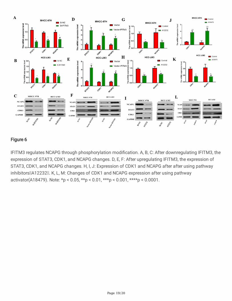
3.5. IFITM3 regulates NCAPG through phosphorylation modi�cationRegarding the speci�c mechanism of action between IFITM3 and NCAPG, our data showed that theremay be phosphorylation modi�cations between IFITM3 and NCAPG, and IFITM3 could regulate NCAPGthrough the STAT3/CDK1 pathway (Fig. 2B). Therefore, we upregulated the expression of IFITM3 andobserved the expression of STAT3/CDK1. We observed that the downregulation of the expression ofIFITM3 decreased the expression of STAT3/CDK1 (Fig. 6A-6F), while the upregulation of IFITM3increased the expression of STAT3/CDK1. Furthermore, we observed that the expression of NCAPG wasupregulated after the use of STAT3/CDK1 activators(A18479) and downregulated after the use ofinhibitors(A12231) (Fig. 6G-6L). This indicated that IFITM3 regulated NCAPG via STAT3/CDK1.
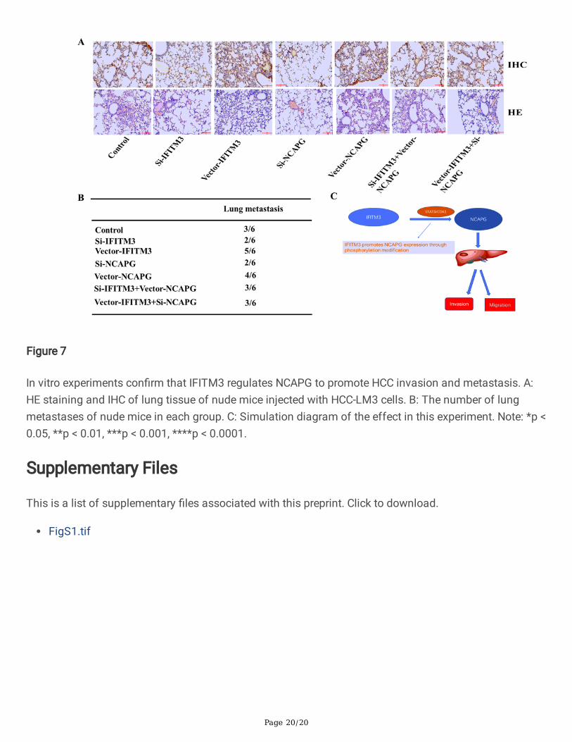
3.6. Con�rmation of results through animal experimentsIn vitro cell experiments indicated that IFITM3 regulated NCAPG to in�uence the invasion and metastasisof HCC. In order to further verify our �ndings, we performed experiments on animals. We observed thatthe degree of lung metastasis in mice injected with cells containing upregulated IFITM3 and NCAPG wassigni�cantly higher than that in the control group and that in mice injected with cells containingdownregulated IFITM3 and NCAPG was signi�cantly lower, while injection with cells containingsimultaneously upregulated IFITM3 and downregulated NCAPG, or downregulated IFITM3 andupregulated NCAPG, showed no signi�cant difference (Fig. 7A-7B). Thus, in vivo experiments reiteratedthat IFITM3 could affect the expression of NCAPG through phosphorylation, which in turn in�uenced theinvasion and metastasis of HCC (Fig. 7C).
4. DiscussionIn this study, we demonstrated that both IFITM3 and NCAPG were highly expressed in HCC tissues andcells, and their expression was closely related to the prognosis and recurrence of liver cancer. We alsodemonstrated for the �rst time that IFITM3 regulated NCAPG through phosphorylation to affect theinvasion and metastasis of HCC.
Several reports have suggested the involvement of IFITM3 in the development of cancer[36, 37]. Forexample, studies have reported that IFITM3 upregulated the expression of c-myc through the ERK1/2signaling pathway and promoted the proliferation of liver cancer cells. Studies have also demonstratedthat IFITM3rs12252-CC is related to the poor differentiation and progression of HCC. Recently, IFITM3
Page 9/20
was reported to play an irreplaceable role in the development of novel coronaviruses [7, 10, 13, 38]. In ourresearch as well, IFITM3 was observed to play a critical role in HCC. The expression of IFITM3 wasdirectly related to the prognosis of HCC patients and could affect the invasion and metastasis of HCC.Western blot and qRT-PCR of the collected liver cancer specimens and adjacent tissues revealed thatIFITM3 was abnormally expressed in liver cancer tissues. After silencing IFITM3, the invasion andmetastatic ability of MHCC-97H and HCC-LM3 decreased, while the opposite was observed after theupregulation of IFITM3. IFITM3 is highly likely to be a novel marker of liver cancer, which represents animportant breakthrough for the diagnosis and treatment of liver cancer.
NCAPG has been shown to play a vital role in biological growth and development[19, 21, 23, 39]. Forinstance, NCAPG plays a role in promoting the recombination of smooth muscle chromosomes and canbe used as a breeding gene to increase fertility in cows. NCAPG may also be used as a therapeutic targetfor the treatment of T2DM. Recent studies have also reported that NCAPG plays a vital role in the genesisand development of tumors. Studies have demonstrated that NCAPG acts as an oncogene in HCC andpromotes cell proliferation and anti-apoptosis by activating the PI3K/AKT/FOXO4 pathway[40–42].Silencing NCAPG inhibits the proliferation of hepatoma cells and induces their apoptosis. We alsocon�rmed by Western blotting and qRT-PCR that NCAPG was abnormally expressed in HCC, and wasclosely related to the invasion and metastasis of liver cancer. Thus, NCAPG can be used as a marker forthe diagnosis and treatment of liver cancer.
Phosphorylation is currently the most widely known post-translational modi�cation[28, 30, 32]. It isestimated that nearly one-third of the proteins in eukaryotes are phosphorylated at any given point oftime. At present, 518 kinds of kinases have been identi�ed in human cells, while the plants Arabidopsisand rice are estimated to possess 1000 and 1467 kinases, respectively. Based on the availabledatabases, about 1–2 million proteins and sites have been estimated to be phosphorylated[33–35, 43]. Inactual research, with the help of mass spectrometry, about 62,679 phosphosites in 11,632 proteins areknown to be phosphorylated in mammals. Thus, we know that phosphorylation is a highly common formof a post-translational modi�cation. The protein encoded by the STAT3 gene is a member of the STATprotein family. STAT family members are phosphorylated by receptor-associated kinases. ActivatedSTAT3 directly affects the activity of its active proteins, which in turn affects the downstream genes. Inour research, we observed that IFITM3 activated STAT3 through phosphorylation. The activated STAT3promoted the phosphorylation of CDK1 and then regulated NCAPG, thereby affecting the invasion andmetastasis of HCC cells.
In conclusion, our study demonstrated that IFITM3 and NCAPG were highly expressed in HCC, and theupregulation of both indicate poor postoperative prognosis. More importantly, we observed that IFITM3could regulate NCAPG through STAT3 to affect the invasion and metastasis of HCC. Therefore, IFITM3and NCAPG may be used as biomarkers for the diagnosis and treatment of HCC.
5. Conclusion
Page 10/20
We observed that the expressions of IFITM3 and NCAPG were signi�cantly upregulated in HCC tissuesand cells and were strongly associated with the prognosis. Downregulation of IFITM3 and NCAPGdecreased the invasion and metastasis ability of HCC cells, while their upregulation showed the oppositeeffect. More importantly, we could con�rm that phosphorylation modi�cation occurs between IFITM3 andNCAPG. IFITM3 regulates the expression of NCAPG through STAT3/CDK1, which in turn affects theinvasion and metastasis of HCC.
AbbreviationsFITM3:Interferon-induced transmembrane protein 3. NCAPG:The non-SMC condensin I complex subunitG. HCC:hepatocellular carcinoma. CO-IP: Co-immunoprecipitation.
DeclarationsAuthor contribution
Project design:WL,LW. data analysis:RZ,EL. article drafting, and revision:WL,EL. and all agreed to thepublication of the �nal version.
Acknowledgments
We sincerely appreciate the experimental facilities provided by the staff and managers of the MolecularMedicine Laboratory of the Second A�liated Hospital of Nanchang University in Jiangxi Province.
Availability of data and materials
This article contains data to support the �ndings of this study. According to reasonable requirements, thedata and materials in this study can be obtained from the corresponding author.
Competing interest
The author declares no con�icts of interest.
Consent for publication
Not applicable.
Ethics approval and consent to participate
All experiments strictly followed the panel's speci�c guidelines for animal feeding, treatment, andeuthanasia. All animal experiments were approved in writing by the Animal Experiment Ethics Committeeof the Second A�liated Hospital of Nanchang University, and all were implemented strictly in accordancewith the "Guidelines for the Care and Use of Laboratory Animals."

Page 11/20
Funding
This research was funded by the National Natural Science Foundation of China (N0.81860431), theJiangxi Natural Science Foundation (NO.20181BBG70025), and the Jiangxi Provincial Health andSanitation Committee (NO.20204266). All expenses are funded by two fundings.
References1. Ono T, Kohro Y, Kohno K, Tozaki-Saitoh H, Nakashima Y, Tsuda M: Mechanical pain of the lower
extremity after compression of the upper spinal cord involves signal transducer and activator oftranscription 3-dependent reactive astrocytes and interleukin-6. Brain, behavior, and immunity 2020.
2. Parisi X, Bergerson J, Urban A, Darnell D, Stratton P, Freeman A: Obstetric and Gynecological Care inPatients with STAT3-De�cient Hyper IgE Syndrome. Journal of clinical immunology 2020.
3. Sebastian N, Miller E, Yang X, Diaz D, Tan Y, Dowell J, Spain J, Rikabi A, Elliott E, Knopp M, WilliamsT: A Pilot Trial Evaluating Stereotactic Body Radiation Therapy to Induce Hyperemia in Combinationwith Transarterial Chemoembolization for Hepatocellular Carcinoma. International journal ofradiation oncology, biology, physics 2020.
4. Wang P, Sun Y, Zhou K, Cheng J, Hu B, Guo W, Yin Y, Huang J, Zhou J, Fan J, et al: Circulating tumorcells are an indicator for the administration of adjuvant transarterial chemoembolization inhepatocellular carcinoma: A single-center, retrospective, propensity-matched study. Clinical andtranslational medicine 2020:e137.
5. CT B, F M, M R, M M, S W, EW T, BH T, SE S, P K, EC H, M M: Correction: Bat IFITM3 restrictiondepends on S-palmitoylation and a polymorphic site within the CD225 domain. Life science alliance2020, 3.
�. DL H, Y L, J L, L Y, M X, XX C, B X, CL T, L L, JS Z: SThe New Salicylaldehyde ,-PropanedithioacetalEster Enables N-to-C Sequential Native Chemical Ligation and Ser/Thr Ligation for Chemical ProteinSynthesis. Journal of the American Chemical Society 2020, 142:8790-8799.
7. H L, LL Y, CC W, Y X, L M, L C, WF Z, ZJ S: Expression and Prognostic Value of IFIT1 and IFITM3 inHead and Neck Squamous Cell Carcinoma. American journal of clinical pathology 2020, 153:618-629.
�. K M, H K: BioID screening of biotinylation sites using the avidin-like protein Tamavidin 2-REVidenti�es global interactors of stimulator of interferon genes (STING). The Journal of biologicalchemistry 2020.
9. L X, J Z, G Q, Z L, Q F, C W, Q W: Recombinant lactobacillin PlnK adjusts the gut microbiomedistribution in broilers. British poultry science 2020.
10. MY H, S AH, IY H, R H, AC S, AA M, Q H: Interferon-Induced Transmembrane Protein (IFITM3) IsUpregulated Explicitly in SARS-CoV-2 Infected Lung Epithelial Cells. Frontiers in immunology 2020,11:1372.

Page 12/20
11. Q S, N L, J L, RB G, Z L, LQ L, Y S, JF G, DY W, YL S: Interferon-induced Transmembrane Protein 3Prevents Acute In�uenza Pathogenesis in Mice. Biomedical and environmental sciences : BES 2020,33:295-305.
12. S B, Q P, A F, C L: Differential pressures of SERINC5 and IFITM3 on HIV-1 envelope glycoprotein overthe course of HIV-1 infection. Journal of virology 2020.
13. S X, K L, C X, M H, A O, D S, B L, J W, Y Q, D M, Z M: Establishment and characterization of the pigtonsil epithelial (PT) cell line as a new model for persist infection of Japanese Encephalitis Virus.Veterinary microbiology 2020, 242:108587.
14. M Z, X Z, S Z, D C, P D, X L, D J, JT G, H Z, H L: Bat SARS-Like WIV1 coronavirus uses the ACE2 ofmultiple animal species as receptor and evades IFITM3 restriction via TMPRSS2 activation ofmembrane fusion. Emerging microbes & infections 2020:1-36.
15. TA G, D H, A S, C K, J O: The Robust Restriction of Zika Virus by Type-I Interferon in A549 Cells Variesby Viral Lineage and Is Not Determined by IFITM3. Viruses 2020, 12.
1�. X L, S Y, P R, X S, M J: Human microRNA-30 inhibits in�uenza virus infection by suppressing theexpression of SOCS1, SOCS3, and NEDD4. Cellular microbiology 2020, 22:e13150.
17. X W, JS S, T D, X Y, C C, Y Z, Y L, Y S, K C, HC H, T P: Site-Speci�c Photo-Crosslinking ProteomicsReveal Regulation of IFITM3 Tra�cking and Turnover by VCP/p97 ATPase. Cell chemical biology2020, 27:571-585.e576.
1�. H Y, Z L, Q S, Q W, J T, Q J, L G: Aberrant expression of cell cycle and material metabolism relatedgenes contributes to hepatocellular carcinoma occurrence. Pathology, research and practice 2017,213:316-321.
19. Q F, F Y, J Z, X Y, T X, G H, J Z, L W, S D, H Y: Bioinformatical identi�cation of key pathways and genesin human hepatocellular carcinoma after CSN5 depletion. Cellular signalling 2018, 49:79-86.
20. J B, EJ F, LM D, A R, M M, M R, C A, MG G, J A, ED M, GD M: A comprehensive study of epigeneticalterations in hepatocellular carcinoma identi�es potential therapeutic targets. Journal of hepatology2019, 71:78-90.
21. Z S, Z X, L H, Y G, J Z, Y L, G S, Q X, D H: The genetic association between type 2 diabetic andhepatocellular carcinomas. Annals of translational medicine 2020, 8:380.
22. Y W, B G, PY T, YA H, K S, A D, VP S, HY O, M S, C X, et al: Genome-wide CRISPR knockout screensidentify NCAPG as an essential oncogene for hepatocellular carcinoma tumor growth. FASEB journal: o�cial publication of the Federation of American Societies for Experimental Biology 2019, 33:8759-8770.
23. Q Z, R S, C S, C G, P W: Non-SMC Condensin I Complex, Subunit G (NCAPG) is a Novel Mitotic GeneRequired for Hepatocellular Cancer Cell Proliferation and Migration. Oncology research 2018, 26:269-276.
24. W L, B L, H L, Y H, X Y, F Z, X Y, Q F, E L, Z Z, L W: Overexpression of non‐SMC condensin I complexsubunit G serves as a promising prognostic marker and therapeutic target for hepatocellularcarcinoma. International journal of molecular medicine 2017, 40:731-738.
Page 13/20
25. L Z, Y D, L K, X Z, Q C: Identi�cation of molecular target genes and key pathways in hepatocellularcarcinoma by bioinformatics analysis. OncoTargets and therapy 2018, 11:1861-1869.
2�. P W, Y X, T G, Y W, S L, L C, Z L: Identifying miRNA-mRNA Pairs and Novel miRNAs fromHepatocelluar Carcinoma miRNomes and TCGA Database. Journal of Cancer 2019, 10:2552-2559.
27. B L, K P, X W: Identifying novel biomarkers in hepatocellular carcinoma by weighted gene co-expression network analysis. Journal of cellular biochemistry 2019.
2�. Buchanan R, Sinclair J: Alcohol use disorder and the liver. Addiction (Abingdon, England) 2020.
29. Li Z, Dai H, Huang X, Feng J, Deng J, Wang Z, Yang X, Liu Y, Wu Y, Chen P, et al: Artesunate synergizeswith sorafenib to induce ferroptosis in hepatocellular carcinoma. Acta pharmacologica Sinica 2020.
30. Li B, Yao J, Guo K, He F, Chen K, Lin Z, Liu S, Huang J, Wu Q, Fang M, et al: Design, synthesis, andbiological evaluation of 5-((8-methoxy-2-methylquinolin-4-yl)amino)-1H-indole-2-carbohydrazidederivatives as novel Nur77 modulators. European journal of medicinal chemistry 2020, 204:112608.
31. Choi W, Lee D, Shim J, Kim K, Lim Y, Lee H, Yoo C, Park S, Ryu M, Ryoo B, Choi J: Effectiveness andSafety of Nivolumab in Child-Pugh B Patients with Hepatocellular Carcinoma: A Real-World CohortStudy. Cancers 2020, 12.
32. Kimura T, Takeda A, Tsurugai Y, Kawano R, Doi Y, Oku Y, Hioki K, Miura H, Nagata Y: A multi-institutional retrospective study of repeated stereotactic body radiation therapy for intra-hepaticrecurrent hepatocellular carcinoma. International journal of radiation oncology, biology, physics2020.
33. Han J, Jang K, Park M, Myung C: Garcinia cambogia suppresses adipogenesis in 3T3-L1 cells byinhibiting p90RSK and Stat3 activation during mitotic clonal expansion. Journal of cellularphysiology 2020.
34. Najm A, Masson F, Preuss P, Georges S, Ory B, Quillard T, Sood S, Goodyear C, Veale D, Fearon U, et al:miR-17-5p reduces in�ammation and bone erosions in collagen induced arthritis mice and directlytargets the JAK-STAT pathway in rheumatoid arthritis �broblast-like synoviocytes. Arthritis &rheumatology (Hoboken, NJ) 2020.
35. Jiang L, Ren L, Chen H, Pan J, Zhang Z, Kuang X, Chen X, Bao W, Lin C, Zhou Z, et al: NCAPG conferstrastuzumab resistance via activating SRC/STAT3 signaling pathway in HER2-positive breast cancer.Cell death & disease 2020, 11:547.
3�. M C, AD K, AC S, RJ F, L Z, A Z, SL L, MZ A, A S, JS Y: Butyrate reprograms expression of speci�cinterferon stimulated genes. Journal of virology 2020.
37. YS A, D Y, G S, S M, K R, A R, AA C: IFITM3 Reduces Retroviral Envelope Abundance and Function andIs Counteracted by glycoGag. mBio 2020, 11.
3�. Y Z, Y H, L W, L H, J Z, X H, Q Q: Grouper interferon-induced transmembrane protein 3 (IFITM3)inhibits the infectivity of iridovirus and nodavirus by restricting viral entry. Fish & shell�shimmunology 2020, 104:172-181.
39. ZK L, RY Z, YL Y, ZY Z, C L, ZN C, H B: Identi�cation of crucial genes based on expression pro�les ofhepatocellular carcinomas by bioinformatics analysis. PeerJ 2019, 7:e7436.
Page 14/20
40. J A, C G, J W, J G, W L, W L, L W: MicroRNA‐181c suppresses growth and metastasis ofhepatocellular carcinoma by modulating NCAPG. Cancer management and research 2019, 11:3455-3467.
41. C X, J G, Y J, J C, Z C, R L, Y C, B H, Q Z: NCAPG Is a Promising Therapeutic Target Across DifferentTumor Types. Frontiers in pharmacology 2020, 11:387.
42. C G, J A, Y F, J G, W L, Q F, W L, L W: NCAPG Promotes The Proliferation Of Hepatocellular CarcinomaThrough PI3K/AKT Signaling. OncoTargets and therapy 2019, 12:8537-8552.
43. de Araujo E, Keserű G, Gunning P, Moriggl R: Targeting STAT3 and STAT5 in Cancer. Cancers 2020,12.
Figures
Figure 1
The upregulation of IFITM3 and NCAPG in HCC is related to poor prognosis. A, B: Expression of IFITM3and NCAPG in HCC in the TCGA database. C, D: Relationship between the expression of IFITM3 andNCAPG in the database and the prognosis of HCC. E, F: Expression of IFITM3 and NCAPG mRNA in livercancer tissues and adjacent tissues. G: Abnormal expression of IFITM3 and NCAPG protein in liver cancertissues and adjacent tissues. H, I: Expression of IFITM3 and NCAPG mRNA in liver cancer cell lines
Page 15/20
(MHCC-97H, HCC-LM3, SMCC-7721, Huh-7) and liver cell lines (HL-7702). Note: *p < 0.05, **p < 0.01, ***p< 0.001, ****p < 0.0001.
Figure 2
Relationship between IFITM3 and NCAPG. A: XY analysis of IFITM3 and NCAPG in the database. B: StringShengxin analysis of the relationship between IFITM3 and NCAPG. C: Volcano graphs of the expressionof IFITM3 and NCAPG in HCC analyzed by Shengxin. D: Analysis of the VENE diagram of downstreamgenes of IFITM3 from the database. E: Relationship between IFITM3 and NCAPG expression in livercancer specimens. F: Co-immunoprecipitation of IFITM3 and NCAPG in HCC-LM3 and MHCC-97Hhepatocellular carcinoma cells. Note: *p < 0.05, **p < 0.01, ***p < 0.001, ****p < 0.0001.
Page 16/20
Figure 3
Positive regulation of NCAPG by IFITM3 in MHCC-97H and HCC-LM3 as the experimental cell lines. A, B,C, D: Expression of NCAPG mRNA after downregulation of IFITM3 expression. E, F: Expression of NCAPGprotein after downregulation of IFITM3 expression. Note: *p < 0.05, **p < 0.01, ***p < 0.001, ****p <0.0001.
Page 17/20
Figure 4
IFITM3 regulates NCAPG and affects the invasion and metastasis of HCC. A, B, C, D: The interferencefragment transfected with IFITM3 and the plasmid of NCAPG were used to observe the expression ofNCAPG. E, F, G, H: Scratch experiments proved that the downregulation of IFITM3 reduced the invasionand metastasis ability of HCC cells, while the upregulation of NCAPG enhanced the invasion andmetastasis ability of HCC cells. Note: *p < 0.05, **p < 0.01, ***p < 0.001, ****p < 0.0001.

Page 18/20
Figure 5
IFITM3 regulates NCAPG and thus affects the invasion and metastasis of HCC. A, B, C, D: Plasmidtransfected with IFITM3 and interference fragment of NCAPG were used to observe the expression ofNCAPG. E, F, G, H: Scratch experiments proved that the upregulation of IFITM3 enhanced the invasion andmetastasis ability of HCC cells, while the downregulation of NCAPG reduced the invasion and metastasisability of HCC cells. Note: *p < 0.05, **p < 0.01, ***p < 0.001, ****p < 0.0001.
Page 19/20
Figure 6
IFITM3 regulates NCAPG through phosphorylation modi�cation. A, B, C: After downregulating IFITM3, theexpression of STAT3, CDK1, and NCAPG changes. D, E, F: After upregulating IFITM3, the expression ofSTAT3, CDK1, and NCAPG changes. H, I, J: Expression of CDK1 and NCAPG after after using pathwayinhibitors A12232 . K, L, M: Changes of CDK1 and NCAPG expression after using pathwayactivator(A18479). Note: *p < 0.05, **p < 0.01, ***p < 0.001, ****p < 0.0001.
Page 20/20
Figure 7
In vitro experiments con�rm that IFITM3 regulates NCAPG to promote HCC invasion and metastasis. A:HE staining and IHC of lung tissue of nude mice injected with HCC-LM3 cells. B: The number of lungmetastases of nude mice in each group. C: Simulation diagram of the effect in this experiment. Note: *p <0.05, **p < 0.01, ***p < 0.001, ****p < 0.0001.
Supplementary Files
This is a list of supplementary �les associated with this preprint. Click to download.
FigS1.tif