Embed Size (px)
Citation preview
FROM POWDER TO SPINAL CAGE PRODUCTION
ADDED VALUE FOR SPINAL IMPLANTS THROUGH ADDITIVE MANUFACTURING
2 GE Additive
The spine segment, which includes spinal fusion and fixation devices, is one of the major segments in the orthopedic industry and accounts for almost one fifth of its overall revenue.
While the Compound Annual Growth Rate (CAGR) for the overall orthopedic market has constantly been between 2-3% over the last couple of years, there is significant adoption of additive technologies, leading to an average growth rate of 20-30% within additively manufactured devices. 1, 2, 3, 4
Additive has shown that it can add value by providing freedom of design and efficient manufacturing, while avoiding some of the limitations of conventional manufacturing. Cages are made in one single component with mesh structures incorporated in the device, as well as new innovative geometries, with multiple design features. Lumbar interbody fusion devices, known as spinal cages, are widely used to treat patients with back pain, caused by degenerative disc disease.
The spinal cages are implanted in minimally invasive procedures and are available in different shapes and sizes to facilitate an appropriate solution for different
indications and techniques, such as DLIF, PLIF, ALIF, TLIF, XLIF. For patients and surgeons, these implants can improve spinal fusion procedures, clinical outcome and recovery time.
Spinal implants have traditionally been milled from titanium or machined from polyether ether ketone (PEEK). The first generation of titanium cages were machined, however the solid cage design caused issues with subsidence and their bulky design restricted the post-operative evaluation due to its limited radiolucency.
In the first decade of the 2000s, most spinal cages were made from PEEK and occasionally further developed with a secondary process, such as plasma-spray coating, to create a rough and porous surface that increases the osseointegration (bone ingrowth). PEEK was selected, partly because of its mechanical properties, more like the human bone than the bulk titanium.
However, in some cases, PEEK implants caused an inflammatory response, promoted fibrous tissue formation instead of bone formation, or wear debris and delamination occurred over time.5, 6, 7
OVERVIEW
GE Additive 3
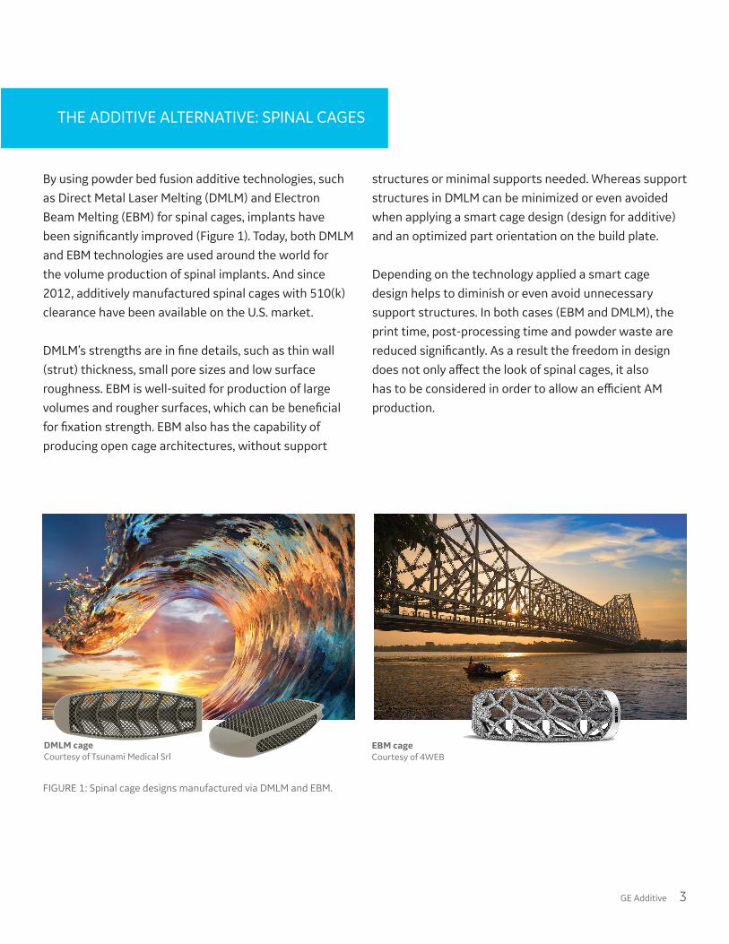
By using powder bed fusion additive technologies, such as Direct Metal Laser Melting (DMLM) and Electron Beam Melting (EBM) for spinal cages, implants have been significantly improved (Figure 1). Today, both DMLM and EBM technologies are used around the world for the volume production of spinal implants. And since 2012, additively manufactured spinal cages with 510(k) clearance have been available on the U.S. market.
DMLM’s strengths are in fine details, such as thin wall (strut) thickness, small pore sizes and low surface roughness. EBM is well-suited for production of large volumes and rougher surfaces, which can be beneficial for fixation strength. EBM also has the capability of producing open cage architectures, without support
structures or minimal supports needed. Whereas support structures in DMLM can be minimized or even avoided when applying a smart cage design (design for additive) and an optimized part orientation on the build plate.
Depending on the technology applied a smart cage design helps to diminish or even avoid unnecessary support structures. In both cases (EBM and DMLM), the print time, post-processing time and powder waste are reduced significantly. As a result the freedom in design does not only affect the look of spinal cages, it also has to be considered in order to allow an efficient AM production.
THE ADDITIVE ALTERNATIVE: SPINAL CAGES
FIGURE 1: Spinal cage designs manufactured via DMLM and EBM.
DMLM cageCourtesy of Tsunami Medical Srl
EBM cageCourtesy of 4WEB
4 GE Additive
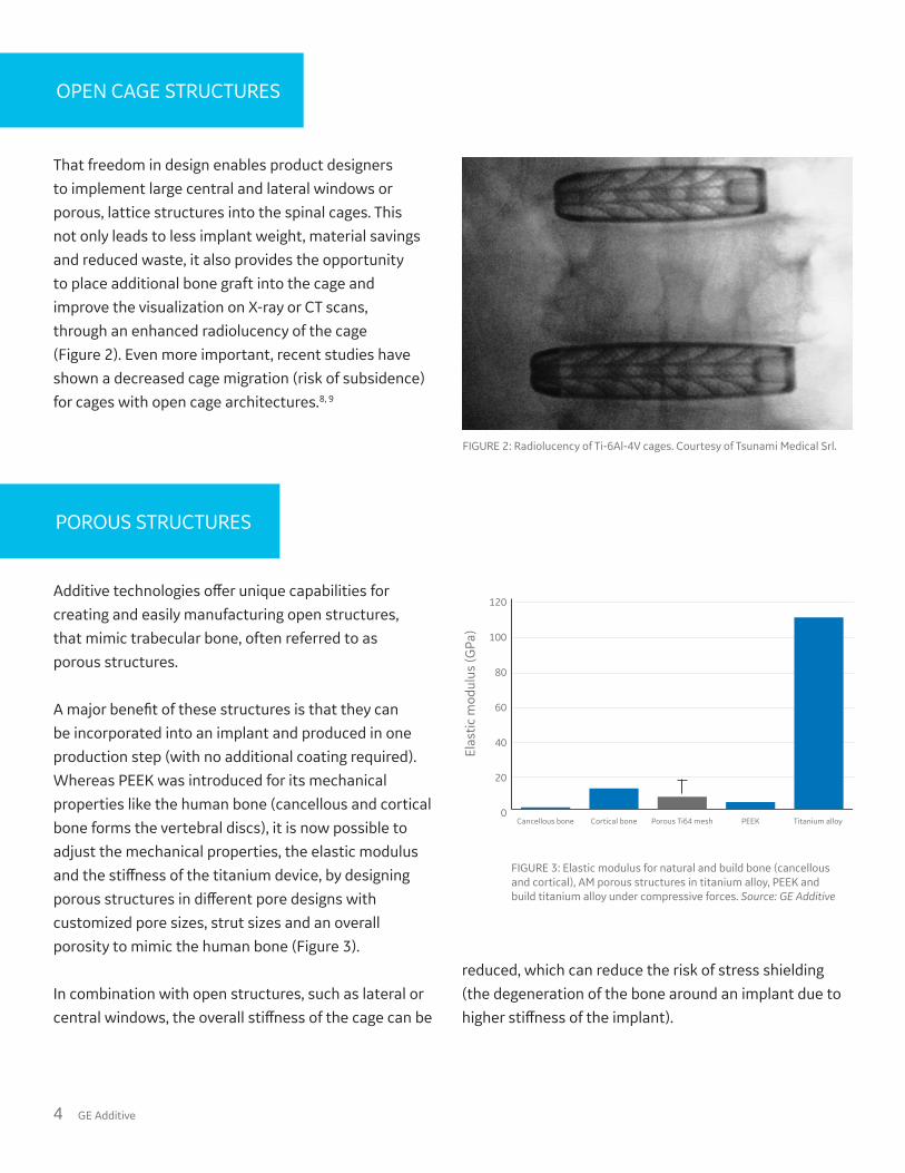
That freedom in design enables product designers to implement large central and lateral windows or porous, lattice structures into the spinal cages. This not only leads to less implant weight, material savings and reduced waste, it also provides the opportunity to place additional bone graft into the cage and improve the visualization on X-ray or CT scans, through an enhanced radiolucency of the cage (Figure 2). Even more important, recent studies have shown a decreased cage migration (risk of subsidence) for cages with open cage architectures.8, 9
OPEN CAGE STRUCTURES
Additive technologies offer unique capabilities for creating and easily manufacturing open structures, that mimic trabecular bone, often referred to as porous structures.
A major benefit of these structures is that they can be incorporated into an implant and produced in one production step (with no additional coating required). Whereas PEEK was introduced for its mechanical properties like the human bone (cancellous and cortical bone forms the vertebral discs), it is now possible to adjust the mechanical properties, the elastic modulus and the stiffness of the titanium device, by designing porous structures in different pore designs with customized pore sizes, strut sizes and an overall porosity to mimic the human bone (Figure 3).
In combination with open structures, such as lateral or central windows, the overall stiffness of the cage can be
reduced, which can reduce the risk of stress shielding (the degeneration of the bone around an implant due to higher stiffness of the implant).
POROUS STRUCTURES
FIGURE 3: Elastic modulus for natural and build bone (cancellous and cortical), AM porous structures in titanium alloy, PEEK and build titanium alloy under compressive forces. Source: GE Additive
FIGURE 2: Radiolucency of Ti-6Al-4V cages. Courtesy of Tsunami Medical Srl.
Elas
tic m
odul
us (G
Pa)
0
20
40
60
80
100
120
Titanium alloy PEEKPorous Ti64 meshCortical boneCancellous bone
GE Additive 5
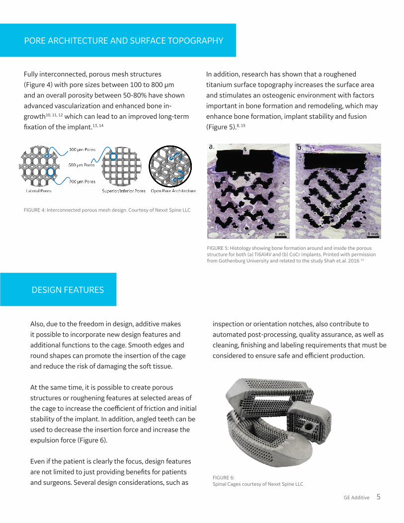
PORE ARCHITECTURE AND SURFACE TOPOGRAPHY
Fully interconnected, porous mesh structures (Figure 4) with pore sizes between 100 to 800 µm and an overall porosity between 50-80% have shown advanced vascularization and enhanced bone in-growth10, 11, 12 which can lead to an improved long-term fixation of the implant.13, 14
In addition, research has shown that a roughened titanium surface topography increases the surface area and stimulates an osteogenic environment with factors important in bone formation and remodeling, which may enhance bone formation, implant stability and fusion (Figure 5).8, 15
FIGURE 4: Interconnected porous mesh design. Courtesy of Nexxt Spine LLC
FIGURE 5: Histology showing bone formation around and inside the porous structure for both (a) Ti6Al4V and (b) CoCr implants. Printed with permission from Gothenburg University and related to the study Shah et.al. 2016 13
FIGURE 2: Radiolucency of Ti-6Al-4V cages. Courtesy of Tsunami Medical Srl.
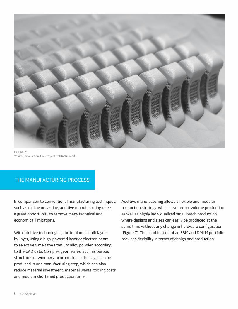
Also, due to the freedom in design, additive makes it possible to incorporate new design features and additional functions to the cage. Smooth edges and round shapes can promote the insertion of the cage and reduce the risk of damaging the soft tissue.
At the same time, it is possible to create porous structures or roughening features at selected areas of the cage to increase the coefficient of friction and initial stability of the implant. In addition, angled teeth can be used to decrease the insertion force and increase the expulsion force (Figure 6).
Even if the patient is clearly the focus, design features are not limited to just providing benefits for patients and surgeons. Several design considerations, such as
inspection or orientation notches, also contribute to automated post-processing, quality assurance, as well as cleaning, finishing and labeling requirements that must be considered to ensure safe and efficient production.
DESIGN FEATURES
FIGURE 6: Spinal Cages courtesy of Nexxt Spine LLC
6 GE Additive
In comparison to conventional manufacturing techniques, such as milling or casting, additive manufacturing offers a great opportunity to remove many technical and economical limitations.
With additive technologies, the implant is built layer-by-layer, using a high-powered laser or electron beam to selectively melt the titanium alloy powder, according to the CAD data. Complex geometries, such as porous structures or windows incorporated in the cage, can be produced in one manufacturing step, which can also reduce material investment, material waste, tooling costs and result in shortened production time.
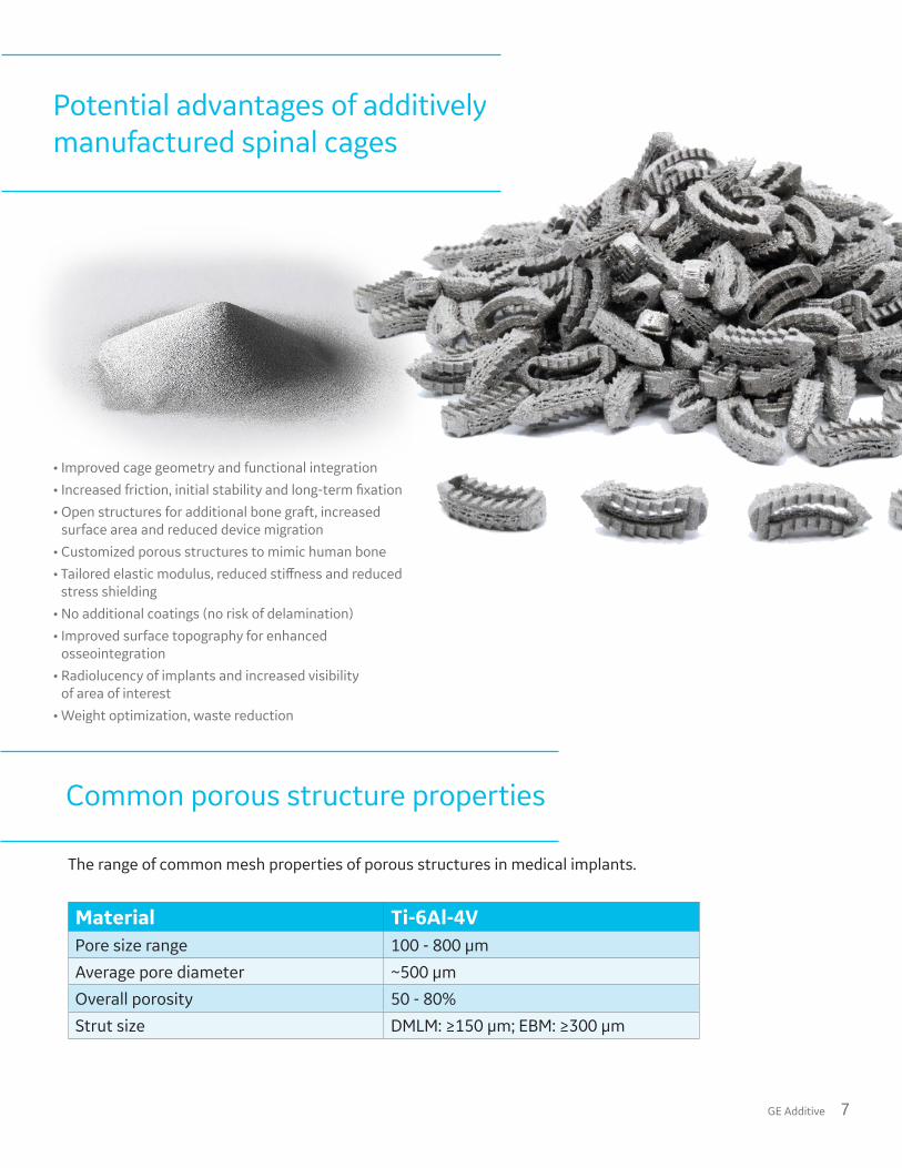
Additive manufacturing allows a flexible and modular production strategy, which is suited for volume production as well as highly individualized small batch production where designs and sizes can easily be produced at the same time without any change in hardware configuration (Figure 7). The combination of an EBM and DMLM portfolio provides flexibility in terms of design and production.
THE MANUFACTURING PROCESS
FIGURE 7: Volume production, Courtesy of FMI Instrumed.
GE Additive 7
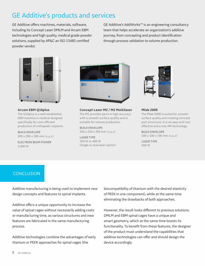
Material Ti-6Al-4VPore size range 100 - 800 µm
Average pore diameter ~500 µm
Overall porosity 50 - 80%
Strut size DMLM: ≥150 µm; EBM: ≥300 µm
The range of common mesh properties of porous structures in medical implants.
Common porous structure properties
• Improved cage geometry and functional integration
• Increased friction, initial stability and long-term fixation
• Open structures for additional bone graft, increased surface area and reduced device migration
• Customized porous structures to mimic human bone
• Tailored elastic modulus, reduced stiffness and reduced stress shielding
• No additional coatings (no risk of delamination)
• Improved surface topography for enhanced osseointegration
• Radiolucency of implants and increased visibility of area of interest
• Weight optimization, waste reduction
Potential advantages of additively manufactured spinal cages
8 GE Additive
Additive manufacturing is being used to implement new design concepts and features to spinal implants.
Additive offers a unique opportunity to increase the value of spinal cages without necessarily adding costs or manufacturing time, as various structures and new features are fabricated in the same manufacturing process.
Additive technologies combine the advantages of early titanium or PEEK approaches for spinal cages (the
biocompatibility of titanium with the desired elasticity of PEEK in one component), while at the same time eliminating the drawbacks of both approaches.
However, the result looks different to previous solutions. DMLM and EBM spinal cages have a unique and smart geometry, which at the same time boosts its functionality. To benefit from these features, the designer of the product must understand the capabilities that additive technologies can offer and should design the device accordingly.
CONCLUSION
GE Additive’s products and services
Arcam EBM Q10plus The Q10plus is a well established EBM machine in medical designed specifically for cost-efficient production of orthopedic implants.
BUILD ENVELOPE 200 x 200 x 180 mm (x,y,z)
ELECTRON BEAM POWER 3,000 W
Concept Laser M2 / M2 MultilaserThe M2 provides parts in high accuracy with a smooth-surface quality and is suitable for volume production.
BUILD ENVELOPE 250 x 250 x 350 mm (x,y,z)
LASER TYPE 200 W or 400 W (Single or dual laser option)
Mlab 200RThe Mlab 200R is suited for smooth-surface quality and creating intricate part structures. It is an easy and cost effective entry into AM technology.
BUILD ENVELOPE100 x 100 x 100 mm (x,y,z)
LASER TYPE200 W
GE Additive offers machines, materials, software, including its Concept Laser DMLM and Arcam EBM technologies and high quality, medical grade powder solutions, supplied by AP&C an ISO 13485 certified powder vendor.
GE Additive’s AddWorks™ is an engineering consultancy team that helps accelerate an organization’s additive journey, from concepting and product identification through process validation to volume production.
CONTRIBUTORSStephan Zeidler, business development manager, Medical, GE AdditiveMaria Öström, PhD, medical product manager, AddWorks™, GE Additive
REFERENCES1 Orthoworld. The Orthopaedic Annual Industry Report. Orthoworld. 20182 Madani A. Tutorial Orthopaedic Market March 2019. Implants 2019.3 Madani A. Tutorial Contract manufacturing Market March 2019. Implants 2019.4 LaWell C. NASS Conversations Reaffirm Spine’s Focus on Additive, Computer-Assisted, Materials. Orthoworld. 2018.
www.orthoworld.com/index.php/publications/orthoknow_content/nass-conversations-spine-focus-on-additive-computer-assisted-mate
5 Kurtz S.M., Devine J.N. PEEK biomaterials in trauma, orthopedic, and spinal implants. Biomaterials. 2007; Volume 28. Number 32: 4845-4869.
6 Olivares-Navarrete R., Hyzy S.L., Slosar P.J. et al. Implant materials generate different peri-implant inflammatory factors: Poly-ether-ether-ketone promotes fibrosis and microtextured titanium promotes osteogenic factors. Spine. 2015; Volume 40. Number 6: 399-404.
7 Kienle A., Graf N., Wilke H.J. Does impaction of titanium-coated interbody fusion cages into the disc space cause wear debris or delamination? The Spine Journal. 2016; Volume 16. Issue 2: 235-242.
8 McGilvray K.C., Easley J., Seim H.B., Bony ingrowth potential of 3D-printed porous titanium alloy: a direct comparison of interbody cage materials in an in vivo ovine lumbar fusion model. The Spine Journal. 2018; Volume 18. Issue 7: 1250-1260.
9 Abbushi A., Cabraja M., Thomale U.W. et al. The influence of cage positioning and cage type on cage migration and fusion rates in patients with monosegmental posterior lumbar interbody fusion and posterior fixation. European Spine Journal. 2019; Issue 18: 1621-1628.
10 Mullen L., Stamp R. C., Fox P. et.al. Selective Laser Melting: A Unit Cell Approach for the Manufacture of Porous, Titanium, Bone In-Growth Constructs, Suitable for Orthopedic Applications. Journal of Biomedical Materials Research. May 2009; Volume 89B. Issue 2: 325-334.
10 Bobyn J.D., Pilliar R.M., Cameron H.U. et.al. The optimum pore size for the fixation of porous surfaced metal implants by the ingrowth of bone. Clin Orthop Relat Res. 1980; Volume 150: 263-270.
12 Hara D., Nakashima Y., Sato T. et.al. Bone bonding strength of diamond-structured porous titanium-alloy implants manufactured using the electron beam-melting technique. Materials Science and Engineering: C. 2016; Volume 59: 1047-1052.
13 Shah F.A., Omar O., Suska F. et.al. Long-term osseointegration of 3D printed CoCr constructs with an interconnected open-pore architecture prepared by electron beam melting. Acta Biomaterialia 2016; Volume 36: 296-309.
14 Ponader S., von Wilmowsky C., Widenmayer M., et al. In vivo performance of selective electron beam-melted Ti-6Al-4V structures. Journal of Biomedical Materials Research. 2010; Volume 92A; Issue 1: 56-62.
15 Olivares-Navarrete R., Hyzy S.L., Gittens R.A., et al. Rough titanium alloys regulate osteoblast production of angiogenic factors. The Spine Journal. 2013, Volume 13. Issue 11: 1563-1570.
REFERENCES

10 GE Additive
When you have a partner every step of the way, anything is possible.
Let’s build anything together.
Learn more at ge.com/additive