Embed Size (px)
Citation preview
© 2017 Dental Press Journal of Orthodontics Dental Press J Orthod. 2017 Jan-Feb;22(1):89-9789
original article
Three dimensional evaluation of alveolar bone changes in response to different rapid palatal expansion activation rates
Brian LaBlonde1, Manuel Lagravere Vich2, Paul Edwards3, Katherine Kula4, Ahmed Ghoneima5
1 Orthodontic Resident, Department of Orthodontics and Oral Facial Genetics, Indiana University School Dentistry, Indianapolis, IN, USA.
2 Clinical Assistant Professor, Department of Dentistry, Orthodontic Graduate Program, University of Alberta, Edmonton, Canada.
3 Professor, Department of Oral Pathology, Medicine and Radiology, Indiana University School of Dentistry, Indianapolis, IN, USA.
4 Chair and Jarabak Endowed Professor, Department of Orthodontics and Oral Facial Genetics, Indiana University School Dentistry, Indianapolis, IN, USA.
5 Assistant professor, Department of Orthodontics and Oral Facial Genetics, Indiana University School of Dentistry, Indianapolis, IN, USA. Lecturer, Department of Orthodontics, Faculty of Dental Medicine, Al-Azhar University, Cairo, Egypt.
Introduction: The aim of this multi-center retrospective study was to quantify the changes in alveolar bone height and thickness after using two different rapid palatal expansion (RPE) activation protocols, and to determine whether a more rapid rate of expansion is likely to cause more adverse effects, such as alveolar tipping, dental tipping, fenestration and dehiscence of anchorage teeth. Methods: The sample consisted of pre- and post-expansion records from 40 subjects (age 8-15 years) who underwent RPE using a 4-banded Hyrax appliance as part of their orthodontic treatment to correct posterior buccal crossbites. Subjects were divided into two groups according to their RPE activation rates (0.5 mm/day and 0.8 mm/day; n = 20 each group). Three-dimensional images for all included subjects were evaluated using Dolphin Imaging Soft-ware 11.7 Premium. Maxillary base width, buccal and palatal cortical bone thickness, alveolar bone height, and root angulation and length were measured. Significance of the changes in the measurements was evaluated using Wilcoxon signed-rank test and comparisons between groups were done using ANOVA. Significance was defined at p ≤ 0.05. Results: RPE activation rates of 0.5 mm per day (Group 1) and 0.8 mm per day (Group 2) caused significant increase in arch width following treatment; however, Group 2 showed greater increases compared to Group 1 (p < 0.01). Buccal alveolar height and width decreased significantly in both groups. Both treatment protocols resulted in significant increases in buccal-lingual angulation of teeth; however, Group 2 showed greater increases compared to Group 1 (p < 0.01). Conclusion: Both activation rates are associated with significant increase in intra-arch widths. However, 0.8 mm/day resulted in greater increases. The 0.8 mm/day activation rate also resulted in more increased dental tipping and decreased buccal alveolar bone thickness over 0.5 mm/day.
Keywords: Rapid palatal expansion. Activation rates. Cone beam computed tomography.
DOI: http://dx.doi.org/10.1590/2177-6709.22.1.089-097.oar
How to cite this article: LaBlonde B, Vich ML, Edwards P, Kula K, Gho-neima A. Three dimensional evaluation of alveolar bone changes in response to different rapid palatal expansion activation rates. Dental Press J Orthod. 2017 Jan-Feb;22(1):89-97. DOI: http://dx.doi.org/10.1590/2177-6709.22.1.089-097.oar
Submitted: June 27, 2016 - Revised and accepted: October 05, 2016
» The authors report no commercial, proprietary or financial interest in the products or companies described in this article.» Patients displayed in this article previously approved the use of their facial and in-traoral photographs, radiographs or CBCT images.
Contact address: Ahmed GhoneimaE-mail: [email protected]
Introdução: o objetivo do presente estudo retrospectivo multicêntrico foi quantificar as alterações na altura e na espessura do osso alveolar após o uso de dois diferentes protocolos de ativação na expansão rápida da maxila (ERM), bem como determinar se uma taxa de expansão mais rápida tem maior probabilidade de causar mais efeitos adversos, tais como inclinação dos dentes e do processo alveolar, fenestração e deiscência dos dentes de ancoragem. Métodos: a amostra consistiu de registros pré- e pós-expansão de 40 indivíduos (com idades entre 8 e 15 anos) que se submeteram à expansão rápida da maxila usando o aparelho Hyrax com quatro bandas como parte de seu tratamento ortodôntico para correção da mordida cruzada posterior. Os indivíduos foram divididos em dois grupos, de acordo com a taxa de ativação na expansão rápida da maxila (0,5 mm/dia e 0,8 mm/dia; n = 20 cada grupo). Imagens tridimensionais de todos os indivíduos da amostra foram avaliadas com o uso do software Dolphin Imaging v. 11.7 Premium. Foram analisadas as seguintes medidas: largura da base da maxila, espessura das corticais ósseas vestibular e lingual, altura do osso alveolar, angulação e comprimento das raízes. A significância das alterações nessas medidas foi avaliada com o teste não paramétrico de Wilcoxon e as comparações entre os grupos foram feitas usando a ANOVA. A significância foi definida como p ≤ 0,05. Resul-tados: as taxas de ativação na ERM de 0,5mm/dia (Grupo 1) e 0,8mm/dia (Grupo 2) causaram aumento significativo na largura da arcada após o tratamento. Porém, o Grupo 2 apresentou maior aumento, se comparado ao Grupo 1 (p < 0,01). A altura e a largura do osso alveolar vestibular diminuíram significativamente em ambos os grupos. Ambos os protocolos de tratamento resultaram em aumento significativo na inclinação ves-tibulolingual dos dentes. Porém, o Grupo 2 apresentou maior aumento, em comparação ao Grupo 1 (p < 0,01). Conclusão: ambas as taxas de ativação estão associadas a um aumento significativo nas larguras intra-arcada; entretanto, a taxa de 0,8mm/dia resultou em maior aumento. Essa mesma taxa de ativação também resultou em maior inclinação dos dentes e em redução mais acentuada da espessura do osso alveolar vestibular.
Palavras-chave: Expansão rápida da maxila. Taxas de ativação. Tomografia computadorizada de feixe cônico.
© 2017 Dental Press Journal of Orthodontics Dental Press J Orthod. 2017 Jan-Feb;22(1):89-9790
Three dimensional evaluation of alveolar bone changes in response to different rapid palatal expansion activation ratesoriginal article
INTRODUCTIONRapid palatal expansion (RPE) is a therapeutic orth-
odontic treatment used to address deficiencies of the maxilla in the transverse dimension such as bilateral crossbite and constricted maxilla, as well as to increase dental arch perimeter in patients with tooth-size and arch-length discrepancies.1,2 Palatal expanders are fre-quently 2- or 4-banded trans-palatal appliances that ex-pand the maxillary arch via a jackscrew mechanism that the patient turns according to the orthodontist’s activa-tion protocol. Heavy, intermittent forces are transmit-ted through the anchorage teeth to cause opening of the midpalatal suture, and thus, expansion of the maxilla.3,4 RPE also opens the circumzygomatic and circummax-illary sutural systems, specifically the nasal, maxillary-zygomatic sutures, and zygomatic-temporal sutures.5,6
RPE causes movement of the maxilla downward and forward during suture opening.7,8 The maxilla and palatine bones move apart during RPE, along with the pterygoid processes of the sphenoid bone.9 Chris-tie et al10 demonstrated that the nasal cavity increased by one-third the width of the opening of the jackscrew appliance. The midpalatal suture opens in an unparallel manner anteroposteriorly and triangularly infero-supe-riorly, with the apex in the nasal cavity and the base of the triangle at the palate.10 The widest portion of skeletal expansion is seen at the anterior nasal spine and dimin-ishes posteriorly towards the posterior nasal spine.9,11,12
Despite these intended skeletal changes, RPE may cause unfavorable changes to the dentition and alveolar bone, such as buccal tipping of the anchorage teeth, de-hiscence, fenestration and root resorption.3,6 Ghoneima et al13 reported that maxillary alveolar width increases more than maxillary base width, supporting the idea that bone tipping might explain the majority of expan-sion.13 Krebs14 indicated that, in adolescents, 65% of the total expansion was shown to be the result of dental movement or tipping.
The palatal expander generates heavy, intermittent forces as much as 10 kg, which initially lead to com-pression of the periodontal ligament, causing bending of alveolar bone and tipping of anchorage teeth.3,15,16 The angulation between molars increases from 1o to 24o during expansions and these changes are due to alveo-lar bending and tipping of the anchorage teeth.17 Buccal alveolar crest levels decrease in all maxillary posterior teeth immediately after RPE, which may be attributed
to the tipping of posterior teeth. This tipping may cause resorption of alveolar crestal bone. In addition, residual loads may cause roots to move buccally towards anchor-age teeth, decreasing buccal cortical bone.18 Rungcha-ressaeng et al19 verified that buccal bone thickness decreases after RPE and that marginal bone loss was considerably apparent three months after expansion.19 RPE also causes root resorption. Langford and Sims20 indicated that root resorption occurs mainly on the buc-cal surface of teeth. However, minor resorption also oc-curs on the apical and coronal parts.21,22
The aim of the current multi-center retrospective study was to measure and quantify changes in alveolar bone height and thickness after two different activa-tion protocols of RPE, using three-dimensional cone beam computed tomography (3D CBCT). The sec-ond aim was to evaluate the adverse effects associ-ated with both activation protocols and to determine whether a more rapid rate of expansion is likely to cause more alveolar tipping, dental tipping, fenestra-tion and dehiscence of anchorage teeth.
MATERIAL AND METHODSThe sample consisted of orthodontic records of forty
patients who underwent RPE using Hyrax appliance as a part of their orthodontic treatment to correct bilateral buccal crossbite. Patients’ age ranged from 8 to 15 years. All forty patients were divided into two groups accord-ing to the activation rates. Group 1 consisted of twenty patients from Alberta, Canada who performed two turns per day (0.25 mm/turn) with a total of 0.5 mm/day and had a CBCT image taken pre-expansion (T1) and 3 months post-expansion (T2). The 4-banded Hyrax ap-pliance (Dentaurum, Ispringen, Germany) was attached to permanent first molars and first premolars. If premo-lars were not present (in two cases from Group 2), the bands were cemented to the deciduous first molars. The size of the wire was 0.036” stainless steel wire. The wires were soldered from the palatal side only and no buccal wires were used in both groups. The CBCT images were acquired with the iCat system (Imaging Sciences International, Hatfield, PA) at 0.3 mm voxel, 8.9 sec, large field of view, at 120 kV and 20 mA. Group 2 consisted of twenty patients from Cairo, Egypt who per-formed four turns per day (0.2 mm/turn) with a total of 0.8 mm/day and had a CT scan taken pre-expansion (T1) and 3 months post-expansion (T2). The CT scans were
© 2017 Dental Press Journal of Orthodontics Dental Press J Orthod. 2017 Jan-Feb;22(1):89-9791
original articleLaBlonde B, Vich ML, Edwards P, Kula K, Ghoneima A
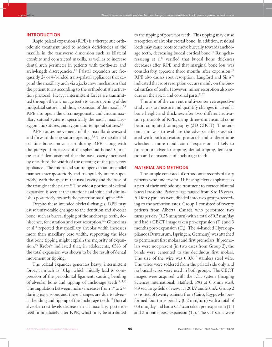
Figure 1 - Orientation in sagittal plane and in coronal plane.
taken with the multiplanar spiral CT machine (X vision EX, General Electric ‘GE’ Corporation Medical Systems Company, New York) at 0.4 mm voxel, 25 cm FOV, 120 kV, and 20 mA, with scanning time of 2 s/section. Expansion in both groups was completed once the max-illary palatal cusps occluded with the mandibular buccal cusps. The average activation time was two weeks. The appliance was left in situ as a passive retainer for three months and then was removed. The digital images were measured using the Dolphin Imaging software v. 11.7 Premium (Dolphin Imaging, Chatsworth, CA). The study was approved by the Institutional Review Board (IRB #1406256293) of Indiana University–Purdue Uni-versity Indianapolis (IUPUI) and written informed con-sent was obtained from all subjects.
Each image was oriented from the sagittal view with the coronal plane passing through the long axis of each tooth, and from the coronal view with the axial plane passing through the lower border of orbital rims and the mid-sagittal plane aligned with the skeletal midline (Fig 1). Coronal slices were used to measure the amount of skeletal and dental expansion, angulation of teeth, buccal bone width and alveolar height. Each CBCT measurement for each tooth was made on standardized slices created parallel to the long axis of the tooth (Fig 1). Measurements were performed using measurement tool
in Dolphin Imaging (Figs 2-4 and Table 1). Measure-ments for the maxillary first molars, first premolars and canines were recorded at the level of CEJ, mid-root and apexes. Maxillary base width and maxillary alveolar width were measured on the coronal sections. Measure-ments of inter-molar, inter-premolar and inter-canine widths were measured on the axial plane. Incidence of fenestrations and dehiscence was verified by means of radiographic examination.
Statistical analysisAll parameters were measured twice by the same ex-
aminer one week apart, to assess intrarater repeatability, which was evaluated using summary statistics for the differences between the repeated measurements, intra-class correlation coefficients (ICCs), and Bland-Altman plots. The two groups were compared for differences in pre-treatment measurements using one-way ANO-VA. The groups were then compared for differences in the post-treatment measurements and measurement changes, using analysis of covariance on the ranks of the data, with the pre-treatment measurements used as the covariants. Significance of the changes in the measure-ments from pre- to post-treatment was evaluated using a Wilcoxon Signed-Rank test separately for each group adopting p ≤ 0.05 as significant.
© 2017 Dental Press Journal of Orthodontics Dental Press J Orthod. 2017 Jan-Feb;22(1):89-9792
Three dimensional evaluation of alveolar bone changes in response to different rapid palatal expansion activation ratesoriginal article
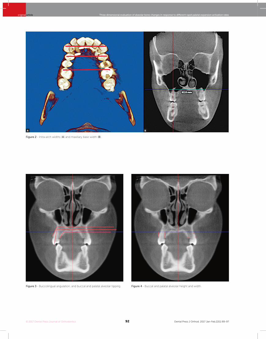
Figure 2 - Intra-arch widths (A) and maxillary base width (B).
Figure 3 - Buccolingual angulation, and buccal and palatal alveolar tipping. Figure 4 - Buccal and palatal alveolar height and width.
A B
© 2017 Dental Press Journal of Orthodontics Dental Press J Orthod. 2017 Jan-Feb;22(1):89-9793
original articleLaBlonde B, Vich ML, Edwards P, Kula K, Ghoneima A
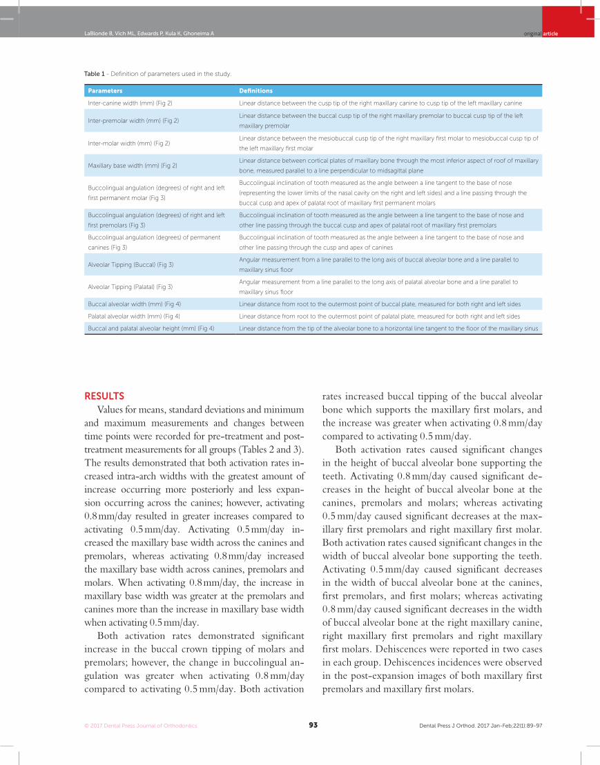
Parameters Definitions
Inter-canine width (mm) (Fig 2) Linear distance between the cusp tip of the right maxillary canine to cusp tip of the left maxillary canine
Inter-premolar width (mm) (Fig 2)Linear distance between the buccal cusp tip of the right maxillary premolar to buccal cusp tip of the left
maxillary premolar
Inter-molar width (mm) (Fig 2)Linear distance between the mesiobuccal cusp tip of the right maxillary first molar to mesiobuccal cusp tip of
the left maxillary first molar
Maxillary base width (mm) (Fig 2)Linear distance between cortical plates of maxillary bone through the most inferior aspect of roof of maxillary
bone, measured parallel to a line perpendicular to midsagittal plane
Buccolingual angulation (degrees) of right and left
first permanent molar (Fig 3)
Buccolingual inclination of tooth measured as the angle between a line tangent to the base of nose
(representing the lower limits of the nasal cavity on the right and left sides) and a line passing through the
buccal cusp and apex of palatal root of maxillary first permanent molars
Buccolingual angulation (degrees) of right and left
first premolars (Fig 3)
Buccolingual inclination of tooth measured as the angle between a line tangent to the base of nose and
other line passing through the buccal cusp and apex of palatal root of maxillary first premolars
Buccolingual angulation (degrees) of permanent
canines (Fig 3)
Buccolingual inclination of tooth measured as the angle between a line tangent to the base of nose and
other line passing through the cusp and apex of canines
Alveolar Tipping (Buccal) (Fig 3)Angular measurement from a line parallel to the long axis of buccal alveolar bone and a line parallel to
maxillary sinus floor
Alveolar Tipping (Palatal) (Fig 3)Angular measurement from a line parallel to the long axis of palatal alveolar bone and a line parallel to
maxillary sinus floor
Buccal alveolar width (mm) (Fig 4) Linear distance from root to the outermost point of buccal plate, measured for both right and left sides
Palatal alveolar width (mm) (Fig 4) Linear distance from root to the outermost point of palatal plate, measured for both right and left sides
Buccal and palatal alveolar height (mm) (Fig 4) Linear distance from the tip of the alveolar bone to a horizontal line tangent to the floor of the maxillary sinus
Table 1 - Definition of parameters used in the study.
RESULTSValues for means, standard deviations and minimum
and maximum measurements and changes between time points were recorded for pre-treatment and post-treatment measurements for all groups (Tables 2 and 3). The results demonstrated that both activation rates in-creased intra-arch widths with the greatest amount of increase occurring more posteriorly and less expan-sion occurring across the canines; however, activating 0.8 mm/day resulted in greater increases compared to activating 0.5 mm/day. Activating 0.5 mm/day in-creased the maxillary base width across the canines and premolars, whereas activating 0.8 mm/day increased the maxillary base width across canines, premolars and molars. When activating 0.8 mm/day, the increase in maxillary base width was greater at the premolars and canines more than the increase in maxillary base width when activating 0.5 mm/day.
Both activation rates demonstrated significant increase in the buccal crown tipping of molars and premolars; however, the change in buccolingual an-gulation was greater when activating 0.8 mm/day compared to activating 0.5 mm/day. Both activation
rates increased buccal tipping of the buccal alveolar bone which supports the maxillary first molars, and the increase was greater when activating 0.8 mm/day compared to activating 0.5 mm/day.
Both activation rates caused significant changes in the height of buccal alveolar bone supporting the teeth. Activating 0.8 mm/day caused significant de-creases in the height of buccal alveolar bone at the canines, premolars and molars; whereas activating 0.5 mm/day caused significant decreases at the max-illary first premolars and right maxillary first molar. Both activation rates caused significant changes in the width of buccal alveolar bone supporting the teeth. Activating 0.5 mm/day caused significant decreases in the width of buccal alveolar bone at the canines, first premolars, and first molars; whereas activating 0.8 mm/day caused significant decreases in the width of buccal alveolar bone at the right maxillary canine, right maxillary first premolars and right maxillary first molars. Dehiscences were reported in two cases in each group. Dehiscences incidences were observed in the post-expansion images of both maxillary first premolars and maxillary first molars.
© 2017 Dental Press Journal of Orthodontics Dental Press J Orthod. 2017 Jan-Feb;22(1):89-9794
Three dimensional evaluation of alveolar bone changes in response to different rapid palatal expansion activation ratesoriginal article
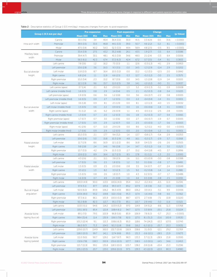
Table 2 - Descriptive statistics of Group 1 (0.5 mm/day): measures changes from pre- to post-expansion.
Group 1 (0.5 mm per day)Pre-expansion Post-expansion Change
(p-Value)Mean (SD) Min Max Mean (SD) Min Max Mean (SD) Min Max
Intra-arch width
Canine 33.1 (7.6) 3.4 40.4 36.4 (3.0) 30.0 41.5 3.3 (6.9) 0.0 31.6 < 0.0001
Premolar 39.5 (3.5) 33.5 46.8 44.1 (3.3) 39.0 50.7 4.6 (2.2) 0.2 8.1 < 0.0001
Molar 47.5 (3.4) 40.2 54.3 52.3 (3.3) 44.4 58.9 4.8 (2.5) -0.5 8.5 < 0.0001
Maxillary base
width
Canine 33.4 (3.9) 27.5 43.2 35.2 (4.8) 26.1 43.5 1.9 (2.7) -3.5 6.4 0.0086
Premolar 39.4 (4.7) 30.1 48.1 41.5 (3.9) 34.6 48.0 2.1 (2.4) -3.3 6.4 0.0006
Molar 56.5 (6.1) 42.3 67.4 57.2 (6.3) 42.4 67.2 0.7 (3.5) -5.4 9.1 0.3833
Buccal alveolar
height
Left canine 7.8 (3.6) 1.2 16.2 7.5 (3.3) 1.1 12.6 -0.3 (1.3) -4.1 2.9 0.0953
Left premolar 10.6 (2.8) 3.6 16.0 9.3 (3.5) 0.0 14.5 -1.3 (2.9) -11.4 2.9 0.0202
Left molar 11.8 (2.0) 8.7 16.4 10.5 (3.2) 0.0 15.2 -1.2 (3.1) -12.5 2.0 0.0842
Right canine 6.8 (2.4) 1.1 11.9 6.6 (2.5) 0.3 12.7 -0.2 (1.2) -3.0 2.5 0.3575
Right premolar 10.2 (3.4) 2.3 15.2 8.7 (3.9) 0.0 14.5 -1.5 (2.8) -11.9 1.4 0.0105
Right molar 12.1 (1.9) 9.6 15.7 11.0 (2.5) 3.8 14.1 -1.0 (2.2) -9.0 3.1 0.0037
Buccal alveolar
width
Left canine (apex) 3.7 (1.4) 2.1 8.2 2.9 (1.0) 1.3 5.2 -0.9 (1.3) -5.1 0.9 0.0008
Left canine (middle third) 1.6 (0.5) 0.8 2.9 1.4 (0.4) 0.5 2.1 -0.2 (0.3) -0.8 0.4 0.0225
Left premolar (apex) 1.9 (0.9) 0.6 3.6 1.2 (0.8) 0.0 3.6 -0.6 (0.7) -2.2 0.8 0.0005
Left premolar (middle third) 1.6 (0.5) 0.9 2.7 1.2 (0.5) 0.0 2.2 -0.4 (0.4) -1.4 0.1 0.0001
Left molar (apex) 3.6 (1.8) 0.9 8.1 2.5 (1.9) 0.0 8.1 -1.0 (1.3) -4.0 1.5 0.0032
Left molar (middle third) 1.5 (0.5) 0.6 2.2 0.9 (0.5) 0.0 1.6 -0.6 (0.6) -1.8 0.1 0.0001
Right canine (apex) 3.1 (1.2) 1.6 6.2 2.6 (0.9) 1.1 4.5 -0.5 (1.1) -2.5 2.8 0.0191
Right canine (middle third) 1.3 (0.4) 0.7 2.0 1.2 (0.3) 0.6 1.8 -0.2 (0.3) -0.7 0.4 0.0065
Right premolar (apex) 1.7 (0.7) 0.7 3.0 1.1 (0.6) 0.0 2.7 -0.6 (0.7) -1.9 0.7 0.0005
Right premolar (middle third) 1.7 (0.5) 0.9 2.9 1.2 (0.7) 0.0 2.5 -0.5 (0.5) -1.7 0.6 0.0003
Right molar (apex) 4.1 (2.6) 0.7 11.0 2.9 (1.9) 0.0 8.6 -1.2 (1.1) -4.0 0.0 < 0.0001
Right molar (middle third) 1.7 (0.6) 0.9 2.9 1.2 (0.5) 0.0 2.0 -0.5 (0.4) -1.2 0.1 0.0001
Palatal alveolar
height
Left canine 10.2 (3.5) 2.1 17.7 9.4 (3.2) 1.4 13.7 -0.8 (1.7) -5.4 2.9 0.0353
Left premolar 10.6 (2.6) 5.0 15.8 10.3 (2.9) 4.8 16.1 -0.2 (1.7) -2.9 5.7 0.1918
Left molar 11.7 (1.9) 8.6 16.9 12.1 (2.2) 8.6 16.8 0.4 (1.5) -2.6 2.6 0.2505
Right canine 9.8 (2.8) 2.4 14.6 9.6 (2.8) 1.4 14.7 -0.2 (1.2) -1.6 3.0 0.2123
Right premolar 10.7 (3.5) 4.2 17.4 10.3 (3.3) 2.7 15.2 -0.4 (1.8) -4.5 3.7 0.2896
Right molar 12.6 (2.1) 8.9 16.4 12.7 (2.5) 8.5 17.8 0.1 (1.1) -1.3 2.0 1.0000
Palatal alveolar
width
Left canine 4.3 (2.6) 2.1 11.1 3.8 (2.5) 1.6 11.1 -0.5 (0.9) -3.0 0.8 0.0388
Left premolar 1.7 (0.5) 0.6 2.5 1.8 (0.5) 1.2 3.0 0.1 (0.6) -0.8 1.7 0.9491
Left molar 1.5 (0.5) 0.9 2.5 2.0 (0.6) 0.8 3.5 0.4 (0.7) -0.7 2.4 0.0049
Right canine 3.5 (2.1) 1.3 8.2 3.3 (2.3) 1.5 9.2 -0.2 (0.8) -1.4 1.4 0.2881
Right premolar 1.5 (0.5) 0.8 2.6 1.8 (0.7) 1.0 4.3 0.2 (0.5) -0.7 1.7 0.0486
Right molar 1.6 (0.9) 0.7 4.9 2.1 (0.8) 1.0 4.4 0.5 (0.8) -0.8 2.5 0.0091
Buccal-lingual
angulation
Left canine 100.9 (4.9) 90.0 113.0 100.6 (4.0) 95.4 111.2 -0.2 (4.1) -6.5 11.0 0.2341
Left premolar 97.4 (5.1) 87.7 105.6 99.3 (4.7) 89.2 107.9 1.8 (3.8) -5.0 10.3 0.0336
Left molar 92.6 (6.0) 83.9 106.2 96.5 (4.9) 88.0 106.2 3.9 (4.1) -5.1 9.9 0.0006
Right canine 103.6 (8.0) 93.2 119.8 102.1 (5.6) 95.3 117.4 -1.6 (4.6) -9.8 6.6 0.1562
Right premolar 97.3 (6.1) 86.1 105.9 100.0 (5.0) 88.7 107.5 2.7 (4.8) -7.1 10.8 0.0220
Right molar 92.2 (8.8) 82.3 113.7 95.1 (7.3) 85.1 110.7 2.9 (4.6) -5.0 11.6 0.0121
Alveolar bone
tipping (buccal)
Left canine 103.5 (5.1) 94.6 114.2 103.9 (5.2) 97.0 114.9 0.4 (5.2) -8.6 11.3 0.5768
Left premolar 105.1 (8.5) 85.4 121.8 108.4 (6.1) 94.7 117.5 3.3 (8.9) -20.1 25.8 0.0224
Left molar 89.1 (7.0) 79.5 103.9 96.8 (5.6) 85.9 106.9 7.8 (6.3) -5.7 25.3 < 0.0001
Right canine 98.4 (31.4) 11.4 130.9 106.5 (7.8) 92.0 127.0 8.1 (31.2) -21.6 100.9 0.9530
Right premolar 101.2 (22.4) 9.1 115.3 108.6 (6.3) 95.0 118.6 7.4 (24.2) -10.3 107.0 0.0743
Right molar 86.0 (9.7) 71.3 104.5 92.2 (11.8) 59.8 116.2 6.2 (10.1) -25.7 20.3 0.0031
Alveolar bone
tipping (palatal)
Left canine 129.6 (10.7) 114.9 160.0 132.7 (10.6) 116.9 158.6 3.1 (9.3) -12.1 29.2 0.1769
Left premolar 118.0 (10.5) 99.7 141.1 117.4 (8.8) 95.5 131.3 -0.6 (10.1) -28.5 21.9 0.9273
Left molar 111.0 (9.0) 90.7 129.2 114.7 (9.7) 98.3 133.3 3.7 (11.3) -28.9 21.8 0.0595
Right canine 132.6 (7.8) 118.3 150.9 131.6 (10.5) 107.7 158.5 -1.0 (10.1) -14.5 34.6 0.2453
Right premolar 115.7 (11.9) 99.1 135.8 118.5 (10.0) 105.7 138.2 2.8 (11.8) -22.4 21.3 0.2196
Right molar 105.1 (23.5) 20.7 133.0 109.6 (10.0) 97.5 139.3 4.5 (22.0) -22.0 80.3 0.4980
© 2017 Dental Press Journal of Orthodontics Dental Press J Orthod. 2017 Jan-Feb;22(1):89-9795
original articleLaBlonde B, Vich ML, Edwards P, Kula K, Ghoneima A
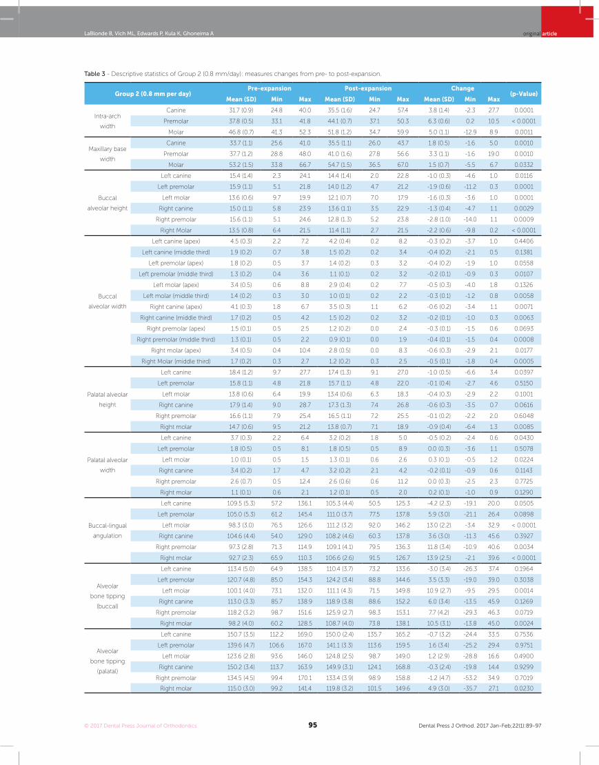
Table 3 - Descriptive statistics of Group 2 (0.8 mm/day): measures changes from pre- to post-expansion.
Group 2 (0.8 mm per day) Pre-expansion Post-expansion Change
(p-Value)Mean (SD) Min Max Mean (SD) Min Max Mean (SD) Min Max
Intra-arch
width
Canine 31.7 (0.9) 24.8 40.0 35.5 (1.6) 24.7 57.4 3.8 (1.4) -2.3 27.7 0.0001
Premolar 37.8 (0.5) 33.1 41.8 44.1 (0.7) 37.1 50.3 6.3 (0.6) 0.2 10.5 < 0.0001
Molar 46.8 (0.7) 41.3 52.3 51.8 (1.2) 34.7 59.9 5.0 (1.1) -12.9 8.9 0.0011
Maxillary base
width
Canine 33.7 (1.1) 25.6 41.0 35.5 (1.1) 26.0 43.7 1.8 (0.5) -1.6 5.0 0.0010
Premolar 37.7 (1.2) 28.8 48.0 41.0 (1.6) 27.8 56.6 3.3 (1.1) -1.6 19.0 0.0010
Molar 53.2 (1.5) 33.8 66.7 54.7 (1.5) 36.5 67.0 1.5 (0.7) -5.5 6.7 0.0332
Buccal
alveolar height
Left canine 15.4 (1.4) 2.3 24.1 14.4 (1.4) 2.0 22.8 -1.0 (0.3) -4.6 1.0 0.0116
Left premolar 15.9 (1.1) 5.1 21.8 14.0 (1.2) 4.7 21.2 -1.9 (0.6) -11.2 0.3 0.0001
Left molar 13.6 (0.6) 9.7 19.9 12.1 (0.7) 7.0 17.9 -1.6 (0.3) -3.6 1.0 0.0001
Right canine 15.0 (1.1) 5.8 23.9 13.6 (1.1) 3.5 22.9 -1.3 (0.4) -4.7 1.1 0.0029
Right premolar 15.6 (1.1) 5.1 24.6 12.8 (1.3) 5.2 23.8 -2.8 (1.0) -14.0 1.1 0.0009
Right Molar 13.5 (0.8) 6.4 21.5 11.4 (1.1) 2.7 21.5 -2.2 (0.6) -9.8 0.2 < 0.0001
Buccal
alveolar width
Left canine (apex) 4.5 (0.3) 2.2 7.2 4.2 (0.4) 0.2 8.2 -0.3 (0.2) -3.7 1.0 0.4406
Left canine (middle third) 1.9 (0.2) 0.7 3.8 1.5 (0.2) 0.2 3.4 -0.4 (0.2) -2.1 0.5 0.1381
Left premolar (apex) 1.8 (0.2) 0.5 3.7 1.4 (0.2) 0.3 3.2 -0.4 (0.2) -1.9 1.0 0.0558
Left premolar (middle third) 1.3 (0.2) 0.4 3.6 1.1 (0.1) 0.2 3.2 -0.2 (0.1) -0.9 0.3 0.0107
Left molar (apex) 3.4 (0.5) 0.6 8.8 2.9 (0.4) 0.2 7.7 -0.5 (0.3) -4.0 1.8 0.1326
Left molar (middle third) 1.4 (0.2) 0.3 3.0 1.0 (0.1) 0.2 2.2 -0.3 (0.1) -1.2 0.8 0.0058
Right canine (apex) 4.1 (0.3) 1.8 6.7 3.5 (0.3) 1.1 6.2 -0.6 (0.2) -3.4 1.1 0.0071
Right canine (middle third) 1.7 (0.2) 0.5 4.2 1.5 (0.2) 0.2 3.2 -0.2 (0.1) -1.0 0.3 0.0063
Right premolar (apex) 1.5 (0.1) 0.5 2.5 1.2 (0.2) 0.0 2.4 -0.3 (0.1) -1.5 0.6 0.0693
Right premolar (middle third) 1.3 (0.1) 0.5 2.2 0.9 (0.1) 0.0 1.9 -0.4 (0.1) -1.5 0.4 0.0008
Right molar (apex) 3.4 (0.5) 0.4 10.4 2.8 (0.5) 0.0 8.3 -0.6 (0.3) -2.9 2.1 0.0177
Right Molar (middle third) 1.7 (0.2) 0.3 2.7 1.2 (0.2) 0.3 2.5 -0.5 (0.1) -1.8 0.4 0.0005
Palatal alveolar
height
Left canine 18.4 (1.2) 9.7 27.7 17.4 (1.3) 9.1 27.0 -1.0 (0.5) -6.6 3.4 0.0397
Left premolar 15.8 (1.1) 4.8 21.8 15.7 (1.1) 4.8 22.0 -0.1 (0.4) -2.7 4.6 0.5150
Left molar 13.8 (0.6) 6.4 19.9 13.4 (0.6) 6.3 18.3 -0.4 (0.3) -2.9 2.2 0.1001
Right canine 17.9 (1.4) 9.0 28.7 17.3 (1.3) 7.4 26.8 -0.6 (0.3) -3.5 0.7 0.0616
Right premolar 16.6 (1.1) 7.9 25.4 16.5 (1.1) 7.2 25.5 -0.1 (0.2) -2.2 2.0 0.6048
Right molar 14.7 (0.6) 9.5 21.2 13.8 (0.7) 7.1 18.9 -0.9 (0.4) -6.4 1.3 0.0085
Palatal alveolar
width
Left canine 3.7 (0.3) 2.2 6.4 3.2 (0.2) 1.8 5.0 -0.5 (0.2) -2.4 0.6 0.0430
Left premolar 1.8 (0.5) 0.5 8.1 1.8 (0.5) 0.5 8.9 0.0 (0.3) -3.6 1.1 0.5078
Left molar 1.0 (0.1) 0.5 1.5 1.3 (0.1) 0.6 2.6 0.3 (0.1) -0.5 1.2 0.0224
Right canine 3.4 (0.2) 1.7 4.7 3.2 (0.2) 2.1 4.2 -0.2 (0.1) -0.9 0.6 0.1143
Right premolar 2.6 (0.7) 0.5 12.4 2.6 (0.6) 0.6 11.2 0.0 (0.3) -2.5 2.3 0.7725
Right molar 1.1 (0.1) 0.6 2.1 1.2 (0.1) 0.5 2.0 0.2 (0.1) -1.0 0.9 0.1290
Buccal-lingual
angulation
Left canine 109.5 (5.3) 57.2 136.1 105.3 (4.4) 50.5 125.3 -4.2 (2.3) -19.1 20.0 0.0505
Left premolar 105.0 (5.3) 61.2 145.4 111.0 (3.7) 77.5 137.8 5.9 (3.0) -21.1 26.4 0.0898
Left molar 98.3 (3.0) 76.5 126.6 111.2 (3.2) 92.0 146.2 13.0 (2.2) -3.4 32.9 < 0.0001
Right canine 104.6 (4.4) 54.0 129.0 108.2 (4.6) 60.3 137.8 3.6 (3.0) -11.3 45.6 0.3927
Right premolar 97.3 (2.8) 71.3 114.9 109.1 (4.1) 79.5 136.3 11.8 (3.4) -10.9 40.6 0.0034
Right molar 92.7 (2.3) 65.9 110.3 106.6 (2.6) 91.5 126.7 13.9 (2.5) -2.1 39.6 < 0.0001
Alveolar
bone tipping
(buccal)
Left canine 113.4 (5.0) 64.9 138.5 110.4 (3.7) 73.2 133.6 -3.0 (3.4) -26.3 37.4 0.1964
Left premolar 120.7 (4.8) 85.0 154.3 124.2 (3.4) 88.8 144.6 3.5 (3.3) -19.0 39.0 0.3038
Left molar 100.1 (4.0) 73.1 132.0 111.1 (4.3) 71.5 149.8 10.9 (2.7) -9.5 29.5 0.0014
Right canine 113.0 (3.3) 85.7 138.9 118.9 (3.8) 88.6 152.2 6.0 (3.4) -13.5 45.9 0.1269
Right premolar 118.2 (3.2) 98.7 151.6 125.9 (2.7) 98.3 153.1 7.7 (4.2) -29.3 46.3 0.0719
Right molar 98.2 (4.0) 60.2 128.5 108.7 (4.0) 73.8 138.1 10.5 (3.1) -13.8 45.0 0.0024
Alveolar
bone tipping
(palatal)
Left canine 150.7 (3.5) 112.2 169.0 150.0 (2.4) 135.7 165.2 -0.7 (3.2) -24.4 33.5 0.7536
Left premolar 139.6 (4.7) 106.6 167.0 141.1 (3.3) 113.6 159.5 1.6 (3.4) -25.2 29.4 0.9751
Left molar 123.6 (2.8) 93.6 146.0 124.8 (2.5) 98.7 149.0 1.2 (2.9) -28.8 16.6 0.4900
Right canine 150.2 (3.4) 113.7 163.9 149.9 (3.1) 124.1 168.8 -0.3 (2.4) -19.8 14.4 0.9299
Right premolar 134.5 (4.5) 99.4 170.1 133.4 (3.9) 98.9 158.8 -1.2 (4.7) -53.2 34.9 0.7019
Right molar 115.0 (3.0) 99.2 141.4 119.8 (3.2) 101.5 149.6 4.9 (3.0) -35.7 27.1 0.0230
© 2017 Dental Press Journal of Orthodontics Dental Press J Orthod. 2017 Jan-Feb;22(1):89-9796
Three dimensional evaluation of alveolar bone changes in response to different rapid palatal expansion activation ratesoriginal article
DISCUSSIONMaxillary expansion has been advocated as the
preferred method for the correction of maxillary arch constriction and for correcting disharmonies between the maxillary and mandibular arches.13,23,24 There is lack of literature describing the changes in buccal bone and potential root resorption due to different rates of activation of RPE that are commonly used in the practice of orthodontics. Faster activation rate is expected to cause more decrease in alveolar bone width and greater incidence of adverse effects than a slower activation rate, possibly because the bone can-not adapt to the heavier forces generated by faster ac-tivation rates of RPE. The present study investigated the changes in alveolar bone height and thickness as well as the adverse effects such as amount of alveolar tipping, dental tipping, fenestration and dehiscence of anchorage teeth associated with using two different RPE activation protocols.
Conventional radiographs, such as cephalometric and panoramic radiographs, are not appropriate for examining buccal bone or periodontal changes after RPE.25 Such radiographs are merely two-dimen-sional representations of three-dimensional struc-tures and do not allow the orthodontist to evaluate and measure changes in buccal bone.25 These radio-graphs have other limitations regarding the superim-position of anatomic structures and difficulty in re-producing angles over time.26 Moreover, the resorp-tion of the buccal plate cannot be distinguished from lingual defects.27 With the development of CBCT, it is now possible to objectively measure skeletal and dental changes in all three dimensions and without superimposition of the neighboring structures.3,25,28
Recent advancements in CBCT technology have also allowed the method to be more affordable for the dental office and to be safer for the patient due to decreased exposure to ionizing radiation.
The results of this study demonstrated that an ac-tivation rate of 0.5 mm/day is effective in increasing intra-arch widths. The activation rate of 0.5 mm/day resulted in an increase in intra-arch widths that are approximately three times greater than the in-crease in maxillary base width, consistent with the findings from other reports.7,13,14 The activation rate of 0.8 mm/day was more effective in increasing in-tra-arch widths compared to activating 0.5 mm/day,
and the increase in intra-arch widths was still ap-proximately three times greater than the increase in maxillary base width. Both activation rates resulted in buccal tipping of the maxillary molars. The great-est amount of tipping occurred in the maxillary first molars. The amount of tipping increased from the anterior region to the molar region, and this was more prominent when activating 0.8 mm/day. This increased tipping associated with 0.8 mm/day activation rate may predispose to significant loss of buccal alveolar bone.
There were also incidences of dehiscence observed in the post-expansion 3D images. Both groups had two patients with incidence of dehiscence. Baysal et al3 re-ported incidence of dehiscence in their study between 2.5% and 55%, which is consistent with the findings from the present study. It may be possible to suggest that the minimal amount of buccal alveolar bone support-ing the teeth may predispose the patient to dehiscence. Clinicians should, thus, carefully assess the amount of alveolar bone supporting the teeth prior to including expansion in the treatment plan for a patient.
Although the treatment outcomes of palatal expan-sion have been reported for many years, the question of which expansion protocol should be used in each case is still controversial. Several studies compared slow and rapid maxillary expansion using Quad-Helix and Hyrax appliances, respectively. They indi-cated that slow maxillary expansion has been related to greater buccal tipping of molars, more physiologic effects on sutural tissues, lower orthopedic effects and better bone formation in the intermaxillary sutures, which minimizes the amount of relapse as compared to rapid maxillary expansion.29-32 The findings of the present study showed that the amount of buccal crown tipping of molars and buccal tipping of the alveolar bone was greater when activating 0.8 mm/day com-pared to activating 0.5 mm/day. These contradictory results could be explained by the difference in force delivery system, since Quad-Helix appliance delivers lighter continuous force while Hyrax appliance deliv-ers heavy interrupted force. This indicates that the force delivery system should carefully be considered when treatment of posterior crossbites is advocated.
In conclusion, the results of this study indicated that both activation rates are effective in increasing intra-arch widths, although 0.8 mm/day was more effective.
© 2017 Dental Press Journal of Orthodontics Dental Press J Orthod. 2017 Jan-Feb;22(1):89-9797
original articleLaBlonde B, Vich ML, Edwards P, Kula K, Ghoneima A
Both activation rates caused significant decreases in the height and width of buccal alveolar bone, and signifi-cant increases in buccal tipping of maxillary first mo-lars. Both activation rates are also associated with the risk of some adverse effects such as alveolar tipping,
dental tipping and dehiscence, although the more rapid activation rates result in more dental tipping. Limitations of the current study that might limit the generability of the findings include the cross-sectional retrospective design and the sample size.
1. Bell RA. A review of maxillary expansion in relation to rate of expansion and
patient’s age. Am J Orthod. 1982 Jan;81(1):32-7.
2. Pangrazio-Kulbersh V, Jezdimir B, de Deus Haughey M, Kulbersh R, Wine P,
Kaczynski R. CBCT assessment of alveolar buccal bone level after RME. Angle
Orthod. 2013 Jan;83(1):110-6.
3. Baysal A, Uysal T, Veli I, Ozer T, Karadede I, Hekimoglu S. Evaluation of alveolar
bone loss following rapid maxillary expansion using cone-beam computed
tomography. Korean J Orthod. 2013 Apr;43(2):83-95.
4. Zimring JF, Isaacson RJ. Forces produced by rapid maxillary expansion. Forces
present during retention. Angle Orthod. 1965 July;35:178-86.
5. Starnbach H, Bayne D, Cleall J, Subtelny JD. Facioskeletal and dental changes
resulting from rapid maxillary expansion. Angle Orthod. 1966 Apr;36(2):152-64.
6. Woller JL, Kim KB, Behrents RG, Buschang PH. An assessment of the maxilla after
rapid maxillary expansion using cone beam computed tomography in growing
children. Dental Press J Orthod. 2014 Jan-Feb;19(1):26-35.
7. Wertz R, Dreskin M. Midpalatal suture opening: a normative study. Am J Orthod.
1977 Apr;71(4):367-81.
8. Smith T, Ghoneima A, Stewart K, Liu S, Eckert G, Halum S, et al. Three-
dimensional computed tomography analysis of airway volume changes
after rapid maxillary expansion. Am J Orthod Dentofacial Orthop. 2012
May;141(5):618-26.
9. Timms DJ. A study of basal movement with rapid maxillary expansion. Am J
Orthod. 1980 May;77(5):500-7.
10. Christie KF, Boucher N, Chung CH. Effects of bonded rapid palatal expansion on
the transverse dimensions of the maxilla: a cone-beam computed tomography
study. Am J Orthod Dentofacial Orthop. 2010 Apr;137(4 Suppl):S79-85.
11. Baydas B, Yavuz I, Uslu H, Dagsuyu IM, Ceylan I. Nonsurgical rapid maxillary
expansion effects on craniofacial structures in young adult females. A
bone scintigraphy study. A bone scintigraphy study. Angle Orthod. 2006
Sept;76(5):759-67.
12. Bishara SE, Staley RN. Maxillary expansion: clinical implications. Am J Orthod
Dentofacial Orthop. 1987 Jan;91(1):3-14.
13. Ghoneima A, Abdel-Fattah E, Eraso F, Fardo D, Kula K, Hartsfield J. Skeletal and
dental changes after rapid maxillary expansion: a computed tomography study.
Aust Orthod J. 2010 Nov;26(2):141-8.
14. Krebs A. Midpalatal suture expansion studies by the implant method over seven-
year period. Rep Congr Eur Orthod Soc. 1964;40:131-42.
15. Baysal A, Karadede I, Hekimoglu S, Ucar F, Ozer T, Veli I, et al. Evaluation of root
resorption following rapid maxillary expansion using cone-beam computed
tomography. Angle Orthod. 2012 May;82(3):488-94.
16. Brunetto M, Andriani JS, Ribeiro GL, Locks A, Correa M, Correa LR. Three-
dimensional assessment of buccal alveolar bone after rapid and slow maxillary
expansion: a clinical trial study. Am J Orthod Dentofacial Orthop. 2013
May;143(5):633-44.
REFERENCES
17. Hicks EP. Slow maxillary expansion. A clinical study of the skeletal versus dental
response to low-magnitude force. Am J Orthod. 1978 Feb;73(2):121-41.
18. Barber AF, Sims MR. Rapid maxillary expansion and external root resorption in
man: a scanning electron microscope study. Am J Orthod. 1981 Jun;79(6):630-
52.
19. Rungcharassaeng K, Caruso JM, Kan JY, Kim J, Taylor G. Factors affecting buccal
bone changes of maxillary posterior teeth after rapid maxillary expansion. Am J
Orthod Dentofacial Orthop. 2007 Oct;132(4):428.e1-8.
20. Langford SR, Sims MR. Root surface resorption, repair, and periodontal
attachment following rapid maxillary expansion in man. Am J Orthod. 1982
Feb;81(2):108-15.
21. Odenrick L, Karlander EL, Pierce A, Kretschmar U. Surface resorption following
two forms of rapid maxillary expansion. Eur J Orthod. 1991 Aug;13(4):264-70.
22. Erverdi N, Okar I, Kucukkeles N, Arbak S. A comparison of two different rapid
palatal expansion techniques from the point of root resorption. Am J Orthod
Dentofacial Orthop. 1994 July;106(1):47-51.
23. Handelman CS, Wang L, BeGole EA, Haas AJ. Nonsurgical rapid maxillary
expansion in adults: report on 47 cases using the Haas expander. Angle Orthod.
2000 Apr;70(2):129-44.
24. McNamara JA. Maxillary transverse deficiency. Am J Orthod Dentofacial Orthop.
2000 May;117(5):567-70.
25. Akyalcin S, Schaefer JS, English JD, Stephens CR, Winkelmann S. A cone-beam
computed tomography evaluation of buccal bone thickness following maxillary
expansion. Imaging Sci Dent. 2013 June;43(2):85-90.
26. Misch KA, Yi ES, Sarment DP. Accuracy of cone beam computed tomography for
periodontal defect measurements. J Periodontol. 2006 July;77(7):1261-6.
27. Rees TD, Biggs NL, Collings CK. Radiographic interpretation of periodontal
osseous lesions. Oral Surg Oral Med Oral Pathol. 1971 July;32(1):141-53.
28. Walker L, Enciso R, Mah J. Three-dimensional localization of maxillary canines
with cone-beam computed tomography. Am J Orthod Dentofacial Orthop.
2005 Oct;128(4):418-23.
29. Bell RA. A review of maxillary expansion in relation to rate of expansion and
patient’s age. Am J Orthod. 1982 Jan;81(1):32-37.
30. Mew J. Relapse following maxillary expansion. Am J Orthod. 1983 Jan;83(1):56-
61.
31. Rungcharassaeng K, Caruso JM, Kan JYK, Kim J, Taylor G. Factors affecting
buccal bone changes of maxillary posterior teeth after rapid maxillary expansion.
Am J Orthod Dentofacial Orthop. 2007 Oct;132: 428.e1-8.
32. Brunetto M, Andriani J, Ribeiro G, Locks A, Correa M, Correa LR. Three-
dimensional assessment of buccal alveolar bone after rapid and slow maxillary
expansion: a clinical trial study. Am J Orthod Dentofacial Orthop. 2013
May;143(5):633-44.

![Title 89 RCW - Washingtonleg.wa.gov › CodeReviser › RCWArchive › Documents › 2016... · (2016 Ed.) [Title 89 RCW—page 1] Title 89 Title 89 89 RECLAMATION, SOIL CONSERVATION,](https://img.pdfslide.us/doc/110x75/5f10d3ff7e708231d44b02f6/title-89-rcw-a-codereviser-a-rcwarchive-a-documents-a-2016-2016.jpg)