Embed Size (px)
Citation preview
University of Nebraska - LincolnDigitalCommons@University of Nebraska - Lincoln
Faculty Publications in the Biological Sciences Papers in the Biological Sciences
2012
Thoughts on Quorum Sensing and FungalDimorphismKenneth W. NickersonUniversity of Nebraska-Lincoln, [email protected]
Audrey L. AtkinUniversity of Nebraska-Lincoln, [email protected]
Jessica C. HargartenUniversity of Nebraska-Lincoln, [email protected]
Ruvini U. PathiranaUniversity of Nebraska-Lincoln, [email protected]
Sahar HasimUniversity of Nebraska-Lincoln, [email protected]
Follow this and additional works at: https://digitalcommons.unl.edu/bioscifacpub
Part of the Biochemistry Commons, and the Biology Commons
This Article is brought to you for free and open access by the Papers in the Biological Sciences at DigitalCommons@University of Nebraska - Lincoln.It has been accepted for inclusion in Faculty Publications in the Biological Sciences by an authorized administrator of DigitalCommons@University ofNebraska - Lincoln.
Nickerson, Kenneth W.; Atkin, Audrey L.; Hargarten, Jessica C.; Pathirana, Ruvini U.; and Hasim, Sahar, "Thoughts on QuorumSensing and Fungal Dimorphism" (2012). Faculty Publications in the Biological Sciences. 622.https://digitalcommons.unl.edu/bioscifacpub/622

Published as a chapter in Biocommunication of Fungi, Guenther Witzany (ed.), pp. 189–204; doi: 10.1007/978-94-007-4264-2_12 Copyright © 2012 Springer Science+Business Media, Dordrecht. Used by permission.
Thoughts on Quorum Sensing and Fungal Dimorphism Kenneth W. Nickerson, Audrey L. Atkin, Jessica C. Hargarten,
Ruvini Pathirana, and Sahar Hasim
School of Biological Sciences, University of Nebraska–Lincoln, Lincoln, Nebraska, USA Corresponding author – Kenneth W. Nickerson, email [email protected] Abstract Farnesol has been best studied for its role in regulating fungal dimorphism. However, farnesol is also a lipid and in this review we analyze data relevant to farnesol’s function and synthesis from the perspective of farnesol and bacterial endotoxins acting as membrane active compounds. This analy-sis implicates the possible roles of: (1) endotoxins in the regulation of farnesol production by C. albicans; (2) farnesol in the interactions between C. albicans and the host during disseminated infections; and (3) ubiquinones in the mechanisms for unusually high resistance to farnesol by some C. albicans cell types. Finally we discuss the implications that the use of farnesol as both a signaling molecule and to antagonize competing microbials species has for the regulation of HMG-CoA reductase, the en-zyme that is the usual rate limiting step in sterol/lipid synthesis. 1. Introduction The role of farnesol in the dimorphism of Candida albicans was discovered by Hornby et al. (2001), reviewed by Nickerson et al. (2006), and updated by Langford et al. (2009) and Ho-gan and Muhlschlegel (2011). Up to now the theme of farnesol research has been farnesol as a signaling molecule and how it affects fungal polymorphism via signal transduction (Nickerson et al. 2006). However, in the process the role of farnesol as a lipid has been somewhat neglected. Thus, the present review will focus on how the lipid nature of farne-sol contributes to its roles in virulence and pathogenicity, mating, and the interactions of C. albicans with both host macrophages and competing microbes.

N I C K E R S O N E T A L . , T H O U G H T S O N Q U O R U M S E N S I N G A N D F U N G A L D I M O R P H I S M (2 0 1 2 )
2
For the fungi, we define quorum sensing as any cell density dependent phenomenon which is mediated by an extracellular molecule which is produced and excreted by the fungus in question. The name is, of course, borrowed from the classic review by Fuqua et al. (1994) which discussed bacterial homoserine lactones. We have extended the terminol-ogy slightly by introducing quorum sensing molecules, or QSMs (Nickerson et al. 2006). Critically, our use of the term QSM does not presuppose anything about its mode of action. In particular, it could include a situation where the fungal role is restricted to modifying an exogenously provided molecule, e.g., linoleic acid to 3-hydroxy tetradecaenoic acid (Nigamet al. 2010). The key point is that fungi also have mechanisms to sense their own population densities. 2. It Must Be Something in the Water This story is both a cautionary tale and a possible area for future study on bacterial-fungal interactions. The first published report on farnesol as a quorum sensing molecule (QSM) for C. albicans (Hornby et al. 2001) was delayed for roughly 2 years by issues of water qual-ity. In 1996 the Nickerson lab moved from an old building soon to be torn down (Lyman Hall) to a new state-of-the-art research center named for University of Nebraska graduate and Nobel Prize winner George W. Beadle. At the time, we were using an activity directed purification scheme to identify the molecule in spent media which blocked germ tube for-mation in C. albicans. The assay worked perfectly in Lyman Hall but did not work in our new facilities. We eventually found that if we purchased bottled distilled water at the local supermarket and used that water to prepare our growth media, then the assay worked perfectly again.
What was different about the distilled waters provided in the two buildings? And did the building distilled/deionized water in Beadle prevent the QSM (farnesol) from being formed or inactivate it after it had been formed? In this regard, we know that farnesol is markedly sensitive to air oxidation and, consequently, we always store our farnesol stock solutions under nitrogen. Shchepin et al. (2003) showed that the 10, 11 epoxide of farnesol has only 3% of farnesol’s QSM activity, and at the time of our move the city of Lincoln had just switched from chlorine to ozone treatment as the penultimate step in its water treat-ment procedures. Could there be any residual ozone carried over? However, this possibil-ity was eliminated by mixing the two spent media, one with QSM activity and one without, and observing that the resulting QSM activity was undiminished.
Thus, we were left with discovering what difference in the two waters regulated QSM/ farnesol production by C. albicans. This question has not yet been fully resolved. A chemical analysis of the respective waters was indicated. There were two precedents. First, waters can differ dramatically in their mineral contents. Consider, for instance, Table 12.1 of Okafor’s text on Industrial Microbiology (Okafor 2007) which compares the mineral content of wa-ter in eight cities noted for their breweries. The concentrations of Ca2+, Mg2+, SO42–, NO3–, Cl–, and HCO3– in the respective waters differed by 44-, 62-, 212-, 62-, 120-, and 31-fold, respectively. Second, Cu2+ and Zn2+ cause morphological shifts in most dimorphic fungi, i.e., Ceratocystis ulmi, Histoplasma capsulatum, Mucor rouxii, Sporothrix schenkii, Ustilago sphaerogena, and C. albicans. The Zn2+ induced shifts are all in the same direction (mycelia
N I C K E R S O N E T A L . , T H O U G H T S O N Q U O R U M S E N S I N G A N D F U N G A L D I M O R P H I S M (2 0 1 2 )
3
to yeasts) but the concentrations of Zn2+ found to be effective varied. C. ulmi was typical in that it required 4–5 mM Zn2+, whereas C. albicans required only 10–20 mM Zn2+ (Yamaguchi 1975; Soll et al. 1981).
Accordingly, we obtained eight types of distilled water locally available in Lincoln and in central Minnesota. Five of the eight permitted QSM accumulation, three did not. These eight water samples were analyzed by atomic absorption for 11 elements including Se and most of the transition metals. At that time ICP-MS was not available to us for the detailed analysis of the elemental composition of the waters. These analyses produced some sur-prises, such as two of the waters having 50-fold more Co2+ than the others, but there was no discernible correlation between elemental composition and QSM formation! We were stumped. At this point we decided to publish our data showing that farnesol was the QSM for C. albicans without having resolved the water issue. Thus, the Hornby et al. (2001) paper specifies that the GPP growth medium was always prepared with Kandiyohi distilled wa-ter (Kandiyohi Bottled Water Co., Willmar, Minnesota). The Minnesota connection re-flected the participation of visiting scientist Professor Ellen Jensen from the College of St. Benedict’s and St. John’s University in Minnesota.
Some years later we learned that our standard water purification system (running dis-tilled water through deionizing columns and activated charcoal columns followed by 0.22 mm filtration) might not remove bacterial endotoxins. We had not considered this possi-bility previously. Accordingly, we purchased an endotoxin detection kit and sampled the distilled/deionized water from three locations in the Beadle Center. They each contained 16–32 endotoxin units per ml. The Kandiyohi distilled water had no endotoxins, and Crys-tal Glen distilled water had 1 endotoxin unit per ml. The other commercial distilled waters were no longer available. Samples of the Beadle distilled water taken prior to filtration contained 10–100 CFU/ml when plated on LB agar. As expected, these bacteria were Gram negative, presumably Pseudomonads. Our current scenario is that: rain water contains ca. 8 mg/L organics, Pseudomonads are notorious for the wide variety of organics they can me-tabolize (Stanier et al. 1966), and it is expected that large volumes of standing water with long residence times will experience some bacterial presence (McCoy 1980). Lyman Hall did not experience these problems because the distilled water had been obtained from con-densed steam and our current supplier Super-Saver has a very short residence time.
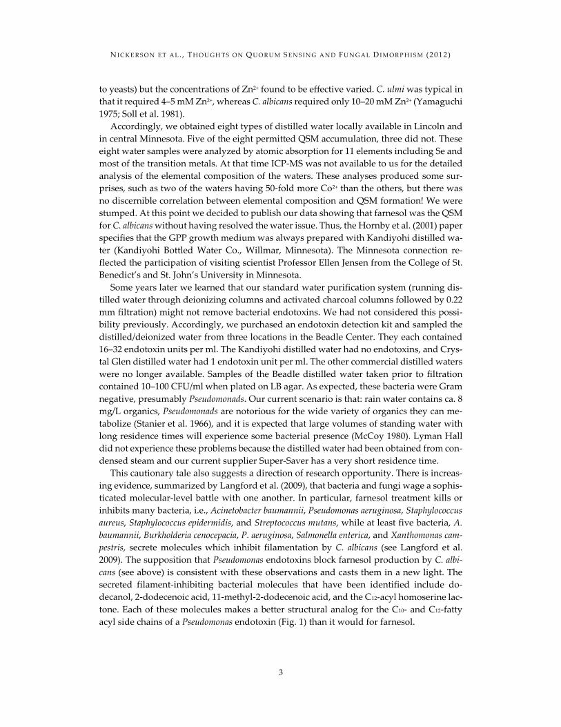
This cautionary tale also suggests a direction of research opportunity. There is increas-ing evidence, summarized by Langford et al. (2009), that bacteria and fungi wage a sophis-ticated molecular-level battle with one another. In particular, farnesol treatment kills or inhibits many bacteria, i.e., Acinetobacter baumannii, Pseudomonas aeruginosa, Staphylococcus aureus, Staphylococcus epidermidis, and Streptococcus mutans, while at least five bacteria, A. baumannii, Burkholderia cenocepacia, P. aeruginosa, Salmonella enterica, and Xanthomonas cam-pestris, secrete molecules which inhibit filamentation by C. albicans (see Langford et al. 2009). The supposition that Pseudomonas endotoxins block farnesol production by C. albi-cans (see above) is consistent with these observations and casts them in a new light. The secreted filament-inhibiting bacterial molecules that have been identified include do-decanol, 2-dodecenoic acid, 11-methyl-2-dodecenoic acid, and the C12-acyl homoserine lac-tone. Each of these molecules makes a better structural analog for the C10- and C12-fatty acyl side chains of a Pseudomonas endotoxin (Fig. 1) than it would for farnesol.
N I C K E R S O N E T A L . , T H O U G H T S O N Q U O R U M S E N S I N G A N D F U N G A L D I M O R P H I S M (2 0 1 2 )
4
Figure 1. Pseudomonas aeruginosa endotoxin structure (p. 245 of Wang and Quinn 2010)
With regard to location, C. albicans is most likely to encounter bacterial endotoxins in
the anaerobic mammalian guts. Thus, it is entirely consistent that under anaerobic growth conditions C. albicans does not produce detectable farnesol or respond to added farnesol (Dumitru et al. 2004). It would be of interest to compare the effects of bacterial endotoxins on C. albicans growing under both aerobic and anaerobic conditions. 3. A Potential Role for Farnesol in C. albicans Host Interactions 3.1 Plasticity of Morphology: A Virulence Factor Biologically speaking, the interactions of C. albicans and the human host are much like a never-ending game of cat-and-mouse: the human host laying down defense mechanisms to keep the fungus in check, and the fungus breaking these walls down for pure survival. Understanding this interplay is important as scientists seek to tip the balance away from the pathogenic C. albicans. Throughout the body, C. albicans has adapted mechanisms to gather, interpret, and respond to signals provided by the host and the diverse terrain the host environment poses, much of which is still unknown. One of the key responses is simply changing its morphology. In vivo, different morphologies of C. albicans have been associated with distinct degrees and locations of infection. From the benign colonization of the skin typically by white or opaque phase cells to benign infections of the oral cavity by white phase cells, chronic vaginal infections by white phase yeast cells, the hyphal and pseudohyphal growth found in the gastrointestinal tract and in many disseminated sys-temic infections, this morphological plasticity appears to act as a tolerance mechanism to counteract the changes in pH, nutrient availability, microflora composition, and oxygen
N I C K E R S O N E T A L . , T H O U G H T S O N Q U O R U M S E N S I N G A N D F U N G A L D I M O R P H I S M (2 0 1 2 )
5
levels the fungus will encounter on and within its human host (Lachke et al. 2003; Sobel 1997; Sudbery 2011).
C. albicans easily colonizes many locations around an immune-competent host without doing much harm to the individual but, in an immunocompromised host, the capability to switch between morphologies acts as a virulence factor and is central to its pathogenicity. However all forms of C. albicans do not convey this level of virulence. Some forms are more susceptible to macrophages and the other defenses of the human immune system than others. This difference in morphological susceptibility could be an important opportunity for the development of antifungal drugs that block systemic Candidiasis. 3.2 Farnesol and Virulence The secretion of farnesol also plays a role in host-pathogen interactions in vivo. Hornby et al. (2001) discovered that in vitro farnesol acted to block the yeast-to-mycelia transition. Thus, in vitro farnesol acted as a QSM. But what would its role be in vivo? At that time we presented two possibilities. Firstly, if farnesol acted in vivo as it did in vitro, then farnesol and its analogs should prove to be effective antifungals because the yeast-to-mycelia tran-sition is essential for virulence. It was on this premise that 50 analogs of farnesol were synthesized and tested for their in vitro QSM potency (Shchepin et al. 2005). However, we also suggested that farnesol production by C. albicans might instead function as a virulence factor (Hornby et al. 2001), and for the mouse-tail–vein injection model that proved to be the case (Navarathna et al. 2007). C. albicans mutants that produced 85% less farnesol were five times less pathogenic to mice than their parent cells. Also, when farnesol was admin-istered orally to the mice prior to infection, their mortality increased, as did the coloniza-tion of kidneys (Navarathna et al. 2007).
These observations pose the dilemma of finding a mechanism whereby a molecule which blocks the yeast-to-mycelia transition can also act as a virulence factor. In this re-gard, we note that there is as yet no evidence for farnesol blocking the yeast-to-mycelia transition in vivo while there is evidence that farnesol behaves differently for surface in-fections, where it is protective (Hisajima et al. 2008), versus systemic infections, where it is a virulence factor (Navarathna et al. 2007). A partial resolution of this dilemma comes from the realization that in vitro, in glass or plastic, excreted farnesol can accumulate, whereas in vivo it would be soaked up by the mammalian cell membranes. Thus, different concen-trations of farnesol should be present in vitro and in vivo. 3.3 A C. albicans Macrophage Chemoattractant: White Versus Opaque Another partial resolution of this in vitro vs. in vivo dilemma concerns how farnesol affects the host innate immune system. The first level of defense the host has against candidal infection is through the innate immune system. Distinct morphologies elicit different re-sponses by the host immune system. Both white and opaque cells are known to attract leukocytes to the site of infection, but only white cells produce and secrete a small molec-ular weight chemoattractant that draws the leukocyte directly toward the white cell (Gei-ger et al. 2004). Lohse and Johnson (2008) took this knowledge a step further by showing that not only were leukocytes more attracted to white cells than opaque cells, but because
N I C K E R S O N E T A L . , T H O U G H T S O N Q U O R U M S E N S I N G A N D F U N G A L D I M O R P H I S M (2 0 1 2 )
6
of the presence of a chemoattractant produced by the white cells, mouse macrophages en-gulfed white C. albicans cells much more efficiently than they did opaque cells (Lohse and Johnson 2008). Not only were the white cells engulfed at a higher rate but they were also less susceptible to killing by human macrophages and neutrophils than were opaque cells, possibly due to their increased capabilities of escape once phagocytosed or possibly due to another effect of the chemoattractant on the macrophages (Kolotila and Diamond 1990). The chemical identity of this chemoattractant is currently unknown, but the reason behind its continued secretion by the white form is intriguing. One likely candidate is farnesol (Langford et al. 2009). Macrophages are capable of detecting and responding to exogenous farnesol, specifically by stimulating secretion of proinflammatory and regulatory cyto-kines (IL-6, IL-1β, IL-10, and TNF-α; Ghosh et al. 2010). The production of these warning signals by macrophages is an important indicator of how the body ultimately hopes to clear the infection. Because of the cytotoxic effects farnesol has on macrophages (ROS and DNA fragmentation), farnesol suppresses the anti-Candida activity of macrophages (Abe et al. 2009), thus making it all the more difficult to eliminate the fungus early in infection.
This chain of events from attraction to engulfment to eventual killing of the macro-phages is mediated at two points by different C. albicans morphologies. It is known that wild type, white cells of C. albicans can escape from mouse macrophages by switching to the hyphal morphology 6–8 h post-engulfment and effectively puncturing the macrophage from within (Ghosh et al. 2009). Those strains with delayed or dysfunctional hyphal for-mation (through disruption of the arginine biosynthetic pathway for instance) were unable to survive within and escape from the macrophage (Ghosh et al. 2009). It remains a per-plexing mystery why certain morphologies such as the opaque cells are better able to elude host immune defenses or retaliate, such as the hyphae, while other morphologies such as the white yeast cells seek to be found through the production of a potent chemoattractant. This aspect of farnesol production by C. albicans is in part counterintuitive of the way we think a fungus should behave but it does have precedence in the form of some pathogenic intracellular bacteria. 3.4 Bacterial Analogs for Host Evasion Direct targeting of tissue phagocytes to the site of infection by pathogenic microorganisms, in the hopes of being phagocytosed, is not a novel concept in the realm of microbial infec-tions. A comprehensive review of bacterial evasion strategies can be found in Flannagan et al. (2009). For some “professional” intracellular bacteria, such as Mycobacterium tubercu-losis, Listeria monocytogenes, and Legionella pneumophila, successful establishment of infec-tion and dissemination throughout the host depends entirely on exploiting the natural responses of phagocytes. Following phagocytosis and entry into the phagosome, these bac-teria have developed mechanisms to prevent further phagocyte killing and digestion, allowing for long-term intraphagosomal survival within host cells, either through interfering with phagolysosome maturation and the secretion of ROS and antibacterial proteases or through counteracting the host cells’ expression of MHC and loading of antigenic bacterial pep-tides, effectively eluding further host immune system detection.
The most researched of these pathogenic microbes that use macrophages to escape im-mune detection is L. monocytogenes. This model system could be used as a possible bacterial
N I C K E R S O N E T A L . , T H O U G H T S O N Q U O R U M S E N S I N G A N D F U N G A L D I M O R P H I S M (2 0 1 2 )
7
analog to understand what strategy C. albicans may utilize during infection. Phagocytosis of L. monocytogenes is mediated by a macrophage scavenger receptor that binds directly to the lipoteichoic acid on this Gram-positive bacterium (Dunne et al. 1994). Once within the phagosome of the macrophage, it uses an array of cholesterol-dependent cytolysins to pre-vent the further maturation of the phagosome by inhibiting its fusion with the lysosome. By sequestering inside the membrane vacuole, it is able to acquire the nutrients it requires to replicate directly from the host without host detection, prior to escape from the macro-phage. Through this comparison, much can be learned about possible intracellular signal-ing interactions between the phagocyte and the intracellular fungi during the phagocytosis process. We note that during the 6 h between their engulfment and escape, C. albicans can spread through the body as the macrophages migrate. 4. Evolutionary Adaptations to Farnesol as a Signaling and Antagonistic Molecule 4.1 C. albicans Resistance to the Antifungal Effects of Farnesol Regardless of their capacity to produce farnesol, many fungi respond to farnesol in that they are inhibited or killed by it, although the exact molecular mechanism of farnesol in-duced cell death is still under investigation. As examples, farnesol induced apoptosis in Aspergillus nidulans (Semighini et al. 2006), inhibited Trichophyton rubrum in co-culture with C. albicans (Jillson and Nickerson 1948), and antagonized many other fungi including S. cerevisiae (Machida et al. 1998). The exceptional resistance to farnesol shown by C. albicans is an interesting issue since it is the only known fungus which tolerates farnesol up to 300 μM while using it as a quorum-sensing molecule. What makes C. albicans different from the rest of the fungi? We assume that C. albicans has a protective mechanism to safeguard itself from excessive farnesol, similar to the self-defense mechanisms used by antibiotic-producing microorganisms to prevent them committing suicide by their own products. What’s more, this resistance of C. albicans to farnesol is not just a constant suit of armor but a subtly variable protection. Anaerobically growing cells are resistant to farnesol right up to its solubility limit of 1–1.2 mM (Dumitru et al. 2004) while resistance is lost entirely in cells which have switched from the white phase to the opaque phase (Dumitru et al. 2007).
In terms of where to look for this variable resistance, the mitochondria are a likely target. As a result of aerobic respiration, all aerobic organisms produce Reactive Oxygen Species (ROS), which leads to oxidative destruction of cells. Consequently, all of these aerobes in-cluding yeasts developed efficient mechanisms to get rid of these unwelcome companions. In a study done to reveal the growth inhibitory effect of farnesol in S. cerevisiae (Machida et al. 1998), the level of farnesol-induced ROS was found to increase in a dose-dependent manner. Further, the inhibition of growth by farnesol could be prevented by the presence of antioxidants in the medium. Thus ROS generation leading to intracellular oxidative stress suggested involvement of the mitochondrial electron transport chain as the target of farnesol. This view on the primary means of farnesol-mediated death in S. cerevisiae by generation of reactive oxygen species was further confirmed by Fairn et al. through a study on the genomic effects of the chemical compounds farnesol and geraniol (chemogenomic profiling; Fairn et al. 2007).
N I C K E R S O N E T A L . , T H O U G H T S O N Q U O R U M S E N S I N G A N D F U N G A L D I M O R P H I S M (2 0 1 2 )
8
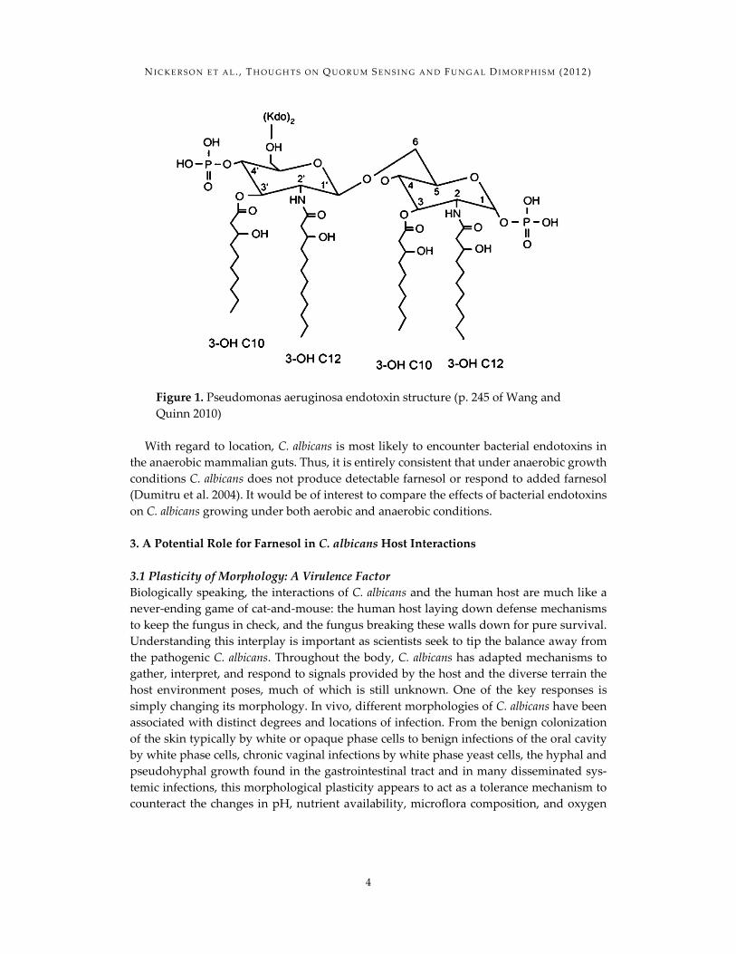
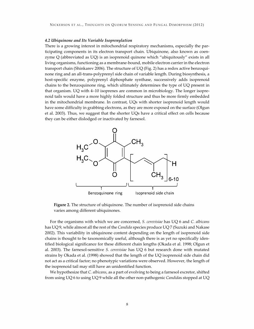
4.2 Ubiquinone and Its Variable Isoprenylation There is a growing interest in mitochondrial respiratory mechanisms, especially the par-ticipating components in its electron transport chain. Ubiquinone, also known as coen-zyme Q (abbreviated as UQ) is an isoprenoid quinone which “ubiquitously” exists in all living organisms, functioning as a membrane-bound, mobile electron carrier in the electron transport chain (Shinkarev 2006). The structure of UQ (Fig. 2) has a redox active benzoqui-none ring and an all-trans-polyprenyl side chain of variable length. During biosynthesis, a host-specific enzyme, polyprenyl diphosphate synthase, successively adds isoprenoid chains to the benzoquinone ring, which ultimately determines the type of UQ present in that organism. UQ with 4–10 isoprenes are common in microbiology. The longer isopre-noid tails would have a more highly folded structure and thus be more firmly embedded in the mitochondrial membrane. In contrast, UQs with shorter isoprenoid length would have some difficulty in grabbing electrons, as they are more exposed on the surface (Olgun et al. 2003). Thus, we suggest that the shorter UQs have a critical effect on cells because they can be either dislodged or inactivated by farnesol.
Figure 2. The structure of ubiquinone. The number of isoprenoid side chains varies among different ubiquinones.
For the organisms with which we are concerned, S. cerevisiae has UQ 6 and C. albicans
has UQ 9, while almost all the rest of the Candida species produce UQ 7 (Suzuki and Nakase 2002). This variability in ubiquinone content depending on the length of isoprenoid side chains is thought to be taxonomically useful, although there is as yet no specifically iden-tified biological significance for these different chain lengths (Okada et al. 1998; Olgun et al. 2003). The farnesol-sensitive S. cerevisiae has UQ 6 but research done with mutated strains by Okada et al. (1998) showed that the length of the UQ isoprenoid side chain did not act as a critical factor; no phenotypic variations were observed. However, the length of the isoprenoid tail may still have an unidentified function.
We hypothesize that C. albicans, as a part of evolving to being a farnesol excretor, shifted from using UQ 6 to using UQ 9 while all the other non-pathogenic Candidas stopped at UQ
N I C K E R S O N E T A L . , T H O U G H T S O N Q U O R U M S E N S I N G A N D F U N G A L D I M O R P H I S M (2 0 1 2 )
9
7. If farnesol-mediated cell death targets the ubiquinones, it is not surprising to observe the resistance of anaerobic cells of C. albicans to farnesol (Dumitru et al. 2004) since they lack mitochondrial respiration. Thus, a key question is whether there is a significant effect of UQ side chain length on farnesol sensitivity during aerobic respiration. This question could be answered by examining the farnesol sensitivity of S. cerevisiae which make UQ 9 instead of UQ 6 or C. albicans which made UQ 6/7 instead of UQ 9. 4.3 Possible Mechanisms for C. albicans White Cell Resistance to Farnesol C. albicans can grow in both aerobic and anaerobic environments and it has two types of cells: white cells and opaque cells. These cell types respond to farnesol in different ways. Neither opaque cells nor anaerobic cells make farnesol (Dumitru et al. 2004, 2007). During anaerobic growth, C. albicans doesn’t produce farnesol or respond to farnesol, even at con-centration as high as 1.2 mM (Dumitru et al. 2004). Similarly, in the presence of farnesol, white cells are prevented from making germ tubes, but they can tolerate farnesol at con-centrations up to 300 μM, while opaque cells are lysed by farnesol at 50 μM (Dumitru et al. 2007). It is desirable to understand why the white cells of C. albicans are highly resistant to farnesol whereas opaque cells are very sensitive to it.
Different phases of white cell growth also differ in their tolerance to farnesol. Farnesol shows different activities toward C. albicans depending on the growth conditions and in-oculum characteristics. In rich growth medium (YPD), C. albicans is very resistant to farne-sol (Langford et al. 2010) while in defined media such as glucose-phosphate-proline (GPP) log-phase cells were significantly more sensitive to farnesol than were stationary phase cells. Consequently, when using an inoculum of stationary-phase cells, the growth curves are similar to those in YPD media (Langford et al. 2010) while using an inoculum of log phase cells resulted in significant delays due to farnesol induced cell lysis (Langford et al. 2010). Thus, the starting growth phase, media, and cell density of the inoculum are critical for the effect of farnesol on the cells. Finally, farnesol tolerance is an energy-dependent process. Cells suspended in a farnesol buffer without an energy source lysed whereas those with an energy source did not (Langford et al. 2010). Together, the influence of cell type, growth conditions and inoculum characteristics suggest resistance to farnesol is an active and regulated process.
C. albicans may detoxify farnesol enzymatically. Using a spent medium assay for quorum sensing activity, i.e. the ability to block hypha formation, Hornby et al. (2001) found that the levels of farnesol present in the spent media roughly paralleled cell mass for 20 h after inoculation but then decreased rapidly after that. Farnesol contains three C═C double bonds and exists in four isomers of which only (E, E) farnesol has QSM activity. Farnesol is a very unstable molecule, and air oxidation results in a hydroxyl or epoxide of farnesol, which causes a dramatic decrease in QSM activity (Shchepin et al. 2003). Thus, the decrease in QSM activity observed by Hornby et al. (2001) could have been either enzymatic or spontaneous. Additionally, the morphological response to farnesol in C. albicans appears to be very sensitive to other minor changes in the structure of farnesol. For instance, farne-sol with three isoprene units has three methyl branches. If the 2-methyl branch is either removed or enlarged to a 2-ethyl branch, then the QSM activity of the resulting farnesol analog is 20-fold lower (Shchepin et al. 2005). Thus, the decreased activity of farnesol with
N I C K E R S O N E T A L . , T H O U G H T S O N Q U O R U M S E N S I N G A N D F U N G A L D I M O R P H I S M (2 0 1 2 )
10
time observed by Hornby et al. (2001) could result from the modification of farnesol to a new compound with a lower QSM activity and/or lower toxicity for the cells. 4.4 Membrane Differences in White and Opaque Cells C. albicans strain WO-1 is capable of switching at high frequencies between two pheno-types, white and opaque cells. Among the many differences between white and opaque cells, it has been shown that these cells have variations in their lipid and sterol contents (Ghannoum et al. 1990). White cells have 4-fold and 7.7-fold higher sterol contents than do opaque cells in mid-exponential and stationary phase, respectively (Ghannoum et al. 1990). Additionally, white cells contain more free sterols and less of the steryl glycoside and steryl ester sterol derivatives (Ghannoum et al. 1990). Finally, the sterols present in mid-exponential and stationary phase cultures of white cells were qualitatively different. The sterols present in mid-exponential phase cultures of white cells were primarily lanosterol (48 wt%) and 24-methylene dihydrolanosterol (26.2 wt%), while for mid-exponential phase opaque cells they were ergosterol (49.3 wt%), lanosterol (33.2 wt%), and squalene (17.5 wt%). During stationary phase, ergosterol was the major sterol in both white and opaque cells (Ghan-noum et al. 1990). These observations suggest the twin hypotheses that a high sterol con-tent in the membrane protects cells from farnesol and that the percentages of total sterols and of ergosterol in particular will differ for mid-exponential phase cells grown with and without farnesol. It also leads to a focus on the regulation of HMG CoA reductase (HMGR), the rate limiting step in sterol biosynthesis.
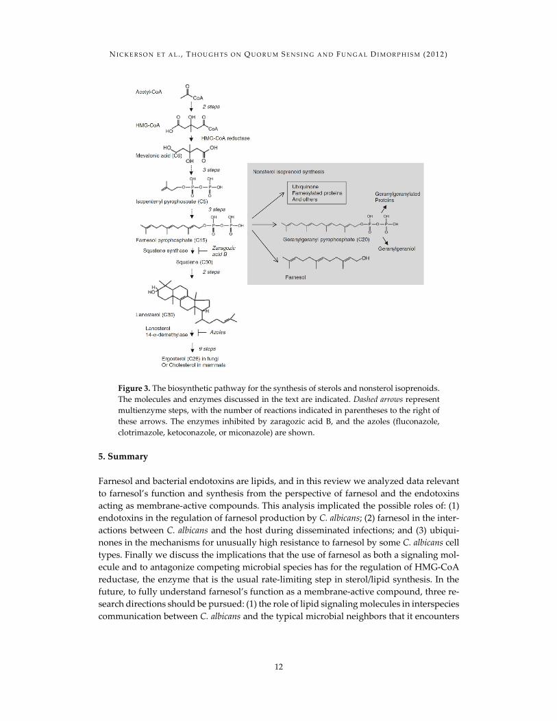
Farnesol is an isoprenoid produced by dephosphorylation of farnesyl pyrophosphate, an intermediate in the isoprenoid pathway (Hornby et al. 2003). The isoprenoid pathway is responsible for synthesis of sterols from acetyl-CoA (Fig. 3). Acetyl-CoA is converted to mevalonate by reduction with NADPH by 3-hydroxy-3-methylglutaryl-CoA (HMG-CoA) reductase (HMGR). Mevalonate leads to the synthesis of farnesyl pyrophosphate, a branch point in the sterol biosynthetic pathway. Farnesol pyrophosphate serves as a precursor for sterol biosynthesis and nonsterol isoprenoids. Sterols are an important structural compo-nent of cellular membranes. Nonsterol isoprenoids include geranylgeranyl pyrophosphate and farnesol. Farnesyl pyrophosphate and geranylgeranyl pyrophosphate are involved in the prenylation of proteins. Isoprenoids are also precursors for the prenyl side chains of ubiquinone.
HMGR is often the rate-limiting enzyme for the sterol biosynthetic pathway. Human HMGR is regulated by transcription, protein degradation, and phosphorylation (Table 1). Phosphorylation decreases the enzyme efficiency. The rapid degradation of mammalian HMGR is triggered by cellular sterols and farnesol, farnesyl pyrophosphate and geranyl-geranyl pyrophosphate (reviewed in Joo and Jetten 2010; Burg and Espenshade 2011). These signals function both in vivo and in vitro as triggers for rapid degradation. In fungi, regulation of HMGR has been studied in the yeast Saccharomyces cerevisiae and the fission yeast Schizosaccharomyces pombe. S. cerevisiae has two HMGR isozymes, Hmg1 and Hmg2. Both isozymes are subject to feedback regulation by nonsterol products of the isoprenoid pathway (Table 1). Hmg1 is the primary source of HMGR in aerobically grown cells, and in these conditions it is mainly regulated at the level of translation (Dimster-Denk et al. 1994). Hmg2, like its mammalian counterpart, is primarily regulated by protein turnover.
N I C K E R S O N E T A L . , T H O U G H T S O N Q U O R U M S E N S I N G A N D F U N G A L D I M O R P H I S M (2 0 1 2 )
11
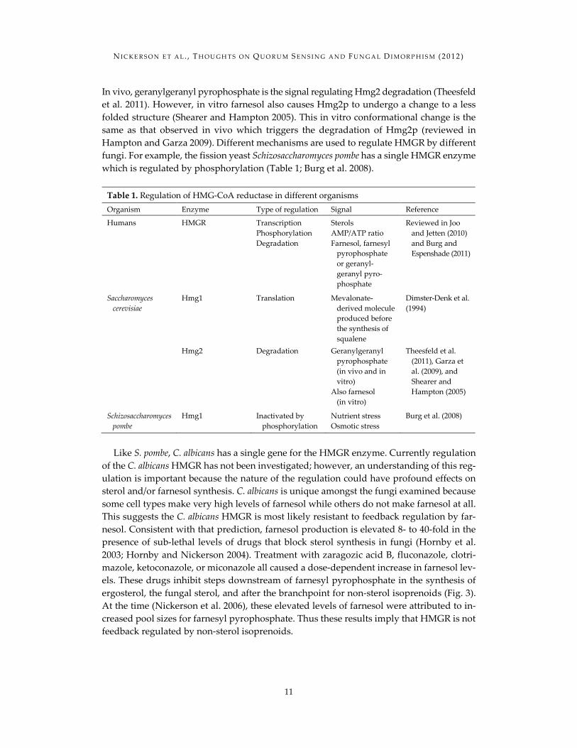
In vivo, geranylgeranyl pyrophosphate is the signal regulating Hmg2 degradation (Theesfeld et al. 2011). However, in vitro farnesol also causes Hmg2p to undergo a change to a less folded structure (Shearer and Hampton 2005). This in vitro conformational change is the same as that observed in vivo which triggers the degradation of Hmg2p (reviewed in Hampton and Garza 2009). Different mechanisms are used to regulate HMGR by different fungi. For example, the fission yeast Schizosaccharomyces pombe has a single HMGR enzyme which is regulated by phosphorylation (Table 1; Burg et al. 2008).
Table 1. Regulation of HMG-CoA reductase in different organisms Organism Enzyme Type of regulation Signal Reference
Humans HMGR Transcription Phosphorylation Degradation
Sterols AMP/ATP ratio Farnesol, farnesyl pyrophosphate or geranyl- geranyl pyro- phosphate
Reviewed in Joo and Jetten (2010) and Burg and Espenshade (2011)
Saccharomyces cerevisiae
Hmg1 Translation Mevalonate- derived molecule produced before the synthesis of squalene
Dimster-Denk et al. (1994)
Hmg2 Degradation Geranylgeranyl pyrophosphate (in vivo and in vitro) Also farnesol (in vitro)
Theesfeld et al. (2011), Garza et al. (2009), and Shearer and Hampton (2005)
Schizosaccharomyces pombe
Hmg1 Inactivated by phosphorylation
Nutrient stress Osmotic stress
Burg et al. (2008)
Like S. pombe, C. albicans has a single gene for the HMGR enzyme. Currently regulation
of the C. albicans HMGR has not been investigated; however, an understanding of this reg-ulation is important because the nature of the regulation could have profound effects on sterol and/or farnesol synthesis. C. albicans is unique amongst the fungi examined because some cell types make very high levels of farnesol while others do not make farnesol at all. This suggests the C. albicans HMGR is most likely resistant to feedback regulation by far-nesol. Consistent with that prediction, farnesol production is elevated 8- to 40-fold in the presence of sub-lethal levels of drugs that block sterol synthesis in fungi (Hornby et al. 2003; Hornby and Nickerson 2004). Treatment with zaragozic acid B, fluconazole, clotri-mazole, ketoconazole, or miconazole all caused a dose-dependent increase in farnesol lev-els. These drugs inhibit steps downstream of farnesyl pyrophosphate in the synthesis of ergosterol, the fungal sterol, and after the branchpoint for non-sterol isoprenoids (Fig. 3). At the time (Nickerson et al. 2006), these elevated levels of farnesol were attributed to in-creased pool sizes for farnesyl pyrophosphate. Thus these results imply that HMGR is not feedback regulated by non-sterol isoprenoids.
N I C K E R S O N E T A L . , T H O U G H T S O N Q U O R U M S E N S I N G A N D F U N G A L D I M O R P H I S M (2 0 1 2 )
12
Figure 3. The biosynthetic pathway for the synthesis of sterols and nonsterol isoprenoids. The molecules and enzymes discussed in the text are indicated. Dashed arrows represent multienzyme steps, with the number of reactions indicated in parentheses to the right of these arrows. The enzymes inhibited by zaragozic acid B, and the azoles (fluconazole, clotrimazole, ketoconazole, or miconazole) are shown.
5. Summary Farnesol and bacterial endotoxins are lipids, and in this review we analyzed data relevant to farnesol’s function and synthesis from the perspective of farnesol and the endotoxins acting as membrane-active compounds. This analysis implicated the possible roles of: (1) endotoxins in the regulation of farnesol production by C. albicans; (2) farnesol in the inter-actions between C. albicans and the host during disseminated infections; and (3) ubiqui-nones in the mechanisms for unusually high resistance to farnesol by some C. albicans cell types. Finally we discuss the implications that the use of farnesol as both a signaling mol-ecule and to antagonize competing microbial species has for the regulation of HMG-CoA reductase, the enzyme that is the usual rate-limiting step in sterol/lipid synthesis. In the future, to fully understand farnesol’s function as a membrane-active compound, three re-search directions should be pursued: (1) the role of lipid signaling molecules in interspecies communication between C. albicans and the typical microbial neighbors that it encounters
N I C K E R S O N E T A L . , T H O U G H T S O N Q U O R U M S E N S I N G A N D F U N G A L D I M O R P H I S M (2 0 1 2 )
13
in its normal habitats, (2) the role of farnesol in the interactions between C. albicans and the human innate immune system, and (3) the role of isoprenoids in regulation of HMG-CoA reductase of C. albicans. We expect that this new understanding will uncover the basic bi-ological principles that underlie interspecies signaling by these lipid molecules. References Abe S, Tsunashima R, Iijima R, Yamada T, Maruyama N et al (2009) Suppression of anti-Candida
activity of macrophages by a quorum-sensing molecule, farnesol, through induction of oxidative stress. Microbiol Immunol 53(6):323–330.
Burg JS, Espenshade PJ (2011) Regulation of HMG-CoA reductase in mammals and yeast. Prog Lipid Res 50(4):403–410.
Burg JS, Powell DW, Chai R, Hughes AL, Link AJ et al (2008) Insig regulates HMG-CoA reductase by controlling enzyme phosphorylation in fission yeast. Cell Metab 8(6):522–531.
Dimster-Denk D, Thorsness MK, Rine J (1994) Feedback regulation of 3-hydroxy-3- methylglutaryl coenzyme A reductase in Saccharomyces cerevisiae. Mol Biol Cell 5 (6):655–665.
Dumitru R, Hornby JM, Nickerson KW (2004) Defined anaerobic growth medium for studying Candida albicans basic biology and resistance to eight antifungal drugs. Antimicrob Agents Chemother 48(7):2350–2354.
Dumitru R, Navarathna DH, Semighini CP, Elowsky CG, Dumitru RV et al (2007) In vivo and in vitro anaerobic mating in Candida albicans. Eukaryot Cell 6(3):465–472.
Dunne DW, Resnick D, Greenberg J, Krieger M, Joiner KA (1994) The type I macrophage scavenger receptor binds to gram-positive bacteria and recognizes lipoteichoic acid. Proc Natl Acad Sci USA 91(5):1863–1867.
Fairn GD, Macdonald K, McMaster CR (2007) A chemogenomic screen in Saccharomyces cerevisiae uncovers a primary role for the mitochondria in farnesol toxicity and its regulation by the Pkc1 pathway. J Biol Chem 282(7):4868–4874.
Flannagan RS, Cosio G, Grinstein S (2009) Antimicrobial mechanisms of phagocytes and bacterial evasion strategies. Nat Rev Microbiol 7(5):355–366.
Fuqua WC, Winans SC, Greenberg EP (1994) Quorum sensing in bacteria: the LuxR-LuxI family of cell density-responsive transcriptional regulators. J Bacteriol 176(2):269–275.
Garza RM, Tran PN, Hampton RY (2009) Geranylgeranyl pyrophosphate is a potent regulator of HRD-dependent 3-hydroxy-3-methylglutrayl-CoA reductase degradation in yeast. J Biol Chem 28(51):35368–35380.
Geiger J, Wessels D, Lockhart SR, Soll DR (2004) Release of a potent polymorphonuclear leukocyte chemoattractant is regulated by white-opaque switching in Candida albicans. Infect Immun 72(2):667–677.
Ghannoum MA, Swairjo I, Soll DR (1990) Variation in lipid and sterol contents in Candida albicans white and opaque phenotypes. J Med Vet Mycol 28(2):103–115.
Ghosh S, Navarathna DH, Roberts DD, Cooper JT, Atkin AL et al (2009) Arginine-induced germ tube formation in Candida albicans is essential for escape from murine macrophage line RAW 264.7. Infect Immun 77(4):1596–1605.
Ghosh S, Howe N, Volk K, Tati S, Nickerson KW et al (2010) Candida albicans cell wall components and farnesol stimulate the expression of both inflammatory and regulatory cytokines in the mu-rine RAW264.7 macrophage cell line. FEMS Immunol Med Microbiol 60(1):63–73.
N I C K E R S O N E T A L . , T H O U G H T S O N Q U O R U M S E N S I N G A N D F U N G A L D I M O R P H I S M (2 0 1 2 )
14
Hampton RY, Garza RM (2009) Protein quality control as a strategy for cellular regulation: lessons from ubiquitin-mediated regulation of the sterol pathway. Chem Rev 109 (4):1561–1574.
Hisajima T, Maruyama N, Tanabe Y, Ishibashi H, Yamada T et al (2008) Protective effects of farnesol against oral candidiasis in mice. Microbiol Immunol 52(7):327–333.
Hogan DA, Muhlschlegel FA (2011) Candida albicans developmental regulation: adenylyl cyclase as a coincidence detector of parallel signals. Curr Opin Microbiol 14(6):682–686.
Hornby JM, Nickerson KW (2004) Enhanced production of farnesol by Candida albicans treated with four azoles. Antimicrob Agents Chemother 48(6):2305–2307.
Hornby JM, Jensen EC, Lisec AD, Tasto JJ, Jahnke B et al (2001) Quorum sensing in the dimorphic fungus Candida albicans is mediated by farnesol. Appl Environ Microbiol 67 (7):2982–2992.
Hornby JM, Kebaara BW, Nickerson KW (2003) Farnesol biosynthesis in Candida albicans cellular re-sponse to sterol inhibition by zaragozic acid B. Antimicrob Agents Chemother 47:2366–2369.
Jillson OF, Nickerson WJ (1948) Mutual antagonism between pathogenic fungi; inhibition of dimor-phism in Candida albicans. Mycologia 40(3):369–385.
Joo JH, Jetten AM (2010) Molecular mechanisms involved in farnesol-induced apoptosis. Cancer Lett 287(2):123–135.
Kolotila MP, Diamond RD (1990) Effects of neutrophils and in vitro oxidants on survival and pheno-typic switching of Candida albicans WO-1. Infect Immun 58(5):1174–1179.
Lachke SA, Lockhart SR, Daniels KJ, Soll DR (2003) Skin facilitates Candida albicans mating. Infect Immun 71(9):4970–4976.
Langford ML, Atkin AL, Nickerson KW (2009) Cellular interactions of farnesol, a quorum-sensing molecule produced by Candida albicans. Future Microbiol 4(10):1353–1362.
Langford ML, Hasim S, Nickerson KW, Atkin AL (2010) Activity and toxicity of farnesol towards Candida albicans are dependent on growth conditions. Antimicrob Agents Chemother 54 (2):940–942.
Lohse MB, Johnson AD (2008) Differential phagocytosis of white versus opaque Candida albicans by Drosophila and mouse phagocytes. PLoS One 3(1):e1473.
Machida K, Tanaka T, Fujita K, Taniguchi M (1998) Farnesol-induced generation of reactive oxygen species via indirect inhibition of the mitochondrial electron transport chain in the yeast Saccharo-myces cerevisiae. J Bacteriol 180(17):4460–4465.
McCoy JW (1980) Microbiology of cooling water. Chemical Publishing Co., New York. Navarathna DH, Hornby JM, Krishnan N, Parkhurst A, Duhamel GE et al (2007) Effect of farnesol
on a mouse model of systemic candidiasis, determined by use of a DPP3 knockout mutant of Candida albicans. Infect Immun 75(4):1609–1618.
Nickerson KW, Atkin AL, Hornby JM (2006) Quorum sensing in dimorphic fungi: farnesol and be-yond. Appl Environ Microbiol 72(6):3805–3813.
Nigam S, Ciccoli R, Ivanov I, Sczepanski M, Deva R (2010) On mechanism of quorum sensing in Candida albicans by 3(R)-hydroxy-tetradecaenoic acid. Curr Microbiol 62(1):55–63.
Okada K, Kainou T, Matsuda H, Kawamukai M (1998) Biological significance of the side chain length of ubiquinone in Saccharomyces cerevisiae. FEBS Lett 431(2):241–244.
Okafor N (2007) Modern industrial microbiology and biotechnology. Science Publishers, Enfield, 530 p. Olgun A, Akman S, Tezcan S, Kutluay T (2003) The effect of isoprenoid side chain length of ubiqui-
none on life span. Med Hypotheses 60(3):325–327. Semighini CP, Hornby JM, Dumitru R, Nickerson KW, Harris SD (2006) Farnesol-induced apoptosis
in Aspergillus nidulans reveals a possible mechanism for antagonistic interactions between fungi. Mol Microbiol 59(3):753–764.
N I C K E R S O N E T A L . , T H O U G H T S O N Q U O R U M S E N S I N G A N D F U N G A L D I M O R P H I S M (2 0 1 2 )
15
Shchepin R, Hornby JM, Burger E, Niessen T, Dussault P et al (2003) Quorum sensing in Candida albicans: probing farnesol’s mode of action with 40 natural and synthetic farnesol analogs. Chem Biol 10(8):743–750.
Shchepin R, Dumitru R, Nickerson KW, Lund M, Dussault PH (2005) Biologically active fluorescent farnesol analogs. Chem Biol 12(6):639–641.
Shearer AG, Hampton RY (2005) Lipid-mediated, reversible misfolding of a sterol-sensing domain protein. EMBO J 24(1):149–159.
Shinkarev VP (2006) Ubiquinone (coenzyme Q10) binding sites: low dielectric constant of the gate prevents the escape of the semiquinone. FEBS Lett 580(11):2534–2539.
Sobel JD (1997) Vaginitis. N Engl J Med 337(26):1896–1903. Soll DR, Bedell GW, Brummel M (1981) Zinc and regulation of growth and phenotype in the infec-
tious yeast Candida albicans. Infect Immun 32(3):1139–1147. Stanier RY, Palleroni NJ, Doudoroff M (1966) The aerobic pseudomonads: a taxonomic study. J Gen
Microbiol 43(2):159–271. Sudbery PE (2011) Growth of Candida albicans hyphae. Nat Rev Microbiol 9(10):737–748. Suzuki M, Nakase T (2002) A phylogenetic study of ubiquinone-7 species of the genus Candida based
on 18S ribosomal DNA sequence divergence. J Gen Appl Microbiol 48(1):55–65. Theesfeld CL, Pourmand D, Davis T, Garza RM, Hampton RY (2011) The sterol-sensing domain
(SSD) directly mediates signal-regulated endoplasmic reticulum-associated degradation (ERAD) of 3-hydroxy-3-methylglutaryl (HMG)-CoA reductase isozyme Hmg2. J Biol Chem 286(30): 26298–26307.
Wang X, Quinn PJ (2010) Endotoxins: structure, function and recognition. Springer, New York. Yamaguchi H (1975) Control of dimorphism in Candida albicans by zinc: effect on cell morphology
and composition. J Gen Microbiol 86(2):370–372.